Neuroimmunology in Clinical Practice - part 2 pdf
Bạn đang xem bản rút gọn của tài liệu. Xem và tải ngay bản đầy đủ của tài liệu tại đây (320.3 KB, 27 trang )
Major components of myelin in the mammalian CNS and PNS 19
of the protein. Cell-type specific alternative splicing
between neurons and myelinating cells accounts
for two of the splice isoforms; neuronal isoforms
include a mucin domain, while myelinating cells
include an additional FNIII domain (Southwood
et al., 2004; Tait et al., 2000). At least two promoters
have been identified and may confer relative cell-type
specific expression in neurons and oligodendrocytes
(unpublished).
Nfasc is a type I glycoprotein with a single trans-
membrane domain. It is an IgSF member belonging
to the L1 subgroup and typically contains six Ig
domains and three FNIII domains in its extracellular
region. Although Nfasc has been studied because
of its neurite outgrowth promoting activity and par-
ticipation in axon–axon interactions (Volkmer et al.,
1996), it has most recently been characterized with
regard to its roles in myelination and node of Ranvier
formation.
In the CNS, both the neuronal and myelin iso-
forms of Nfasc are targeted to paranodal regions of
myelin sheaths where they participate in formation
of axoglial junctions along with contactin and Caspr
(Sherman et al., 2005; Tait et al., 2000). Neuronal
splicing of the Nfasc gene encodes a 186 kD form of
the protein (NF186) while oligodendrocytes and
Schwann cells synthesize NF155. NF186 is also
targeted to nodes of Ranvier, where it may par-
ticipate in macromolecular complexes to stabilize
the association of astrocyte processes with the nodal
axonal membrane. In the PNS, NF155 synthesized
by Schwann cells is targeted to myelin paranodes
and axons target NF186 only to nodes of Ranvier.
Deletion of the Nfasc gene in mice results in the
absence of axoglial junctions at myelin paranodes,
the failure of Schwann cell microvilli adhesion to
the nodal axon, reduced saltatory conduction in a
subpopulation of myelinated fibers, and early death
(Sherman et al., 2005).
Myelin lipids
Traditionally, scholarly contributions from brain
lipid research to our understanding of the molecular
components of the nervous system have been promin-
ent, although a switch to proteinaceous components
triggered by recombinant DNA technologies in recent
decades has shifted the focus of neurochemistry.
A renaissance of lipid biochemistry in the nervous
system is in progress and has yielded very important
insights into function, particularly at the level of the
myelin sheath (Taylor et al., 2004).
To underscore their importance, lipids comprise
37% of total rat brain dry weight, but in purified
myelin it exceeds 70% and is more than 50% complex
lipids and cholesterol. Indeed, myelin is one of the
most protein-poor membranes known (Norton and
Cammer, 1984). Recent studies show that, like pro-
teins, myelin lipids do not simply form the amorphous
structures that were envisioned in the fluid mosaic
model (Singer and Nicolson, 1972), but rather are
assembled into highly organized domains that regu-
late structural protein clustering, receptor signaling
activity, protein–protein and cell–cell interactions.
The most intensively studied of these domains are lipid
rafts, which are detergent resistant and enriched in
glycolipids and cholesterol (Taylor et al., 2002).
Several knockout mice have been generated to
ablate different classes of myelin glycolipids and these
have focussed on inactivating key enzymes in the
biosynthetic pathways. Thus, ablation of the genes
encoding ceramide sulfotransferase (CST), to eliminate
sulfated glycolipids, or ceramide galactosyltrans-
ferase (CGT) to eliminate galactosyl and sulfated gly-
colipids, cause axoglial junction phenotypes largely
limited to the CNS (Coetzee et al., 1996; Honke et al.,
2002). These junctions form during myelinogenesis
but eventually dissipate and cause myelin paranodal
loops to dissociate from the axon with variable loss
of compartmentalization and mixing of nodal and
juxtaparanodal ion channels. Elimination of complex
gangliosides by ablating GM2/GD2 synthase also
causes myelination defects, although the phenotype
is mild and appears to be more like a late-onset pro-
gressive disorder related more to motorneuron dys-
function and Wallerian degeneration than to myelin
sheath abnormalities (Chiavegatto et al., 2000).
Myelin glycolipids are also of importance to disease
involving the immune system, particularly Guillain–
Barré syndrome and other inflammatory neuropathies
which lead to PNS myelin or neuromuscular dys-
function (Hughes and Cornblath, 2005; Overell and
Willison, 2005). Thus, molecular mimicry stemming
from infectious illnesses (often Hemophilus influenzae
and Campylobacter jejuni infections) leads to the pro-
duction of antibodies that cross-react with PNS gan-
gliosides (GD1, GD3, or GQ1b) and myelin proteins
that may disrupt myelin paranodes (Kwa et al., 2003).
Transcriptional regulation of myelin genes
Transcriptional regulation of myelin genes has been
an area of study for relatively few laboratories in the
myelin field and, in general, the data are relatively
NICP_C02 04/05/2007 12:27PM Page 19
20 ALEXANDER GOW
difficult to obtain. Working with primary oligoden-
drocyte cultures is difficult because large numbers of
cells are not easily obtained, particularly from mice,
and transfection efficiencies are low. A few cell lines
have been generated for myelinating cells; however,
these studies yield data of variable quality and should
be interpreted with a healthy dose of skepticism as
illustrated below. Accordingly, I only deal with two
transcription factors for which in vivo data are avail-
able from knockout mouse studies. Importantly,
these data provide genetic evidence of genes that are
downstream of the transcription factor activity; they
do not demonstrate that the transcription factor
binds to the promoters/enhancers of those down-
stream genes.
Nkx6-2 (Gtx)
The transcription factor, Nkx6-2, is a homeodomain
protein expressed in neurons during development and
in oligodendrocytes postnatally (Awatramani et al.,
1997; Cai et al., 1999; Komuro et al., 1993). From
oligodendrocyte cell culture experiments, Nkx6-2 was
found to act as a repressor of the PLP1 and MBP genes
(Awatramani et al., 2000) and several consensus
Nkx6-2 binding sites are present in the proximal
promoter regions of these genes. Using an in silico
approach, Farhadi and colleagues identified evolu-
tionarily conserved binding sites in the MBP pro-
moter/enhancer (Farhadi et al., 2003). However,
expression of these genes is unperturbed in Nkx6-2-
null mice (Cai et al., 2001; Southwood et al., 2004),
indicating that the transfection data are largely in
vitro artifact. Consistent with the cell culture experi-
ments, Nkx6-2 appears to act as a repressor in oligo-
dendrocytes in vivo, but this transcription factor
actually regulates at least two genes associated with
axoglial junction formation, NF155 and contactin
(Southwood et al., 2004).
Olig1 and Olig2
The transcription factors, Olig1 and Olig2, are basic
helix-loop-helix proteins coordinately expressed in
neural progenitor cells and oligodendrocytes during
development and in oligodendrocytes postnatally.
Both proteins appear to regulate expression of the
same genes in oligodendrocytes and each can parti-
ally compensate for the other. However, Olig1 func-
tion is far more important in brain than spinal cord
and the converse is true for Olig2 (Lu et al., 2002,
Xin et al., 2005).
In Olig1-null mice, oligodendrocyte progenitors
born in the brain are able to migrate, proliferate, and
differentiate to the point of recognizing and making
contact with axons; however, myelinogenesis is
arrested at this point which is just prior to expression
of major myelin genes such as MAG, PLP1, and MBP
(Xin et al., 2005). Arnett and colleagues (Arnett et al.,
2004) suggest that Olig1 is only required for remye-
lination in the brain; however, this partial pheno-
type likely stems from a technical problem with the
knockout construct that masks the developmental
phenotype by enabling Olig2 to compensate for the
absence of Olig1 during myelinogenesis. Thus, Olig1
is genetically upstream of a number of myelin-specific
genes in vivo, although it seems unlikely that these
genes are direct targets of Olig1. In contrast, Olig1-
null oligodendrocytes in primary cell culture can
override this arrest in myelinogenesis and can syn-
thesize myelin membrane and at least some myelin
proteins (Xin et al., 2005). In Olig2-null mice, spinal
cord oligodendrocyte precursor cells are apparently
never born so it is unclear if this transcription factor
regulates myelin gene expression (Lu et al., 2002).
The notion of myelin as an immune-privileged
compartment
Originally, the concept of immune privilege arose
from transplantation studies because of the relative
lack of immune system activation toward grafted
tissue in specific locations in the body such as the
brain and the eye (reviewed by Bechmann, 2005). In
light of the discovery that adaptive immunity, to dis-
tinguish “self” from “non-self”, is established perinat-
ally in at least some mammals, the immune-privileged
compartment concept was expanded to account for
the absence of immune activation toward proteins
that are not expressed until well after birth.
From early morphological studies on Sertoli cells
in the testis and subsequently in oligodendrocyte
myelin sheaths, a common perception about the
function of tight junctions assembled in these cells
was that they defined immune-privileged compart-
ments (reviewed in Abraham, 1991; Mugnaini and
Schnapp, 1974). Thus, spermatocyte- and myelin-
specific proteins that are not expressed in the peri-
natal period during the establishment of immune
self-tolerance require lifelong sequestration from
the immune system to avoid recognition as foreign
antigens.
This notion is consistent with the pathogenesis
of autoimmune orchiditis in the testis and multiple
NICP_C02 04/05/2007 12:27PM Page 20
Major components of myelin in the mammalian CNS and PNS 21
sclerosis in the CNS, which were postulated to stem
from the dysfunction of tight junctions and the
release of “protected antigens” into the interstitium
where they could be recognized by the immune
system. However, the phenotype of claudin 11-null
mice, which includes male sterility and reduced
saltatory conduction velocity in the CNS, does not
include signs of autoimmune disease in the testis
or CNS, either in terms of infiltrating immune cells
or the production of autoimmune antibodies (Gow
et al., 1999). Accordingly, this mutant casts doubt
on the longstanding notion that myelin proteins are
shielded from the immune system by myelin tight
junctions to protect against the induction of multiple
sclerosis.
Acknowledgments
This work was supported by grants from NINDS,
NIH (NS43783) and the National Multiple Sclerosis
Society (RG2891).
References
Abraham, M., 1991. The male germ cell protective bar-
rier along phylogenesis. Int Rev Cytol, 130, 111–90.
Arnett, H.A., Fancy, S.P., Alberta, J.A. et al. 2004.
bHLH transcription factor Olig1 is required to repair
demyelinated lesions in the CNS. Science, 306,
2111–15.
Arroyo, E.J. and Scherer, S.S. 2000. On the molecular
architecture of myelinated fibers. Histochem Cell Biol,
113, 1–18.
Awatramani, R., Beesley, J., Yang, H. et al. 2000. Gtx,
an oligodendrocyte-specific homeodomain protein,
has repressor activity. J Neurosci Res, 61, 376–87.
Awatramani, R., Scherer, S., Grinspan, J. et al. 1997.
Evidence that the homeodomain protein Gtx is
involved in the regulation of oligodendrocyte myeli-
nation. J Neurosci, 17, 6657–68.
Ballenthin, P.A. and Gardinier, M.V. 1996. Myelin/
oligodendrocyte glycoprotein is alternatively spliced
in humans but not mice. J Neurosci Res, 46, 271–81.
Banerjee, S.A. and Patterson, P.H. 1995. Schwann
cell CD9 expression is regulated by axons. Mol Cell
Neurosci, 6, 462–73.
Bartsch, S., Montag, D., Schachner, M. and Bartsch, U.
1997. Increased number of unmyelinated axons
in optic nerves of adult mice deficient in the myelin-
associated glycoprotein (MAG). Brain Res, 762,
231–4.
Bechmann, I. 2005. Failed central nervous system
regeneration: A downside of immune privilege?
Neuromolecular Med, 7, 217–28.
Berglund, E.O., Murai, K.K., Fredette, B. et al. 1999.
Ataxia and abnormal cerebellar microorganization
in mice with ablated contactin gene expression.
Neuron, 24, 739–50.
Bernard, C.C., Johns, T.G., Slavin, A. et al. 1997. Myelin
oligodendrocyte glycoprotein: A novel candidate
autoantigen in multiple sclerosis. J Mol Med, 75,
77–88.
Bhat, M.A., Rios, J.C., Lu, Y. et al. 2001. Axon–glia
interactions and the domain organization of myelin-
ated axons requires neurexin iv/caspr/paranodin.
Neuron, 30, 369–83.
Bosse, F., Zoidl, G., Wilms, S., Gillen, C.P., Kuhn, H.G.
and Muller, H.W. 1994. Differential expression
of two mRNA species indicates a dual function of
peripheral myelin protein PMP22 in cell growth and
myelination. J Neurosci Res, 37, 529–37.
Boucheix, C., Benoit, P., Frachet, P. et al. 1991.
Molecular cloning of the CD9 antigen. A new family
of cell surface proteins. J Biol Chem, 266, 117–22.
Boyle, M.E., Berglund, E.O., Murai, K.K., Weber, L.,
Peles, E. and Ranscht, B. 2001. Contactin orches-
trates assembly of the septate-like junctions at the
paranode in myelinated peripheral nerve. Neuron,
30, 385–97.
Braun, P.E. 1984. Molecular organization of myelin.
In P. Morell (ed.), Myelin, Plenum Press, New York,
pp. 97–116.
Burger, D., Steck, A.J., Bernard, C.C. and Kerlero
de Rosbo, N. 1993. Human myelin/oligodendrocyte
glycoprotein: A new member of the L2/HNK-1
family. J Neurochem
, 61, 1822–7.
Cai, J., Qi, Y., Wu, R. et al. 2001. Mice lacking the
Nkx6.2 (Gtx) homeodomain transcription factor
develop and reproduce normally. Mol Cell Biol, 21,
4399–403.
Cai, J., St Amand, T., Yin, H. et al. 1999. Expression and
regulation of the chicken Nkx-6.2 homeobox gene
suggest its possible involvement in the ventral neural
patterning and cell fate specification. Dev Dyn, 216,
459–68.
Cai, Z., Sutton-Smith, P., Swift, J. et al. 2002. Tomacula
in MAG-deficient mice. J Peripher Nerv Syst, 7,
181–9.
Campagnoni, A.T., Pribyl, T.M., Campagnoni, C.W.
et al. 1993. Structure and developmental regulation
of Golli-mbp, a 105-kilobase gene that encompasses
the myelin basic protein gene and is expressed in
cells in the oligodendrocyte lineage in the brain. J Biol
Chem, 268, 4930–8.
Chance, P.F., Alderson, M.K., Leppig, K.A. et al. 1993.
DNA deletion associated with hereditary neuropathy
with liability to pressure palsies. Cell, 72, 143–51.
Chiavegatto, S., Sun, J., Nelson, R.J. and Schnaar, R.L.
2000. A functional role for complex gangliosides:
Motor deficits in GM2/GD2 synthase knockout mice.
Exp Neurol, 166, 227–34.
Coetzee, T., Fujita, N., Dupree, J. et al. 1996.
Myelination in the absence of galactocerebroside and
sulfatide: Normal structure with abnormal function
and regional instability. Cell, 86, 209–19.
NICP_C02 04/05/2007 12:27PM Page 21
22 ALEXANDER GOW
de Ferra, F., Engh, H., Hudson, L. et al. 1985. Altern-
ative splicing accounts for the four forms of myelin
basic protein. Cell, 43, 721–7.
Delarasse, C., Daubas, P., Mars, L.T. et al. 2003. Myelin/
oligodendrocyte glycoprotein-deficient (MOG-deficient)
mice reveal lack of immune tolerance to MOG in
wild-type mice. J Clin Invest, 112, 544–53.
Duncan, I.D., Hammang, J.P., Goda, S. and Quarles,
R.H. 1989. Myelination in the jimpy mouse in the
absence of proteolipid protein. Glia, 2, 148–54.
D’Urso, D. and Muller, H.W. 1997. Ins and outs of
peripheral myelin protein-22: Mapping transmem-
brane topology and intracellular sorting. J Neurosci
Res, 49, 551–62.
Edgar, J.M., McLaughlin, M., Barrie, J.A., McCulloch,
M.C., Garbern, J. and Griffiths, I.R. 2004a. Age-related
axonal and myelin changes in the rumpshaker
mutation of the Plp gene. Acta Neuropathol (Berl),
107, 331–5.
Edgar, J.M., McLaughlin, M., Yool, D. et al. 2004b.
Oligodendroglial modulation of fast axonal transport
in a mouse model of hereditary spastic paraplegia.
J Cell Biol, 166, 121–31.
Einheber, S., Zanazzi, G., Ching, W. et al. 1997. The
axonal membrane protein Caspr, a homologue of
neurexin IV, is a component of the septate-like para-
nodal junctions that assemble during myelination.
J Cell Biol, 139, 1495–506.
Farhadi, H.F., Lepage, P., Forghani, R. et al. 2003. A
combinatorial network of evolutionarily conserved
myelin basic protein regulatory sequences confers
distinct glial-specific phenotypes. J Neurosci, 23,
10214–23.
Feng, J.M., Fernandes, A.O., Campagnoni, C.W., Hu, Y.H.
and Campagnoni, A.T. 2004. The golli-myelin basic
protein negatively regulates signal transduction in
T lymphocytes. J Neuroimmunol, 152, 57–66.
Fournier, A.E., GrandPre, T. and Strittmatter, S.M. 2001.
Identification of a receptor mediating Nogo-66 in
hibition of axonal regeneration. Nature, 409, 341–6.
Gennarini, G., Cibelli, G., Rougon, G., Mattei, M.G. and
Goridis, C. 1989. The mouse neuronal cell surface
protein F3: A phosphatidylinositol-anchored member
of the immunoglobulin superfamily related to
chicken contactin. J Cell Biol, 109, 775–88.
Giese, K.P., Martini, R., Lemke, G., Soriano, P. and
Schachner, M. 1992. Mouse P0 gene disruption
leads to hypomyelination, abnormal expression of
recognition molecules, and degeneration of myelin
and axons. Cell, 71, 565–76.
Gow, A. 2004. Protein misfolding as a disease deter-
minant. In R.A. Lazzarini (ed.), Myelin Biology and
Disorders Vol. 1, Elsevier, Amsterdam, pp. 877–85.
Gow, A., Davies, C., Southwood, C.M. et al. 2004.
Deafness in Claudin 11-null mice reveals the critical
contribution of basal cell tight junctions to stria
vascularis function. J Neurosci, 24, 7051–62.
Gow, A., Gragerov, A., Gard, A., Colman, D.R. and
Lazzarini, R.A. 1997. Conservation of topology, but
not conformation, of the proteolipid proteins of the
myelin sheath. J Neurosci, 17, 181–9.
Gow, A. and Smith, R. 1989. The thermodynamically
stable state of myelin basic protein in aqueous solu-
tion is a flexible coil. Biochem J, 257
, 535–40.
Gow, A., Southwood, C.M., Li, J.S. et al. 1999. CNS
myelin and Sertoli cell tight junction strands are
absent in Osp/Claudin 11-null mice. Cell, 99, 649–59.
GrandPre, T., Nakamura, F., Vartanian, T. and
Strittmatter, S.M. 2000. Identification of the Nogo
inhibitor of axon regeneration as a Reticulon protein.
Nature, 403, 439–44.
Holz, A., Schaeren-Wiemers, N., Schaefer, C., Pott, U.,
Colello, R.J. and Schwab, M.E. 1996. Molecular and
developmental characterization of novel cDNAs of
the myelin-associated/oligodendrocytic basic pro-
tein. J Neurosci, 16, 467–77.
Honke, K., Hirahara, Y., Dupree, J. et al. 2002. Paranodal
junction formation and spermatogenesis require sul-
foglycolipids. Proc Natl Acad Sci USA, 99, 4227–32.
Huang, J.K., Phillips, G.R., Roth, A.D. et al. 2005. Glial
membranes at the node of Ranvier prevent neurite
outgrowth. Science, 310, 1813–17.
Hudson, L.D. and Nadon, N.L. 1992. Amino acid sub-
stitutions in proteolipid protein that cause dysmyeli-
nation. In R.E. Martenson (ed.), Myelin: Biology and
Chemistry, CRC Press, Boca Raton, pp. 677–702.
Hughes, R.A. and Cornblath, D.R. 2005. Guillain-
Barre syndrome. Lancet, 366, 1653–66.
Hunt, D., Coffin, R.S. and Anderson, P.N. 2002. The
Nogo receptor, its ligands and axonal regeneration in
the spinal cord; a review. J Neurocytol, 31, 93–120.
Ishibashi, T., Ding, L., Ikenaka, K. et al. 2004.
Tetraspanin protein CD9 is a novel paranodal com-
ponent regulating paranodal junctional formation.
J Neurosci, 24, 96–102.
Jacobs, E.C. 2005. Genetic alterations in the mouse
myelin basic proteins result in a range of dysmyeli-
nating disorders. J Neurol Sci, 228, 195–7.
Jacobs, E.C., Pribyl, T.M., Kampf, K. et al. 2005. Region-
specific myelin pathology in mice lacking the golli
products of the myelin basic protein gene. J Neurosci,
25, 7004–13.
Kim, T., Fiedler, K., Madison, D.L., Krueger, W.H.
and Pfeiffer, S.E. 1995. Cloning and characteriza-
tion of MVP17: A developmentally regulated myelin
protein in oligodendrocytes. J Neurosci Res, 42,
413–22.
Kitajiri, S., Miyamoto, T., Mineharu, A. et al. 2004.
Compartmentalization established by claudin-11-
based tight junctions in stria vascularis is required
for hearing through generation of endocochlear
potential. J Cell Sci, 117, 5087–96.
Klugmann, M., Schwab, M.H., Puhlhofer, A. et al.
1997. Assembly of CNS myelin in the absence of
proteolipid protein. Neuron, 18, 59–70.
Komuro, I., Schalling, M., Jahn, L. et al. 1993. Gtx: A
novel murine homeobox-containing gene, expressed
specifically in glial cells of the brain and germ cells
NICP_C02 04/05/2007 12:27PM Page 22
Major components of myelin in the mammalian CNS and PNS 23
of testis, has a transcriptional repressor activity in
vitro for a serum-inducible promoter. Embo J, 12,
1387–401.
Kottis, V., Thibault, P., Mikol, D. et al. 2002.
Oligodendrocyte-myelin glycoprotein (OMgp) is
an inhibitor of neurite outgrowth. J Neurochem, 82,
1566–9.
Kroepfl, J.F., Viise, L.R., Charron, A.J., Linington, C.
and Gardinier, M.V. 1996. Investigation of myelin/
oligodendrocyte glycoprotein membrane topology.
J Neurochem, 67, 2219–22.
Kwa, M.S., van Schaik, I.N., De Jonge, R.R. et al.
2003. Autoimmunoreactivity to Schwann cells
in patients with inflammatory neuropathies. Brain,
126, 361–75.
Landry, C.F., Ellison, J.A., Pribyl, T.M., Campagnoni, C.,
Kampf, K. and Campagnoni, A.T. 1996. Myelin basic
protein gene expression in neurons: Developmental
and regional changes in protein targeting within
neuronal nuclei, cell bodies, and processes. J Neurosci,
16, 2452–62.
Le Naour, F., Rubinstein, E., Jasmin, C., Prenant, M.
and Boucheix, C. 2000. Severely reduced female
fertility in CD9-deficient mice. Science, 287, 319–21.
Li, C., Trapp, B., Ludwin, S., Peterson, A. and Roder, J.
1998. Myelin associated glycoprotein modulates glia-
axon contact in vivo. J Neurosci Res, 51, 210–17.
Li, C., Tropak, M.B., Gerlai, R. et al. 1994. Myelination
in the absence of myelin-associated glycoprotein.
Nature, 369, 747–50.
Li, S., Kim, J.E., Budel, S., Hampton, T.G. and
Strittmatter, S.M. 2005. Transgenic inhibition of
Nogo-66 receptor function allows axonal sprouting
and improved locomotion after spinal injury. Mol Cell
Neurosci, 29, 26–39.
Linsley, P.S., Peach, R., Gladstone, P. and Bajorath, J.
1994. Extending the B7 (CD80) gene family. Protein
Sci, 3, 1341–3.
Lu, Q.R., Sun, T., Zhu, Z. et al. 2002. Common develop-
mental requirement for Olig function indicates
a motor neuron/oligodendrocyte connection. Cell,
109, 75–86.
MacKenzie, M.L., Ghabriel, M.N. and Allt, G. 1984.
Nodes of Ranvier and Schmidt–Lanterman incisures:
An in vivo lanthanum tracer study. J Neurocytol, 13,
1043–55.
Manfioletti, G., Ruaro, M.E., Del Sal, G., Philipson, L.
and Schneider, C. 1990. A growth arrest-specific
(gas) gene codes for a membrane protein. Mol Cell
Biol, 10, 2924–30.
Martin-Belmonte, F., Martinez-Menarguez, J.A.,
Aranda, J.F., Ballesta, J., de Marco, M.C. and
Alonso, M.A. 2003. MAL regulates clathrin-mediated
endocytosis at the apical surface of Madin-Darby
canine kidney cells. J Cell Biol, 163, 155–64.
Martini, R., Mohajeri, M.H., Kasper, S., Giese, K.P. and
Schachner, M. 1995a. Mice doubly deficient in the
genes for P0 and myelin basic protein show that both
proteins contribute to the formation of the major
dense line in peripheral nerve myelin. J Neurosci, 15,
4488–95.
Martini, R., Zielasek, J., Toyka, K., Giese, K. and
Schachner, M. 1995b. Protein zero (P0)-deficient
mice show myelin degeneration in peripheral nerves
characteristic of inherited human neuropathies.
Nature Genet, 11, 281–6.
Matsunami, N., Smith, B., Ballard, L. et al. 1992.
Peripheral myelin protein-22 gene maps in the
duplication in chromosome 17p11.2 associated with
Charcot-Marie-Tooth 1A. Nat Genet, 1, 176–9.
McCallion, A.S., Stewart, G.J., Montague, P., Griffiths,
I.R. and Davies, R.W. 1999. Splicing pattern,
transcript start distribution, and DNA sequence of
the mouse gene (Mobp) encoding myelin-associated
oligodendrocytic basic protein. Mol Cell Neurosci, 13,
229–36.
McKerracher, L. and Winton, M.J. 2002. Nogo on the
go. Neuron, 36, 345–8.
Mikol, D.D., Rongnoparut, P., Allwardt, B.A., Marton,
L.S. and Stefansson, K. 1993. The oligodendrocyte-
myelin glycoprotein of mouse: Primary structure
and gene structure. Genomics, 17, 604–10.
Miyado, K., Yamada, G., Yamada, S. et al. 2000.
Requirement of CD9 on the egg plasma membrane
for fertilization. Science, 287, 321–4.
Miyamoto, T., Morita, K., Takemoto, D. et al. 2005.
Tight junctions in Schwann cells of peripheral myeli-
nated axons: A lesson from claudin-19-deficient
mice. J Cell Biol, 169, 527–38.
Montag, D., Giese, K.P., Bartsch, U. et al. 1994. Mice
deficient for the myelin-associated glycoprotein show
subtle abnormalities in myelin. Neuron, 13, 229–46.
Morita, K., Sasaki, H., Fujimoto, K., Furuse, M. and
Tsukita, S. 1999. Claudin-11/OSP-based tight junc-
tions of myelin sheaths in brain and Sertoli cells in
testis. J Cell Biol, 145, 579–88.
Mugnaini, E. and Schnapp, B. 1974. Possible role of
zonula occludens of the myelin sheath in demyeli-
nating conditions. Nature, 251, 725–7.
Nakamura, Y., Iwamoto, R. and Mekada, E. 1996.
Expression and distribution of CD9 in myelin of the
central and peripheral nervous systems. Am J Pathol,
149, 575–83.
Newman, S., Kitamura, K. and Campagnoni, A.T.
1987. Identification of a cDNA coding for a fifth form
of myelin basic protein in mouse. Proc Natl Acad Sci
USA, 84, 886–90.
Nie, D.Y., Zhou, Z.H., Ang, B.T. et al. 2003. Nogo-A
at CNS paranodes is a ligand of Caspr: Possible
regulation of K(+) channel localization. Embo J, 22,
5666–78.
Norton, W.T. and Cammer, W. 1984. Isolation and
characterization of myelin. In P. Morell (ed.), Myelin,
Plenum Press, New York, pp. 147–95.
Oertle, T., Klinger, M., Stuermer, C.A. and Schwab, M.E.
2003. A reticular rhapsody: phylogenic evolution
and nomenclature of the RTN/Nogo gene family.
Faseb J, 17, 1238–47.
NICP_C02 04/05/2007 12:27PM Page 23
24 ALEXANDER GOW
Omlin, F., Webster, H.d.F., Pulkovits, C.G. and Cohen,
S.R. 1982. Immunocytochemical localization of BP
in major dense line regions of central and peripheral
myelin. J Cell Biol, 95, 242–8.
Overell, J.R. and Willison, H.J. 2005. Recent develop-
ments in Miller Fisher syndrome and related dis-
orders. Curr Opin Neurol, 18, 562–6.
Park, J.B., Yiu, G., Kaneko, S. et al. 2005. A TNF
receptor family member, TROY, is a coreceptor with
Nogo receptor in mediating the inhibitory activity of
myelin inhibitors. Neuron, 45, 345–51.
Patel, P.I. and Lupski, J.R. 1994. Charcot-Marie-Tooth
disease: A new paradigm for the mechanism of inher-
ited disease. Trends Genet, 10, 128–33.
Patel, P.I., Roa, B.B., Welcher, A.A. et al. 1992. The
gene for the peripheral myelin protein PMP-22 is a
candidate for Charcot-Marie-Tooth disease type 1A.
Nat Genet, 1, 159–65.
Peles, E., Nativ, M., Lustig, M. et al. 1997. Identifica-
tion of a novel contactin-associated transmembrane
receptor with multiple domains implicated in
protein–protein interactions. Embo J, 16, 978–88.
Perez, P., Puertollano, R. and Alonso, M.A. 1997.
Structural and biochemical similarities reveal a family
of proteins related to the MAL proteolipid, a compon-
ent of detergent-insoluble membrane microdomains.
Biochem Biophys Res Commun, 232, 618–21.
Pham-Dinh, D., Della Gaspera, B., Kerlero de Rosbo, N.
and Dautigny, A. 1995a. Structure of the human
myelin/oligodendrocyte glycoprotein gene and multiple
alternative spliced isoforms. Genomics, 29, 345–52.
Pham-Dinh, D., Jones, E.P., Pitiot, G. et al. 1995b.
Physical mapping of the human and mouse MOG
gene at the distal end of the MHC class Ib region.
Immunogenetics, 42, 386–91.
Poliak, S., Gollan, L., Martinez, R. et al. 1999. Caspr2, a
new member of the neurexin superfamily, is localized
at the juxtaparanodes of myelinated axons and asso-
ciates with K+ channels. Neuron, 24, 1037– 47.
Pribyl, T.M., Campagnoni, C.W., Kampf, K. et al. 1993.
The human myelin basic protein gene is included
within a 179-kilobase transcription unit: Expression
in the immune and central nervous systems. Proc
Natl Acad Sci USA, 90, 10695–9.
Puertollano, R. and Alonso, M.A. 1999. MAL, an inte-
gral element of the apical sorting machinery, is an
itinerant protein that cycles between the trans-Golgi
network and the plasma membrane. Mol Biol Cell,
10, 3435–47.
Rancano, C., Rubio, T. and Alonso, M. 1994. Altern-
ative splicing of human T-cell-specific MAL mRNA
and its correlation with the exon/intron organiza-
tion of the gene. Genomics, 21, 447–50.
Roa, B.B., Garcia, C.A., Suter, U. et al. 1993. Charcot-
Marie-Tooth disease type 1A. Association with a
spontaneous point mutation in the PMP22 gene.
N Engl J Med, 329, 96–101.
Schachner, M. and Bartsch, U. 2000. Multiple func-
tions of the myelin-associated glycoprotein MAG
(siglec-4a) in formation and maintenance of myelin.
Glia, 29, 154–65.
Schaeren-Wiemers, N., Bonnet, A., Erb, M. et al. 2004.
The raft-associated protein MAL is required for
maintenance of proper axon–glia interactions in the
central nervous system. J Cell Biol, 166, 731–42.
Schaeren-Wiemers, N., Valenzuela, D.M., Frank, M.
and Schwab, M.E. 1995. Characterization of a rat gene,
rMAL, encoding a protein with four hydrophobic
domains in central and peripheral myelin. J Neurosci
,
15, 5753–64.
Shapiro, L., Doyle, J.P., Hensley, P., Colman, D.R. and
Hendrickson, W.A. 1996. Crystal structure of the
extracellular domain from P0, the major structural
protein of peripheral nerve myelin. Neuron, 17,
435–49.
Sherman, D.L., Tait, S., Melrose, S. et al. 2005. Neuro-
fascins are required to establish axonal domains for
saltatory conduction. Neuron, 48, 737–42.
Singer, S.J. and Nicolson, G.L. 1972. The fluid mosaic
model of the structure of cell membranes. Science,
175, 720–31.
Smith, R. 1992. The basic protein of CNS myelin:
Its structure and ligand binding. J Neurochem, 59,
1589–608.
Southwood, C., He, C., Garbern, J., Kamholz, J., Arroyo,
E. and Gow, A. 2004. CNS myelin paranodes require
Nkx6-2 homeoprotein transcriptional activity for
normal structure. J Neurosci, 24, 11215–25.
Spiegel, I., Salomon, D., Erne, B., Schaeren-Wiemers, N.
and Peles, E. 2002. Caspr3 and caspr4, two novel
members of the caspr family are expressed in the
nervous system and interact with PDZ domains. Mol
Cell Neurosci, 20, 283–97.
Stecca, B., Southwood, C.M., Gragerov, A., Kelley, K.A.,
Friedrich, V.L.J. and Gow, A. 2000. The evolution of
lipophilin genes from invertebrates to tetrapods:
DM-20 cannot replace PLP in CNS myelin. J Neurosci,
20, 4002–10.
Steinman, L. 1993. Connections between the immune
system and the nervous system [comment]. Proc Natl
Acad Sci USA, 90, 7912–14.
Stoeckli, E.T., Kuhn, T.B., Duc, C.O., Ruegg, M.A. and
Sonderegger, P. 1991. The axonally secreted protein
axonin-1 is a potent substratum for neurite growth.
J Cell Biol, 112, 449–55.
Sun, J., Link, H., Olsson, T. et al. 1991. T and B cell
responses to myelin-oligodendrocyte glycoprotein in
multiple sclerosis. J Immunol, 146, 1490–5.
Suter, U., Snipes, G.J., Schoener-Scott, R. et al. 1994.
Regulation of tissue-specific expression of altern-
ative peripheral myelin protein-22 (PMP22) gene
transcripts by two promoters. J Biol Chem, 269,
25795–808.
Suter, U., Welcher, A.A., Ozcelik, T. et al. 1992.
Trembler mouse carries a point mutation in a myelin
gene. Nature, 356, 241–4.
Tait, S., Gunn-Moore, F., Collinson, J.M. et al. 2000.
An oligodendrocyte cell adhesion molecule at the
NICP_C02 04/05/2007 12:27PM Page 24
Major components of myelin in the mammalian CNS and PNS 25
site of assembly of the paranodal axo-glial junction.
J Cell Biol, 150, 657–66.
Takahashi, N., Roach, A., Teplow, D.B., Prusiner, S.B.
and Hood, L. 1985. Cloning and characterization
of the myelin basic protein gene from mouse: one
gene can encode both 14 kd and 18.5 kd MBPs by
alternate use of exons. Cell, 42, 139–48.
Taylor, C.M., Coetzee, T. and Pfeiffer, S.E. 2002.
Detergent-insoluble glycosphingolipid/cholesterol
microdomains of the myelin membrane. J Neurochem,
81, 993–1004.
Taylor, C.M., Marta, C.B., Bansal, R. and Pfeiffer, S.E.
2004. The transport, assembly and function of myelin
lipids. In R.A. Lazzarini (ed.), Myelin Biology and
Disorders Vol. 1, Elsevier, Amsterdam, pp. 57–88.
Teng, F.Y. and Tang, B.L. 2005. Why do Nogo/
Nogo-66 receptor gene knockouts result in inferior
regeneration compared to treatment with neutraliz-
ing agents? J Neurochem, 94, 865–74.
Traka, M., Goutebroze, L., Denisenko, N. et al. 2003.
Association of TAG-1 with Caspr2 is essential for the
molecular organization of juxtaparanodal regions of
myelinated fibers. J Cell Biol, 162, 1161–72.
Volkmer, H., Leuschner, R., Zacharias, U. and Rathjen,
F.G. 1996. Neurofascin induces neurites by hetero-
philic interactions with axonal NrCAM while NrCAM
requires F11 on the axonal surface to extend
neurites. J Cell Biol, 135, 1059–69.
Vourc’h, P. and Andres, C. 2004. Oligodendrocyte
myelin glycoprotein (OMgp): Evolution, structure
and function. Brain Res Brain Res Rev, 45, 115–24.
Vourc’h, P., Moreau, T., Arbion, F., Marouillat-Vedrine,
S., Muh, J.P. and Andres, C. 2003. Oligodendrocyte
myelin glycoprotein growth inhibition function
requires its conserved leucine-rich repeat domain,
not its glycosylphosphatidyl-inositol anchor. J Neuro-
chem, 85, 889–97.
Waehneldt, T.V. 1990. Phylogeny of myelin proteins.
Ann N Y Acad Sci, 605, 15–28.
Wang, K.C., Koprivica, V., Kim, J.A. et al. 2002.
Oligodendrocyte-myelin glycoprotein is a Nogo
receptor ligand that inhibits neurite outgrowth.
Nature, 417, 941–4.
Waterhouse, R., Ha, C. and Dveksler, G.S. 2002.
Murine CD9 is the receptor for pregnancy-specific
glycoprotein 17. J Exp Med, 195, 277–82.
Willard, H.F. and Riordan, J.R. 1985. Assignment
of the gene for myelin proteolipid protein to the X
chromosome: Implications for X-linked myelin dis-
orders. Science, 230, 940–2.
Xin, M., Yue, T., Ma, Z., Wu, F.F., Gow, A. and Lu, Q.R.
2005. Myelinogenesis and axonal recognition by
oligodendrocytes in brain are uncoupled in Olig1-
null mice. J Neurosci, 25, 1354–65.
Yamamoto, Y., Mizuno, R., Nishimura, T. et al. 1994.
Cloning and expression of myelin-associated oligo-
dendrocytic basic protein. A novel basic protein
constituting the central nervous system myelin.
J Biol Chem, 269, 31725–30.
Yamamoto, Y., Yoshikawa, H., Nagano, S. et al. 1999.
Myelin-associated oligodendrocytic basic protein
is essential for normal arrangement of the radial
component in central nervous system myelin.
Eur J Neurosci, 11, 847–55.
Yamashita, T., Fujitani, M., Yamagishi, S., Hata, K. and
Mimura, F. 2005. Multiple signals regulate axon
regeneration through the nogo receptor complex.
Mol Neurobiol,
32, 105–11.
Yin, X., Crawford, T.O., Griffin, J.W. et al. 1998.
Myelin-associated glycoprotein is a myelin signal
that modulates the caliber of myelinated axons.
J Neurosci, 18, 1953–62.
Yoo, D., Fang, L., Mason, A., Kim, B.Y. and Welling,
P.A. 2005. A phosphorylation-dependent export
structure in ROMK (Kir 1.1) channel overrides
an endoplasmic reticulum localization signal. J Biol
Chem, 280, 35281–9.
NICP_C02 04/05/2007 12:27PM Page 25
The condition recognized today as multiple sclerosis
(MS) was first described in the early nineteenth
century (Cruveilhier, 1829–42; Carswell, 1838;
Frerichs, 1849). Systematic clinical and patholo-
gical characterizations of the disease, and the name
“la sclerose en plaques” were provided by Charcot
(1868). Comprehensive reviews of contemporary
clinical and pathological observations were pub-
lished by Charcot’s pupils, Bourneville and Guerard
(1869) and Bourneville (1892). The subsequent
development of microscopic techniques resulted in
thorough analyses of inflammation, demyelination,
and neuronal injury in the central nervous system
(CNS), whereas advances in neurophysiology led
to a better understanding of the protean clinical
presentations of the disease. The etiology of MS,
however, appeared elusive and most investigators
explored toxic or microbial causes (Dejong, 1970).
Autoimmunity as a prevailing hypothesis arose in
the early twentieth century, when postvaccinal
leukoencephalitis was observed in a proportion of
patients who received vaccines against viral diseases,
particularly rabies. The complication was initially
attributed to the attenuated virus grown in rabbit
brains. However, when Rhesus macaques injected
with normal rabbit brain homogenate also developed
a condition similar to postvaccinal leukoencephalitis,
the autoantigen-triggered and T-cell mediated nature
of the process gained support (Rivers and Schwentker,
1935). The animal model became known as experi-
mental allergic (or autoimmune) encephalomyelitis
(EAE), and it was reproduced in various species for
studying immune-mediated pathways of demyelina-
tion. After a long-lasting influence of this paradigm,
activated myelin-specific T cells are not uniformly
accepted any more as a primary cause of lesion
development in typical MS. Several alternative hypo-
theses of etiology are under investigation, but no deci-
sive conclusion has been reached (Lassmann, 2005).
The inspiring development of biotechnology and
the resultant extraordinary amount of information
in molecular immunology and genetics, clinical
neurology, pathology, and imaging, are expected to
reveal new correlations of data and a better under-
standing of MS pathogenesis. Classical natural his-
tory data serve today as reference information for
evaluating disease heterogeneity and the response
to therapy (Krementchutzky et al., 1999, 2006).
The first disease-modifying drug was approved by the
Food and Drug Administration (FDA) in 1993 (The
IFNB Multiple Sclerosis Study Group, 1993). Since
then, the methodology of designing, monitoring,
and interpreting clinical trials has itself evolved into
a new science while numerous new pharmaceutical
agents were developed and tested. Novel strategies
also continuously emerge in the area of molecular
therapies (Imitola et al., 2006; Polman et al., 2006;
Rudick et al., 2006a).
The following sections summarize the most
up-to-date observations concerning epidemiology
and genetics, immune pathogenesis, histology, clin-
ical and paraclinical features, and current therapies
of MS and related immune-mediated disorders in
the CNS.
3.1 Epidemiology and genetics (Bernadette
Kalman)
Epidemiology
Epidemiological data of MS have accumulated since
the early twentieth century. Davenport (1922) and
Limburg (1950) demonstrated that a geographic
distribution of MS exists. A north to south gradient
was noted on the northern hemisphere including
Europe, North America, and Japan (Kurtzke, 1975a,b,
1993; Kuroiwa et al., 1983), while a south to north
gradient was observed in Australia and New Zealand
on the southern hemisphere (McLeod et al., 1994;
Miller et al., 1990; Skegg et al., 1987). Prevalence
surveys from the 1960s to date distinguished high
prevalence (30 or more / 100,000, e.g. north, western,
3
Multiple sclerosis
Bernadette Kalman
NICP_C03 04/05/2007 12:26PM Page 29
30 BERNADETTE KALMAN ET AL.
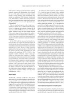
Table 3.1 Epidemiological studies support both environmental and genetic etiology of MS.
Evidence for environmental factors
MS in immigrants occurs with a rate similar
to that in the indigenous population when
the immigration is before teenage years
Epidemics of MS (e.g. on the Faroe Islands)
Increasing prevalence and decreasing age
of onset of MS in populations with stable
genetics
Evidence for genetic factors
• Ethnic groups (genetic isolates) with varying
susceptibility to MS
• Increased familial recurrence
• Higher concordance in monozygotic than in dizygotic
twins
• Higher risk for MS in full-sibs than in half-sibs; the
presence of a maternal parental effect
• Highly increased risk of MS in siblings of index cases
from consanguineous parents
• A similar risk of MS for nonbiological relatives and
individuals in the general population
and central Europe), medium (5–29/100,000, e.g.
south Europe) and low prevalence regions (less than
5/100,000, e.g. most Asian countries) (Kurtzke,
2005). These distributions may be related to both
environmental (climate, viruses, UV irradiation, and
diet) and genetic factors (Table 3.1).
Migration studies, history of epidemics, and serial
epidemiological updates support the existence of
environmental effects. European immigrants in South
Africa develop MS with a similar frequency as the
indigenous population, while an opposite trend is
observed for offspring of individuals immigrating
from India, South Africa, and the West Indies to the
United Kingdom (Dean, 1967; Elian et al., 1990). A
migration before mid-teenage years seems to confer
to the migrant the recipient country’s risk for MS,
possibly related to the effects of childhood infections
on immune maturation (Alter et al., 1966).
MS occurred on the Faroe Islands in four epi-
demics between 1943 and 1990. These epidemics
were attributed to a primary infectious agent imported
into the islands by the occupying British forces during
World War II, and to its transmission to subsequent
generations (Kurtzke 1975a,b, 1993, 2005).
Serial epidemiological updates suggest that the
relative risk for MS is increasing in certain groups over
time (Kurztke, 2005). This observation is well illus-
trated in Sardinia, where the mean annual incidence
rate significantly increased from 1.1/100,000 in
1965–9 to 5.8/100,000 in 1995–9 (Pugliatti et al.,
2005). Estimates of MS in cohorts from World War II
and the Korean conflict show a relative risk of 0.44
for African American men and 0.22 for other men as
compared to white men, while estimates in similar
ethnic cohorts from the Vietnam war and up to 1994
reveal a relative risk of 0.67 and 0.3, respectively
(Kurtzke, 2005). The risk of MS for white women as
compared to white men was 1.79 in the earlier cohorts,
which also significantly increased in the more recent
cohorts. Women of all races now have a risk ratio near
to 3:1 as compared to white men (Kurtzke, 2005).
Anticipation of age at onset may be another indic-
ator for the involvement of environmental factors.
Anticipation was demonstrated in two-generational
MS families and longitudinal surveys of sporadic cases
in Sardinia, where the mean age of onset decreased
from the most remote to the most recent decade of
birth from 41 to 21 years (Cocco et al., 2004). Inter-
estingly, in another subset of the Sardinian popula-
tion an increasing age of onset was noted (Pugliatti
et al., 2005).
In contrast to data supporting the involvement of
environmental effects, ethnic, family, and twin studies
suggest the involvement of genetic factors in MS
(Table 3.1). While the highest prevalence rates (100/
100,000 and beyond) correlate with the worldwide
distribution of individuals of Scandinavian descent
(Poser, 1994), several ethnic isolates with resistance
to MS live in geographic locations where the disease
is generally common. Examples include gypsies in
Hungary (Gyodi et al., 1981), Indians and Orientals
in North America (Ebers, 1983; Kurtzki et al., 1979),
Lapps in Scandinavia (Gronning and Mellgren, 1985),
Maoris in New Zealand (Skegg et al., 1987) and
Aborigines in Australia (McLeod et al., 1994). The
varying prevalence rates of MS in the genetically dis-
tinct but geographically close populations of Malta,
Sicily, and Sardinia also implicate genetic factors
NICP_C03 04/05/2007 12:26PM Page 30
Multiple sclerosis 31
(Elian et al., 1987; Rosati 1986). In addition, some
ethnic groups (e.g. Orientals and African Blacks) are
characterized by very low occurrence of MS (Dean,
1967; Poser, 1994).
Familial recurrence of MS was recognized long
ago (Eichorst, 1896). The observed inheritance pat-
terns are incompatible with Mendelian autosomal
dominant, recessive, and X-linked or mitochondrial
transmission patterns. MS is a complex trait disorder
with the involvement of several genes, each exerting
small effect, and probably in an interaction with the
environment. There is an excess in the mother-to-child
as compared to the father-to-child transmissions in
families with vertical concordance (Sadovnick et al.,
1991). The age-adjusted empirical recurrence risk
for first-degree relatives is 3 to 5%, which is 30 to
50 times the 0.1% rate in the general population
(Sadovnick et al., 1991, 1998). Individuals with a
greater “genetic loading” have an earlier age of onset,
and “genetic loading” is increased in individuals
with affected parents (Sadovnick et al., 1998). In a
population-based analysis of MS index cases and
their siblings whose parents were related, Sadovnick
et al. (2001) found a recurrence risk of 9% for sibs,
which is significantly higher than the risk for sibs of
MS index cases from nonconsanguineous parents.
Data from several large twin studies consistently
demonstrated a higher concordance rate of MS
among monozygotic (21.05% to 40%) as compared
to dizygotic twins (0 to 4.7%), strongly suggesting a
genetic effect (Bobowick et al., 1978; Hansen et al.,
2005; Heltberg and Holm, 1982; Kinnunen et al.,
1988; Mumford et al., 1994; Sadovnick et al., 1993).
The concordance rate among dizygotic twins (4.7%)
is similar to that observed among siblings (5.1%)
(Sadovnick et al., 1993).
Further confirmation of genetic effects is gained
from studies on adoptees revealing that the frequency
of MS among first-degree nonbiological relatives
living with an index case is not greater than expected
from the general population (Ebers et al., 1995). The
largest half-sib study (Ebers et al., 2004) defines an
age adjusted recurrence risk of 3.11% and 1.89%
for full-siblings and half-siblings, respectively. The
moderately significant excess of maternal vs. pater-
nal half-sibling risk (2.35% vs. 1.31%, respectively)
suggests a maternal effect on susceptibility to MS.
Early case–control candidate gene association
studies
Associations of MS with polymorphic alleles of
candidate genes involved in immune regulation and
myelin production have been extensively investigated
based on the autoimmune hypothesis of demyelina-
tion (Table 3.2). The first association noted with the
haplotype of class I human leukocyte antigen (HLA)
A3 and B7 alleles was extended to the Class II DR2
allele in both population and family studies ( Jersild
et al., 1973; Stewart et al., 1979). Since then, the
association of MS with the HLA A3, B7, DR2, Dw2
haplotype has been the most consistent finding
in Caucasians (Francis et al., 1991; Gyodi et al.,
1981; Olerup and Hillert, 1991), while HLA DR4
was detected in Sardinians and Jordanian Arabs,
and DR6 was described in Japanese and Mexicans
(Gorodezky et al., 1986; Kurdi et al., 1977; Marrosu
et al., 1988; Naito et al., 1978). Further studies
revealed that the DR15, DQ6 alleles define the MS-
associated DR2, DW2 haplotype, which is described
today in DNA-based terminology as DRB1*1501,
DQA1*0102, DQB1*0602 haplotype (Hillert et al.,
Table 3.2 Studies in MS.
Region of interest
Approach
Major finding
Case–control association
Polymorphic alleles of
candidate genes
Hypothesis-based
MHC DRB1 alleles define a
major proportion of genetic
susceptibility and resistance
to MS
Linkage
Full genome or regional
scans
Hypothesis-free
Multiple susceptibility loci
with small effect including
1q44, 2q35, 5p15–5q13,
6p21, 17q11, 17q22,
18p11, 19q13
LD mapping
Full genome or regional scans
Hypothesis-free or
hypothesis-directed
Distribution of LD genome
wide; identification of
chromosomal segments
carrying MS susceptibility
genes and variants in
progress
NICP_C03 04/05/2007 12:26PM Page 31
32 BERNADETTE KALMAN ET AL.
1994). Whether the primary MS-specific allele is the
DRB1*1501 or the DQB1*0602 could not be sorted
out because of the strong linkage disequilibrium
(LD) in this region. However, recent family and case–
control studies in African Americans suggested a
selective association of MS with the DRB1*1501
allele and a primary role for the DRB1 locus. This
finding appeared unlikely to be secondary to an
admixture with Caucasians, since several African
American MS susceptibility haplotypes were found
within chromosomal segments of African origin
(Oksenberg et al., 2004).
Additional studies in the major histocompatibil-
ity complex (MHC or HLA) region tested the associa-
tion of polymorphic alleles of HLA DP (Roth et al.,
1991), complement C4, Bf, C2 components (MHC III)
(Hauser et al., 1989), protein transporters (LMP,
TAP1, TAP2) (Bell and Ramachandran, 1995; Liblau
et al., 1993) and of the TNF α and TNF β genes with
MS (Braun et al., 1996; Roth et al., 1994). A poly-
morphic CA repeat within the gene of myelin oligo-
dendrocyte glycoprotein (MOG), telomeric to the
MHC, was also tested in MS (Malfroy et al., 1995)
(Fig. 3.1). However, the overall outcome of these
studies reflected inconsistent observations and did
not reveal independent associations of MS with genes
outside of the MHC class II subregion (Fig. 3.1).
Analyses of sequence variations within germline
elements of the T-cell receptor (TCR) α and β chain
genes as well as tests for a preferential utilization
of certain TCR V–J–D gene segments in the re-
arranged mRNA seemed to reveal promising data
in several studies. Nevertheless, the involvement of
TCR genes in MS susceptibility could not be consis-
tently supported (Fugger et al., 1990; Hashimoto
et al., 1992; Oksenberg et al., 1989; Seboun et al.,
1989). Linkage analyses also excluded that a TCR
α or β gene would contribute to MS susceptibility
(Lynch et al., 1991, 1992). Similarly, both associa-
tion and linkage studies of immunoglobulin genes
revealed conflicting observations (Feakes et al.,
1998; Hashimoto et al., 1993). Additional analyses
of immune response genes included ligands and
receptors in the cytokine, chemokine, and adhesion
molecule networks, but also without unequivoval
conclusion (Crusius et al., 1995; Epplen et al., 1997;
He et al., 1998a). Candidates related to myelin
production included the myelin basic protein (MBP)
promoter, MOG on chromosome 6 and genes of
oligodendrocyte growth factors or their receptors.
However, associations or linkage detected in some
studies were not confirmed in subsequent analyses
(Boylan et al., 1990; He et al., 1998b; Mertens et al.,
1998; Wood et al., 1994).
Despite the involved conceptual and technical dif-
ficulties (e.g. matching controls to patients in order
to avoid population stratification or selecting genetic
candidates), the case–control design continues to be
a widely used approach in MS. With the recognition
of more and more intercellular and subcellular path-
ways in pathogenesis, the number of molecular can-
didates permanently grows (Achiron et al., 2004;
Chiocchetti et al., 2005; Kantarci et al., 2005; Leyva
et al., 2005; Michailova et al., 2005; Oksenberg and
Barcellos, 2005). Today, however, both full-genome
scans and expression profiling assist a focused selec-
tion of candidates, and a comprehensive list of sequence
variations is provided for association studies by the
Human Genome Project (see below).
Linkage studies
In contrast to the hypothesis-driven case–control
association approach, the method of linkage can
identify susceptibility loci genome wide without a
preconceived idea of disease pathogenesis (Table 3.2).
Four full-genome scans with microsatellite markers
in MS families showed linkage to multiple suscept-
ibility loci, each with a minor effect (λ
s
= ഛ2) (Ebers
et al., 1996; Kuokkanen et al., 1997; The Multiple
DP
0
Centromere
1000
DN DO
Class II Class III Class I
DQ DR Hsp
TNF HLA-B HLA-C HLA-X HLA-E
HLA-H
HLA-A HLA-F
HLA-G MOG
αβ
21-OH
C4
BFC2
2000 3000 4000
Telomere
kb
Fig. 3.1 The figure depicts the MHC class II, class III, and class I regions encompassing 4 MB in chromosome 6p21.3.
MS-associated haplotypes have been consistently detected in the DRB1–DQB1 subregion in Caucasians.
NICP_C03 04/05/2007 12:26PM Page 32
Multiple sclerosis 33
Sclerosis Genetics Group, 1996; Sawcer et al., 1996).
Among the reported provisional sites, the 6p21,
5p15, 5q13, 17q22, and 19q13 regions were con-
sistently positive in more than one study. Additional
susceptibility loci at 1q44, 2q35, and 18p11 were
recently suggested (Kenealy et al., 2006). A meta-
analysis of combined, raw genotype data of three
full-genome scans underscored the importance of
17q11 and 6p21 in MS (The Transatlantic Multiple
Sclerosis Genetics Cooperative Study, 2001). Within
many regions with the highest scores, clusters of
immune regulatory genes are encoded (e.g. 6p21 –
MHC cluster; 17q11 – β-chemokine cluster). There
are, however, also loci likely involved in neuro-
degeneration. An association of the epsilon 4 allele
of the ApoE gene in 19q13 was suggested with both
susceptibility and progression rate of MS in several
studies, but a recent meta-analysis of available
worldwide data showed negative outcome (Burwick
et al., 2006; Schmidt et al., 2002).
Comprehensive analyses of the MHC region further
confirmed its importance in MS. While Haines et al.
(1998) proposed that linkage to the MHC was limited
to families segregating DR2, Ligers et al. (2001) also
detected linkage to this region in a larger cohort of
DRB1*15-negative families. This latter observation
led to the conclusion that the DRB1*15 may not be
the only HLA determinant of MS. The association
with DRB1*15 may be secondary to LD with a nearby
locus, or disease susceptibility alleles can be present
in DRB1*15-negative haplotypes. Analyses of the
DRB1 allelic heterogeneity in a large number of MS
families showed the involvement of several suscept-
ibility (e.g. DRB1*15 and DRB1*17) and protective
(e.g. DRB1*14) alleles suggesting trans interactions
among DRB1*15-positive and -negative genotype
combinations (Dyment et al., 2005). Altogether, asso-
ciation and linkage studies unambiguously established
that MHC class II genotypes determine a major pro-
portion of genetic susceptibility and resistance to MS.
Further refinements of MS susceptibility loci
by LD mapping
The method of linkage in complex disorders usually
identifies large (2–20 cM) chromosomal regions of
interest, but does not have the power to confine these
regions to single candidate genes with small effects.
For fine mapping, the use of modern association
methods within linkage-defined chromosomal regions
or the entire genome may be considered. The Human
Genome Project provided the means for this new
approach. The data revealed that two human
genomes are approximately 99.9% identical, leaving
still millions of different base pairs among the total
of 3.2 billion. The 0.1% difference is attributed to the
presence of a single nucleotide polymorphism (SNP)
at approximately every 1000 nucleotides. SNPs are
not only responsible for the interindividual pheno-
typic differences, but also for the variations in sus-
ceptibility to common diseases (Table 3.2, Fig. 3.2)
(The International SNP MAP Working Group, 2001).
Because of the abundance of SNPs, these varia-
tions may be used as markers in comprehensive
association studies. SNP marker alleles align in
haplotypes which tend to be inherited together in
a given population. This correlation between paired
SNP markers is called LD. The genome-wide distribu-
tion of LD is influenced by many factors and varies
among ethnic groups. A recent study of selected
chromosomal regions revealed that half of the
SNP haplotypes are 22 kb or larger in African and
African-American samples, and 44 kb or larger in
European and Asian samples (Gabriel et al., 2002).
However, it is important to note that the distribution
of LD shows great regional variations. LD between
a marker allele and a disease-specific allele may
allow us to identify mutations or variants with patho-
genic significance if SNP markers are genotyped
with sufficient density in a selected region (e.g. in a
linkage-defined susceptibility locus). Fewer markers
may be used in a two-stage study, which aims first to
identify the disease-associated haplotypes, and then
attempts (with a more dense marker distribution) to
reveal disease-relevant mutations within or in the
proximity of these haplotypes (Fig. 3.2). Using this
strategy in the 17q11 region, we first defined and
then refined MS-associated haplotypes in two inde-
pendent sets of families. Sequence analyses of these
haplotypes and their flanking regions will reveal if
disease-causing mutations are present in the co-
localizing CC chemokine genes (Vyshkina et al.,
2005; Vyshkina and Kalman, 2005).
With a high-density SNP panel encompassing the
MHC and flanking genomic regions in over 1100 MS
families, Lincoln et al. (2005) detected strong asso-
ciations with blocks in the class II region, and the
strongest association with the DRB1. Conditioning
on HLA DRB1 found no additional block or SNP
association independent of the class II genomic
region. Thus, this study established that MHC-
associated susceptibility in MS is defined by HLA class
II alleles, their interactions, and closely neighbor-
ing variants.
NICP_C03 04/05/2007 12:26PM Page 33
34 BERNADETTE KALMAN ET AL.
Full-genome LD mapping studies are also in progress
and are expected to identify novel susceptibility genes
with small effects. Mapping by admixture linkage
disequilibrium (MALD) offers another new approach
for the identification of disease-relevant genes with
approximately 100 times fewer SNP markers than
would be required for whole-genome haplotype scans.
This method implies that the genomes of different
ethnic groups have chromosomal segments of differ-
ent origin due to a historic gene flow between them
(e.g. African Americans have chromosomal segments
of African as well as European origin) (Smith et al.,
2004). The strategy involves the identification of a
genomic region from one ancestral population with
the highest occurrence of the disease. MALD can be
particularly powerful in finding genes for a disease
that differs profoundly in frequency among popula-
tions. Highly informative MALD markers have been
recently defined (Smith et al., 2004) and successfully
used to identify MS-relevant loci in African Americans
(Reich et al., 2005).
In summary, recent genetic data reflect a signifi-
cant increase in power achieved by the use of new
association-based methods and densely packed SNP
markers in MS. In addition to the identification of
disease-associated allelic variants, further explora-
tions of epistatic gene interactions as well as of
transcriptional and post-transcriptional regulatory
mechanisms are needed to better understand the
disease pathogenesis and to identify the best targets
for therapy.
Mitochondrial (mt)DNA in MS
A group of corollary studies aimed to clarify mito-
chondrial genetics in MS. MtDNA is a small (16.5 kb)
extranuclear part of the human genome. Numerous
point mutations and deletions in mtDNA have
been identified in association with neuromuscular
and multiorgan disorders. A possible involvement
of mtDNA in MS was postulated because there is a
higher transmission of the disease from mother to
child than from father to child (Sadovnick et al.,
1991), and because of the observed association
between Leber’s hereditary optic neuropathy (LHON),
a mitochondrial disease, and MS (Harding et al.,
1992; Lee et al., 1964). The identification of mtDNA
mutations at nt 11,778, 3460, and 14,484 with
primary pathogenic significance for blindness made
possible the objective evaluation of the association
between MS and LHON (Howell et al., 1991; Johns
et al., 1992; Wallace et al., 1988). Such mutations
were reported in a number of patients with prom-
inent optic neuritis (PON) and MS (Flanigan et al.,
1993; Harding et al., 1992; Kellar-Wood et al.,
1994), while inflammatory demyelination was also
found in LHON patients more often than expected
by chance (Riordan-Eva et al., 1995). However,
C
HA
R
A
C
TE
R
I
S
TI
CS
O
F THE H
U
MAN
G
EN
O
M
E
S
NP
s
HAPL
O
TYPE
S
A
• 3.2 BILLION BASEPAIR
S
• 30
,
000 GENE
S
• EVE
R
Y TW
O
G
EN
O
ME
S
A
RE
99.9% IDENTICAL
• A SNP IS PRESENT AT EVERY
1000 NUCLEOTIDE
• SNPs CONTRIBUTE TO
PHENOTYPIC DIFFERENCES
AND SUSCEPTIBILITY TO
COMMON DISEASES
TCAATGTCTGCATA
TCCATGACTGCGTA
GATCCTGGACTGC
GATCGTGAACTGA
ACGTTTACGTCGC
ACGTATAGGTCGC
acgt aTgtac gtacgt
acgt acgtac Atacgt
Gcgt aTgtac Ata*Ggt
Gcgt aTgtac Ata*Ggt
Gcgt aTgtac Ata*Ggt
Gcgt acgtac gtacgt
Gcgt acgtac gtacgt
B
• SNP ALLELES ALIGN IN
HAPLOTYPES
• WHEN A MUTATION ARISES,
IT DOES SO IN A SPECIFIC
HAPLOTYPE
• EACH MUTATION CAN BE
TRACKED IN A POPULATION
BY IDENTIFYING THE
CORRESPONDING
ANCESTRAL CHROMOSOMAL
SEGMENT ON WHICH IT
AROSE
*G:disease-related mutation
SNPs:A/G C/T G/A
Fig. 3.2 The Human Genome
Project provided the means for
LD mapping in complex
disorders.
NICP_C03 04/05/2007 12:26PM Page 34
Multiple sclerosis 35
comprehensive screening and sequencing of mtDNA
in large patient cohorts revealed that primary LHON
mutations are very rare in typical MS or PON, and
no other pathogenic mtDNA mutations contribute
to the pathogenesis of MS (Kalman et al., 1995,
1996, 1999).
Postgenomics
The recently developed cDNA microarray techno-
logy led to the identification of hundreds of gene
products potentially involved in disease pathogenesis
based on their differential expressions in patients and
controls. Comparisons between mRNA repertoires
in leukocytes as well as brain tissues of MS patients
and controls have been carried out in several studies
(Lindberg et al., 2004; Mandel et al., 2004; Satoh et al.,
2005). In addition, mRNA repertoires in chronic and
acute plaques were compared to those in the corres-
ponding normal appearing white-matter regions
in patients (Tajouri et al., 2003). The differentially
expressed molecules can be groupped into functional
clusters that include mediators of inflammation and
apoptosis, regulators of cell cycle, nuclear factors, and
molecules involved in subcellular signaling, myelin
development, and protection against oxidative stress.
The cDNA microarray technology has also been used
to investigate more clinically oriented questions such
as regulation of gene expression by disease-modifying
drugs, or differences in gene expression regulation in
responders and nonresponders to a drug (Koike et al.,
2003). Proteomic identification and quantitation of
proteins represent the next level of molecular analyses,
which aim to define disease-related changes in
various tissues of patients (Newcombe et al., 2005).
A simultaneous determination of genetic, transcrip-
tional, and proteomic profiles in correlation with
clinical, imaging, and histological phenotypes may
represent a comprehensive approach that will enable
us to better capture the mechanism and control the
natural history of this complex disease.
3.2 Immunopathogenesis (Thomas P. Leist)
Lesion characteristics and model systems
There have been advances in the understanding
of the pathogenesis of MS over recent years. These
advances have been fostered by observations in
animal models, and by ex vivo studies using speci-
mens of human origin and by development of new
imaging techniques. The experimental work done
to elucidate the mode of action of the currently avail-
able therapies also should not to be underestimated
as a source of new insights into the disease process.
In turn these advances have pointed to new molecu-
lar targets that may warrant development of future
therapies.
White-matter plaques in the CNS, particularly in
the optic nerve, brainstem, spinal cord, and periven-
tricular regions, are a cardinal pathological feature
of MS (Ikuta et al., 1976). It is now clear that disease-
induced changes are not restricted to the white
matter alone but also occur in gray matter (Dalton
et al., 2004). Inflammation and demyelination are
the histological hallmarks of MS, but astrogliosis,
neuronoaxonal injury, and degeneration also con-
tribute to the overall pathology (Raine, 1984). There
is a considerable heterogeneity of actively demyeli-
nating lesions. A classification to distinguish four
lesion types was proposed based on pathological fea-
tures including myelin protein loss, oligodendrocyte
involvement, complement activation, and types of
inflammatory infiltrates. Type I is characterized by
demyelination, T cell and macrophage infiltration,
and presence of macrophage-related products such
as tumor necrosis factor. In Type II, immunoglobulin
and complement are also present in addition to
the mononuclear cell infiltration. Type III lesions are
characterized by the dying-back type of oligodendro-
cytopathy with a preferential loss of myelin-associated
glycoprotein but with a preservation of proteolipid
lipoprotein and myelin basic protein, and lack of
remyelination in the absence of overt inflammation
or immunoglobulin and complement deposition.
Type IV is characterized by apoptosis oligodendro-
cytes (Lucchinetti et al., 2000). It is a central issue
in MS whether different immunological pathways
sequentially active over time result in demyelination
in an individual patient or mechanisms of demyelina-
tion differ from patient to patient defined by genetic
factors. The ultimate answers will have significant
therapeutic implications. Barnett and Prineas (2004)
have demonstrated that lesions from a given patient
can contain features of more than one of the above
lesion types. The matter of lesion classification remains
therefore a process in evolution.
At cellular level, neurons and their projections and
oligodendrocytes and the myelin sheets they generate
can be damaged and destroyed through a number of
immunopathological mechanisms. These mechanisms
include direct interactions with cytotoxic immune
cells and their products named cyto- and chemokines
with cytotoxic potential, and demyelinating antibodies.
NICP_C03 04/05/2007 12:26PM Page 35
36 BERNADETTE KALMAN ET AL.
Remyelination is an important feature of MS lesions
and re-expression of oligodendrocyte transcrip-
tion factor 1 can be seen during remyelination. To
what degree apoptosis, signaled through members
of the family of tumor necrosis factor receptors, is re-
sponsible for oligodendrocyte drop out remains to be
elucidated. Molecules associated with recruitment
of myelinating cells and remyelination include CXC
and CC chemokine receptors, which were recently
described on oligodendrocytes (Omari et al., 2005).
Recruitmentof oligodendrocyte-precursor cells appears
to be normal in MS lesions. There appears to be
inhibition of differentiation and growth of these
precursors. Several substances have been identi-
fied in the gliotic scar that stunt differentiation of
oligodendrocytes and local myelin repair. A leucine-
rich–repeat and immunoglobulin-domain-containing
Nogo receptor has been identified which may re-
present a target for improved myelin repair (Mi et al.,
2005). The axonal compromise observed in MS is,
however, not just a function of inflammation and
demyelination but is also reflective of abnormal expres-
sion of ion channels including sodium channels. As a
result there is an increased entry of sodium, slowing
of nerve conduction, and conduction block.
These effector mechanisms have also been shown
to be operative in EAE models. Distinct pathological
presentations similar to those recognized in active
MS lesions can be produced dependent on the animal
strain used, and the employed protocol of active or
passive immunization. EAE in rodents predominantly
affects the spinal cord, and therefore, the observed
clinical deficits almost stereotypically reflect the
extent of inflammation and demyelination. In the per-
sistent virus model of inflammatory demyelination
induced with Theiler’s virus, the correlation between
the distribution of cellular immune mediators and
demyelination and the clinical deficit is also limited.
However, immunopathological changes that are
characteristic of specific mechanisms can be identi-
fied during the acute destruction of myelin. Inflam-
matory characteristics of active lesions include
standard elements of inflammation such as CD4+
and CD8+ T cells along with activated microglia,
macrophages, and B cells (Fig. 3.3).
Induction of an autoreactive immune response
It is not known how and when MS is initially induced
but there is substantial evidence to support the hypo-
thesis that genetics determines a person’s susceptibil-
ity to MS, and that environmental factors modulate
the risk. It is not clear when induction of autoreactive
immune cells occurs but migration studies suggest
that this may occur before or around puberty (Nose-
worthy et al., 2000). Though these self-reactive cells
have the potential to cross-react with CNS antigens,
it appears that they remain dormant and sequestered
outside of the CNS. In this model, an unidentified
external trigger is postulated to deliver a second
IFNγ / IL-17
Y
microglia
macrophage
T cell
Invasion/
Diapodesis
Adhesion
T cell
Postcapillary
venule
Adhesion
molecules
BBB
IL-12/23
MMPs
Reactivation/Proliferation
B Cell
Axon
Myelin
Y
Y
Y
Antibodies
macrophage
Oligo
Y
Y
Y
Fig. 3.3 Recruitment of T cells into the CNS.
NICP_C03 04/05/2007 12:26PM Page 36
Multiple sclerosis 37
signal that will lead to the activation of resting self-
reactive cells. This second signal is believed to be
delivered after puberty. Once activated, these cells are
capable of traversing the blood–brain barrier (BBB).
Characteristics of the inflammatory response
In rodent and primate model systems of EAE, CD4+
or CD8+ T cells reactive with constituent proteins of
the myelin can induce inflammatory demyelination
in the CNS (Ando et al., 1989; Huseby et al., 2001;
Pettinelli and McFarlin, 1981). Myelin basic protein
is one of these proteins. T cells reactive with myelin
basic protein can be demonstrated in individuals
with MS and in normal controls. The myelin-specific
T cells from patients with MS generally exhibit a
memoryor activated phenotype, whereas those from
healthy persons have a naive phenotype. The most
direct evidence that myelin-reactive T cells can induce
inflammatory demyelination comes from one of the
clinical studies using an altered peptide ligand as a
potentially disease-attenuating treatment. While a
reduced disease activity was observed on magnetic
resonance imaging (MRI) in one of the studies using
a low dose of the altered peptide ligand (Kappos et al.,
2000), an increased clinical and MRI activity was
noted in several patients treated with a higher dose
of the altered peptide ligand (Bielekova et al., 2000).
Patients exhibited a surge of T cells reactive with the
corresponding myelin basic protein peptide at the time
of clinical activity. This finding supports the view
that a self-reactive inflammatory process directed
against myelin constituents plays a central role in
MS. The cytokine profile produced by myelin-specific
T cells determines the ability of these cells to initiate
inflammationin the CNS.
Myelin-reactive T cells from patients with MS pro-
duce cytokines more consistent with a proinflammat-
ory Th1-mediated response, whereas myelin-reactive
T cells from healthy persons are more likely to pro-
duce Th2 cytokines which exert immunoregulatory
response. Cytokines associated with a Th2 response
include interleukin (IL-4) andIL-5.
IL-12 is a cytokine that is strongly associated with
proinflammatory Th1 responses. It activates the trans-
cription factor Stat-4 in human T cells and promotes
their differentiation towards a proinflammatory,
pathogenic Th1 phenotype. Some data suggest that
interferon-β, which isused to treat MS, causes a shift
from a Th1-mediated to a Th2-mediated response.
However, microarray studies demonstrate that inter-
feron therapy induces the upregulation of a number
of genes which are associated with a differentia-
tion into Th1rather than Th2 phenotype (Wandinger
et al., 2001). IL-12 and IL-23 are heterodimers and
share the same p40 subunit. They affect regulation
of T-cell responses in a way that may be relevant to
the disease process of MS (Trinchieri et al., 2003).
Developmentof EAE depends on a sequencial expres-
sion of distinct cytokine patterns. Mice deficient in
both IL-12 and IL-23 are resistant to EAE, whereas
animals deficient in IL-12 alone develop severe dis-
ease. Interleukin-23 induces IL-17 production by T
cells. IL-17 is expressed in brain lesions and appears
to be a regulator of CNS inflammation.
T cells have been viewed as central players in the
autoimmune process of MS. However, when instructed
by T cells, B cells differentiate and produce autoreact-
ive antibodies which contribute to the pathobiology
of the disease by binding of antigens and activation
of complement. The intrathecal synthesis of immuno-
globulins is characteristic of MS: cerebrospinal
fluid (CSF)-restricted oligoclonal bands (OCB) and
an increased IgG can be observed in most patients.
In addition, B-cell clones restricted to the CNS can
be found (Monson et al., 2005). The presence of anti-
myelin oligodendrocyte glycoprotein (MOG) antibodies
in patients with clinically isolated demyelinating
syndromes was suggested to have predictive value in
identifying those patients who will go on to develop
clinically definite MS. Further studies are needed to
evaluate this finding. There is an interest in targeting
B cells as an alternative strategy to the approaches
that are directed against T cells and their products.
Marcophages and microglia are the primary medi-
ators of the inflammatory response in active early
MS plaques. They are generally abundant at the
outer rim of the lesion and release cytokines, reactive
oxygen species, and nitric oxide, which cause tissue
injury including damage to the myelin sheath. In
hyperacute and highly destructive lesions, particu-
larly in variant forms such as neuromyelitis optica and
Marburg’s type of MS, granulocytes and eosinophils
may be part of the inflammatory reaction.
Recruitment of inflammatory cells into the CNS
Migration of autoreactive leukocytes from the sys-
temic circulation into the CNS is thought to play an
important role in the formation of inflammatory MS
lesions. The leukocyte extravasation occurs in con-
secutive steps (Fig. 3.3). The initial step is marked by
tethering and rolling of inflammatory cells on the
endothelium. E-selectin and sialyl Lewis (SLe) are
NICP_C03 04/05/2007 12:26PM Page 37
38 BERNADETTE KALMAN ET AL.
thought to play an important role in this stage. The
ensuingfirm adhesion is mediated by β
2
integrins and
includes vascular cell adhesion molecule (VCAM)-1,
very late antigen (VLA)-4, intracellular adhesion mole-
cule (ICAM)-1, and lymphocyte function-associated
antigen (LFA)-1 (Miller et al., 2003; Yednock et al.,
1992). Antibodies against LFA-1 and ICAM-1 were
shown suppressing EAE in some studies, and a rise in
ICAM-1 levels was seen in the same model just prior
to onset of clinical symptoms.
Inflammatory cells diapedese through the endo-
thelial layer and the extracellular matrix. Matrix
metalloproteases (MMPs) produced by activated
leukocytes assist the breakdown of the BBB and
invasion of the brain parenchyma. In both EAE and
MS, levels of gelatinase B (MMP-9) and matrilysin
(MMP-7) are increased in blood, CSF, and brain during
periods of inflammatory disease activity associated
with enhancing MRI lesions. Chemokines form a
chemo-attractant gradient that is perceived by T cells
and assists their invasion into the CNS. Chemokines,
including interferon γ-inducible protein-10 and its
receptor CXCR3, are elevated in patients with disease
activity. The sequence of tethering, adhesion, and
diapedesis is a choreographed process dependent on
a number of interacting molecules each of which
may represent a potential target for therapeutic inter-
vention. Macrophage activation antigens, MRP14
and 27E10, hold promise as markers for the iden-
tification of actively demyelinating lesions in MS as
they are only transiently expressed after emigration
from the vasculature.
Once in the CNS, lymphocytes interact with antigen-
presenting cells (microglia, macrophages, and B cells
as well as other antigen-presenting cells) that bear
the appropriate antigenic epitope embedded in HLA
class I or II molecules, and undergo local clonal
proliferation if appropriate costimulatory molecules
are expressed. In turn, activated T cells will recruit
macrophages and control the synthesis of immuno-
globulins that can bind to epitopes on the myelin
sheath. T cells reactivated within the CNS also secrete
cytokines that in turn stimulate activation of T and
B cells as well as monocytes. These immune inter-
actions result in a vitious circle of local inflammation
in the CNS.
Extent of inflammatory changes
The white-matter regions distant from plaques are
frequently abnormal even though usually referred
to as “normal appearing white matter”. While some
of these white-matter changes in MS patients with
longstanding disease may partly be due to other
coincidental diseases affecting the CNS (e.g. vascular
or neurodegenerative disorders), findings on MR
spectroscopy at the time of a first demyelinating event
emphasize that the process of MS itself causes early
and significant changes in the “normal appearing
white matter”. This diffuse pathology in the “normal
appearing white matter” may be explained by a
reaction of brain tissue to the chronic inflammation
and axonal degeneration, or by a diffuse molecular
pathology present in myelin prior to the formation of
overt demyelinating lesions. Gray- and white-matter
atrophy and gliosis are characteristic features in
MS cases with severe or longstanding disease and
with significant cognitive involvement. The damage
occurring in plaques results in anterograde and
retrograde effects along the fiber tracts, and thus
contributes to tissue changes remote to the lesion. In
addition, the “normal appearing white matter” often
contains small focal inflammatory and demyelinat-
ing lesions that elude macroscopic detection.
The mechanisms of demyelination may vary among
patients, and the complexity of MS pathogenesis
cannot be explained on the basis of a T-cell mediated
immune response alone. The involvement of demye-
linating antibodies is suggested by the close similar-
ity between MS and MOG-induced EAE in rodents
and primates, by the demonstration of complement
activation in active lesions and by the presence of
anti-MOG antibodies in some patients. Deposition
of anti-MOG antibodies can be observed in MOG-
induced EAE.
While the destructive nature of inflammation is
much appreciated, its tissue protective features are
often neglected. Evidence suggests that inflammation
can induce removal of debris, which may be a neces-
sary step in the induction of reparative mechanisms.
Subsets of activated lymphocytes control inflam-
mation while others produce nerve growth factors.
Therefore, a complete abrogation of all inflam-
matory responses at a given time point in the disease
process may not always provide the most effective
route towards a maximal treatment efficacy.
3.3 Courses and diagnosis of MS
(Bernadette Kalman)
3.3.1 Courses of MS
Analyses of clinical and pathological phenotypes
suggest that MS represents a spectrum rather than
NICP_C03 04/05/2007 12:26PM Page 38
Multiple sclerosis 39
a single entity of inflammatory demyelination and
neurodegeneration. Natural history studies confirm
heterogeneity of the disease and define discrete
clinical subtypes with characteristic course, accu-
mulation of disability, and long-term outcome. While
phenotype-specific biomarkers are not yet available,
a standardized terminology describes four clinical
courses that are routinely used in practice (Lublin
and Reingold, 1996).
This empirical classification, in agreement with
natural history studies, distinguishes relapsing-
remitting (RR), secondary progressive (SP), primary
progressive (PP), and progressive-relapsing (PR) forms
of MS (Fig. 3.4). Eighty to eighty-five percent of patients
have an RR onset with a substantial proportion con-
verting into SP-MS over time. The remaining patients
present with PP-MS characterized by a later onset, less
female predominance, and a more rapid deteri-
oration (Cottrell et al., 1999). The median time from
disease onset to reach an expanded disability status
scale (EDSS) score of 4, 6, and 7 is longer in RR than
in PP-MS. However, the time to reach EDSS 6 from
EDSS 4 is similar in the SP- and PP-MS groups
(Confavreux et al., 2000), and the time to major dis-
ability milestones after the onset of progression at
DSS2 or less is also similar in all progressive forms
(Kremenchutzky et al., 2006). These observations
suggest that the rates of accumulation of irrevers-
ible pathological changes are different in RR- and
PP-MS, but are markedly similar in SP- and PP-MS
(Confavreux et al., 2000; Ebers 2004; Kremenchutzky
et al., 2006). Analyses of long-term outcomes (time
to EDSS 3, 6, 8, and 10) suggest that patients with
relapsing-progressive (RP) course can be reassigned
either to SP- or PP-MS, and patients with PR-course
can be reassigned to PP-MS (Kremenchutzky et al.,
1999). The younger the age of onset, the younger
the age of reaching disability milestones (Confavreux
and Vukusic, 2006).
In addition to these patterns of disease course,
“benign” and “aggressive” labeling of MS have been
used, but with varying definitions. McAlpine (1961)
proposed “benign” disease for those patients who,
after a follow up of 10 or more years, were without
restriction of normal activity in professional and
private life, but not necessarily without neurological
symptoms. Hawkins and McDonell (1999) identified
a subgroup with “benign” MS representing 19.9% of
their patient cohort when defining EDSS = р3.0 at
least 10 years after onset. Pittock et al. (2004) found
that patients with EDSS = 2 or less for 10 years or
longer have a greater than 90% chance of remaining
stable in the subsequent decade. This subgroup repres-
ented 17% of their original (year 1991) prevalence
cohort. These studies suggest that approximately a
fifth of patients with MS have little or no disability
for long periods of time. However, the EDSS-based
definitions of benign MS are not without pitfalls, as
EDSS is weighted for motor disability without appre-
ciating cognitive impairments, conventional MRI
activity of the disease, and nonconventional MRI
measures of the disease burden.
Overall, a favorable disease course is associated
with female gender, early onset, presentation with ON
and sensory symptoms. Acute onset, little residual
disability after each relapse, and long inter-relapse
periods are also indicators of a benign course. Data
from the Optic Neuritis Study Group (2004) revealed
that 65% of patients, who were enrolled in the Optic
Neuritis Treatment Trial (ONTT) between 1988 and
1991 and developed clinically definite MS (CDMS),
had an EDSS less than 3.0 at least 10 years after the
initial ON, underscoring that ON at onset is a favor-
able prognostic factor.
In contrast, unfavorable prognostic indicators
include male gender, later age of onset, progressive
course from onset, frequent exacerbations in the
first two years, poor recovery from relapses and early
development of progressive deficit, involvement of
cerebellar and motor pathways, and initial presenta-
tion with multisystem involvement.
There are also “fulminant” forms of MS that are
less well defined than the benign forms, but generally
have a rapid progression rate and high mortality.
“Marburg type” of MS is reserved for a rapidly progress-
ive monophasic disease reaching severe disability or
even death in a few weeks to months, as was the case
Time Time
DisabilityDisability
RR-MS
Time
PP-MS
DisabilityDisability
SP-MS
Time
PR-MS
Fig. 3.4 Course of MS (after Lublin and Reingold, 1996).
NICP_C03 04/05/2007 12:26PM Page 39
40 BERNADETTE KALMAN ET AL.
described by Otto Marburg (1906). Patients usually
have acute lesions affecting eloquent regions of the
brainstem and widespread inflammatory lesions of
the optic nerves, spinal cord, and hemispheral white
matter. The lesions are usually extensive and show
signs of intense inflammation (Johnson et al., 1990;
Marburg, 1906). With the currently available anti-
inflammatory and disease-modifying medications, the
management of this subtype became less dismal.
The “tumefactive” form of MS typically is related
to a single mass-like plaque which is both clinically
and radiologically indistinguishable from a brain
tumor. The clinical presentation may be a subacute
neurological deterioration reflecting the lesion loca-
tion. A tumefactive lesion may develop in an indi-
vidual with an established diagnosis, or sometimes as
an initial presentation of MS. Response of the lesion
to corticosteroid treatment and other paraclinical
data including CSF immune work up, conventional
magnetic resonance imaging (MRI), and spectroscopy
(MRS) monitoring may help to establish the diagnosis
without biopsy in some of these patients. However,
this presentation often represents a diagnostic chal-
lenge and makes brain biopsy unavoidable (Capello
et al., 2001; Khoshyomn et al., 2002).
Another potentially severe variant, Balo’s disease,
is both radiologically and pathologically a distinct
one, and has been predominantly observed in Asians.
In Balo’s type of concentric sclerosis, large hemi-
spheral lesions occur with alternating rings of pre-
served and damaged myelin in MRI and pathological
studies (Balo, 1928; Stadelmann et al., 2005). Typical
MS plaques are also usually present. The clinical
course may be fulminant with obscuration of con-
sciousness or even development of coma and death,
if not treated aggressively. Recent molecular studies
implicate the upregulation of neuroprotective mach-
inery in regions of subtotal, inflammation-induced
mitochondrial impairment leading to the preserva-
tion of myelinated tissue at the edges of inflammation
and demyelination. Inflammation, however, may
overcome neuroprotection at the edges of preserved
tissue segments, resulting in alternating damaged
and preserved tissue layers. This mechanism of
hypoxic deconditioning is similar to that described
in acute ischemic injury (Stadelmann et al., 2005).
3.3.2 Diagnosis of MS
The diagnosis of MS has always relied on object-
ive evidence for a dissemination of clinical events
and pathological lesions in time and space, and on
a simultaneous exclusion of alternative diagnoses
(Poser et al., 1983; Schumacher et al., 1965). The
Poser’s criteria (1983), still in use (particularly in
repositories and retrospective data analyses), pro-
vide different levels of diagnostic certainty including
clinically definite or laboratory-supported definite,
and clinically probable or laboratory-supported prob-
able, and possible MS. The more recently devel-
oped McDonald’s criteria (2001) omit the terms of
“clinically definite” and “probable MS,” and retains
only “MS” or “not MS,” leaving diagnostic uncer-
tainties for the category of “possible MS”. A major
advantage of the McDonald’s criteria is that by the
integration of MRI work up into clinical and para-
clinical methods, the diagnosis can be established
much earlier than before. When no signs of dis-
semination in time and space have occurred yet, but
are expected with high probability in the future, clinic-
ally isolated syndrome (CIS) or monosymptomatic
disease may be diagnosed.
The McDonald’s criteria allow establishing the dia-
gnosis of MS exclusively based on clinical observa-
tions if there is objective evidence of the separation of
lesions in time and space. An acute episode of neuro-
logical dysfunction that lasts for at least 24 hours in
the absence of fever or metabolic disturbance can be
called an attack, exacerbation, or relapse. All events
within 30 days of the initial event are considered
to be part of the same exacerbation. For dissemina-
tion in space, the involvement of distinct regions of
the CNS needs to be demonstrated. MRI criteria
for the demonstration of dissemination in space and
time are summarized in Boxes 3.1 and 3.2, respect-
ively. Information from MRI, laboratory analyses
of the CSF, and visual evoked potentials (VEP) will
become critical when the clinical presentation alone
is not sufficient to establish the diagnosis. Among
these methods, MRI has the highest degree of specifi-
city and sensitivity. CSF can be supportive when the
clinical presentation is unusual and the MRI altera-
tions are insufficient to fulfill the criteria. VEP also
can be supportive when the number (e.g. progressive
myelopathy in PP-MS) or the specificity (e.g. elderly
patients with possible microvascular disease) of MRI
lesions is in question. Other evoked potentials con-
tribute little to the diagnosis (McDonald et al., 2001)
(Tables 3.3 and 3.4).
The distinct characteristics of PP-MS (Cottrell et al.,
1999; Montalban 2005) prompted investigators to
develop separate diagnostic criteria for this subgroup
of patients. Thompson et al. (2000) state that clinical
progression from onset at least for one year must be
NICP_C03 04/05/2007 12:26PM Page 40
Multiple sclerosis 41
Three of four of the following
requirements:
1 One gadolinium-enhancing lesion or
nine T2-weighted hyperintense lesions
if there is no enhancing lesion.
2 One infratentorial lesion.
3 One juxtacortical lesion.
4 Three periventricular lesions.
Note: One spinal cord lesion can be substituted for one brain
lesion. After Barkhof et al. (1997) and Tintore et al. (2000).
The 2005 revision by Polman et al. (2005) confirmed these
criteria and provided clarification for the incorporation of
spinal cord lesions into the criteria (see text).
Box 3.1 MRI determination of dissemination of lesions in space
(McDonald et al., 2001).
(a) MRI criteria for dissemination of lesions in
time (McDonald et al., 2001)
If the first scan is performed three months or
more after the clinical onset, a gadolinium-
enhancing lesion (not at the site involved in
the original clinical event) is needed; if no
enhancing lesion is present, a follow-up scan
with new T2- or gadolinium-enhancing lesion
in another three months or later is needed to
fulfill the criterion for dissemination in time.
(b) MRI criteria for lesion dissemination in
time (Polman et al., 2005)
1 A gadolinium-enhancing lesion at a site
not involved in the initial event, and at
least three months after the onset of the
initial clinical event is needed; or
2 A new T2 lesion at any time compared
to the reference scan performed at least
30 days after the clinical onset is needed
to fulfill the criterion.
Box 3.2 MRI determination of lesion dissemination in time.
Table 3.3 Diagnostic criteria for MS (McDonald et al., 2001; Polman et al., 2005).
Clinical presentation
Two or more attacks
Objective clinical evidence of two or
more lesions
Two or more attacks
Objective clinical evidence of one lesion
One attack
Objective clinical evidence of two or
more lesions
One attack
Objective clinical evidence of one lesion
(monosymptomatic presentation)
Notes: The diagnosis is “MS” if the criteria fulfilled; the diagnosis is “possible MS” if the criteria are not completely fulfilled;
the diagnosis is “not MS” if the criteria are fully explored and not fulfilled.
Table 3.3 is published with the permission of the Legal Department of John Wiley & Sons, Inc.
Additional data needed for MS diagnosis
None required
(If MRI or CSF are done, the results should be consistent with MS)
Dissemination in space demonstrated by MRI (Box 3.1) or
Two or more MRI lesions consistent with MS plus positive CSF
(OCB by isoelectrofocusing or raised IgG index) or
Another clinical attack involving a different site
Dissemination in time, demonstrated by MRI (Box 3.2) or
Second clinical attack
Dissemination in space, demonstrated by MRI (Box 3.1) or
Two or more MRI lesions consistent with MS plus positive CSF
(OCB by isoelectrofocusing or raised IgG index) and
Dissemination in time demonstrated by MRI (Box 3.2) or
Second clinical attack
NICP_C03 04/05/2007 12:26PM Page 41
42 BERNADETTE KALMAN ET AL.
documented before the diagnosis of PP-MS is made.
Most patients with PP-MS present with spastic para-
paresis, while the remaining patients have progressive
cerebellar, brainstem, hemiparetic, visual, or cognitive
involvement. Based on the Thompson et al. (2000)
criteria adopted by McDonald et al. (2001) for the
diagnosis of definite PP-MS, the presence of intra-
thecal IgG synthesis with one of three MRI criteria
was required: (i) nine brain lesions; (ii) two spinal cord
lesions; or (iii) 4–8 brain lesions and one cord lesion.
The McDonald’s criteria were studied in retro-
spective and prospective analyses, and proven to be
superior in sensitivity, specificity, and clinical utility
compared to previous criteria, particularly in adult
western populations with classical MS. These criteria
have not been adequately tested in pediatric popula-
tions and in ethnic groups other than western popu-
lations (e.g. Asian or Latin American patients). In
2005, the International Panel reviewed available data
concerning the McDonald’s criteria and proposed a
revision based on refined consensus (Polman et al.,
2005). While this revision reiterates the essential clin-
ical requirements for the diagnosis of MS, it simplifies
and clarifies the original definitions in three areas.
1 MRI criteria for dissemination in time: A new T2
lesion (not only new enhancing lesion) at any time
point after a reference scan performed at least
30 days after the onset of the initial clinical event
would meet the imaging criteria of dissemination
in time.
2 Incorporation of spinal cord lesions into the
imaging requirements: A spinal lesion charac-
teristic of MS (little or no swelling in the cord,
hyperintense on T2, at least 3 mm in size, less than
two vertebral segment in length and occupies part
of the cord’s cross-section) is helpful to eliminate
alternative diagnosis or to confirm MS when no
dissemination in space is detected on brain MRI.
A spinal cord lesion can substitute for a brain
infratentorial (but not for periventricular or
juxtacortical) lesion. An enhancing cord lesion
can count doubly (for both an enhancing lesion
and an infratentorial lesion) in fulfilling the cri-
teria. Individual spinal cord and brain lesions
together may reach the required nine T2 lesions.
Although an MS lesion may occur as a diffuse
cord lesion (particularly in PP-MS), for the purpose
of diagnostic evaluation, a discrete focal lesion is
required. Repeated spinal cord MRI has generally
a low yield, and is only recommended when a
clinical event suggests a new spinal cord lesion.
3 Diagnosis of PP-MS: A positive CSF finding is no
longer necessary for the diagnosis of PP-MS
(Polman et al., 2005) (See revised definitions in
Box 3.2, Tables 3.3 and 3.4).
3.4 Clinical features (Bernadette Kalman)
The stochastic distribution of CNS lesions results in
a great variety of clinical symptoms usually reflect-
ing multisystem involvement in MS. Nevertheless,
Table 3.4 Diagnostic criteria for PP-MS (McDonald et al., 2001; Polman et al., 2005).
Clinical presentation
Insidious neurological
progression suggestive
of MS (PP-MS)
Table 3.4 is published with the permission of the Legal Department of John Wiley & Sons, Inc.
Additional data
(Polman et al., 2005)
One year of disease progression
plus two of the following:
– Positive brain MRI (nine T2
lesions or four or more T2
lesions with positive VEP)
– Positive cord MRI (two focal
T2 lesions)
– Positive CSF
Additional data (McDonald et al., 2001)
Positive CSF (OCB by isoelectrofocusing and
raised IgG) and
Dissemination in space, demonstrated by
– Nine or more T2 lesions in brain or
– Two or more lesions in spinal cord or
– Four to eight brain lesions plus one spinal
lesion; or
Abnormal VEP with four to eight brain lesions
or with fewer than four brain lesions plus one
spinal cord lesion demonstrated by MRI; and
Dissemination in time demonstrated by MRI
(Box 3.2); or
Continued progression for one year
NICP_C03 04/05/2007 12:26PM Page 42
Multiple sclerosis 43
macroscopic lesions in noneloquent CNS regions may
remain asymptomatic, while a single small lesion in
an eloquent region (optic nerve, spinal cord, brainstem)
can lead to severe neurological disability. A wide-
spread microscopic lesion load may also remain clin-
ically silent until a critical sum of tissue loss is reached,
from which point deficits (e.g. spastic paraparesis or
cognitive decline) will progressively accumulate.
Among the individually variable neurological
phenotypes, there are a number of rather stereotypic
presentations in MS. Lhermitte’s sign is often noted
at onset or during the course of the disease. Optic
neuritis (ON) either alone or in combination with other
system involvements is another frequent presentation.
The distinct combination of ON with myelitis will
be discussed in the separate chapter on neuromyelitis
optica. Oculomotor abnormalities and internuclear
ophthalmoplegia may present alone, but are often
associated with signs of corticospinal, spinothalamic,
or spinocerebellar involvement in patients with
brainstem lesions. Some MS patients have almost
exclusively sensory, while others have predomin-
antly motor disability at onset. Cerebellar symptoms
can also dominate the clinical picture. Autonomous
dysfunctions rarely occur in isolation at the initial
presentation, but commonly develop during the
course of disease and accompany other symptoms.
A great proportion of patients has central and neuro-
pathic or neuralgiform pain. Cognitive and emo-
tional dysfunctions had been somewhat overlooked
until recently, even though they may represent the
most significant components of disability.
The list of idiosyncratic presentations is endless.
Paroxysmal symptoms, dystonia, hearing loss,
aphasia, pruritus, hyperpathia, or allodynia may be
mentioned among the more frequent ones.
Optic neuritis
Idiopathic ON is most commonly observed in young
adults (20–50 years of age) with an incidence of 3/
10
5
in the US (Frohman et al., 2005). ON can be an
isolated event, but it is the initial presentation of MS
in about a quarter of patients. In its typical forms, a
partial or complete unilateral loss of vision develops
over a few days up to 7–10 days. Impairment of color
vision often occurs in the early stage. The visual loss
is painful, particularly with eye movements, in the
majority (92%) of patients (Optic Neuritis Study Group,
1991). The recovery usually starts in two weeks and
the improvement may proceed for several months.
In an acute stage, the ophthalmoscopic exam may
be unrevealing, but swollen disc (papillitis), typically
without hemorrhages or exudates, can be seen in a
third of patients. In retrobulbar neuritis, the head
of the optic nerve appears normal. Visual field exam
usually reveals large central scotoma and centrocecal
scotoma (involving the macula and blind spot) or
variable field defects. The afferent pupillary defect
(APD) is a critical finding.
Chronic ON is associated with neuro-ophthalmo-
logical abnormalities including disc pallor, pupil-
lary abnormalities, and field defects. Exclusion of
superimposed glaucoma or infiltrating lesions may
be necessary in some cases. Atrophy of optic nerves
may be noticeable on MRI, and the atrophy of the
retinal nerve fiber layer (RNFL) can be detected by
optical coherence tomography (OCT).
Neuro-ophthalmological tests used in the ONTT
(Beck et al., 1992) included visual acuity (retroillu-
minated Snellen charts at 4 m), color vision (pseudo-
isochromatic Ishihara plates – 11 plates and the
Farnsworth–Munsell 100-hue test), contrast sensit-
ivity (Pelli–Robson letter chart at 1 m), and a visual
field test (Humphrey Field Analyzer Program 30-2).
Most neurologists in bedside settings limit the assess-
ment to acuity, confrontational field evaluation, pupil-
lary light reflexes, color vision assessment, and
ophthalmoscopy. VEP studies may be useful in atyp-
ical and chronic cases, or when a discrimination of
retinal and optic nerve disease is needed (Frohman
et al., 2005). While orbital MRI with gadolinium
enhancement can support the diagnosis of acute
ON, a T2-weighted or FLAIR study of the brain has
prognostic values (see below). Likewise, a CSF work
up may not only facilitate the diagnosis and differ-
ential diagnosis but also serve as a prognostic marker
in patients with ON. OCT is a recently introduced
method that allows quantifying the axonal loss in
the RNFL along with the assessment of secondary
retinal ganglion cell loss (Trip et al., 2005). A signi-
ficant reduction of the RNFL thickness and macular
volume was noted in affected eyes with incomplete
recovery as compared to the unaffected fellow eyes
or to the eyes of normal controls (Trip et al., 2005).
While the visual loss completely recovers in about
half of patients with ON and significantly improves
in most of the remaining cases, atrophy of the optic
nerve (optic disc pallor, MRI finding) and of the RNFL
(OCT assessment) are usual findings in chronic stages.
Approximately, one in every eight patients will have
relapsing ON, and a few patients will also have ON
in the fellow eye. Relapsing ON is greatly associated
with increased risk for CDMS.
NICP_C03 04/05/2007 12:26PM Page 43
44 BERNADETTE KALMAN ET AL.
The ONTT revealed that patients with no MRI
lesions had 16% risk for CDMS, while those with
three or more MRI lesions had 51% risk at five years
(Optic Neuritis Study Group, 1997). Lack of pain, optic
disk swelling, and mild loss of visual acuity were
associated with a low risk for CDMS in patients with
negative MRI and history of no neurological sym-
ptoms or ON in the fellow eye. The analyses of data
from the 10-year follow up established that even
one brain lesion predicts a risk of 56% for CDMS as
compared to the risk of 22% in patients with neg-
ative MRI (Optic Neuritis Study Group, 2003). Inflam-
matory CSF profile with OCB at onset also doubled
the risk for CDMS in patients presenting with ON
(Nilsson et al., 2005).
In less typical cases, ON presents with progressive
visual loss over weeks, resembling optic neuropathy
of genetic, toxic-metabolic, chronic infectious, or com-
pressive origin. In such cases and in the appropriate
clinical setting, Leber’s hereditary optic neuropathy
(LHON), Lyme disease, sarcoidosis, syphilis, lupus,
West Nile virus, B12 deficiency, toxic causes, and in-
filtrative processes should be considered. The clinical
presentation of anterior ischemic optic neuropathy
may also overlap with that of ON. Similarities include
the rate and severity of visual deterioration or altitu-
dinal field defect, although this latter is rare in ON. In
addition to the appropriate laboratory tests, orbital
MRI with gadolinium enhancement around the optic
nerve sheaths may clarify diagnostic uncertainties.
LHON is a subacute, painless visual loss in young
adults, most commonly in men. LHON not only re-
presents a differential diagnostic problem in MS, but
can also co-occur with inflammatory demyelination
(see Section 3.1). While family history of a maternally
transmissed visual loss and neuro-ophthalmological
observations raise the suspicion for LHON, com-
mercially available tests for pathogenic mtDNA
mutations at nt 11,778, 3460 and 14,484 can unam-
biguously define the diagnosis (Howell et al., 1991;
Johns et al., 1992; Wallace et al., 1988).
Uveitis
Uveitis is another ophthalmological complication
detected more often than expected by chance in MS.
The estimated frequency of uveitis varies in the range
of 0.4% and 27% in this population due to the use
of different diagnostic methods and criteria. The
diagnosis of MS may precede, follow, or be concomit-
ant with the diagnosis of uveitis (Zein et al., 2004).
Pars planitis (intermediate uveitis) and panuveitis
are the most commonly encountered presentations,
but anterior uveitis may also occur in MS (Biousse
et al., 1999). Associated symptoms include retinal
inflammation presenting as periphlebitis retinae.
Bilateral pars planitis without significant visual loss
in white females is particularly often associated with
MS. Slit lamp exam and dilated fundoscopy need to
be included in the routine neuro-ophthalmological
work up to reveal the characteristic abnormalities of
uveitis when it is suspected in patients with MS.
Oculomotor abnormalities associated with
brainstem and cerebellar lesions
Internuclear ophthalmoplegia (INO) is one of the
commonest brainstem signs in MS that is caused by
the involvement of the medial longitudinal fasciculi
(MLF) within the dorsomedial pontine or midbrain
tegmentum. INO is characterized by slowing or par-
esis of the medial rectus on an attempted lateral gaze
and by nystagmus in the abducting eye. During hor-
izontal gaze, burst cells in the pontine paramedian
reticular formation (PPRF) innervate the abducens
(VI) nucleus. The abducens nerve innervates the
ipsilateral lateral rectus muscle, while axons from
the abducens interneurons cross to the contralateral
side and form the MLF that innervates the medial
rectus subnucleus of the oculomotor complex
(Frohman et al., 2005). The impairment of binocular
fusion may lead to transient oscillopsia, diplopia,
reading fatigue, and loss of stereopsis. INO can be
unilateral, but it is frequently bilateral in MS.
Other forms of ophthalmoparesis (related to oculo-
motor, abducens, or rarely trochleal nerve lesions)
may occur alone or in combination with long tract
sensory and motor symptoms. INO in one direction
may be associated with horizontal gaze paresis in
the other direction, presenting as one-and-a-half
syndrome. Many patients with bilateral INO also
have abnormal vertical eye movements, impaired
vestibulo-ocular reflex, and impaired optokinetic and
pursuit responses. Skew deviation is characterized
by supranuclear vertical misalignment and changes
in ocular torsion, when the eye in the higher position
is ipsilateral to the lesion in the pons or midbrain,
while the eye in the lower position is usually ipsilat-
eral to a medullary lesion. Unilateral and bilateral
horizontal gaze palsy related to lesions in the PPRF
can also occur in MS (Frohman et al., 2005).
Nystagmus can be caused by lesions in various
anatomical locations. Frohman et al. (2005) pro-
pose to approach nystagmus as disorders of the
NICP_C03 04/05/2007 12:26PM Page 44
Multiple sclerosis 45
gaze-holding networks in the brainstem, cerebellum,
and the inputs to them (e.g. the vestibular system).
Essential structures of this neuronal integrator
system are located in the medulla for horizontal gaze
(medial vestibular nuclei and the adjacent nucleus
propositus hypoglossi) and in the mesencephalon for
vertical gaze (the interstitial nucleus of Cajal). The
superior vestibular nuclei may also influence vertical
gaze by their connections via the MLF to the inter-
stitial nucleus of Cajal (Frohman et al., 2005). The
nuclei in the PPRF are important for ocular motor
integration. The brainstem components of the gaze-
holding network are connected to the cerebellar
flocculus and paraflocculus involved in fine-tuning
of brainstem integrators (Frohman et al., 2005).
Gaze-evoked nystagmus is a common finding in
MS, and is related to a lesion in the neuronal integ-
rators presenting with a slow drift in one, and a
resetting saccade in the other direction.
Pendular nystagmus is characterized by a back
and forth slow-phase oscillation, and is also frequently
seen in MS. Pendular nystagmus may be related to
increased conduction time in demyelinated fibers,
visual loss, and lesions in the Guillian–Mollaret triangle
composed of the dentate nucleus, superior cerebellar
peducle, red nucleus, central tegmental tract, inferior
olive, and inferior cerebellar peduncle (Frohman et al.,
2005). It is often associated with palatal tremor (also
called palatal myoclonus).
Impaired fixation is caused by saccadic intrusions
related to lesions in the pause-cell neurons in the
pontine raphe which tonically inhibit saccadic pre-
motor burst neurons in the PPRF. The saccadic
intrusions may present as square-wave jerks (1–5
degree eye movements away and back to the neutral
position with intersaccadic latency), ocular flutter
(horizontal back-to-back saccades without inter-
saccadic latency), and opsoclonus (both horizontal
and vertical back-to-back saccades).
Hypermetric or hypometric saccades may be associ-
ated with lesions in the cerebellar dorsal vermis and
posterior fastigial nuclei. Plaques in the cerebellar
peduncules cause hypermetric saccades towards the
side of a lateral medullary lesion or away from a
lesion in the Hook Bundle region close to the superior
cerebellar peduncle. Floccular and para-floccular
lesions cause horizontal gaze-evoked nystagmus,
neutral-position downbeat nystagmus, impaired pur-
suit with corrective saccades, rebound nystagmus,
postsaccadic drift, and loss of vestibulo-ocular reflex
suppression (Frohman et al., 2005). Ocular contra-
pulsion presents in a triad of abnormalities including:
(i) hypermetric saccadic eye movements in the direc-
tion opposite to the lesion; (ii) hypometric saccades
towards the side of the lesion; and (iii) oblique saccades
directed away from the lesion on attempted vertical
saccades. The relevant pathology is in the uncinate
fasciculus at the level of superior cerebellar peduncle
(Frohman et al., 2005).
The vestibulo-ocular reflex is normally suppressed
to allow a concurrent smooth head and eye move-
ment. In case of an impaired suppression, “catch-up”
saccades develop to maintain fixation of the target
moving with the head while the unsuppressed reflex
drives the eyes in the opposite direction of the head
movement. Impaired suppression of the vestibulo-
ocular reflex is usually associated with impaired
smooth pursuit (Frohman et al., 2005).
Parinaud’s syndrome is related to lesions in the
dorsal midbrain and is characterized by diminished
upward saccades, convergent retraction nystag-
mus on attempted upward saccades, and near-light
dissociation.
Other brainstem symptoms
Vestibular involvement is rather common but
deafness rarely occurs in MS. Dysarthria, dysphasia,
and tongue movement difficulties usually develop later
during the course of the disease, and can be related
to supranuclaer tract lesions or to direct nuclear
and fascular lesions in the brainstem. The corti-
cospinal, spinothalamic, and spinocerebellar tracts
are frequently affected in brainstem lesions. In most
severe forms and advanced disease, communication
difficulties may develop in association with extensive
brainstem lesions. High cervical–low bulbar lesions
rarely have the size to cause respiratory problems
in MS, while a life-threatening respiratory arrest is
associated with a high mortality in Dévic’s disease.
Sensory symptoms
Initial presentations of MS frequently include pure
sensory symptoms such as paraesthesia (pins and
needles), numbness and decreased temperature
sensation on the face, extremities, and trunk, or belt-
like pain usually at the low thoracic level with or
without sensory abnormalities below. The quality of
sensory abnormality may be difficult to describe (e.g.
“I feel that the bottom of my foot is round shaped”
or “cannot stand wearing a shirt”). Abnormalities
in proprioception may present with unsteadiness,
gait difficulties, clumsiness in fine movements, and
NICP_C03 04/05/2007 12:26PM Page 45
46 BERNADETTE KALMAN ET AL.
sensory ataxia. Isolated involvement of the dorsal
columns may be present in some patients.
Lhermitte’s sign is a classical sign associated
with but not specific for MS, and typically indicates
a cervical cord lesion causing a stereotypic sharp
electric sensation radiating into the upper or lower
extremities upon bending forward the neck. Al-Araji
and Oger (2005) found that 41% of their 300 MS
patients and none of 100 normal controls experi-
enced Lhermitte’s sign. In 53% of those who had
Lhermitte’s sign, this abnormality appeared in the
first three years. It was an isolated symptom at onset
in 64% of patients, and polysymptomatic in 36%.
Cervical MRI lesions were highly associated with
Lhermitte’s sign.
Pain
About a third of MS patients describe pain as one of
the worst symptoms of the disease (Svendsen et al.,
2005), and about half of MS patients experience pain
some time during the course of the disease (Osterberg
et al., 2005). The clinical presentations are complex
and include musculoskeletal pain, painful spasm,
neuralgias, neuropathic and central pain. Musculo-
skeletal pain may be related to associated conditions
or develop secondary to immobility. Trigeminal
neuralgia is caused by a demyelinatined plaque in the
root-entry zone of the sensory division of the V. nerve,
leading to ectopic generation of action potentials
and/or emphatic neuronal transmission. Similar
mechanisms may result in neuralgiform pain in the
distribution of other cranial nerves and spinal roots.
The mechanism of central pain is the least under-
stood and this pain type is also the most difficult to
control. It is detected in about half of MS patients with
pain (Svendsen et al., 2005). Disinhibition of pain
pathways by an injury to the spinothalamic tract and
imbalance of different sensory inputs has been implic-
ated in the pathogenesis, but hyperexcitability caused
by a CNS lesion may also contribute to it. Quant-
itative sensory testing reveals that abnormal pain
and temperature sensitivity are frequently present.
Allodynia and mechanical and thermal hyperalgesia
are sensory qualities associated with central pain.
Lesions in the posterior columns, however, are also
frequently detected in patients with central pain.
Motor symptoms
Signs of nuclear and fascicular involvements of the
motor divisions of cranial nerves (e.g. VII – Bell’s
palsy, or III, IV, and VI – ophthalmoparesis) may be
seen alone or in association with other brainstem
symptoms (as described above). Depending on the
anatomical location of lesions, the involvement of
corticospinal tract may present as a spastic hemi-
paretic, paraparetic, or tetraparetic syndrome. The
weakness occasionally follows a pseudoradicular or
pseudoperipheral nerve distribution. Spasticity is a
significant component of upper motor neuron syn-
dromes, and is associated with increased responses
to rapid stretch, flexor, extensor, and adductor spasms,
and simultaneous contraction of agonist–antagonist
muscles and clonus. The stretch-reflex response
at rest is normally mediated by IA afferents with
monosynaptic connection to the spinal motoneuron.
Other reflex components, including group II spindle
afferents and transcortical pathways, are also evoked
when the muscle is activated. Modulation of stretch
reflexes by task reflects changes in motoneuronal
and spinal inhibitory interneuronal activity that is
also controlled by descending supraspinal and peri-
pheral inputs (Thompson et al., 2005). In spasticity,
an enhanced and prolonged response to stretch
occurs, with the involvement of both group IA and
group II afferents via mono- and polysynaptic circuits.
Abnormal modulation of stretch reflexes is related to
abnormalities in the supraspinal control. In associa-
tion with spasticity, exaggerated deep tendon reflexes
and pathological reflexes (e.g. Babinski) are seen.
Fatigue and perceived heaviness of an extremity are
also frequently noted. Even mild degrees of spasticity
and weakness can significantly interfere with normal
daily activity, personal hygiene and mobility, and
result in a decreased quality of life. In most extreme
conditions, the severe spasticity causes limited range
of motion in a joint, contractures, pain, decubitus, and
infectious complications (Haselkorn et al., 2005).
Cerebellar symptoms
Oculomotor abnormalities caused by cerebellar lesions
are detailed above. A pure cerebellar presentation
is relatively uncommon, and may include vertigo,
unsteadiness of gait, and change of speech in addi-
tion to nystagmus and eye-movement abnormalities.
Over time, many of these patients develop severe
intention tremor, coordination difficulties in the
extremities, ataxia of trunk, difficulties with alternat-
ing movements, overshooting, and scanning speech.
The Charcot’s triad includes nystagmus, intention
tremor, and scanning speech, and is usually seen
in advanced disease. Cerebellar symptoms are often
NICP_C03 04/05/2007 12:26PM Page 46