Neuroimmunology in Clinical Practice - part 4 pot
Bạn đang xem bản rút gọn của tài liệu. Xem và tải ngay bản đầy đủ của tài liệu tại đây (304.15 KB, 27 trang )
76 BERNADETTE KALMAN ET AL.
disease-modifying treatment in MS: What do we
need and what can we expect in the future? J Neurol,
251(5), V57–64.
Kappos, L., Moeri, D., Radue, E.W. et al. 1999.
Predictive value of gadolinium-enhanced magnetic
resonance imaging for relapse rate and changes in
disability or impairment in multiple sclerosis: A
meta-analysis. Gadolinium MRI Meta-analysis Group.
Lancet, 353, 964–9.
Keegan, M., Konig, F., McClelland, R. et al. 2005.
Relation between humoral pathologic changes
in multiple sclerosis and response to therapeutic
plasma exchange. Lancet, 366, 579–82.
Keiper, M.D., Grossman, R.I., Hirsch, J.A. et al. 1998.
MR identification of white matter abnormalities in
multiple sclerosis: A comparison between 1.5 T and
4 T. AJNR Am J Neuroradiol, 19, 1489–93.
Keir, G.L.R. and Thompson, E.J. 1990. Isoelectric
focusing of cerebrospinal fluid immunoglobulin G:
An annotated update. Ann Clin Biochem, 27, 436–43.
Kellar-Wood, H., Robertson, N., Govan, G.G.,
Compston, D.A.S. and Harding, A.E. 1994. Leber’s
hereditary optic neuropathy mitochondrial DNA
mutations in multiple sclerosis. Ann Neurol, 36,
109–12.
Kenealy, S.J., Herrel, L.A., Bradford, Y. et al. 2006.
Examination of seven candidate regions for multiple
sclerosis: strong evidence of linkage to chromosome
1q44. Genes Immun, 7, 73–6.
Khoshyomn, S., Braff, S.P. and Penar, P.L. 2002.
Tumefactive multiple sclerosis plaque. Neurological
picture. J Neurol Neurosurg Psychiatry, 73, 85.
Kinnunen, E., Juntunen, J., Ketonen, L. et al., 1988.
Genetic susceptibility to multiple sclerosis: A co-twin
study of a nation-wide series. Arch Neurol, 45,
1108–11.
Koike, F., Satoh, J., Miyake, S. et al. 2003. Microarray
analysis identifies interferon beta-regulated genes
in multiple sclerosis. J Neuroimmunol, 139, 109–18.
Korn-Lubetzki, I., Kahana, E., Cooper, G. and
Abramsky, O. 1984. Activity of multiple sclerosis
during pregnancy and puerperium. Ann Neurol, 16,
229–31.
Kornek, B. and Lassmann, H. 2003. Neuropathology
of multiple sclerosis – new concepts. Brain Res Bull,
61, 321–6.
Kremenchutzky, M., Cottrell, D., Rice, G. et al. 1999.
The natural history of multiple sclerosis: A geo-
graphically based study. 7. Progressive-relapsing
and relapsing-progressive multiple sclerosis: a re-
evaluation. Brain, 122(10), 1941–50.
Kremenchitzky, M., Rice, G.P.A., Baskerville, J.,
Wingerchuck, D.M. and Ebers, G.C. 2006. The
natural history of multiple sclerosis: A geographic-
ally based study 9: Observations on the progressive
phase of the disease. Brain, 129, 584–94.
Krupp, L.B. 2004. Fatigue in Multiple Sclerosis. A
Guide to Diagnosis and Management. Demos Medical
Publishing Inc., New York.
Krupp, L.B., Christodoulou, C., Melville, P., Scherl, W.F.,
MacAllister, W.S. and Elkins, L.E. 2004. Donepezil
improved memory in multiple sclerosis in a random-
ized clinical trial. Neurology, 63(9), 1579–85.
Kuokkanen, S., Sundvall, M., Terwilinger, J.D. et al.
1996. A putative vulnerability locus to multiple
sclerosis maps to 5p14-p12 in a region syntenic to
the murine locus Eae2. Nature Genet, 13, 477–80.
Kuokkanen, S., Gschwend, M., Rioux, J.D. et al. 1997.
Genomewide scan of multiple sclerosis in Finnish
multiplex families. Am J Hum Genet, 61, 1379–87.
Kurdi, A., Ayesh, I., Abdallat, A. and Maayta, U. 1977.
Different B lymphocyte alloantigens associated with
multiple sclerosis in Arabs and Northern Europeans.
Lancet, i, 1123–5.
Kuroiwa, Y., Shibasaki, H. and Ikeda, M. 1983.
Prevalence of multiple sclerosis and its north to south
gradient in Japan. Neuroepidemiol, 2, 62–9.
Kurtzke, J.F. 1975a. A reassessment of the distribution
of multiple sclerosis. Part One. Acta Neurol Scand, 51,
110–36.
Kurtzke, J.F. 1975b. A reassessment of the distribution
of multiple sclerosis. Part Two. Acta Neurol Scand, 51,
137–57.
Kurtzke, J.F. 1993. Epidemiologic evidence for mul-
tiple sclerosis as an infection. Clin Microbiol Rev, 6,
382–427.
Kurtzke, J.F. 2005. Epidemiology and etiology of multiple
sclerosis. Phys Med Rehab Clin N Am, 16327–49.
Kutzelnigg, A., Lucchinetti, C.F., Stadelmann, C. et al.
2005. Cortical demyelination and diffuse white
matter injury in multiple sclerosis. Brain, 128,
2705–12.
La Rocca, N.G. 2000. Cognitive and emotional disorders.
In J.S. Burks and K.P. Johnson (eds.), Multiple Sclerosis.
Diagnosis, Medical Management, and Rehabilitation,
Demos, New York, pp. 405–21.
Lassmann, H. 2005. Multiple sclerosis pathology:
Evolution of pathogenetic concepts. Brain Pathology,
15, 217–22.
Lassmann, H., Raine, C.S., Antel, J. and Prineas, J.W.
1998. Immunopathology of multiple sclerosis: Report
on an international meeting held at the Institute
of Neurology of the University of Vienna. J Neuro-
immunol, 86, 213–17.
Laule, C., Vavasour, I.M., Whittall, K.P. et al. 2003.
Evolution of focal and diffuse magnetisation transfer
abnormalities in multiple sclerosis. J Neurol, 250,
924–31.
Lee, F., MacDonald, A M. and Aldren Turner, J.W.
1964. Leber’s disease with symptoms resembling
disseminated sclerosis. J Neurol Neurosurg Psychiatry,
27, 415–21.
Leyva, L., Fernandez, O., Fedetz, M. et al. 2005. IFNAR1
and IFNAR2 polymorphisms confer susceptibility to
multiple sclerosis but not to interferon-beta treatment
response. J Neuroimmunol, 163, 165–71.
Li, D.K., Held, U., Petkau, J. et al. 2006. MRI T2
lesion burden in multiple sclerosis: A plateauing
NICP_C03 04/05/2007 12:26PM Page 76
Multiple sclerosis 77
relationship with clinical disability. Neurology, 66,
1384–9.
Liblau, R., van Endert, P.M., Sandberg-Wollheim, M.
et al. 1993. Antigen processing gene polymorph-
isms in HLA-DR2 multiple sclerosis. Neurology, 43,
1192–7.
Ligers, A., Dyment, D.A., Willer, C.J. et al.; Canadian
Collaborative Study Groups. 2001. Evidence of linkage
with HLA-DR in DRB1*15-negative families with
multiple sclerosis. Am J Hum Genet, 9, 900–3.
Limburg, C.C. 1950. The geographic distribution of
multiple sclerosis and its estimated prevalence in
the United States. Proc Assoc Res Nerv Ment Dis, 28,
15–24.
Lindberg, R.L., De Groot, C.J., Certa, U. et al. 2004.
Multiple sclerosis as a generalize CNS disease-
comperative microarray analysis of normal appear-
ing white matter and lesions in secondary progressive
MS. J Neuroimmunol, 152, 154–67.
Lincoln, M.R., Montpetit, A., Cader, M.Z. et al. 2005.
A predominant role for the HLA class II region in
the association of the MHC region with multiple
sclerosis. Nat Genet, 37, 1108–12.
Link, H. and Tibbling, G. 1977. Principles of albumin
and IgG analyses in neurological disorders. III.
Evaluation of IgG synthesis within the central
nervous system in multiple sclerosis. Scand J Clin
Lab Invest, 37, 397– 40.
Lublin, F.D. 2005. Clinical features and diagnosis of
multiple sclerosis. Neurologic Clinics, 23, 1–15.
Lublin, F.D. and the Hu 23F2G MS Study Group. 1999.
A Phase II trial of anti-cd11/cd18 monoclonal anti-
body in acute exacerbations of multiple sclerosis.
Neurology, 52, A290.
Lublin, F.D. and Reingold, S.C. 1996. Defining the
clinical course of multiple sclerosis: Results of an
international survey. National Multiple Sclerosis
Society (USA) Advisory Committee on Clinical Trials
of New Agents in Multiple Sclerosis. Neurology,
46, 907–11.
Lucchinetti, C., Bruck, W., Parisi, J., Scheithauer, B.,
Rodriguez, M. and Lassmann, H. 2000. Heterogeneity
of multiple sclerosis lesions: Implications for the
pathogenesis of demyelination. Ann Neurol, 47,
707–17.
Lucchinetti, C.F., Bruck, W., Rodriguez, M. and
Lassmann, H. 1996. Distinct patterns of multiple
sclerosis pathology indicates heterogeneity on
pathogenesis. Brain Pathology, 6, 259–74.
Lucchinetti, C.F., Parisi, J. and Bruck, W. 2005. The
pathology of multiple sclerosis. Neurol Clin, 23,
77–105.
Lynch, S.G., Rose, J.W., Petajan, J.H., Stauffer, D.,
Kamerath, C. and Leppert, M. 1991. Discordance of
T-cell receptor β-chain genes in familial multiple
sclerosis. Ann Neurol, 30, 402–10.
Lynch, S.G., Rose, J., Petajan, J.H. and Leppert, M. 1992.
Discordance of the T cell receptor alpha-chain in
familial multiple sclerosis. Neurology, 42, 839–44.
Malfroy, R., Viratelle, C., Coppin, H., Borot, N. and
Roth, M.P. 1995. Polymorphic tri- and tetranu-
cleotide repeats in exon 1 and 8 of the myelin oligo-
dendrocyte glycoprotein (MOG) gene. Hum Genet,
96, 737–8.
Mandel, M., Gurevich, M., Pauzner, R., Kaminski, N.
and Achiron, A. 2004. Autoimmunity gene expres-
sion portrait: specific signature that intersects or
differentiates multiple sclerosis and systemic lupus
erythematosus. Clin Exp Immunol, 138, 164–70.
Mao-Draayer, Y. and Panitch, H. 2004. Alexia without
agraphia in multiple sclerosis: Case report with
magnetic resonance imaging localization. Mult Scler,
10, 705–7.
Marburg, O. 1906. Die sogennante “acute Multiple
Sklerosis” (Encephalomyelitis periaxialis scleroticans).
J Psychiat Neurol, 27, 211–312.
Marrosu, M.G., Muntoni, F., Murru, M.R. et al. 1988.
Sardinian multiple sclerosis is associated with HLA-
DR4: A serologic and molecular analysis. Neurology,
38, 1749–53.
Matsumoto, J., Morrow, D., Kaufman, K. et al. 2001.
Surgical therapy for tremor in multiple sclerosis: An
evaluation of outcome measures. Neurology, 57(10),
1876–82.
McAlpine, D. 1961. The benign form of multiple sclerosis.
A study based on 241 cases seen within 3 years of
onset and follow up until the tenth year or more
of the disease. Brain, 84, 186–203.
McDonald, W.I., Compston, A., Edan, G. et al. 2001.
Recommended diagnostic criteria for multiple
sclerosis: guidelines from the International Panel on
the diagnosis of multiple sclerosis. Ann Neurol, 50,
121–7.
McDougall, A.J. and McLeod, J.G. 2003. Autonomic
nervous system function in multiple sclerosis.
J Neurol Sci, 215, 79–85.
McLeod, J., Hammond, S. and Hallpike, J. 1994.
Epidemiology of multiple sclerosis in Australia.
Med J Aust, 160, 117–22.
Mertens, C., Brassat, D., Reboul, J. et al. 1998. A
systemic study of oligodendrocyte growth factors
as candidates for genetic susceptibility to MS.
Neurology, 51, 748–53.
Mi, S., Miller, R.H., Lee, X. et al. 2005. LINGO-1 neg-
atively regulates myelination by oligodendrocytes.
Nat Neurosci, 8, 745–51.
Michailova, S., Ivanova, M., Mihaylova, A., Quin, L.,
Mikova, O. and Naumova, E. 2005. Pro- and anti-
inflammatory cytokine gene polymorphism profiles
in Bulgarian multiple sclerosis patients. J Neuro-
immunol, 168, 138–43.
Miller, D., Hammond, S., McLeod, J., Purdie, G. and
Skegg, D.C. 1990. Multiple sclerosis in Australia
and New Zealand: Are the determinants genetic or
environmental? J Neurol Neurosurg Psychiatry, 53,
903–5.
Miller, D.H., Barkhof, F., Frank, J.A., Parker, G.J. and
Thompson, A.J. 2002. Measurement of atrophy in
NICP_C03 04/05/2007 12:26PM Page 77
78 BERNADETTE KALMAN ET AL.
multiple sclerosis: pathologic basis, methodological
aspects and clinical relevance. Brain, 125, 1676–95.
Miller, D.H., Khan, O.A., Sheremata, W.A. et al.;
International Natalizumab Multiple Sclerosis Trial
Group. 2003. A controlled trial of natalizumab for
relapsing multiple sclerosis. N Engl J Med, 348,
15–23.
Miller, D.M., Weinstock-Guttman, B., Bethoun, F. et al.
2000. A meta-analysis of methylprednisolone in
recovery from multiple sclerosis exacerbations. Mult
Scler, 6(4), 267–73.
Minneboo, A., Uitdehaag, B.M., Ader, H.J., Barkhof, F.,
Polman, C.H. and Castelijns, J.A. 2005. Patterns of
enhancing lesion evolution in multiple sclerosis are
uniform within patients. Neurology, 65, 56–61.
Monson, N.L., Brezinschek, H.P., Brezinschek, R.I. et al.
2005. Receptor revision and atypical mutational
characteristics in clonally expanded B cells from the
cerebrospinal fluid of recently diagnosed multiple
sclerosis patients. J Neuroimmunol, 158, 170–81.
Montalban, X. 2005. Primary progressive multiple
sclerosis. Curr Opin Neurol, 18, 261–6.
Morales, Y., Parisi, J.E. and Lucchinetti, C.F. 2006.
The pathology of multiple sclerosis: Evidence for
heterogeneity. Adv Neurol, 98, 27–45.
Morgen, K., Jeffries, N.O., Stone, R. et al. 2001. Ring-
enchancement in multiple sclerosis: Marker of dis-
ease severity. Mult Scler, 7, 167–71.
Moseley, I.F., Miller, D.H. and Gass, A. 1998. The
contribution of magnetic resonance imaging to
the assessment of optic nerve and spinal cord
involvement in multiple sclerosis. J Neurol Neurosurg
Psychiatry, 64(1), S15–20.
MS Council for Clinical Practice Guidelines. 1998. Fatigue
in Multiple Sclerosis, Paralyzed Veterans Association,
Washington, DC.
Mumford, C.J., Wood, N.W., Kellar-Wood, H.,
Thorpe, J.W., Miller, D.H. and Compston, D.A. 1994.
The British Isles survey of multiple sclerosis in twins.
Neurology, 44, 11–15.
Naito, C., Kuroiwa, Y., Itoyama, T. et al. 1978. HLA
and Japanese multiple sclerosis. Tissue Antigens, 12,
19–24.
Narayana, P.A. 2005. Magnetic resonance spectroscopy
in the monitoring of multiple sclerosis. J Neuroimaging,
15, 46S–57S.
Narayanan, S., Francis, S.J., Sled, J.G. et al. 2006.
Axonal injury in the cerebral normal-appearing
white matter of patients with multiple sclerosis
is related to concurrent demyelination in lesions
but not to concurrent demyelination in normal-
appearing white matter. Neuroimage, 29, 637–42.
Nelson, N., Eeg-Olofsson, O., Larsson, L. and Öhman, S.
1986. The diagnostic and predictive value of cere-
brospinal fluid lactate in children with meningitis.
Its relation to current diagnostic methods. Acta
Pediatr Scand, 75, 52–7.
Newcombe, J., Eriksson, B., Ottevald, J., Yang, Y. and
Franzen, B. 2005. Extraction and proteomic analysis
of proteins from bormal and multiple sclerosis post-
mortem brain. J Chromatogr B Analyt Technol Biomed
Life Sci, 815
, 191–202.
Nilsson, P., Larsson, E.M., Maly-Sundgren, P.,
Perferkt, R. and Sandberg-Wollheim, M. 2005.
Predicting the outcome of optic neuritis evaluation
risk factors after 30 years of follow-up. J Neurol, 252,
396–402.
Noseworthy, J.H., Lucchinetti, C., Rodriguez, M. and
Weinshenker, B.G. 2000. Multiple sclerosis. N Engl J
Med, 343, 938–52.
Noseworthy, J., Miller, D. and Compston, A. 2005.
Disease-modifying treatments in multiple sclerosis.
In A. Compston, C. Confavreaux, H. Lassmann
et al. (eds.), McAlpine’s Multiple Sclerosis, Churchill
Livingston, London, pp. 729–802.
O’Connor, P.W., Goodman, A., Willmer-Hulme, A.J. et al.;
Natalizumab Multiple Sclerosis Trial Group. 2004.
Randomized multicenter trial of natalizumab in acute
MS relapses: Clinical and MRI effects. Neurology,
62(11), 2038–43.
Oksenberg, J.R. and Barcellos, L.F. Multiple sclerosis
genetics: Leaving no stone unturned. 2005. Genes
Immunol, 6, 375–87.
Oksenberg, J.R., Sherritt, M., Begovich, A.B. et al.
1989. T-cell receptor V alpha and C alpha alleles
associated with multiple and myasthenia gravis.
Proc Natl Acad Sci USA, 86, 988–92.
Oksenberg, J.R., Barcellos, L.F., Cree, B.A. et al. 2004.
Mapping multiple sclerosis susceptibility to the
HLA-DR locus in African Americans. Am J Hum
Genet, 74, 160–7.
Olerup, O. and Hillert, J. 1991. HLA class II-associated
genetic susceptibility in multiple sclerosis: A critical
evaluation. Tissue Antigens, 38, 1–15.
Omari, K.M., John, G.R., Sealfon, S.C. and Raine, C.S.
2005. CXC chemokine receptors on human oligo-
dendrocytes: implications for multiple sclerosis.
Brain, 128, 1003–15.
Optic Neuritis Study Group. 1991. The clinical profile of
acute optic neuritis: Experience of the Optic Neuritis
treatment trial. Arch Ophthalmol, 109, 1673–8.
Optic Neuritis Study Group. 1997. The 5-year risk of
MS after optic neuritis: Experience of the Optic Neuritis
Treatment Trial. Neurology, 49, 1404–13.
Optic Neuritis Study Group. 2003. High- and low-risk
profiles for the development of multiple sclerosis
within 10 years after optic neuritis: Experience of
the optic neuritis treatment trial. Arch Ophthalmol,
121, 944–9.
Optic Neuritis Study Group. 2004. Neurologic impair-
ment 10 years after optic neuritis. Arch Neurol, 61,
1386–9.
Osterberg, A., Boivie, J. and Thomas, K. 2005. Central
pain in multiple sclerosis – prevalence and clinical
characteristics. Eur J Pain, 9, 531–42.
Ozaki, I., Suzuki, C., Baba, M. and Matsunaga, M. 2003.
Multiple sclerosis manifesting as a Brown–Sequard
syndrome. Eur J Neurol, 10, 187–92.
NICP_C03 04/05/2007 12:26PM Page 78
Multiple sclerosis 79
Pantano, P., Mainero, C. and Caramia, F. 2006. Func-
tional brain reorganization in multiple sclerosis:
Evidence from fMRI studies. J Neuroimaging, 16,
104–4.
Paty, D.W. and Li, D.K. 1993. Interferon beta-1b is
effective in relapsing-remitting multiple sclerosis. II.
MRI analysis results of a multicenter, randomized,
double-blind, placebo-controlled trial. UBC MS/MRI
Study Group and the IFNB Multiple Sclerosis Study
Group. Neurology, 43, 662–7.
Pettinelli, C.B. and McFarlin, D.E. 1981. Adoptive
transfer of experimental allergic encephalomyelitis
in SJL/J mice after in vitro activation of lymph node
cells by myelin basic protein: Requirement for Lyt 1+
2- T lymphocytes. J Immunol, 127, 1420–3.
Pittock, S.J., McClelland, R.L., Mayr, W.T. et al. 2004.
Clinical implications of benign multiple sclerosis:
A 20-year population-based follow-up study. Ann
Neurol, 56, 303–6.
Pittock, S.J. McClelland, R.L. Achenbach, S.J. et al.
2005. Clinical course, pathologic correlations, and
outcome of biopsy proved inflammatory demyelinat-
ing disease. J Neurol Neurosurg Psychiatr, 76, 1693–7.
Pittock, S.J., Weinshenker, B.G., Noseworthy, J.H. et al.
2006. Not every patient with multiple sclerosis
should be treated at time of diagnosis. Arch Neurol,
63, 611–14.
Paolino, E., Fainardi, E., Ruppi, P. et al. 1996. A
prospective study on the predictive value of CSF
oligoclonal bands and MRI in acute isolated neuro-
logical syndromes for subsequent progression to
multiple sclerosis. J Neurol Neurosurg Psychiatry, 60,
572–5.
Polman, C.H., Reingold, S.C., Edan, G. et al. 2005.
Diagnostic criteria for multiple sclerosis: 2005
revisions to the “McDonald Criteria”. Ann Neurol, 58,
840–6.
Polman, C.H., O’Connor, P.W., Havrdova, E. et al. 2006.
A randomized, placebo-controlled trial of natalizumab
for relapsing multiple sclerosis. N Engl J Med, 354(9),
899–910.
Poser, C.M. 1994. The dissemination of multiple sclerosis:
a Viking saga? A historical essay. Ann Neurol, 36(S2),
S231–43.
Poser, C.M., Paty, D.W., Scheinberg, L. et al. 1983. New
diagnostic criteria for multiple sclerosis: Guidelines
for research protocols. Ann Neurol, 13, 227–31.
Prineas, J.W. Kwon, E.E. Cho, E.S. et al. 2001. Immuno-
pathology of secondary-progressive multiple sclerosis.
Ann Neurol, 50, 646–57.
Pugliatti, M., Riise, T., Sotgiu, M.A. et al. 2005.
Increasing incidence of multiple sclerosis in the
province of Sassari, northern Sardinia. Neuroepi-
demiology, 25, 129–34.
Raine, C.S. 1984. Biology of disease. Analysis of auto-
immune demyelination: Its impact upon multiple
sclerosis. Lab Invest, 50, 608–35.
Rao, S.M., Leo, G.J., Bernardin, L. and Unverzagt, F.
1991. Cognitive dysfunction in multiple sclerosis. I.
Frequency, patterns, and prediction. Neurology, 41,
685–91.
Reddy, H., Narayanan, S., Arnoutelis, R. et al. 2000.
Evidence for adaptive functional changes in the
cerebral cortex with axonal injury from multiple
sclerosis. Brain, 123(11), 2314–20.
Reiber, H. and Peter, J. 2001. Cerebrospinal fluid ana-
lysis – disease-related data patterns and evaluation
programs. J Neurol Sci, 184, 101–22.
Reich, D., Patterson, N., Jager, P.L. et al. 2005. A
whole-genome admixture scan finds a candidate
locus for multiple sclerosis susceptibility. Nat Genet,
37, 1113–18.
Riordan-Eva, P., Sanders, M.D., Govan, G.G., Sweeney,
M.G., Da Costa, J. and Harding, A.E. 1995. The clin-
ical features of Leber’s hereditary optic neuropathy
defined by the presence of a pathogenic mitochon-
drial DNA mutation. Brain, 118, 319–37.
Rivers, T.M. and Schwentker, F.F. 1935. Encephal-
omyelitis accompanied by myelin destruction experi-
mentally produced in monkeys. J Exp Med, 61,
689–702.
Rodriguez, M., Karnes, W.E., Bartelson, J.D. and
Pineda, A.A. 1993. Plasmapheresis in acute episodes
of fulminant CNS inflammatory demyelination.
Neurology, 43(6), 1100–4.
Rosati, G., Aiello, I., Granieri, E. et al. 1986. Incidence
of MS in Macomer, Sardinia, 1912–1981. Neurology,
36, 1419.
Roth, M.P., Coppin, H., Descoins, P., Ruidavets, J.B.,
Cambon-Thomsen, A. and Clanet, M. 1991. HLA-
DPB1 gene polymorphism and multiple sclerosis:
a large case-control study in the southwest of France.
J Neuroimmunol, 34, 215–22.
Roth, M.P., Nogueira, L., Coppin, H., Clanet, M.,
Clayton, J. and Cambon-Thomsen, A. 1994. Tumor
necrosis factor polymorphism in multiple sclerosis:
no additional association independent of HLA. J
Neuroimmunol, 51, 93–9.
Rovaris, M., Agosta, F., Sormani, M.P. et al. 2003.
Conventional and magnetization transfer MRI
predictors of clinical multiple sclerosis evolution:
A medium-term follow-up study. Brain, 126,
2323–32.
Rovaris, M., Gallo, A., Valsasina, P. et al. 2005a. Short-
term accrual of gray matter pathology in patients
with progressive multiple sclerosis: An in vivo
study using diffusion tensor MRI. Neuroimage, 24,
1139–46.
Rovaris, M., Gass, A., Bammer, R. et al. 2005b.
Diffusion MRI in multiple sclerosis. Neurology, 65,
1526–32.
Roychowdhury, S., Maldjian, J.A. and Grossman, R.I.
2000. Multiple sclerosis: Comparison of trace
apparent diffusion coefficients with MR enhance-
ment pattern of lesions. AJNR Am J Neuroradiol, 21,
869–74.
Rudick, R.A., Lee, J.C., Simon, J. and Fisher, E. 2006a.
Significance of T2 lesions in multiple sclerosis:
NICP_C03 04/05/2007 12:26PM Page 79
80 BERNADETTE KALMAN ET AL.
A 13-year longitudinal study. Ann Neurol, 60(2),
236–42.
Rudick, R.A., Stuart, W.H., Calabresi, P.A. et al. 2006b.
Natalizumab plus interferon beta-1a for relapsing
multiple sclerosis. N Engl J Med, 354(9), 911–23.
Sadovnick, A.D., Armstrong, H., Rice, G.P. et al. 1993.
A population-based twin study of multiple sclerosis
in twins: Update. Ann Neurol, 33, 281–5.
Sadovnick, A.D., Bulman, D. and Ebers, G.C. 1991.
Parent–child concordance in multiple sclerosis. Ann
Neurol, 29, 252–5.
Sadovnick, D.A., Yee, I., Ebers, G.C. and Risch, N.J. 1998.
Effect of age at onset and parental disease status on
sibling risks for MS. Neurology, 50, 719–23.
Sadovnick, D.A., Yee, I.M., Ebers, G.C.; Canadian
Collaborative Study Group. 2001. Recurrence risks
to sibs of MS index cases: impact of consanguineous
matings. Neurology, 56, 784–5.
Sailer, M., O’Riordan, J.I., Thompson, A.J. et al. 1999.
Quantitative MRI in patients with clinically isolated
syndromes suggestive of demyelination. Neurology,
52, 599–606.
Sandberg-Wollheim, M., Frank, D., Goodwin, T.M. et al.
2005. Pregnancy outcomes during treatment with
interferon beta-1a in patients with multiple sclerosis.
Neurology, 65(6), 802–6.
Sanfilipo, M.P., Benedict, R.H., Sharma, J., Weinstock-
Guttman, B. and Bakshi, R. 2005. The relation-
ship between whole brain volume and disability in
multiple sclerosis: A comparison of normalized gray
vs. white matter with misclassification correction.
Neuroimage, 26, 1068–77.
Sanfilipo, M.P., Benedict, R.H.B., Weinstock-Guttman,
B. and Bakshi, R. 2006. Gray and white matter
brain atrophy and neuropsychological impairment
in multiple sclerosis. Neurology, 66, 685–92.
Sastre-Garriga, J., Ingle, G.T., Chard, D.T. et al. 2005.
Grey and white matter volume changes in early
primary progressive multiple sclerosis: A longitud-
inal study. Brain, 128(6), 1454–60.
Satoh, J., Nakanishi, M., Koike, F. et al. 2005. Microarray
analysis identifies an aberrant expression of apoptosis
and DNA damage-regulatory genes in multiple
sclerosis. Neurobiol Dis, 18, 537–50.
Sawcer, S., Jones, H.B., Feakes, R. et al. 1996. A genome
screen in multiple sclerosis reveals susceptibility
loci on chromosome 6p21 and 17q22. Nat Genet, 13,
464–8.
Seboun, E., Robinson, M.A., Doolittle, T.H., Ciulla, T.A.,
Kindt, T.J. and Hauser, S.L. 1989. A susceptibility
locus for multiple sclerosis is linked to the T cell receptor
β chain complex. Cell, 57, 1095–100.
Secondary Progressive Efficacy Clinical Trial of
Recombinant Interferon-beta-1a in MS (SPECTRIMS)
Study Group. 2001. Randomized controlled trial
of interferon-beta-1a in secondary progressive MS:
Clinical results. Neurology, 56(11), 1496–504.
Schmidt, S., Barcellos, L.F., Desombre, K. et al.; Multiple
Sclerosis Genetics Group. 2002. Association of
polymorphisms in the apolipoprotein E region with
susceptibility to and progression of multiple sclerosis.
Am J Hum Genet, 70, 708–17.
Schmierer, K., Scaravilli, F., Altmann, D.R., Barker, G.J.
and Miller, D.H. 2004. Magnetization transfer ratio
and myelin in postmortem multiple sclerosis brain.
Ann Neurol,
56, 407–15.
Schumacher, F.A., Beeve, G.W. and Kibler, R.F. 1965.
Problem of experimental trials of therapy in multiple
sclerosis. Ann NY Acad Sci, 122, 552–68.
Sharma, J., Jaisani, Z., Fabiano, A.J., Singh, B.,
Horsfield, M. and Bakshi, R. 2006. A magnetization
transfer MRI study of deep gray matter involvement
in multiple sclerosis. J Neuroimaging, in press.
Sicotte, N.L., Voskuhl, R.R., Bouvier, S., Klutch, R.,
Cohen, M.S. and Mazziotta, J.C. 2003. Comparison of
multiple sclerosis lesions at 1.5 and 3.0 Tesla. Invest
Radiol, 38, 423–7.
Simon, J.H., Li, D., Traboulsee, A. et al. 2006.
Standardized MR imaging protocol for multiple
sclerosis: Consortium of MS Centers consensus
guidelines. AJNR Am J Neuroradiol, 27, 455–61.
Sipski, M.L., Rosen, R.C., Alexander, C.J. and Hamer, R.M.
2000. Sildenafil effects on sexual and cardiovascular
responses in women with spinal cord injury. Urology,
55(6), 812–15.
Skegg, D., Corwin, P., Craven, R., Malloch, J.A. and
Pollock, M. 1987. Occurrence of multiple sclerosis
in the north and south of New Zealand. J Neurol
Neurosurg Psychiatry, 50, 134–9.
Smith, M.W., Patterson, N., Lautenberger, J.A. et al.
2004. A high-density admixture map for disease gene
discovery in African Americans. Am J Hum Genet, 74,
1001–13.
Snow, B.J., Tsui, J.K., Bhatt, M.H., Varelas, M.,
Hashimoto, S.A. and Calne, D.B. 1990. Treatment
of spasticity with botulinum toxin: a double-blind
study. Ann Neurol, 28(4), 512–15.
Solaro, C., Lunardi, G.L., Capello, E. et al. 1998. An
open-label trial of gabapentin treatment of paroxysmal
symptoms in multiple sclerosis patients. Neurology,
51(2), 609–11.
Srinivasan, R., Sailasuta, N., Hurd, R., Nelson, S. and
Pelletier, D. 2005. Evidence of elevated glutamate in
multiple sclerosis using magnetic resonance spectros-
copy at 3 T. Brain, 128, 1016–25.
Stadelmann, C., Ludwin, S., Tabira, T. et al. 2005. Tissue
preconditioning may explain concentric lesions in
Balo’s type of multiple sclerosis. Brain, 128, 979–87.
Stewart, G.J., Basten, A. and Kirk, R.L. 1979. Strong
linkage disequilibrium between HLA-Dw2 and BfS
in multiple sclerosis ad in normal population. Tissue
Antigens, 14, 86–97.
Stolp-Smith, K.A., Carter, J.L., Rohe, D.E. and
Knowland, D.P. III. 1997. Management of impair-
ment, disability, and handicap due to multiple
sclerosis. Mayo Clin Proc, 72(12), 1184–96.
Svendsen, K.B., Jensen, T.S., Hansen, H.J. and Bach,
F.W. 2005. Sensory function and quality of life in
NICP_C03 04/05/2007 12:26PM Page 80
Multiple sclerosis 81
patients with multiple sclerosis and pain. Pain, 114,
473–81.
Tajouri, L., Mellick, A.S., Ashton, K.J. et al. 2003.
Quantitative and qualitative changes in gene expres-
sion patterns characterize the activity of plaques
in multiple sclerosis. Brain Res Mol Brain Res, 119,
170–83.
Tartaglia, M.C. and Arnold, D.L. 2006. The role of
MRS and fMRI in multiple sclerosis. Adv Neurol, 98,
185–202.
The IFBN Multiple Sclerosis Study Group. 1993.
Interferon beta-1b is effective in relapsing-remitting
multiple sclerosis. I. clinical results of a multicenter,
randomized, double-blind, placebo-controlled trial.
Neurology, 43(4), 655–61.
The International SNP Map Working Group. 2001.
A map of human genome sequence variation con-
taining 1.42 million single nucleotide polymorphisms.
Nature, 409, 928–33.
The Multiple Sclerosis Genetics Group. 1996. A com-
plete genomic screen for multiple sclerosis under-
scores a role for the major histocompatibility complex.
Nat Genet, 13, 469–71.
The Transatlantic Multiple Sclerosis Genetics Cooper-
ative. 2001. A meta-analysis of genomic screens in
multiple sclerosis. Mult Scler, 7, 3–11.
Transverse Myelitis Consortium Working Group. 2002.
Proposed diagnostic criteria and nosology of acute
transverse myelitis. Neurology, 59, 499–505.
Trapp, B.D., Peterson, J., Ransohoff, R.M., Rudick, R.,
Mork, S. and Bo, L. 1998. Axonal transection in
the lesions of multiple sclerosis. N Engl J Med, 338,
278–85.
Trapp, B.D., Ransohoff, R. and Rudick, R. 1999.
Axonal pathology in multiple sclerosis: Relation-
ship to neurologic disability. Curr Opin Neurol, 12,
295–302.
Thompson, A.J., Montalban, X., Barkhof, F. et al. 2000.
Diagnostic criteria for primary progressive multiple
sclerosis: a position paper. Ann Neurol, 47, 831–5.
Thompson, A.J., Jarett, L., Lockley, L., Marsden, J.
and Stevenson, V.L. 2005. Clinical management of
spasticity. J Neurol Neurosurg Psychiatry, 76, 459–63.
Thronton, A.E. and Naftail, R. 1997. Memory impairment
in multiple sclerosis: A quantitative review. Neuro-
psychology, 11, 357–66.
Tintore, M., Rovira, A., Martinez, M.J. et al. 2000.
Isolated demyelinating syndromes: comparison of
different MR imaging criteria to predict conversion
to clinically definite multiple sclerosis. AJNR Am J
Neuroradiol, 21, 702–6.
Tourtellotte, W.W. 1970. Cerebrospinal fluid in
multiple sclerosis. In P. Vinken and G.W. Bruyn
(eds.), Handbook of Clinical Neurology, Vol. 9. North-
Holland Publishing Co., Amsterdam, pp. 324–82.
Trinchieri, G., Pflanz, S. and Kastelein, R.A. 2003. The
IL-12 family of heterodimeric cytokines: New players
in the regulation of T-cell responses. Immunity, 19,
641–4.
Trip, S.A., Schlottmann, P.G., Jones, S.J. et al. 2005.
Retinal nerve fiber layer axonal loss and visual
dysfunction in optic neuritis. Ann Neurol, 58,
383–91.
Valsasina, P., Benedetti, B., Rovaris, M., Sormani, M.P.,
Comi, G. and Filippi, M. 2005. Evidence for pro-
gressive gray matter loss in patients with relapsing-
remitting MS. Neurology, 65, 1126–8.
van Buchem, M.A., Udupa, J.K., McGowan, J.C. et al.
1997. Global volumetric estimation of disease
burden in multiple sclerosis based on magnetiza-
tion transfer imaging. AJNR Am J Neuroradiol, 18,
1287–90.
Vukusic, S., Hutchinson, M., Hours, M. et al.; Pregnancy
In Multiple Sclerosis Group. 2004. Pregnancy
and multiple sclerosis (the PRIMS study): Clinical
predictors of post-partum relapse. Brain, 127,
1353–60.
Vyshkina, T., Shugart, Y.Y., Birnbaum, G., Leist, T.P.
and Kalman, B. 2005. Association of haplotypes in
the β-chemokine locus with multiple sclerosis. Eur J
Hum Genet, 13, 240–7.
Vyshkina, T. and Kalman, B. 2005. Haplotypes within
genes of β-chemokines are associated with multiple
sclerosis: a second phase study. Hum Genet, 118,
67–75.
Wallace, D.C., Singh, G., Lott, M.T. et al. 1988.
Mitochondrial DNA mutation associated with
Leber’s hereditary optic neuropathy. Science, 242,
1427–30.
Wandinger, K.P., Sturzebecher, C.S., Bielekova, B.
et al. 2001. Complex immunomodulatory effects of
interferon-beta in multiple sclerosis include the
upregulation of T helper 1-associated marker genes.
Ann Neurol, 50, 349–57.
Waubant, E., Alize, P., Tourbah, A. and Agid, Y. 2001.
Paroxysmal dystonia (tonic spasm) in multiple
sclerosis. Neurology, 57, 2320–1.
Waxman, S.G. 2005. Cerebellar dysfunction in multiple
sclerosis: Evidence for an acquired channelopathy.
Prog Brain Res, 148, 353–65.
Weinshenker, B.G., O’Brien, P.C., Petterson, T.M. et al.
1999. A randomized trial of plasma exchange in acute
central nervous system inflammatory demyelinating
disease. Ann Neurol, 46(6), 878–86.
Werring, D.J., Brassat, D., Droogan, A.G. et al. 2000.
The pathogenesis of lesions and normal-appearing
white matter changes in multiple sclerosis: a serial
diffusion MRI study. Brain, 123(8), 1667–76.
Werring, D.J., Clark, C.A., Barker, G.J., Thompson, A.J.
and Miller, D.H. 1999. Diffusion tensor imaging
of lesions and normal-appearing white matter in
multiple sclerosis. Neurology, 52, 1626–32.
Wingerchuk, D.M. 2006. Multiple sclerosis disease-
modifying therapies: adverse effect surveillance and
management. Expert Rev Neurotherapeutics, 6(3),
333–46.
Wingerchuk, D.M., Benarroch, E.E., O’Brien, P.C. et al.
2005. A randomized controlled crossover trial of
NICP_C03 04/05/2007 12:26PM Page 81
82 BERNADETTE KALMAN ET AL.
aspirin for fatigue in multiple sclerosis. Neurology,
65(7), 1267–9.
Wood, N.W., Holmans, P., Clayton, D., Robertson, N.
and Compston, D.A. 1994. No linkage or association
between multiple sclerosis and the myelin basic pro-
tein gene in affected sibling pairs. J Neurol Neurosurg
Psychiatry, 57, 1191–4.
Yednock, T.A., Cannon, C., Fritz, L.C., Sanchez-Madrid, F.,
Steinman, L. and Karin, N. 1992. Prevention of
experimental allergic encephalomyelitis by anti-
bodies against alpha 4 beta 1 integrin. Nature, 356,
63–6.
Yousry, T.A., Major, E.O., Ryschkewitsch, C. et al. 2006.
Evaluation of patients treated with natalizumab for
progressive multifocal leukoencephalopathy. N Engl
J Med, 354(9), 924–33.
Zein, G., Berta, A. and Foster, C.S. 2004. Multiple
sclerosis-associated uveitis. Ocul Immunol Inflamm,
12, 137–42.
Zeman, A., McLean, B., Keir, G., Luxton, R., Sharief, M.
and Thompson, E. 1993. The significance of serum
oligoclonal bands in neurological diseases. J Neurol
Neurosurg Psychiatry, 56, 32–5.
Zenzola, A., De Mari, M., De Blasi, R., Carella, A. and
Lamberti, P. 2001. Paroxysmal dystonia with
thalamic lesion in multiple sclerosis. Neurol Sci, 22,
391–4.
Zivadinov, R., Bagnato, F., Nasuelli, D. et al. 2004.
Short-term brain atrophy changes in relapsing-
remitting multiple sclerosis. J Neurol Sci, 223,
185–93.
Zivadinov, R. and Bakshi, R. 2004a. Central nervous
system atrophy and clinical status in multiple sclerosis.
J Neuroimaging, 14, S27–35.
Zivadinov, R. and Bakshi, R. 2004b. Neurologic dis-
ability and central nervous system atrophy in
multiple sclerosis. In R. Zivadinov and R. Bakshi
(eds.), Brain and Spinal Cord Atrophy in Multiple
Sclerosis, Nova Biomedical Books, Hauppauge, NY,
pp. 113–36.
Zivadinov, R. and Bakshi, R. 2004c. Role of MRI in
multiple sclerosis I: Inflammation and lesions. Front
Biosci, 9, 665–83.
Zivadinov, R. and Bakshi, R. 2004d. Role of MRI in
multiple sclerosis II: Brain and spinal cord atrophy.
Front Biosci, 9, 647–64.
Zivadinov, R., Cookfair, D., Locatelli, L. et al. 2006.
Interferon-beta-1a slows down progression of brain
atrophy in relapsing-remitting multiple sclerosis by
reducing predominantly grey matter atrophy pro-
gression. Mult Scler, in press.
Zivadinov, R. and Leist, T.P. 2005. Clinical-magnetic
resonance imaging correlations in multiple sclerosis.
J Neuroimaging, 15, 10S–21S.
Zivadinov, R., Rudick, R.A., De Masi, R. et al. 2001a.
Effects of IV methylprednisolone on brain atrophy in
relapsing-remitting MS. Neurology, 57, 1239–47.
Zivadinov, R., Sepcic, J., Nasuelli, D. et al. 2001b. A
longitudinal study of brain atrophy and cognitive
disturbances in the early phase of relapsing-
remitting multiple sclerosis. J Neurol Neurosurg
Psychiatry, 70, 773–80.
NICP_C03 04/05/2007 12:26PM Page 82
The association of a spinal cord disease and blindness
was first reported by Albutt (1880). Eugene Dévic
described his own patient with bilateral optic neuritis
and myelitis, and reviewed 16 similar cases from
the literature (Dévic, 1894). His student, Ferdinand
Gault, summarized the available knowledge on the
subject in his thesis (1894) and introduced the term
neuromyelitis optica (NMO) that later also became
known as Dévic’s disease. A great proportion of recent
clinical and paraclinical studies of NMO was reported
by investigators at the Mayo Clinic (Lennon et al.,
2003, 2004, 2005; Lucchinetti et al., 2002; Pittock
et al., 2005, 2006a,b; Wingerchuck et al., 1999,
2003, 2005, 2006).
Clinical phenotype
Prior to the availability of a biomarker, the question
persisted as to whether the distinct clinical, magnetic
resonance imaging (MRI), and pathological features
of the NMO phenotype reflect a separate entity or
rather a subtype within the spectrum of multiple
sclerosis (MS). Clinically, patients with NMO present
with isolated or simultaneous symptoms of optic
neuritis (ON) and myelitis. The index events at nadir
are usually associated with severe visual loss and/or
paraplegia, sensory impairment, and loss of bowel
and bladder control. Patients, who only have one
index event at onset, typically develop all index events
(myelitis and bilateral ON) within a few days to a
few years.
Reviewing medical records and MRI data between
1950 and 1993, and adding personal observations
of patients between 1993 and 1997 at the Mayo Clinic,
Wingerchuck et al. (1999) summarized demographics
and clinical, laboratory, and MRI characteristics of
NMO. Seventy-one predominantly Caucasian patients
were retained in the analyses based on strict criteria
(bilateral ON and myelitis, occurring within two years
of one another without signs of disease outside of optic
nerves and spinal cord) or not meeting strict criteria
(unilateral ON or development of a second index
event in more than two years). In this series, NMO
presented with monophasic and relapsing pheno-
types in 23 and 48 patients, respectively. The female
to male ratio was close to 1:1 in the monophasic and
5:1 in the relapsing group. The median onset age of
29 years in the monophasic cohort was 10 years
earlier than that in the relapsing cohort. While there
was no difference in the rate of preceding viral illnesses
or immunizations, 30% of patients with the relapsing
form of NMO also had another autoimmune disorder.
In relapsing NMO, optic neuritis or myelitis alone
was the index event in 48% and 42% of cases, respect-
ively, while 31% of patients with monophasic NMO
presented with simultaneous bilateral optic neuritis
and myelitis (Wingerchuck et al., 1999).
The clinical severity of index events tended to be
more severe at presentation and at recovery in the
monophasic as compared to the relapsing form of
NMO, but patients with the relapsing form also
accumulated severe disability over time. Respiratory
failure caused by cervical myelitis was noted 19 times
in 16 relapsing patients and twice in two monophasic
patients, and contributed to a 93% mortality rate in
the relapsing group. The survival rate at five years
was 90% in the monophasic and 68% in the relaps-
ing group (Wingerchuck et al., 1999).
Predictors of relapsing course included longer
interattack intervals between the first two clinical
(index) events, older age at onset, female gender, and
less severe motor impairment with the myelitis.
Mortality due to relapsing NMO was related to the
history of other autoimmune disease, higher attack
rate in the first two years, and a better motor
recovery after the index myelitis (Wingerchuck and
Weinshenker, 2003).
The original Wingerchuck et al. (1999) criteria
proposed that only clinical symptoms and imaging
signs of lesions affecting the optic nerves/chiasm and
the spinal cord (but not the brain) are compatible
with the diagnosis of NMO. These clinical criteria
4
Dévic’s disease
Bernadette Kalman
NICP_C04 03/05/2007 10:35 AM Page 83
84 BERNADETTE KALMAN
agreed with the opticospinal restriction of pathology
emphasized by Mandler et al. (1993). However, in a
more recent study using newer MRI and laboratory
methods, Wingerchuck et al. (2005) found that
20% of their 84 patients with NMO also had neuro-
logical symptoms suggesting disease outside of the
optic nerves and spinal cord. Variables with high
discriminative power included the NMO-IgG (see
below) and distinct T2-weighted MRI features of the
spinal cord lesion (see below) (Wingerchuck et al.,
2005). The possible involvement of the central ner-
vous system (CNS) in addition to lesions in the optic
nerves and spinal cord was re-emphasized in the
new NMO diagnostic criteria that incorporate the
highly disease-specific NMO-IgG biomarker status
(Wingerchuck et al., 2006).
Cerebrospinal fluid characteristics
In the acute cerebrospinal fluid (CSF), a moderate
pleocytosis and increased proteins can usually be
seen. The presence of oligoclonal bands (OCB) and
elevated IgG index are less characteristic in NMO
than in MS. In the survey by Wingerchuck et al.
(1999), the median values of white blood cells were
12 and 28/mm
3
with 50% and 60% neutrophils
in the monophasic and relapsing groups, respec-
tively. Generally, >50 white blood cells/ml and >5
neutrophils/ml was proposed to support the diagnosis
of NMO. The median CSF protein level was higher
(84 mg/dl) in the relapsing as compared to the mono-
phasic group (54 mg/dl). Elevated IgG index was
detected in only 20% of each group, and OCBs were
found in 43% and 33% of the monophasic and relaps-
ing groups, respectively (Wingerchuck et al., 1999).
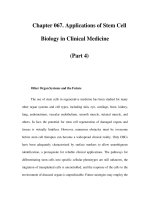
MRI characteristics
Imaging hallmarks of NMO are demonstrated in
Fig. 4.1. Typically, a longitudinal cervical lesion
(often affecting both the gray and white matter)
extending across three or more vertebrae can be seen.
Cord swelling and gadolinium enhancement occur
in more than half of the patients. Cervical lesions
may also extend into the medulla, the high thoracic
region or even the entire cord, and eventually show
signs of necrosis and cavitation. The diffuse enhance-
ment of optic nerves and chiasm in acute stages is
usually followed by atrophy in chronic stages of the
disease (Wingerchuck et al., 1999). While the presence
of cerebral lesions was previously considered incom-
patible with the diagnosis of NMO (Wingerchuck et al.,
1999), Pittock et al. (2005) found in a follow-up
study of 60 patients that 50% of them also had
positive brain MRI with nonspecific lesions in most,
and with features suggestive of MS in 10%. A small
subgroup (8%, mostly children) had atypical, conflu-
ent cerebral hemispheral, brainstem or diencephalic
(thalamic/hypothalamic) lesions, uncharacteristic
of MS (Pittock et al., 2005). The revised diagnostic
criteria reflect these observations (Wingerchuck
et al., 2006). Magnetization transfer and diffusion
tensor imaging techniques also revealed that micro-
scopic pathology may be present in the normal
appearing gray matter in the brain of patients with
NMO (Rocca et al., 2004).
Pathological characteristics
A modern and comprehensive pathological evalua-
tion of NMO was reported by Mandler et al. (1993)
using hematoxylin-eosin, Luxol fast blue-hematoxylin,
periodic acid-Schiff and Bodian’s silver staining. Five
of the studied eight patients died between one and
four years after the onset. The spinal cord was affected
throughout most of its length. Microscopically, cavit-
ation and necrosis were noted in both the gray and
white matter. Necrotic lesions were associated with
the presence of macrophages and prominent blood
vessels. Vessel walls were thickened and hyalinized
with scarce nuclei. The perivascular and parenchymal
infiltrate included macrophages but no lymphocytes
or plasma cells. The anterior optic pathway showed
signs of demyelination, gliosis, and cavitation (Mandler
et al., 1993). Using a panel of markers for immuno-
histochemistry, Lucchinetti et al. (2002) characterized
82 lesions from nine autopsy cases of clinically definite
NMO. All patients had extensive demyelination
(a) (b)
Fig. 4.1 MRI images of Dévic’s disease. The lesion
extends across the entire cervical and upper thoracic
cord with swelling of the cervical cord on the T2-weighted
scan (a). The axial T1-weighted postgadolinium image (b)
shows enhancement of the optic nerves.
NICP_C04 03/05/2007 10:35 AM Page 84
Dévic’s disease 85
associated with cavitation, necrosis and acute
axonal spheroids across multiple vertebral levels
in both the gray and white matter. The depletion of
oligodendrocytes was profound. In addition to macro-
phages, large numbers of neutrophils and eosinophils
were seen in acute parenchymal and meningeal
infiltrates, with a rare appearance of CD3 and CD8
positive T cells. Striking deposition of immunoglobu-
lins (mostly IgM) and complement C9 neoantigen was
associated with vascular fibrosis and hyalinization
in active and inactive lesions (Lucchinetti et al.,
2002). The involvement of humoral immunity in
the pathogenesis of NMO was further confirmed
by demonstrating the increased numbers of myelin-
oligodendrocyte glycoprotein (MOG)-specific B cells
that produced increased amounts of IL-5, IL-6, IgG,
and IgM in the CSF (Correale and Fiol, 2004). The
number of IgM-secreting B cells was much higher
than that of the IgG-producing cells. Chemokines
chemoattractant for eosinophils were also increased
in the CSF of patients with NMO.
Genetics
NMO presents as a sporadic disease and has been
observed in both Caucasians and non-Caucasians.
Nevertheless, its preferential occurrence in ethnic
groups (i.e. Asians, Africans, Canadian Aboriginals,
and French Afro-Caribbeans) with lower prevalence
rates of typical MS was recognized (Cabre et al.,
2001; Kuroiwa, 1985; Mirsattari et al., 2001; Misu
et al., 2002; Osuntokun, 1981). While the relative
risk for developing MS is 0.64 in African Americans
(AA) compared to that in Caucasian Americans (CA),
AAs are more likely to present with the opticospinal
form or with transverse myelitis and have a more
aggressive disease course than Caucasians (Cree et al.,
2004). The DRB1*1501 and DRB5*0101 alleles
associated with Western-type of MS are absent in
patients with NMO or Asian-type of the disease in
Japan (Kira et al., 1996). In contrast, these patients
have an increased frequency of the HLA-DPB1*1501
allele (Yamasaki et al., 1999). Whole-genome admix-
ture analyses using polymorphic markers in AA
individuals aimed to differentiate chromosomal
segments of African and European origin, and defined
that 79% of the composite ancestry is African and
21% is European in origin. The association of MS
with both the HLA-DRB1*1501 allele of Caucasian
origin and the DRB1*1503 allele of African origin
revealed that MS susceptibility was not simply related
to the Northern-European gene influx into the AA
population (Oksenberg et al., 2004). Genetic markers
associated with the opticospinal disease in AA indi-
viduals, however, remain to be identified. Full sequence
analyses of mitochondrial DNA in three Caucasian
NMO patients excluded the possibility that the necrotic
nature of pathology was related to inherited point
mutations or deletions in this extranuclear part of
the genome (Kalman and Mandler, 2002).
NMO-IgG, a biomarker for NMO
In accordance with histological data, recent serolo-
gical studies also add support to the involvement of a
humoral mechanism by identifying a new immuno-
globulin specific for NMO (Lennon et al., 2003). Sera
from patients with NMO, MS, and other autoimmune
disorders were used against mouse brain sections
in an indirect immunofluorescence assay. A distinct
IgG binding pattern associated with capillaries and
the blood–brain barrier in the cerebellar cortex,
midbrain, pia, and a subpial mesh (prominent in the
midbrain) was noted in 54% of patients with NMO.
This pattern of staining was also identified with
sera from eight patients among several thousands
screened in a blinded fashion at the Mayo Clinic.
Breaking the code revealed that all these eight
patients had definite or possible NMO. Follow-up
studies using this assay confirmed a sensitivity of 83%
and a specificity of 91% for NMO, and a sensitivity
of 58% and specificity of 100% for the Asian form of
opticospinal MS (Lennon et al., 2004). This study,
thus, established that the NMO-IgG is a biological
marker that distinguishes NMO from typical (or
western-type of ) MS, and confirmed the immunolo-
gical similarity between NMO and the Asian form of
opticospinal MS. The NMO-IgG is positive in about
40% of longitudinally extensive transverse myelitis
at the first event and predicts recurrence in 50%
of patients in one year (Weinshenker et al., 2006).
Lennon et al. (2005) recently identified the aquaporin-4
water channel as the antigenic target of NMO-IgG.
Aquaporin-4 is a component of the dystroglycan
protein complex located in the abluminal surface
of blood vessels and the foot processes of astrocytes
at the blood–brain barrier. Pittock et al. (2006a)
observed in a subgroup of NMO patients that the dis-
tribution of MRI abnormalities in the hypothalamic
and periventricular areas corresponds to cereb-
ral regions with high aquaporin-4 water channel
expression. These findings raise the possibility that
NMO may belong to a new class of autoimmune
channelopathies and is biologically distinct from
NICP_C04 03/05/2007 10:35 AM Page 85
86 BERNADETTE KALMAN
MS. Nevertheless, the direct pathogenic role of anti-
aquaporin-4 IgG remains to be experimentally
evaluated. Of note, this antibody frequently coexists
with other antineuronal and antimuscle autoanti-
bodies, and the occurrence of several autoimmune
diseases including myasthenia gravis (2.6%) has been
observed in Dévic’s disease (Pittock et al., 2006b).
Diagnostic criteria
The revised diagnostic criteria for definite NMO
(Wingerchuck et al., 2006) invariably require the
presence of optic neuritis and myelitis, but allow the
presence of clinical and MRI evidence for neurolo-
gical disease outside of the optic nerves and spinal
cord. In addition to optic neuritis and myelitis, the
best diagnostic combination (with 99% sensitivity
and 90% specificity for NMO) also consists of at
least two of the three following supportive criteria: a
continuous spinal cord lesion extending over three
or more vertebral segments on T2-weighted MRI;
onset brain MRI nondiagnostic for MS; or NMO-IgG
seropositivity.
Treatment of NMO
Corticosteroids remain the choice of treatment in
acute attacks, and corticosteroid dependence has been
noted in relapsing patients (Wingerchuck et al., 1999).
Plasma exchange is recommended for corticosteroid
unresponsive patients (Keegan et al., 2002). For
long-term treatment, azathioprine and cyclophos-
phamide have been used with some benefit (Mandler
et al., 1998; Wingerchuck et al., 1999). In a pro-
spective pilot trial, Mandler et al. (1998) treated
eight newly diagnosed NMO patients with prednisone
and azathioprine. The Expanded Disability Status
Scale (EDSS) score significantly improved in all patients
and no relapse occurred during the 18 months follow
up. The involvement of immunoglobulins and com-
plement in the pathogenesis of NMO suggests that
immunosuppression and plasma exchange should
be used for these patients rather than immuno-
modulation approved for MS patients. Targeting the
B-cell lineage may also modify the natural history
of the disease. Initial data from an open-label uncon-
trolled trial using rituximab, a chimeric murine/
human monoclonal antibody directed against the
CD20 antigen on precursor and mature B cells
(Biogen-Idec, Cambridge, MA and Genentech, South
San Francisco, CA), revealed promising results in
eight patients with worsening NMO (Cree et al.,
2005). Six of eight patients were relapse free and the
median relapse rate dropped from 2.6 to 0 attack/
patient/year. Seven patients experienced signific-
ant improvement during the one-year follow up.
The pretreatment EDSS of 8.5 improved to 5.5.
Rituximab was well tolerated and no major adverse
event occurred.
Summary
Based on clinical, imaging, laboratory, and his-
tological characteristics, NMO has always been
considered to be a distinct entity with a molecular
pathogenesis different from that of MS. This view
recently gained firm support by the identification
of a new biomarker, the NMO-IgG specific for the
aquaporin-4 water channel. The NMO-IgG has moder-
ate sensitivity and high specificity for NMO, and is
absent in typical MS. Clinical data suggest a distinct,
but not exclusive, optic nerve and spinal cord dis-
tribution of lesions. The pathology involves both the
gray and white matter and shows demyelination,
cavitation, necrosis, hyalinization of small vessels,
and a presence of macrophages, neutrophils and
eosinophils, along with the deposition of IgM and
IgG classes of immunoglobulins and activated com-
plement components in the proximity of blood–
brain barrier. MRI correlates of these histological
changes are T2-weighted lesions affecting more
than three segments of the spinal cord and optic
nerves with more frequent than previously appreci-
ated cerebral involvement. A moderate pleocytosis
typically without OCB or increased IgG synthesis
may be seen in the CSF. The identification of NMO-
IgG has significantly reduced diagnostic ambiguities
and raised the possibility that NMO may belong to a
new class of autoimmune channelopathies. Further
refinements of the diagnostic evaluations may be
necessary to capture the true spectrum of NMO
including inaugural symptoms and other limited
variants such as recurrent myelitis with negative
brain MRI, recurrent isolated optic neuritis, and
isolated longitudinal myelitis with or without sys-
temic autoimmunity (e.g. Sjögren’s syndrome or SLE)
associated with NMO-IgG seropositivity.
References
Albutt, T.C. 1880. On the ophthalmoscopic signs of
spinal disease. Lancet, 1, 86–8.
Cabre, P., Heinzlef, O., Merle, H. et al. 2001. MS and
neuromyelitis optica in Martinique (French West
Indies). Neurology, 56, 508–14.
NICP_C04 03/05/2007 10:35 AM Page 86
Dévic’s disease 87
Correale, J. and Fiol, M. 2004. Activation of humoral
immunity and eosinophils in neuromyelitis optica.
Neurology, 63, 2363–80.
Cree, B.A., Khan, O., Bourdette, D. et al. 2004. Clinical
characteristics of African Americans vs. Caucasian
Americans with multiple sclerosis. Neurology, 63,
2039–45.
Cree, B.A., Lamb, S., Morgan, K., Chen, A., Waubant, E.
and Genain, C. 2005. An open-label study of the
effects of rituximab in neuromyelitis optica. Neurology,
64, 1280–2.
Dévic, E. 1894. Myelite subaigue compliquee de nevrite
optique (Abstract). Bull Med (Paris), 8, 1033–4.
Gault, F. 1894. De la Neutomyelite Optique Aigue. Thesis,
Lyon, France.
Kalman, B. and Mandler, R.N. 2002. Studies on mito-
chondrial (mt)DNA in Dévic’s disease reveal no
pathogenic mutations but polymorphisms also found
in association with multiple sclerosis. Ann Neurol,
51, 661–2.
Keegan, M., Pineda, A.A., McClelland, R.L., Darby,
C.H., Rodriguez, M. and Weinshenker, B.G. 2002.
Plasma exchange for severe attacks of CNS demy-
elination: predictors of response. Neurology, 58,
143–6.
Kira, J., Kanai, T., Nishimura, Y. et al. 1996. Western
versus Asian types of multiple sclerosis: immuno-
genetically and clinically distinct disorders. Ann
Neurol, 40, 569–84.
Kuroiwa, Y. 1985. Neuromyelitis optica (Dévic’s
disease, Dévic’s syndrome). In J.C. Koetsier (ed.),
Handbook of Clinical Neurology, Chapter 13. Elsevier,
Amsterdam.
Lennon, V.A., Kryzer, T.J., Pittock, S.J., Verkman, A.S.
and Hinson, S.R. 2005. IgG marker of optic-spinal
multiple sclerosis binds to the aquaporin-4 water
channel. J Exp Med, 202, 483–8.
Lennon, V.A., Lucchinetti, C.F. and Weinshenker, B.G.
2003. Identification of a marker autoantibody of
neuromyelitis optica. Neurology (Suppl 1), A519.
Lennon, V.A., Wingerchuk, D.M., Kryzer, T.J. et al.
2004. A serum autoantibody marker of neuro-
myelitis optica: Distinction from multiple sclerosis.
Lancet, 364, 2106–12.
Lucchinetti, C.F., Mandler, R.N., McGavern, D. et al.
2002. A role for humoral mechanisms in the patho-
genesis of Dévic’s neuromyelitis optica. Brain, 125,
1450–61.
Mandler, R.N., Davis, L.E., Douglas, R.J. and Kornfeld,
M. 1993. Dévic’s neuromyelitis optica: A clinico-
pathological study of 8 patients. Ann Neurol, 34,
162–8.
Mandler, R.N., Ahmed, W. and Dencoff, J.E. 1998.
Dévic’s neuromyelitis optica: A prospective study
of seven patients treated with prednisone and
azathioprine. Neurology, 51, 1219–20.
Mirsattari, S.M., Johnston, J.B., McKenna, R. et al.
2001. Aboriginals with multiple sclerosis: HLA
types and predominance of neuromyelitis optica.
Neurology, 56(3), 318–23.
Misu, T., Fujihara, K., Nakashima, I. et al. 2002. Pure
optic-spinal form of multiple sclerosis in Japan. Brain,
125, 2460–8.
Oksenberg, J.R., Barcellos, L.F., Cree, B.A. et al. 2004.
Mapping multiple sclerosis susceptibility to the
HLA-DR locus in African Americans. Am J Hum
Genet, 74, 160–7.
Osuntokun, B.O. 1981. The pattern of neurological
illness in tropical Africa: Experience at Ibadan,
Nigeria. J Neurol Sci, 12, 418–42.
Pittock, S.J., Weinshenker, B.G., Lucchinetti, C.F.,
Wingerchuck, D.M., Corboy, J.R. and Lennon, V.A.
2006a. Neuromyelitis optica brain lesions localized
at sites of high aquaporin 4 expression. Arch Neurol,
63, 964–8.
Pittock, S.J., Weinshenker, B.G., Wingerchuk, D.M.,
Lucchinetti, C.F. and Lennon, V.A. 2006b. Auto-
immune neurological accompaniments of neuro-
myelitis optica (NMO). Ann Neurol, 60(Suppl 10),
M-24.
Pittock, S.J., Wingerchuck, D.M., Krecke, K., Lennon,
V.A., Lucchinetti, C.F. and Weinshenker, B.G. 2005.
Brain abnormalities in patients with neuromyelitis
optica (NMO). Neurology, 64, A39.
Rocca, M.A., Agosta, F., Mezzapesa, D.M. et al. 2004.
Magnetization transfer and diffusion tensor MRI
show gray matter damage in neuromyelitis optica.
Neurology, 62, 486–8.
Weinshenker, B.G., Wingerchuck, D.M., Vukusic, S.,
Pittock, S.J. and Lennon, V.A. 2006. Neuromyelitis
optica IgG predicts relapse after longitudinally extens-
ive transverse myelitis. Ann Neurol, 59, 566–9.
Wingerchuck, D.M., Hogancamp, W.F., O’Brien, P.C.
and Weinshenker, B.G. 1999. The clinical course of
neuromyelitis optica (Dévic’s syndrome). Neurology,
53, 1108–14.
Wingerchuck, D.M., Lennon, V.A., Pottock, S.J.,
Lucchinetti, C.F. and Weinshenker, B.G. 2006.
Revised diagnostic criteria for neuromyelitis optica.
Neurology, 66, 1485–9.
Wingerchuck, D.M., Pittock, S.J., Lennon, V.A.,
Lucchinetti, C.F. and Weinshenker, B.G. 2005.
Neuromyelitis optica diagnostic criteria revisited:
Validation and incorporation of NMO-IgG serum
antibody. Neurology, 64, A38.
Wingerchuk, D.M. and Weinshenker, B.G. 2003.
Neuromyelitis optica: Clinical predictors of a relaps-
ing course and survival. Neurology, 60, 848–53.
Yamasaki, K., Horiuchi, I., Minohara, M. et al. 1999.
HLA-DPB1*0501-associated opticospinal multiple
sclerosis: Clinical, neuroimaging and immuno-
genetic studies. Brain, 122, 1689–96.
NICP_C04 03/05/2007 10:35 AM Page 87
Acute disseminated encephalomyelitis (ADEM) and
multiple sclerosis (MS) are nonvasculitic inflammat-
ory diseases of the central nervous system (CNS) that
bear striking clinical, pathological, and pathophysi-
ological resemblance not only to one another, but
also to the research model, experimental allergic
encephalomyelitis (EAE). Some of the pathophysio-
logical features that these three entities share are
listed in Box 5.1. Detailed discussion of the consider-
able amount of information that is known about
pathogenesis of ADEM and MS falls outside of the scope
of this clinical review; many current sources for that
information are available (Hohlfeld and Wekerle,
2001; Johnson, 1998; Lucchinetti et al., 2001; Poser,
2000; Pouly and Antel, 1999; Pouly et al., 2000;
Rust, 2000; Rust and Fleming, 1996; Tellis, 1998).
Despite a large and increasing body of literature, it is
not yet clear why ADEM is an inflammatory illness
with a generally favorable outcome that usually
remains monophasic, while MS is an inflammatory
illness that follows a chronic degenerative course
(Poser, 2000; Pouly and Antel, 1999; Pouly et al.,
2000).
ADEM must be considered in relationship not
only to MS, but also to other designated clinical
entities that appear to constitute a spectrum between
ADEM and MS. As is true of ADEM and MS, most
of these have no specific diagnostic test, although
recent development of a specific test for neuromyelitis
optica (NMO) suggests that in the future the current
classification will be replaced by a less equivocal
system incorporating specific tests and greater under-
standing of the pathophysiological differences of the
various entities. It is of particular importance to keep
in mind that the collections of illnesses currently
labeled “ADEM” or “childhood MS” likely contain
some subtypes for which distinctive names will in
time be selected as biomarkers for these particular
subtypes are identified. This likelihood is supported
by the fact that many examples of one clinical sub-
type that has formerly been included under either
of these collective headings, neuromyelitis optica,
are now discretely identified by the diagnostic bio-
logical marker, NMO-IgG. This is true for the clinical
entities such as those shown in Box 5.2 that are
currently without discrete biomarkers. These are
5
Acute disseminated encephalomyelitis and
related conditions
Robert S. Rust
1. Earliest stages of inflammation mediated
by stimulated clones of T-helper cells
sensitized to autoantigens such as
myelin proteins (Oleszak et al., 2001).
2. Ensuing complex inflammatory cascade
entails the local action of lymphokines
as well as lymphokine-induced
chemotaxis of other cellular mediators
of inflammation (other T-cell lines,
B cells, microglia, phagocytes).
3. Tumor necrosis factor (TNF)-α, soluble
TNF receptor 1, and interleukins (IL)
10 and 6, each of which may be elevated
in cerebrospinal fluid (CSF), likely involved
in pathogenesis.
4. IL-6 and TNF-α are likely proinflammatory
and TNF-α may play a particular role in
demyelination (Ichiyama et al., 2002).
5. Disturbance of blood–brain barrier
function is likely to be very important.
6. Lymphocytic perivenular inflammation
is prominent.
7. Patchy perivenular demyelination occurs
with relative preservation of axons.
8. Microglial cells are characteristic elements
of the inflammatory exudate.
Box 5.1 Similarities between ADEM and MS.
NICP_C05 04/05/2007 12:26PM Page 88
Acute disseminated encephalomyelitis 89
• Acute hemorrhagic leukoencephalopathy
(AHLE)
• Acute multiple sclerosis
• Acute necrotic encephalomyelitis
• ADEM without prodromal phase
• Bickerstaff’s encephalitis
• Childhood limbic encephalomyelitis
• Concentric sclerosis, Balò type
• Dévic syndrome (neuromyelitis optica)
• Encephalomyeloradiculoneuriopathy
• Miller–Fisher syndrome
• Optic neuritis
• “Recurrent” or “multiphasic” ADEM
• Recurrent herpes encephalitis
• Schilder disease
• Steroid-dependent hyper-recurrent
disseminated encephalomyelitis
• Transverse myelitis
Box 5.2 Syndromes – some examples of which may be
or are ADEM or MS variants.
clinical syndromes that in some instances occur as
types of ADEM while in others as types of MS.
ADEM usually arises in close association with
some exogenous stimulus, such as a febrile illness
suggestive of viral infection, sometimes rendering
the distinction of ADEM from “viral meningoence-
phalitis” troublesome. There are many other condi-
tions that sometimes produce a clinical appearance
so suggestive of ADEM as to be mistaken for it. These
conditions constitute the differential diagnosis for
ADEM, listed in Box 5.3 (Rust, 2000; Hartung and
Grossman, 2001). Many of these conditions are also
in the differential diagnosis of MS and will be termed
in this chapter “other neurological diseases,” or ONDs.
It is possible that some of the conditions in Box 5.3
may share certain pathophysiological mechanisms
with ADEM or MS, accounting for similarities not
only in clinical manifestations but also in imaging
appearance and in various laboratory results includ-
ing cerebrospinal fluid (CSF) immune profile testing.
These CSF tests, shown in Box 5.4, are of consider-
able importance in the evaluation of patients sus-
pected of having MS, since approximately 95% of all
individuals who have MS can be expected to show
abnormalities of most or all of these tests upon their
second clinical bout and thereafter. Although CSF
myelin basic protein (MBP) is not, strictly speaking,
an immune profile test, it is included because it is
a very useful indicator of inflammatory injury to
white matter. ADEM, which produces abnormalities
of the other tests in only a minority of cases, is usually
associated with an abnormal MBP value. Abnormal-
ities of the CSF immune profile may be seen in some
of the conditions listed in Box 5.2 as well as in some
ONDs – as is shown in Box 5.5 (Trotter and Rust,
1989).
As will be seen, similar caution must be exerted
in the interpretation of imaging findings obtained in
children or adolescents who may have ADEM or its
possible variants, MS, or various ONDs. The fact that
there are indeterminate boundaries between these
various conditions, which give rise to uncertainties
concerning the results of immune profile testing or
brain imaging, should not be surprising given the
probability that there are areas of mechanistic overlap
between these conditions. This is to be expected, con-
sidering the overlap in pathophysiological mechan-
isms. Additional specific tests are available for refine-
ment of diagnosis of conditions that resemble ADEM
or MS, with the particularly important recent addition
of the Wingerchuk assay for neuromyelitis optica.
Acute disseminated encephalomyelitis
(ADEM)
What might be termed “typical ADEM” is an acute
monophasic, multifocal CNS disturbance arising
in the wake of an exogenous stimulus (e.g. febrile
illness or vaccination). It is chiefly encountered in
prepubertal children, although it may occur in adults.
Strictly speaking, it is a pathologically defined entity,
consisting of inflammatory perivenular demyelina-
tion with relative sparing of axons. Nonetheless, as
is the case with MS, it is a disease that affects both
white matter and gray matter. The gray matter
manifestations of ADEM tend to be much more
prominent during an acute bout of ADEM than one
of MS. However, these ADEM manifestations tend
to resolve with typical ADEM, while MS is, as has
recently become clear, a progressive gray matter
degeneration. The MRI manifestations of typical
ADEM tend to be characteristic and quite supportive
NICP_C05 04/05/2007 12:26PM Page 89
90 ROBERT S. RUST
• CSF IgG synthetic rate
(Tourtellotte, 1970)
• CSF IgG/Albumin ratio
• CSF oligoclonal bands
• CSF: serum IgG index
• Myelin basic protein
Box 5.4 CSF immune profile tests.
• Acute cerebellar ataxia
• Aicardi–Goutieres syndrome
• AIDS
• Atlanto-occipital instability
• Behçet syndrome
• Childhood ataxia with cerebral
hypomyelination
• Chronic fatigue syndrome
• Cysticercosis
• Echinococcosis
• Embolic/thrombotic vascular disease
• Encephalitis
• Glioblastoma multiforme/other
glial tumors
• Hashimoto encephalopathy
• Heritable leukodystrophies
• Hereditary spastic paraplegia
• Hypersensitivity vasculitides
• Leber’s hereditary optic atrophy
• Leukemia/lymphoma, other
cerebral tumors
• Lyme encephalomyelitis/neuroborreliosis
• Marchiafava–Bignami disease
• Medium-chain acyl dehydrogenase
deficiency
• Mesencephalic
leukoencephalopathy/SC cysts
• Meningitis
• Migraine
• Monoclonal gammopathy
• Moyamoya
• Neurobrucellosis
• Neuroaxonal dystrophy
• Opsoclonus-myoclonus
• Paraneoplastic syndromes
• Pelizaeus–Merzbacher disease
• Primary CNS vasculitis
• Progressive multifocal
leukoencephalopathy
• Progressive rubella panencephalitis
• Rabies
• Sarcoidosis
• Schilder’s myelinoclastic diffuse sclerosis
• Schindler disease
• Sinovenous thrombosis
• Sjögren syndrome
• Spinal stenosis
• Spinocerebellar degenerations
• Subacute sclerosing panencephalitis
• Sydenham chorea/PANDAS
• Syphilis
• Syringomyelia
• Systemic lupus erythematosus
•
Chiari malformation/tethered cord
• Toluene leukoencephalopathy
(“glue sniffing”)
• Toxic subacute myelopticoneuropathy
• Toxoplasmosis
• Tropical spastic paraparesis
(HTLV-1-associated myelopathy
TSP/HAM))
• Vascular malformations – brainstem or
spinal cord
• Whipple disease
Box 5.3 Other ADEM/MS differential considerations.
of diagnosis, as are those of typical MS, from which
they differ. Although pathological confirmation is
seldom obtained, “typical, monophasic ADEM” is not
so difficult to recognize and will be the chief subject of
this review. Should an ADEM-like illness recur, as in
some of the conditions listed in Box 5.2 (excepting
NMO-IgG positive Dévic syndrome), it is currently
unsettled as to whether to classify such an illness
as ADEM, MS, or something else (Cole et al., 1995).
The discussion that follows is based not only on
cited references, but also on more than 300 cases of
various forms of the conditions considered in this
review that we have studied including approx-
imately 150 cases of “typical” ADEM.
NICP_C05 04/05/2007 12:26PM Page 90
Acute disseminated encephalomyelitis 91
Clinical aspects
Data from our patient series and another large recent
series (Tenembaum et al., 2002) show that although
ADEM may develop at any point from the first year
of life to late middle age, most cases occur in children
3–10 years of age (mean age approximately 6 ± 4
years). Contrary to what is seen in either childhood-
or adult-onset MS and possibly adult-onset ADEM
(Hollinger et al., 2002) boys are at higher risk than
girls for childhood-onset ADEM (boy:girl ratio 1.3–
1.8:1). It is not clear whether the white:black ratio
of approximately 6:1 in our series merely reflects
referral bias (Rust et al., 1989a).
A wide variety of possible exogenous provocations
have been identified, many of which are listed in
Box 5.6 (Campistol et al., 2001; Miller et al., 1956;
Rust, 2000; Shintani et al., 2001; Tselis, 2001). In
• Acute bacterial, viral, parasitic, tuberculous,
fungal meningitis/encephalitis
• Acute or chronic inflammatory
demyelinating polyneuropathy
• ADEM
• Chronic rubella panencephalitis
• CNS tumor
• Lyme disease
• MS
• Neurosyphilis
• Sarcoidosis
• SSPE
• Stroke
• Systemic lupus erythematosus
Box 5.5 Diseases that may provoke abnormalities
of the CSF immune profile.
• Antihelminthic medication
treatments
• Bartonella henselae
• BCG vaccine
• Campylobacter
• Chlamydia pneumoniae
• Clostridium tetani and tetanus
vaccine
• Corynebacterium diphtheria
• Coxsackie B virus
• Cryptococcus neoformans
(Jaing et al., 2001)
• Cytomegalovirus
• Echoviruses
• Enteroviruses
• Epstein–Barr virus
• Hepatitis A or B
• Human herpes virus 1 or 6
• Influenza A or B viruses/vaccines
• Japanese B virus/vaccine
(Plesner et al., 1998)
• Legionella (Sommer et al., 2000)
• Lyme disease
• Malaria
• Measles virus/vaccine
• Mumps virus/vaccine
• Mycoplasma pneumoniae
(Hasegawa, 2001)
• Organ/bone marrow transplant
(Au et al., 2002)
• P. malusia (J Am Phys Ind 49:756, 2001)
• Parainfluenza virus (Au et al., 2002)
Periodic acid-Schiff treatment
• Pertussis
• Polio virus/Salk or Sabin vaccines
(Arya, 2001)
• Puumala virus (Toivanen et al., 2002)
• Rabies virus/Pasteur rabies vaccine
• Rickettsia rickettsiae
(Rocky Mountain Spotted Fever)
• Rubella virus and vaccine
• Rubeola virus and vaccine
• Serum administration
• Streptococci, Group A beta-hemolytic
(Ito, 2002)
• Streptococci, pyogenes
• Sulfonamide treatment
• Typhoid and paratyphoid
vaccines
• Vaccinia virus/vaccine
• Varicella virus
• Variola virus
Box 5.6 Infections and other exogenous stimuli associated with ADEM.
NICP_C05 04/05/2007 12:26PM Page 91
92 ROBERT S. RUST
our series slightly more than half of all cases occur
in the wake of respiratory infections, 15–20% follow
gastroenteritis, while the remainder are due to
nonspecific febrile illnesses, vaccines, or no clearly
identifiable provocation. It is presumed that most
ADEM cases are provoked by viruses, although specific
viruses are seldom identified. Increased prevalence
of ADEM in winter months (Dale et al., 2000; Rust
et al., 1989; Tenembaum et al., 2002) may be due
to increased prevalence during that time of year
of particularly provocative pathogens, particularly
large envelope-bearing viruses.
Fortunately most of the various childhood illnesses
due to viruses, which figured prominently in older
series of ADEM, are currently prevented by immun-
ization in large parts of the industrialized world. Some
of these viruses (e.g. measles, mumps, rubeola, rubella,
varicella, variola) were capable of provoking severe
and sometimes fatal encephalomyelitis. However,
it must be remembered that these same potentially
lethal agents continue to cause widespread and
severe childhood illnesses including ADEM in less
medically favored regions of the world. Antibiotic
therapy or changes in immunogenicity may account
for reduced prevalence of streptococcal infection as
the cause of illnesses prodromal to the development
of ADEM, at least in many industrialized nations.
Agents for which serological or other evidence can
most often be found in contemporary series include
Epstein–Barr virus, Bartonella, and Mycoplasma.
The statistical prominence of the last of these is due
in part to the unsatisfactory specificity of available
serological tests.
“Recurrent” herpes encephalitis is at least in some
instances a severe form of ADEM from which recovery
is slow but may be complete. Particularly severe and
permanent manifestations of an ADEM-like illness
continue to arise in the wake of Lyme disease and
Brucellosis. It is possible that the hyperergic and severe
illnesses such as cerebral malaria and Dengue fever,
which account for millions of cases yearly around the
world, share elements of ADEM pathogenesis. Various
vaccines have been suggested, sometimes quite con-
troversially, as the exogenous provocations of cases
of ADEM, most clearly older versions of the Pasteur
rabies vaccine. Currently immunizations account for
no more than 3–6% of ADEM cases (Dale et al., 2000;
Hynson et al., 2001; Rust et al., 1989; Tenembaum
et al., 2002). As many as 14–26% of all ADEM cases
are “cryptogenic” with no definite antecedent cause.
Neurological dysfunction of ADEM usually arises
1–30 days after exogenous stimulation, typically with
a fever-free interval of hours to weeks separating the
two phases of illness (Croft, 1969; de Vries, 1960;
Miller et al., 1957; Scott, 1967; Tenembaum et al.,
2002). The outside limit of 30 days is an artificial
one as it remains unclear whether longer latencies
between febrile prodrome and ADEM occur, although
experience with herpes and other infections of vaccines
suggests that it is possible (Sacconi et al., 2001). How-
ever, the more remote the presumed provocation the
more difficult to prove cause and effect (Rust et al.,
1989; Tenembaum et al., 2002). Very short intervals,
on the other hand, may obscure the distinction
between possible exogenous stimulus and inflamma-
tory results, which may in turn prompt diagnosis of
infectious meningoencephalitis rather than ADEM.
Interestingly, however, a hiatus of at least a few
hours is usually found between a febrile prodrome
and onset of ADEM, even in cases where the febrile
prodromal illness was of many weeks’ duration.
It remains to be determined what percentage if
any of cases of presumed meningoencephalitis
without identifiable pathogen are actually cases of
ADEM. The question is important since as many as
70% of all cases of clinically diagnosed meningo-
encephalitis in North America occur without an
identifiable pathogen. Similar diagnostic problems
arise in cases of presumed postinfectious vasculitis,
cases which must themselves be distinguished from
ADEM. Approximately 15% of cases in our series
thought to represent ADEM have no clearly defined
prodrome.
Irritability, lethargy, fever recurrence, vomiting,
and various other neurological manifestations mark
the onset of most cases of ADEM. These features and
other features listed in Table 5.1 readily distinguish
most first bouts of ADEM from MS. The approximate
prevalence of various specific clinical findings of ADEM
is shown in Table 5.2. In bouts of either ADEM or
childhood MS, neurological manifestations may evolve
very dramatically over intervals as short as minutes.
However, slower evolution is more common, usually
involving 2–6 days, although in some less common
instances abnormalities unfold over intervals as long
as 6–8 weeks. The evolution of abnormalities tends
to proceed longer in ADEM than in a discrete bout of
childhood-onset MS. Improvement tends to occur
only after the abnormalities have fully developed.
ADEM seldom if ever manifests the fluctuation in
degree of certain dysfunctions that is characteristic
of MS, such as Uthoff ’s phenomenon (transient
heat-induced worsening of vision, other sensory or
motor abnormalities). On the other hand, findings
NICP_C05 04/05/2007 12:26PM Page 92
Acute disseminated encephalomyelitis 93
of acute encephalopathy (e.g. confusion, agitation,
obtundation, coma, aphasia, personality change,
hallucinations, psychosis, seizures) are much more
common in childhood ADEM than in childhood or
adult MS or even “adult-onset” ADEM (many cases
of which evolve to earn an MS diagnosis). Seizures
occur in 30–40% of cases of childhood ADEM, usually
with localization-related manifestations. Occasionally,
focal or secondary generalized status epilepticus
occurs (Dale et al., 2000; Hung et al., 2001; Hynson
et al., 2001; Rust et al., 1989; Schwarz et al., 2001;
Tenembaum et al., 2002).
Many of the distinctive sensory disturbances that
are common in MS (e.g. hemisensory or isolated
posterior column deficits, band or girdle dysesthesiae,
Lhermitte sign) are seldom detected in childhood
ADEM. However, visual loss due to optic neuritis
(ON) occurs in approximately one-quarter of ADEM
cases. When it occurs, it is bilateral in 70–80%,
although the involvement of the second eye may
lag behind the first by weeks to months. This con-
trasts with either childhood or adult MS, where ON is
typically unilateral. Extrapyramidal findings, chorea
more commonly than dystonia, are seen in 10–15%
of ADEM cases. ADEM occurring in the wake of
HSV encephalitis may produce especially malignant
extrapyramidal syndromes, in some instances
associated with the development of Parkinsonian
manifestations that evolve into chorea and may
be associated with a prominent Kluever-Bucy
syndrome (Tulyapronchote and Rust, 1992). ADEM-
related ataxia tends to be appendicular, setting it
apart from the uniformly gait/trunk ataxia charac-
teristic of acute cerebella ataxia (ACA). The ataxia of
ADEM is not associated with opsoclonus (Dale and
Beachey, 1984; Hynson et al., 2001; Rust et al.,
1989; Schwarz et al., 2001). Most cases of ADEM
can be placed into one of seven reasonably distinct
clinical syndromes based upon the predominant
signs, as shown in Table 5.3.
Table 5.1 Prevalence of various features, first bout childhood or adolescent MS or ADEM.
Prevalence of features in first disease bout – Childhood
Disease
Features MS ADEM
Age >11 <11
Febrile illness/vaccination precedes No Yes
Seizure/encephalopathy Rare Common
Optic neuritis Often unilateral Often bilateral
Nonvisual sensory symptoms Common Rare*
Posterior column signs Common Rare*
Abnormal CSF immune profile Common Rare
Abnormal EEG Rare Common
Transient sensory paroxysms Common Rare
*Unless transverse myelitis.
Table 5.2 Clinical findings in bouts of childhood
ADEM and MS.
ADEM MS
Fever 50–80% 10%
Irritability 65–81% 13%
Vomiting 40–50% 13%
Headache 45–65% 16%
Meningismus 20–55% 13%
Encephalopathy 50–75% 4%
Language disturbances 10–20%
Seizures 20–30% 7%
Psychiatric disturbances 30–50% 20%
Weakness 50–75%
Long tract signs 45–55%
Cranial nerve palsies 35–50%
Optic neuritis 10–30%
Nonvisual sensory 15–24% 60–70%
Ataxia 35–50%
Extrapyramidal syndromes 10–15%
MS findings from Boiko et al., 2002.
NICP_C05 04/05/2007 12:26PM Page 93
94 ROBERT S. RUST
Laboratory tests
Modest elevations of white blood cell (WBC) counts
may be found in blood, especially in the wake of
convulsive seizures. The blood sedimentation rate is
sometimes elevated. Modest to moderate elevation of
CSF white blood cell counts may be found in child-
hood ADEM, usually less than 200 WBC/mm
3
. Red
blood cell counts may be slightly to greatly elevated
in CSF. In some instances this is likely to be due to
the severe hyperacute form of ADEM termed acute
hemorrhagic leukoencephalopathy of Hurst.
Only rarely do cultures of CSF obtained from indi-
viduals with ADEM yield pathogens. The significance
of such positive cultures remains uncertain as does
the issue of whether such cases should all be desig-
nated meningitis or meningoencephalitis. Elevated
CSF titers are occasionally found for HSV, Borrelia,
measles, Mycoplasma, HHV6, or other agents. In
such instances, serum sampling for acute and con-
valescent titers should be performed to determine
whether a four-fold rise or fall occurs. In instances
where such a titer rise or fall is observed, the altern-
ative diagnosis of meningitis or meningoencephalitis
may be entertained. Serological testing for evidence
of pertinent ONDs (Box 5.3) should be negative.
CSF immune profile testing (Box 5.4), employing
age-appropriate normative data (Rust et al., 1988),
is positive in less than 20% of prepubertal children
with ADEM (Rust et al., 1989; Tenembaum et al.,
2002). Oligoclonal bands (CSF bands present in
equivalent or greater concentration than in simultan-
eously obtained serum) are seldom positive in ADEM
unless ADEM occurs in the wake of Lyme disease
or measles encephalitis. As noted, the CSF immune
profile is positive after the second clinically apparent
bout of illness in more than 95% of childhood or
adult cases of MS. As has been stated, CSF immune
profile tests, most of which are usually negative in
ADEM, may also be positive in a number of ONDs
that may resemble ADEM clinically or radiographic-
ally including leukodystrophies, tumors, and infect-
ions. CSF myelin basic protein concentration, usually
positive in ADEM, may also be elevated in ONDs,
including not only infections, but also tumors. Rates
of positivity of these tests as compared to clinically
definite MS (CDMS), probable or possible MS (P/PMS),
non-MS inflammatory illnesses (NMSI/I) or ONDs
are shown in Table 5.4.
The EEG is abnormal at the onset of as many as
90% of ADEM cases, a finding that sets childhood
ADEM apart from most or possibly all childhood-
onset cases of MS. The prevalence and severity of EEG
abnormalities depends to some extent on the degree
of clinical encephalopathy, although an abnormal
EEG may be found even in patients who have no such
signs. EEG slowing (generalized >> focal) is found
in 75–80% of ADEM cases. Abnormalities of sleep
architecture may be found. Sharp waves, rhythmic
Table 5.3 Clinical subtypes of ADEM.
ADEM Subtype
Mild encephalopathy, disseminated CNS
dysfunction
Severe encephalopathy, quadriparesis or
double hemiparesis
Brainstem*
Hemiparesis
Myelitis/Dévic syndrome
Mixed upper motor neurone (UMN) and
lower motor neurone (LMN) signs**
Ataxic***
*Some classifiable as Bickerstaff encephalitis.
**Includes encephalomyeloradiculoneuropathy, some
Miller-Fisher syndrome.
***Excludes acute cerebellar ataxia.
Table 5.4 Approximate rates of positivity, various CSF immune profile tests.
Diseases
CSF immune profile tests CDMS P/PMS ADEM NMSI/I OND
CSF IgG/Albumin ratio 60–80% 21–24% 16–20% 14–42% 6–10%
CSF/Serum IgG index 88–94% 44–55% 15–20% 43–57% 3–18%
IgG synthetic rate 88–96% 27–67% 16–18% 29% 4–12%
Oligoclonal bands 83–100% 24–89% 10–12% 27–72% 2–29%
NICP_C05 04/05/2007 12:26PM Page 94
Acute disseminated encephalomyelitis 95
delta, or spikes may be found in the waking state
during the early stages of as many as 2–7% of ADEM
cases, features that also distinguish ADEM from
childhood-onset MS. The absence of such abnor-
malities during the first bout of acute disseminated
demyelinating illness in a child significantly increases
the risk for ultimate MS diagnosis (Rust et al., 1989).
EEG abnormalities are not found in adult ADEM
(Hollinger et al., 2002).
Imaging studies
Focal low-density abnormalities are found in the
brain computed tomography (CT) scans of 60–80%
of cases of ADEM. These abnormal areas always
correspond to much larger areas of abnormality
detectable on MRI scans. Due to the fact that the CT
technique is far less sensitive than MRI for the detec-
tion of the lesions associated with ADEM, MRI usu-
ally identifies numerous additional smaller areas of
abnormality. MRI scans detect abnormalities of vari-
ous size on T2, proton density, and fluid attenuation
inversion recovery weighted sequences in more than
90% of ADEM cases. CT scans are superior to MRI
scans only in the detection of hemorrhagic changes
that define acute hemorrhagic leukoencephalopathy.
Unlike MS plaques, the characteristic lesions of
ADEM are centrifugal. Nearly 90% of individuals with
ADEM are found to harbor lesions at the junction of
deep cortical gray and subadjacent white matter.
The indistinct and irregular margins of childhood
ADEM lesions tend to suggest a “smudge” rather
than the clearly demarcated plaque margin charac-
teristic of MS. Lesions similar to those characteristic
of childhood ADEM are found in less than 40% of
cases that are labeled as “adult” ADEM – a diagnosis
that often changes to MS as time goes on. In childhood
or adolescent ADEM, additional unilateral or bilateral
lesions may be found in deeper white matter, optic
nerves, basal ganglia (30–40%), thalamus (12– 40%),
brainstem (45–55%), cerebellum (30– 40%), and
spinal cord. Periventricular lesions (30–45%) and
corpus callosum lesions (10–15%) are much less
common in childhood ADEM than in MS where they
are so characteristic. Various unusual lesions identified
by MRI imaging in the nervous system of individuals
with ADEM are listed in Box 5.7. Diseases that pro-
duce changes on MRI that have been misdiagnosed
as ADEM are shown in Box 5.8.
In approximately two-thirds of ADEM cases lesions
are small (<5 mm) and asymmetrically distributed
in white matter admixed with a few larger lesions.
Periventricular and callosal lesions if found are usu-
ally considerably outnumbered by subcortical lesions
and the characteristic lesions spanning cortical gray
and subcortical white matter. Few if any lesions are
• Solitary lesions in deep white matter
or deep gray nuclei
• Massive symmetrical frontal white
matter “butterfly” lesions
• Symmetrical linear, posteriorly
emphasized lesions resembling
leukodystrophy
• Large, asymmetrically located lesions
with central pseudo-cavitation
• Lesions containing hemorrhage,
suggesting HSV2 encephalitis
• Ring-enhancing lesions
• Lesions with mass effect resembling
tumor or cysticercosis
Box 5.7 Atypical T2 bright imaging changes in ADEM or presumed ADEM
(Baum et al., 1994; Caldemeyer et al., 1994; van der Meyden, 1994).
• Encephalitis
• Glioblastoma multiforme
• CNS lymphoma
• MELAS
• Cysticercosis
• Multiple sclerosis
• Sarcoidosis
• Vasculitis
• CNS histiocytic lymphangiomatosis
• Brain abscess
Box 5.8 Diseases mistaken for ADEM on the basis of
T2 changes on imaging studies.
NICP_C05 04/05/2007 12:26PM Page 95
96 ROBERT S. RUST
oriented in the “Dawson’s finger” fashion of MS
plaques, that is the long axis of typical ovoid plaques
is perpendicular to the lateral ventricular wall. Quite
large confluent lesions are found in 15–25% of
cases, lesions that may be mistaken for tumor or
leukodystrophy. Uncommonly very large unilateral
or asymmetrical bilateral lesions are found with
necrotic-appearing cores. Often, this core is in fact
pseudonecrotic, resolving with resolution of the bout
of ADEM. In about 10% of cases, white matter lesions
are accompanied by bithalamic lesions in a pattern
that is sometimes suggestive of sinovenous throm-
bosis (Apak et al., 1999; Atlas et al., 1986; Baum
et al., 1994; Caldemeyer et al., 1994; Cusmai et al.,
1994; Dale et al., 2000; Hasegawa, 2001; Hesselink
et al., 1986; Hung et al., 2000; Hynson et al., 2001;
Kesselring et al., 1990; Marcu et al., 1979; Suwa
et al., 1999; Tenembaum et al., 2002).
As many as 90% of childhood ADEM lesions
enhance with gadolinium. The degree of contrast
enhancement of ADEM lesions is typically uniform
and usually not very dense. If enhancement occurs,
most lesions tend to enhance. Rarely, some lesions
will show enhancement while others do not. In con-
trast, MS plaques tend to vary in degree of contrast
enhancement and may at times enhance quite
densely. As many as 30% of ADEM lesions may show
ring enhancement (Apak et al., 1999; Atlas et al.,
1986; Baum et al., 1994; Caldemeyer et al., 1994;
Cusmai et al., 1994; Dale et al., 2000; Hasegawa,
2001; Hesselink et al., 1986; Hung et al., 2000;
Hynson et al., 2001; Kesselring et al., 1990; Marcu
et al., 1979; Suwa et al., 1999; Tenembaum et al.,
2002).
Some patients with ADEM have normal MRI
scans on initial presentation but display typical
abnormalities of ADEM if the scan is repeated several
weeks later, despite interim clinical improvement
(Honkaniemi et al., 2001). Thus, a normal MRI scan
does not exclude the ADEM diagnosis. Moreover,
it must be presumed that the appearance of new
lesions during recuperation from ADEM may not
represent recrudescence of disease (Hollinger et al.,
2002; Jacobson et al., 1990; Kesselring et al., 1990;
Ormerod et al., 1984). Magnetization-transfer MRI,
single photon emission CT, or NMR spectroscopy
will prove helpful in distinguishing ADEM from
alternative diagnoses (Hung et al., 2000; Takahashi
et al., 1992). None of these approaches is likely to
render a pathognomonic finding of ADEM or MS and
diagnosis of ADEM should always rest on clinical
grounds (Hollinger et al., 2002).
Treatment
Treatment of ADEM involves providing whatever
support is indicated based upon the clinical circum-
stances. In as many as 30–45% of cases considerable
intensive care is provided, including mechanical
ventilation in as many as 10% of cases. In some
severe cases treatment for swelling of brain or spinal
cord is necessary. Acute anticonvulsant treatment is
indicated in 20–35% of cases.
ADEM is often treated with high-dose intraven-
ous corticosteroids, to which most cases appear to
be acutely responsive (Pasternak, 1980). Slower
responses are seen in very widespread brain or
brainstem ADEM, and especially ADEM-related ON
or myelitis. The slow response in the latter conditions
may de due to injury produced by swelling within a
rigidly enclosing space or in the case of spinal cord
disease because of the usual uncertainty (in cases of
isolated myelitis) of pathogenesis. In such cases the
important advantage that urgently administered
corticosteroids at high doses may have over other
forms of treatment is their efficacy in closing the
blood–brain barrier and preventing swelling rather
than some primary anti-inflammatory effect. This
action of corticosteroids may be of particular import-
ance as well in very young patients with ADEM, who
appear prone to greater degrees of ADEM-related
swelling.
There has as yet been no well-designed pro-
spective study of the use of corticosteroids or any
other anti-inflammatory therapy for ADEM. Our data
suggest that it is likely that corticosteroids hasten the
onset and early stages of recovery. Recovery of some
functions, including vision, mental status, or brain-
stem deficits, is sometimes observable within hours
of treatment even where the deficits have been pres-
ent for weeks. Further study is of great importance
to confirm these widely held clinical impressions
and to study the as yet quite unanswered question
as to whether and how corticosteroids or other
treatments influence the long-term outcome or the
amount of time required to achieve maximal recovery
in ADEM. The difficulty in answering such a question
is in large part because of the fact that except for
cases in young children or after certain severe pro-
vocations, most children with ADEM experience a
complete recovery even after falling to a very low
nadir of clinical function.
A widely employed treatment for ADEM is 20–
30 mg/kg/day of intravenous methylprednisolone
(or the equivalent dose of some other type cortico-
NICP_C05 04/05/2007 12:26PM Page 96
Acute disseminated encephalomyelitis 97
steroid) administered daily (we do so once in the
morning) for 3–5 days. We generally administer
no more than one gram daily except in the case of
rapidly progressive malignant edematous myelitis,
where “spinal cord trauma doses” are administered.
Onset of improvement is generally observed within
several days although it may be observed within
hours. An oral taper (initially methylprednisolone
1–2 mg/kg/day, maximum 60 mg) for three weeks
or some other interval is often appended. Taper-
related recurrence occurs in 5–15% of cases of
prepubertal ADEM. Risk is related to speed of the
taper and the sensitivity of an individual patient varies.
Resolution of this problem may in some instances
require slight prolongation of the taper, while in
others (particularly those with ADEM-related extra-
pyramidal syndromes) very gradual tapering over
many months is in some instances necessary. Par-
ticular restraint should be exercised in assigning
diagnostic significance to such recurrences.
The chief alternative therapy is IVIg (Hahn et al.,
1996; Kleiman and Brunquell, 1995; Nishikawa
et al., 1999). In our hands, it has appeared that IVIg
induces onset of recovery and hastens the early
phase of recovery of ADEM in a manner similar to
that of corticosteroids. It does not appear to have the
same theoretical benefit with regard to tissue swelling.
It has been suggested that IVIg may be preferable in
instances where meningoencephalitis cannot be ex-
cluded, based upon the hypothesis that corticosteroids
might worsen the course of infection (Nishikawa et
al., 1999). Some investigators have combined corti-
costeroid and IVIg therapy. As with corticosteroids,
although there are laudatory case reports and small
series, there are as yet no data from well-designed
prospective studies to support clinical impressions
concerning efficacy.
Severe ADEM and cases labeled “recurrent ADEM”
have been treated, seemingly successfully, with such
alternative approaches as cyclosporin, cyclophos-
phamide, or plasma exchange/plasmapheresis (Kanter
et al., 1995; Stricker et al., 1992). Some have advo-
cated undertaking – in severe cases – successive treat-
ment with corticosteroids, IVIg, and plasmapheresis
waiting three days after each form of treatment and
adding the next if no significant improvement is
observed. There is as yet no clear evidence upon which
to base such an approach, especially as significant
recovery may be observed after longer latencies with
a single form of therapy while little recovery may
be observed after completing each phase of this
empirical therapeutic plan. Others have advocated
combinations such as corticosteroids and IVIg from
the outset in severe cases. This too remains an
empirical therapeutic approach without hard data
to indicate whether such treatment is beneficial or
possibly deleterious. Other forms of treatment are
contemplated or in development (Miller et al., 1991;
Sugita et al., 1993). Various hypothetical conclusions
concerning treatment of ADEM, based upon our own
experience and published data, are listed in Box 5.9.
Prognosis
The mortality rate of 1.5% in our series, lower than
rates found in series that were reported decades ago,
1. High-dose methylprednisolone and
IVIg are usually interchangeably
effective in speeding the onset of
recovery from ADEM.
2. It is possible that exceptional patients
respond to one better than another,
although the “response” to the second
drug administered may be a pseudo
response related to passage of time.
3. There is as yet no convincing evidence
that degree of final recovery or time
required to achieve that final outcome
is influenced by corticosteroid or IVIg
therapy.
4. One possible exception is exceedingly
high-dose corticosteroids for infants
with severe forms of ADEM and brain
swelling, or individuals with spinal
cord disease with severe swelling.
Efficacy in this case, if true, may be
related to treatment of edema rather
than of ADEM inflammatory disease
per se.
5. There is no compelling evidence that
either form of therapy favorably or
unfavorably influences the risk of
recurrence or ultimate diagnosis
of MS.
Box 5.9 Hypotheses concerning corticosteroid or IVIg treatment of ADEM.
NICP_C05 04/05/2007 12:26PM Page 97
98 ROBERT S. RUST
likely reflects the effect of control by immunizations
in some but not all parts of the world of childhood
infectious illnesses such as measles that were cap-
able of producing in susceptible individuals severe and
even lethal ADEM as noted above (Epperson et al.,
1988; Johnson et al., 1984). Moreover, the control
of illnesses that produce severe ADEM has reduced
the danger of permanent deficits after ADEM. Very
nearly 90% of children or adolescents with ADEM
experience complete recovery. Death or poor recovery
has tended to be confined to children under the age of
30 months, with such residual deficits as impaired
vision and psychomotor retardation. In older indi-
viduals, those with optic neuritis may have milder
degrees of permanent visual impairment, while those
with transverse myelitis may also have permanent
deficits – most commonly involving urinary or sexual
function. Bithalamic lesions on initial scans have also
been associated with poorer outcome (Tenembaum
et al., 2002).
The improvement of some ADEM-related deficits
(e.g. visual, myelitic, extrapyramidal) may require
more than one year. Mild impairment of visual acuity
persists in approximately 6% of ADEM cases, or about
20–24% of those who have ADEM with optic neuritis.
ADEM-associated transverse myelitis may result, in
a minority of cases, in permanent bladder dysfunc-
tion. Mild to severe hemiparesis persists in less than
10% of ADEM cases, paraparesis in less than 4%,
mental retardation in about 4%, epilepsy (usually
partial) in 5–8% (Tenembaum et al., 2002). Most of
these deficits are found after ADEM in the very young.
Although the ADEM-related movement disorders
that appear chiefly in older children or adolescents
may cause considerable problems over many months,
gradual complete resolution can be anticipated in
many cases.
“Recurrent ADEM”
A recurrent or progressive course of illness after
an initial bout is necessary for the diagnosis of MS.
Some, including many highly experienced experts
who care for adults with multiple sclerosis, have
tended to regard a single recurrence of presumed
ADEM as strongly indicative or diagnostic of MS.
However, highly experienced child neurologists
caring for children with demyelinating illnesses tend
to accept the view that recurrence of an ADEM-like
illness does not necessarily presage the development
of MS (Dale et al., 2000; Tenembaum et al., 2002). It
is indeed true that in some instances a prepubertal
illness suggesting ADEM may earn the designation
MS even after a hiatus as long as 8–10 years. Such
instances are uncommon with prepubertal onset of
disease but are quite common after puberty.
Recurrence or progression of findings during steroid
taper are generally regarded as complications of the
initial bout and the appearance of new lesions within
the first month or two is similarly regarded as con-
tinued expression of an initial bout, particularly where
the newly appearing lesions are small and were pos-
sibly below the limits of detection on the first scan.
Resumption of steroid treatment with slower taper
is a reasonable approach to such instances as after
having reassessed the possibility of an alternative
diagnosis.
With additional recurrences of an ADEM-like illness,
alternative diagnoses such as ONDs or MS must be
considered. Central to this evaluation is the reassess-
ment of the CSF immune profile and myelin basic
protein as well as additional tests pertinent to the
exclusion of ONDs. Among the ONDs most com-
monly encountered in our experience are tumors,
inflammatory vasculopathies, and leukodystrophies.
In some instances such illnesses will appear to be
steroid responsive both clinically and radiograph-
ically prior to recurrence. In such cases biopsies
may be necessary to clarify the diagnosis. Recurrent
involvement of spinal cord or optic nerves suggests
the importance of excluding NMO-IgG positivity. As
noted above, it is not uncommon for a second optic
nerve to manifest neuritis weeks to months after the
ADEM has occurred with unilateral optic neuritis.
This bilaterality, even with such staggered onset, is
more characteristic of ADEM than MS.
With recurrence, the risk for development of MS is
strongly influenced by age of the individual at first
bout of illness, with further refinement based upon
the number of recurrences. Recurrences during
steroid taper (which occurs in less than 5% of ADEM
cases), indeed most recurrences noted in the first
few months after an ADEM bout, do not appear to
influence risk for MS. Having excluded ONDs (Boxes
5.4, 5.5) and steroid taper-related relapses, those rare
prepubertal children who may have as many as
two or rarely three relapses preceded by febrile pro-
dromata have less than a 10% chance of manifesting
postpubertal MS even if followed for intervals of more
than a decade. We term this group oligorecurrent
ADEM, although others have designated it multi-
phasic ADEM (MDEM).
Most prepubertal children who experience recur-
rence after a febrile illness and who have normal CSF
NICP_C05 04/05/2007 12:26PM Page 98
Acute disseminated encephalomyelitis 99
immune profile studies have just one recurrence.
Therefore, the suggestion based upon a single case
(Hahn et al., 1996) that such children should be
treated after the first relapse with IVIg in order to
assure that there will be no further relapses remains
tentative. Most children with a single relapse will have
no further disease if treated instead with steroids,
although it is also unclear whether either medica-
tion influences the ensuing natural history of disease.
However, where there are more than three relapses,
a situation that arises chiefly postpuberty and with-
out prodromal fever, it is highly likely that the CSF
immune profile will be positive and in the absence of
some OND, MS is the most likely diagnosis.
However, in very rare instances, a single bout
of prepubertal ADEM-like illness (febrile prodrome,
ADEM-like imaging results, normal CSF immune
indices) may be followed, after a latency as long as
eight years, by a postpubertal bout of MS (no febrile
prodrome, MS-like imaging results, abnormal CSF
immune indices). Factors that influence the likeli-
hood of an MS diagnosis in prepubertal children are
shown in Table 5.5. Several additional prebupertal
illnesses of uncertain standing vis-à-vis the diagnosis
of MS or ADEM are to be considered below. Post-
pubertal relapses render the diagnosis of MS much
more likely – as does the occurrence of any illness
suggesting differentiation between MS and ADEM.
In most if not all of these individuals, the recurrence
is not preceded by fever, is usually not associated
with abnormalities of higher cortical functions or
EEG, and CSF immune profile results are abnormal.
A system for classifying the various entities that fall
into something on an “ADEM-MS spectrum” is shown
in Box 5.10.
Hyper-recurrent ADEM (steroid mediated
inflammatory leukoencephalopathy)
There is a very rare syndrome of toddlers or young
children whose initial ADEM-like bout of illness with
febrile prodrome is followed by a persistent “hyper-
recurrent” course. The CSF immune profile remains
normal despite recurrence. However, myelin basic
protein is usually elevated in the CSF. Mental status
changes accompany recurrences (which tend to occur
when the chronic oral prednisone treatment is tapered
to some particular threshold, such as alternate
day doses of between 12 and 16 mg of prednisone).
Progressive visual loss may occur and epilepsy may
develop. Some children tolerate steroid taper after an
initial bout but develop “steroid dependency” after
the second or third bout (Campistol et al., 2001).
IVIg administration may lead to temporary improve-
ment during a recurrence, but does not alleviate the
“steroid dependency.”
However, these children do respond to chronic
cyclophosphamide or to immunomodulatory treat-
ment. MRI scans more closely resemble the classic
findings of ADEM, than MS. In some instances the
Table 5.5 Factors that modify risk for subsequent diagnosis of MS at the time of initial or recurrent prepubertal bouts of
presumed ADEM (ONDs – see Boxes 5.3–5.5 – having been excluded).
Factors that increase risk Degree of risk increase
No febrile prodrome preceded within 30 prior days Great
Abnormality of CSF immune profile Great
Normal EEG in acute stage Moderate
More than three recurrences Moderate to great?
Appearance of new MRI lesions >2 months after first bout* Moderate to great?
Factors that decrease risk Degree of risk decrease
Clear febrile prodrome within 30 prior days Moderate
Normal CSF immune profile Moderate
Moderately abnormal EEG at onset (slow or paroxysmal) Great
Fever and constitutional signs/symptoms at onset Great
Elevated platelet count or sedimentation rate Moderate
*As has been noted in the text, the appearance of new lesions within two months after an initial bout is of uncertain
significance.
NICP_C05 04/05/2007 12:26PM Page 99
100 ROBERT S. RUST
scan appearance suggests the diagnosis of Schilder
disease. Whether this group represents MS or some
other illness is unclear, although in some instances
an inflammatory angiopathy may be identified on
biopsy. ONDs must be particularly scrupulously
excluded. We are aware of cases of sarcoidosis, “CNS
vasculitis,” histiocytic lymphangiomatosis, leuko-
dystrophies, and glioblastoma multiforme that have
exhibited a similar pattern of recurrence and “steroid
responsiveness/dependency.”
ADEM without prodrome
Categories 1B and 3B in Box 3.10 suggest the import-
ance of prodromal illness in assigning diagnosis of
ADEM as compared to MS or OND and in estimating
the risk of subsequent diagnosis of MS. Approxim-
ately 15% of childhood cases for which the diag-
nosis of ADEM is suggested by clinical features and
imaging lack the history of a prodromal illness or
vaccination (Dale et al., 2000; Hynson et al., 2001;
Rust, 2000). Depending upon what definitions of
ADEM are applied, no prodrome is found at the onset
of more than 30% of adolescent or 45% of adult
“ADEM” cases (Schwarz et al., 2001). Possible reasons
why initial or recurrent bouts of presumed ADEM
should not have such identifiable provocations are
listed in Box 5.11. There is likely a considerable
risk in such cases for the ultimate diagnosis of MS
(Schwarz et al., 2001).
Acute MS
The first bout of acute demyelinating CNS illness in
the prepubertal child may be associated with unusu-
ally severe or even bizarre presentations, sometimes
labeled “acute MS” (Cole et al., 1995). Similar cases
in adults have rarely been reported (Vliegenthart et al.,
1985). Acute encephalopathy is typically associated
with mental status changes ranging from confusion
to coma; convulsions and prominent pyramidal tract
abnormalities are usually found (Shaw and Alvord,
1987). More common in very young children, the
ensuing course may be one of rapidly progressive
• Milder/subclinical or more remote
infectious illnesses may provoke ADEM
• Post-vaccination ADEM may occur after
a long latency
• ADEM may occur without a preceding
infectious illness or vaccination
• The illness in question is not
ADEM; even where there is no
further recurrence it is rather an
example of monophasic MS or
some other form of illness other
than ADEM
Box 5.11 Possible reasons why an ADEM-like illness can occur without
identifiable infectious prodrome (or potentially provocative vaccination).
1 Monophasic, nonrecurrent
1A Clearly identifiable febrile
prodromal illness
1B No clearly identifiable febrile
prodromal illness
2 Recurrent during taper of
anti-inflammatory therapy
(usually corticosteroids)
2A Resolution without recurrence
after slower corticosteroid recurrence
(“steroid-withdrawal ADEM”)
2B Obligate recurrence each time oral
corticosteroid dose is tapered below
a “minimum controlling dose” (MCD).
“Hyper recurrent ADEM”
3 Remotely recurrent (i.e. not related to
taper of anti-inflammatory therapy)
3A Clearly identifiable febrile prodromal
illness associated with recurrence
3A* CSF IgG index or oligoclonal band
abnormalities found
3B No clearly identifiable febrile
prodromal illness associated with
recurrence
3B* CSF IgG index or oligoclonal band
abnormalities found
Box 5.10 Descriptive categories of recurrent illness
resembling ADEM or MS.
NICP_C05 04/05/2007 12:26PM Page 100