Neuroimmunology in Clinical Practice - part 7 pps
Bạn đang xem bản rút gọn của tài liệu. Xem và tải ngay bản đầy đủ của tài liệu tại đây (374.48 KB, 27 trang )
Autoimmune myasthenic syndromes 159
Immunotherapy
Immunotherapy is necessary for most patients with
MG and is generally more effective than symptomatic
therapy. Immunotherapy targets the autoimmune
pathophysiology: either by reducing pathogenic
antibody production or by reducing damage to the
end plate caused by pathogenic antibodies. Immun-
otherapies in MG can be divided into two groups
based on their onset and duration of response: those
that provide rapid improvement but have short-lived
benefits; and those that have relatively slower onset
but provide long-term benefits. The therapeutic
responses of the various therapies dictate their
strategic use in the treatment of patients with MG.
Long-term immunotherapy
Thymectomy
Thymectomy has two roles in the management
of MG. Thymectomy is indicated for all patients
with thymoma in order to prevent local spread
and invasion of the tumor. Thymectomy also is an
accepted therapy for nonthymomatous MG. The
rationale is based on the presumed role of the thymus
gland in the initiation and/or maintenance of the
immune dysfunction in MG. Numerous studies give
support for beneficial effects of thymectomy on the
clinical course of MG. An evidenced-based review
of 21 studies conducted between 1953 and 1998
found that thymectomy led to clinical improve-
ments and two times the likelihood of achieving
a medication-free clinical remission (Gronseth and
Barohn, 2000). The onset of benefit tends to occur
6 to 12 months following surgery, and the maximal
benefit may require 2 to 5 years (Masaoka et al., 1996).
Unfortunately, all studies have suffered from signific-
ant confounding factors: none were randomized, many
were not controlled. Thus, significant limitations and
controversies exist regarding their interpretations.
Currently, most practitioners consider thymectomy
more effective if performed within the first years
of symptom onset for patients who are younger
(usually less than 60 years old), and if more invasive
and “complete” operative procedures are performed
(Durelli et al., 1991; Maggi et al., 1989). Seronegat-
ive patients may benefit from thymectomy but small
retrospective series suggest that anti-MuSK positive
patients may not benefit from thymectomy (Sanders
et al., 2003). The therapeutic response to thymectomy
may not be as favorable for patients with thymoma
compared to patients with thymic hyperplasia, but
the myasthenic symptoms of thymoma-related MG
are similarly responsive to medical therapy.
Immunosuppressants
Corticosteroids (usually prednisone) are the most
commonly used form of maintenance immunotherapy
in patients with MG. Steroids have numerous
immunosuppressive effects on the immune system.
They provide a reliable and rapid onset of action,
but produce the most side effects of the immuno-
suppressive therapies. They are typically indicated
for patients whose symptoms respond inadequately
to AChE inhibitors and are significantly disabling or in
danger (i.e., have respiratory or bulbar weakness) so
as to warrant the high risk of side effects that occur
with chronic steroid use. Prednisone can be initiated
at 60 to 100 mg per day with the expectation of an
initial response within two to four weeks and a
maximal response in about six months. With this
regimen, transient steroid-induced weakness can
occur in about one-third of patients. Lower dose,
slowly escalating regimens reduce this risk but take
longer to induce a response. After adequate improve-
ment is achieved, the dose should be minimized very
slowly and cautiously. Aggressive measures should
be taken to prevent and monitor for the side effects
common to chronic steroid use.
Several immunosuppressant drugs are commonly
used in the management of MG. These therapies
typically take a few months to achieve a response,
and many more months to reach maximum benefit.
They are usually employed in combination with cor-
ticosteroids as steroid-sparing agents or to produce a
greater response than corticosteroids alone. Patients
who are mildly disabled and stable enough to wait
several months for treatment effect may be able
to use immunosuppressant monotherapy. Patients
who tolerate corticosteroids poorly, such as diabetics
or patients with gastric ulcer disease, also may bene-
fit from this option. While side effects can occasion-
ally be significant, they are usually better tolerated
than long-term corticosteroid therapy.
Azathiopurine is a general immune suppressant
that is beneficial for MG (Kuks et al., 1991). This
purine analog inhibits DNA synthesis and thus
reduces T- and B-cell proliferation. Its onset of action
takes a few months, with maximal benefit sometimes
requiring up to one to two years. Mycophenolate
NICP_C09 04/05/2007 12:25PM Page 159
160 ANDREW SYLVESTER AND ARMISTEAD WILLIAMS
mofetil is becoming increasingly popular due to its
efficacy and favorable side-effect profile (Meriggioli
et al., 2003). Its predominant action is blocking
purine synthesis selectively in activated T and B
lymphocytes by blocking the de novo pathway of
purine synthesis upon which only lymphocytes rely.
Patients sometimes show improvement within two
months (Ciafaloni et al., 2000) but typically require
many more months to achieve maximum benefits.
Cyclosporine A is an immunosupressant that blocks
interleukin-2 activation of T-helper cells by inhibit-
ing calcineurin. While a controlled, double-blind
clinical trial has shown its effectiveness in MG
(Tindell et al., 1993), significant side effects and drug
interactions usually render it less preferable than
azathioprine and mycophenolate mofetil for most
patients. Onset of action is often four to eight weeks,
with benefits increasing over many months. Other
immunosuppressive agents sometimes considered
for severe treatment-resistant MG patients include
cyclophosphamide (Gustavo De Feo et al., 2002),
tacrolimus (Evoli et al., 2002), and rituximab (Zara
et al., 2000).
Short-term immunotherapy
The two short-term immunotherapies used in the
treatment of MG are plasmapheresis (therapeutic
plasma exchange) and intravenous immunoglo-
bulin (IVIg). These treatments have a rapid onset of
action, usually within one week, and short durations
of benefit, usually between one and two months.
They are used in four clinical settings:
1 in myasthenic crisis or severe exacerbations to
produce rapid improvement;
2 prior to surgery (including thymectomy) in order
to maximize strength and reduce postoperative
morbidity;
3 as bridging therapy for treatment-resistant MG
or steroid-intolerant patients, administered every
month or so until slow-onset long-term therapy
takes effect
4 when initiating corticosteroid therapy to minimize
the risk of transient steroid-induced weakness.
The efficacy of plasmapheresis has been demon-
strated in several uncontrolled studies (Pinching et al.,
1976, 1977). Plasma exchanges typically remove
one to two plasma volumes roughly every other day
for a total of five to six treatments. The mechanism
of action of plasmapheresis is the bulk removal of
pathogenic antibodies and immune complexes. Small
series of patients with anti-MuSK antibodies report
good response to plasmapheresis (Evoli et al., 2003).
With IVIg, polyclonal human Ig is administered at
2 g/kg over two to five days. Efficacy has been
demonstrated in several nonplacebo-controlled studies
(Arsura, 1989; Cosi et al., 1991). While IVIg is known
to have numerous effects on the immune system,
those responsible for the therapeutic response in
MG have not been established. A randomized study
comparing IVIg to plasmapheresis showed equal
efficacy, but IVIg had fewer and less severe side
effects (Gajdos et al., 1997).
Lambert–Eaton myasthenic syndrome
Introduction
Lambert–Eaton syndrome (LES) is a rare disease that
causes fatigable muscle weakness and mild autonomic
dysfunction. It is caused by an immune-mediated
attack against the voltage-gated calcium channels
(VGCC) on the presynaptic motor nerve terminal.
In about 40–50% of patients, LES occurs as a para-
neoplastic syndrome (P-LES), usually associated with
small-cell lung cancer (SCLC). Although paraneo-
plastic syndromes are rare in neurology, LES is the
most common and one of the best characterized.
Nonparaneoplastic LES (NP-LES) accounts for app-
roximately 50–60% of LES cases and occurs as an
idiopathic autoimmune disease of unknown etiology.
History
In 1953, Anderson and colleagues described a patient
with oat cell lung cancer who had myasthenic symp-
toms (Anderson et al., 1953). From 1956 to 1961,
Lambert and his colleagues described a series of
patients who suffered from fatigable muscle weak-
ness that differed from myasthenia gravis. These
patients had a different distribution of weakness,
areflexia, and autonomic dysfunction. Their neuro-
physiological profile was also distinctive with facil-
itation of both muscle strength after exercise and
the amplitude of compound muscle action potential
(CMAP) after high-frequency repetitive electrical
stimulation (Eaton and Lambert, 1957; Lambert et al.,
1956, 1961). In most of these patients (10 out of 17),
their myasthenic syndrome was associated with
malignancies, especially small-cell lung cancer.
Elmqvist and Lambert were the first to identify that
the pathophysiology of LES involved dysfunction
NICP_C09 04/05/2007 12:25PM Page 160
Autoimmune myasthenic syndromes 161
of the presynaptic motor nerve terminal with reduc-
tion of quantal release of acetylcholine (Elmqvist
and Lambert, 1968; Lambert and Elmqvist, 1971).
In 1972, Gutmann noted an association of autoim-
mune disorders in patients with LES without malig-
nancies, and theorized an autoimmune etiology for
LES (Gutman et al., 1972). The autoimmune basis
for LES was supported by the development of clinical
and physiological features of LES in mice receiving
IgG from patients with LES (Fukunaga et al., 1983;
Kim, 1986; Lambert and Lennon, 1988; Lang et al.,
1981), the discovery that serum IgG from patients
with LES interacts with VGCC in cell cultures of
human small-cell carcinoma (Roberts et al., 1985),
and, in 1989, the development of a diagnostic radio-
immunoassay that binds the pathogenic VGCC-
directed autoantibodies in LES (Lennon and Lambert,
1989). Based on its known pathophysiology, several
treatments have been developed that positively impact
the lives of patients with LES. These treatments include
those that enhance the release of acetylcholine from
the presynaptic motor nerve terminal, plasmaphere-
sis, IVIg, and immunosuppressive medications, as
well as surgical removal of associated malignancies.
Clinical features
The characteristic clinical presentation of patients
with LES consists of subacute progressive proximal
limb muscle weakness and fatigability, diminished
or reduced muscle stretch reflexes, and autonomic
dysfunction. The diagnosis is often delayed for months
or even years because the symptoms often begin
insidiously and findings on physical examination
may go undetected early in the course of the disease.
The typical distribution of weakness involves the hip
flexors and other hip girdle muscles, the proximal
muscles of the upper extremity, neck muscles, and
the interossei muscles. The hip girdle muscles usu-
ally are affected more prominently than those of
the upper extremities, and account for the majority
of disability. In one series of 50 patients, weakness
began in the lower limbs in 65% (O’Neill et al., 1988);
12% began with generalized weakness, but with hip
girdle muscles involved more than other muscles of
the body. Patients often complain of difficulty arising
from a sitting position and climbing stairs, and some-
times even fall. Patients commonly report that the
weakness transiently worsens on repeated muscle
exertion and improves with rest. Occasionally, patients
experience transient improvement of strength follow-
ing brief exertion, followed by increasing weakness
with continued exertion; eliciting this history, though,
is not common. Muscle atrophy is rare. Enhance-
ment of depressed reflexes after brief exercise or
repeated elicitation of the reflex is strongly sug-
gestive of the diagnosis and is more readily demon-
strated at the bedside than facilitation of muscle
strength. Patients may experience aching pain in
their hips and posterior thigh. Approximately 25%
have cranial nerve involvement. Ptosis, facial weak-
ness, dysphagia, dysarthria, and difficulty chewing
can occur but are usually milder and tend to occur
later in the disease course than in MG. Respiratory
involvement is less common and usually signific-
antly milder than in MG. Respiratory failure is rare.
Approximately 80% of patient with LES have sym-
ptoms of autonomic dysfunction. In 6%, autonomic
dysfunction is the presenting symptom (O’Neill et al.,
1988). The most common autonomic symptoms
are erectile dysfunction in men and xerostoma (dry
mouth) in both sexes. Other features include slow
pupillary reaction to light, gastrointestinal dysmotil-
ity, orthostatic hypotension, and urinary retention.
Autonomic testing may reveal abnormalities in sweat-
ing, cardiovagal reflexes, and salivation.
As mentioned earlier, early medical description
noted a clear association of malignancy in approxim-
ately 40–50% of patients diagnosed with LES. The
majority of the patients had small-cell lung cancer
(SCLC). While several other types of cancer have been
reported in patients with LES, only lymphoma has
shown a possible paraneoplastic association with
LES. The symptoms of LES can precede the diagnosis
of malignancy by several years, but not usually more
than five years. Symptoms of lung cancer itself are
uncommon at the time of diagnosis of LES. The sen-
sory system is always spared in LES. If sensory deficits
are present, their origin may lie in the presence of an
additional paraneoplastic disease such as sensory
neuronopathy, peripheral neuropathy, or myelopathy.
Other paraneoplastic syndromes associated with
LES include subacute cerebellar ataxia and encephalo-
myelitis. Organ-specific autoimmune diseases such as
pernicious anemia and autoimmune thyroid disease
are common in patients with NP-LES.
Natural history
The course of LES tends to be slowly progressive
in the first year. Fluctuations of symptoms are less
pronounced and spontaneous remissions are less
common than with MG. The course of NP-LES can
vary considerably. Approximately half of NP-LES
NICP_C09 04/05/2007 12:25PM Page 161
162 ANDREW SYLVESTER AND ARMISTEAD WILLIAMS
patients achieve sustained remissions, usually with
chronic immunotherapy but sometimes spontan-
eously (Maddison et al., 2001). The other half suffers
various degrees of long-term disability. Immuno-
therapy is effective in reducing disability, however,
less so than with MG. In patients with P-LES, the
course may be more heterogeneous compared to those
with NP-LES. The symptoms of P-LES often improve
following effective treatment of the underlying cancer,
with 70% of patients achieving clinical remission
(Chalk et al., 1990; Maddison et al., 2001). The
overall prognosis for these patients is related to that
of the underlying cancer, which for SCLC is usually
fatal. One report notes SCLC in patients with LES
tends to be less aggressive and has a greater response
to therapy than patients without LES (Maddison et al.,
1999). When coexisting paraneoplastic syndromes
of peripheral neuropathy and subacute cerebellar
ataxia occur, their symptoms tend to be more pro-
minent and disabling than LES.
Epidemiology
Figures for the incidence and prevalence of LES are
unknown due to its rarity. Idiopathic LES occurs more
often in females and can occur at any age (Maddison
et al., 2001). There is a frequent association with organ-
specific autoimmune disorders in these patients and
family members. Certain HLA-gene products are
found in higher prevalence than in patients with
P-LES (Parsons et al., 2000; Willcox et al., 1985).
P-LES occurs more often in men and in older
populations at higher risk of cancer. Cancer is found
in 45% of patients with LES, with SCLC accounting
for 90% (O’Neill et al., 1988). Other cancers reported
include lymphoproliferative disorders, thymoma,
renal cell cancer, and tumors of the reproductive
tract (Argov et al., 1995; Burns et al., 1999; Collins,
1999; Gutmann et al., 1992; O’Neill et al., 1988;
Oyaizu, 2001). Occasionally patients with these
cancers harbor occult SCLC as well. Primary small-
cell carcinoma of extrapulmonary locations can
occur in patients with LES, especially when no risk
factors for lung cancer are present. In patients with
SCLC, LES occurs in approximately 6% (Croft and
Wilkinson, 1965; De La Monte et al., 1984; Hawley
et al., 1980). Eighteen percent of patients with
SCLC are seropositive for antibodies to the P/Q-type
calcium channel without clinical evidence of LES.
Pathophysiology
The Lambert–Eaton myasthenic syndrome is an
immune-mediated disease caused by antibodies
directed at the α
1A
subunit of the VGCC located on
the presynaptic nerve terminal. As described in the
pathophysiology section on myasthenia gravis, a
nerve impulse induces presynaptic calcium influx
via VGCCs, which triggers the release of acetyl-
choline into the synaptic cleft. Electrophysiological
studies of LES patients show a presynaptic abnormal-
ity in neuromuscular transmission (Lambert et al.,
1961). The miniature end-plate potentials (MEPP)
have normal amplitude and frequency in LES. There
is a reduced number of ACh vesicles released, leading
to a reduction of end-plate potential (EPP) amplitude.
This produces a decrease in the safety margin of
neuromuscular transmission. High-frequency repet-
itive nerve stimulation or bathing a muscle sample in
high-calcium solutions increases the EPP amplitude.
These features support the concept that VGCC anti-
bodies in LEMS patients reduce calcium flux through
VGCCs on the presynaptic membrane.
While the specific disruptive mechanism or mech-
anisms of these antibodies requires further study,
significant evidence implicates VGCC antibodies that
bind and cross-link adjacent VGCCs. This causes an
acceleration in the rate of VGCC degradation, known
as antigenic modulation (Fukuoka, 1987; Prior,
1985). This reduces the number of available VGCCs.
This process is IgG-mediated, and does not involve
complement.
Significant ultrastructural abnormalities occur in
the presynaptic membrane in patients with LES that
further reduce the safety margin of neuromuscular
transmission. Several types of VGCC exist which are
distinguished by differences in their biomolecular
and pharmacological properties. The P/Q-type VGCC
are normally arranged in parallel arrays in the active
zones of the presynaptic motor nerve terminal and
some preganglionic autonomic synapses. Most patients
with LES have antibodies directed at the P/Q-type
VGCC. In patients with LES, there is a reduction
in the density of active zones in the motor nerve
terminals and the normally formed parallel arrays
of P/Q-type VGCCs become disordered (Fukunaga
et al., 1982). These anatomical changes reduce the
number of functional calcium channels leading to a
decrease in the release of acetylcholine.
The autonomic symptoms involved in LES may
be mediated by a similar immunological process
(Waterman, 2001). The neurons of the autonomic
nervous system contain a different array of VGCCs.
The subtype of VGCCs varies among specific tissues
and the neurotransmitters utilized. The predominant
NICP_C09 04/05/2007 12:25PM Page 162
Autoimmune myasthenic syndromes 163
subtype found in the autonomic nervous system is
the N-type VGCC, but P/Q-type and R-type VGCCs
are present to lesser degrees. Autoantibodies from
patients with LES may impair neurotransmitter release
through downregulation of one or more subtypes of
VGCCs at the presynaptic sympathetic and parasym-
pathetic autonomic nerve terminal. This inhibition
of autonomic nervous system transmission likely is the
basis for their autonomic symptoms. Interestingly,
most patients with autonomic symptoms in LES do
not possess antibodies to the N-type VGCC, whereas
the majority possess antibodies to the P/Q-type recep-
tor. The degree of autonomic dysfunction does not
seem to correlate with the presence of the P/Q-type
or the N-type VGCC.
In P-LEMS, evidence supports the hypothesis that
antigens expressed on the underlying neoplasm may
provoke and maintain the autoimmune response
towards the VGCCs. First, P/Q-type VGCCs are ex-
pressed in SCLC cells (McCann et al., 1981; Roberts
et al., 1985). Immunoglobulins obtained from LEMS
patients with SCLC bind to P/Q-type VGCCs and pro-
duce downregulation of these channels in SCLC
cells. Patients with LEMS often experience improve-
ments in muscle strength with effective treatment of
the underlying cancer. This evidence suggests that
VGCCs on the SCLC cells trigger an autoimmune
process in which pathogenic antibodies cross-react
with VGCCs on the SCLC cells and the presynaptic
nerve terminals.
In NP-LES, the etiology of the autoimmune dys-
regulation has not been elucidated. Patients with
NP-LES are known to have an increased frequency
of autoimmune disease and autoantibodies in their
personal or family history compared to patients with
P-LES (Lennon et al., 1982; O’Neill et al., 1988).
Clinically, no major features differ between NP-LES
and P-LES except the age and sex differences noted
above and the presence of P/Q-type VGCCs is
more commonly found in P-LES. While an immune-
mediated process has been established as the cause,
the primary cause of this immune dysregulation has
not been found.
Diagnosis
Clinical manifestations
The basis for the diagnosis of LES is the clinical triad
of fatigable proximal limb muscle weakness, reduced
or absent muscle stretch reflexes, and autonomic
dysfunction. Ancillary tests for LES are distinctive and
usually provide reliable confirmation of the diagnosis.
Cases of prolonged apnea following surgery with
exposure to neuromuscular blocking agents should
prompt suspicion of LES (as well as MG). LES should
also be considered in patients with another lung
cancer associated paraneoplastic syndrome who
develop significant weakness or have antibodies
to the P/Q-type calcium channel. Sometimes LES is
diagnosed in patients previously misdiagnosed with
seronegative MG.
Electrodiagnostic studies
Electrodiagnostic studies provide the most specific
and rapid confirmation of the diagnosis of LEMS.
The distal hand muscles often provide the most pro-
nounced findings. CMAPs have a low amplitude at
baseline. CMAPs display a decremental response at
slow rates of repetitive nerve stimulation and marked
postactivation facilitation of greater than 100–200%
following high-frequency repetitive nerve stimulation
(at rates of 20–50 Hz) or about 10 seconds of brief
exercise (Harper, 1999). Single-fiber electromyo-
graphy demonstrates increased jitter and blocking
that transiently improve with high firing rates. This
combination of findings is the hallmark of LES.
Serum testing
Antibodies directed towards the P/Q-type VGCC are
detectable in greater than 90% of patients with LES
(Lennon et al., 1995). In LES patients with SCLC, up
to 100% have antibodies to VGCC, whereas 50–90%
of LES patients without associated cancer have anti-
bodies to VGCC. In P-LEMS, antibodies directed to
the N-type VGCC are present in 75% of patients with
SCLC and generally not seen in patients with other
types of cancer. Forty percent of NP-LES have anti-
bodies to the N-type VGCC. Antibody titers tend to
diminish with improving disease severity, the admini-
stration of immunotherapy, and effective treatment
of an underlying malignancy. It is, thus, imperative
to test for VGCC antibodies early in the evaluation of
all patients under consideration for LES.
The interpretation of positive serum antibody tests
requires correlation with the clinical picture. Anti-
bodies to VGCC can occur in other conditions not in
association with clinical LEMS. In SCLC patients
without LEMS, 18% have P/Q-type VGCC antibodies
and 22% have N-type VGCC antibodies. VGCC anti-
bodies occur in 15–40% of patients with paraneo-
plastic cerebellar ataxia, and rarely in autoimmune
NICP_C09 04/05/2007 12:25PM Page 163
164 ANDREW SYLVESTER AND ARMISTEAD WILLIAMS
MG or other autoimmune disorders (Yu et al., 2001).
In 13% of patients with LEMS, muscle or ganglionic
nicotinic AChR antibodies or striational antibodies
are present.
Differential diagnosis
While the characteristic clinical features of LES
are quite distinctive, LES is frequently misdiagnosed
initially due to its insidious onset and rarity. The dif-
ferential diagnosis of LES includes such disorders
as MG, myopathies, polymyalgia rheumatica, and
botulism. Several features distinguish LES from these
disorders. With MG, the distribution of weakness
and fatigability with early involvement of ocular and
bulbar muscles, as well as typical electrodiagnostic
and autoimmune serological profile differentiate it
from LES. Myopathies may present with a similar
distribution of weakness. However, the lack of auto-
nomic dysfunction, different electrodiagnostic findings,
and reflexes which are proportional to the degree
of weakness and do not exhibit potentiation after
brief voluntary exertions, distinguish them from LES.
Botulism causes progressive weakness, autonomic
dysfunction, and some similar electrodiagnostic fea-
tures. The course, however, is more fulminant, and
prominent respiratory involvement, early pupillary
involvement, and a descending progression of weak-
ness are not typical features of LEMS. Polymyalgia
rheumatica can present with muscle pain, but an
elevated erythrocyte sedimentation rate (ESR) and
the lack of true muscle weakness and fatigability dif-
ferentiate it from LEMS.
Treatment and management
Once the diagnosis of LES has been established, an
extensive investigation for an underlying malignancy
should be implemented. For patients with an under-
lying malignancy, symptoms of LES often improve
with treatment of the malignancy. If the evaluation
detects no malignancy, repeated testing should be
performed at regular intervals for at least five years,
especially in persons with significant risk factors for
cancer. In patients with P-LES, treatment should be
directed towards the underlying malignancy. Patients
in whom the underlying malignancy is not effect-
ively treated tend not to improve substantially with
immunotherapy. Conversely, effective treatment of
the underlying malignancy usually leads to signific-
ant improvement of the neurological symptoms, and,
in some cases, to complete remission. Some of these
patients may not require further treatment for LES.
Immunotherapy should be considered for patients
whose malignancies respond to therapy but their
neurological symptoms not improve adequately. It
is also indicated for patients without malignancy
who suffer from significant disability despite symp-
tomatic therapy. Therapy of LES must be indi-
vidualized, with consideration given to the degree of
disability, associated underlying medical conditions,
and life expectancy. The therapeutic strategies for
immunotherapy are generally similar to those used
with MG, although the response to treatment is often
less dramatic.
Symptomatic treatment
3,4-Diaminopyridine (3,4-DAP) has been shown to
improve symptoms in patients with LES in placebo-
controlled prospective trials (Lundh, 1990; McEvoy,
1989). Many practitioners consider 3,4-DAP first-
line therapy for LES. 3,4-DAP blocks voltage-gated
potassium channels in the nerve terminal, which leads
to prolongation of the action potential and opening
of VGCC, increased calcium entry and, ultimately,
to enhanced ACh release. Dosing schedules vary,
and repeated dosages are titrated to optimize patient
response (Lundh et al., 1993; Sanders, 1998). 3,4-
DAP is usually well tolerated but it is not approved
for clinical use in the United States. Thus, it is avail-
able only for compassionate use as an investiga-
tional drug.
Guanidine hydrochloride also enhances the
release of ACh from the presynaptic nerve terminal.
Guanidine increases calcium within the nerve ter-
minal by inhibiting uptake of calcium by subcellular
organelles (Kamenskaya et al., 1975). While guani-
dine is occasionally utilized in the management of
LES, its use has been significantly limited due to
potential hematopoietic and renal toxicity.
Cholinesterase inhibitors are sometimes adminis-
tered in LES. Potential benefits are limited, perhaps
because it is an attempt to slow the metabolism of an
already-reduced amount of ACh. Clinically, CIs are
not particularly effective as monotherapy. Enhanced
benefits can occur when CIs are combined with med-
ications that facilitate the release of acetylcholine,
such as the 3,4-diaminpyridine or guanidine.
Immunotherapy
For patients who fail to respond adequately to symp-
tomatic treatments, immunotherapy is an option.
NICP_C09 04/05/2007 12:25PM Page 164
Autoimmune myasthenic syndromes 165
Therapeutic plasma exchange and IVIg are both
used for short-term immunotherapy because they
provide relatively quick onset but short-term benefits
(Bain et al., 1996; Newsom-Davis and Murray, 1984).
Clinical response to plasma exchange occurs around
10 days and for IVIg around 2 weeks, somewhat
slower than with MG. The benefit of both lasts about
6–8 weeks. Plasma exchange and IVIg both are
typically utilized for patients with severe weakness,
or bulbar or respiratory muscle involvement. Repeated
treatments sometimes have been employed for
maintenance therapy or until long-term immuno-
therapy takes effect.
Patients with long-standing disabling symptoms
should consider chronic immunosuppressive therapy.
In patients with known malignancy or at high risk
for malignancy, it is important to be contemplative
of the theoretical risk of immunosuppression pro-
moting tumor growth. Although not considered high
risk, some practitioners reserve immunosuppress-
ive therapy only for the most disabled patients who
fail to respond to other therapies. Prednisone is often
the choice immunosuppressive (Lundh et al., 1990).
It is administered at dosages of 0.75–1 mg/kg/day,
typically 60–80 mg per day. After clinical response
occurs, the dosing schedule may be converted to
alternate days (i.e. 100–120 mg every other day).
Starting on alternate-day dosing usually delays the
onset of the clinical response. The dose of prednisone
should be gradually tapered over many months to
the minimal dose required for adequate disease
control. The administration of an additional im-
munosuppressive agent may help minimize steroid
exposure and is usually better tolerated than steroids.
Azathioprine is often utilized and has shown benefit
in this role in a retrospective study (Lee et al., 2001).
Azathioprine also can be used as monotherapy for
those patients who can wait months for clinical
response. The therapeutic efficacies of mycopheno-
late mofetil, cyclosporine, and other immunosup-
pressive agents have not been adequately investigated
for use in patients with LES. Some practitioners have
employed these medications on the theoretical basis
that treatments efficacious in MG should show simi-
lar efficacy in LES.
References
Anderson, H.J., Churchill-Davidson, H.C. and
Richardson, A.T. 1953. Bronchial neoplasm with
myasthenia: Prolonged apnea after administration
of succinylcholine. Lancet, 2, 1291–3.
Argov, Z., Shapira, Y., Averbuch-Heller, L. and
Wirguin, I. 1995. Lambert–Eaton myasthenic syn-
drome (LEMS) in association with lymphoproliferat-
ive disorders. Muscle Nerve, 18, 715–19.
Arsura, E.L. 1989. Experience with intravenous im-
munoglobulins in myasthenia gravis. Clin Immunol
Immunopathol, 53, S170.
Bain, P.G., Motomura, M., Newsom-Davis, J. et al. 1996.
Effects of intravenous immunoglobulin in Lambert–
Eaton myasthenic syndrome. Neurology, 47, 678–83.
Blalock, A., Harvey, A.M., Ford, F.D. et al. 1941.
Treatment of myasthenia gravis by removal of the
thymus gland. JAMA, 117, 1529–33.
Boneva, N., Frenkian-Cuvelier, M., Bidault, J., Brenner,
T. and Berrih-Aknin, S. 2006. Major pathogenic
effects of anti-MuSK antibodies in myasthenia gravis.
J Neuroimmun, 177, 119–31.
Burns, T.M., Juel, V.C., Sanders, D.B. and Phillips, L.H.
1999. Neuroendocrine lung tumors and disorders of
the neuromuscular junction. Neurology, 52, 1490–1.
Chalk, C.H., Murray, N.M., Newsom-Davis, J., O’Neill, J.H.
and Spiro, S. 1990. Response of the Lambert–Eaton
myasthenic syndrome to treatment of associated
small cell lung carcinoma. Neurology, 40, 1552–6.
Cristensen, P.B., Jensen, T.S., Tsiropoulos, I. et al. 1995.
Associated autoimmune diseases in myasthenia
gravis. A population-based study. Acta Neurol Scand,
92(6), 503–4.
Ciafaloni, E., Massey, J.M., Tucker-Lipscomb, B.
and Sanders, D.B. 2001. Mycophenolate mofetil for
myasthenia gravis: An open label study. Neurology,
56, 97–9.
Collins, D.R., Connolly, S., Burns, M., Offiah, L.,
Grainger, R. and Walsh, J.B. 1999. Lambert–Eaton
myasthenic syndrome in association with trans-
itional cell carcinoma: A previously unrecognized
association. Urology, 54(1), 162.
Conti-Fine, B. and Pia Protti, M. 1997. Historical notes.
In B. Conti-Fine and M. Pia Protti (eds.), Myasthenia
Gravis: The Immunobiology of an Autoimmune Disease,
Landes Bioscience.
Cosi, V., Lombardi, M., Piccolo, G. and Erbetta, A. 1991.
Treatment of myasthenia with high-dose intra-
venous immunoglobulin. Acta Neurol Scand, 84, 81–4.
Croft, P.B. and Wilkinson, M. 1965. The incidence of
carcinomatous neuromyopathy with special refer-
ence to lung and breast carcinoma. In R. Brain and
F.H. Norris (eds.), The Remote Effects of Cancer on the
Nervous System, Grune and Stratton, New York,
pp. 44–64.
De La Monte, S., Hutchins, G.M. and Moore, G.W. 1984.
Paraneoplastic syndromes in prediction of metastatic
behavior of small cell carcinoma of the lung. Am
J Med, 77, 851–7.
Durelli, L., Maggi, G., Casadio, C., Ferri, R., Rendine, S.
and Bergamini, L. 1991. Actuarial analysis of the
occurrence of remissions following thymectomy for
myasthenia gravis in 400 patients. J Neurol Neuro-
surg Psychiatry, 54, 406–11.
NICP_C09 04/05/2007 12:25PM Page 165
166 ANDREW SYLVESTER AND ARMISTEAD WILLIAMS
Eaton, L.M. and Lambert, E.H. 1957. Electromyogra-
phy and electrical stimulation of nerves in diseases of
the motor unit: observations on a myasthenic syn-
drome associated with malignant tumours. JAMA,
163, 1117–24.
Elmqvist, D. and Lambert, E.H. 1968. Detailed analysis
of neuromuscular transmission in a patient with the
myasthenic syndrome sometimes associated with bron-
chogenic carcinoma. Mayo Clin Proc, 43, 689–713.
Evoli, A., Di Schimo, C., Marsili, F. and Punzi, C. 2002.
Successful treatment of myasthenia gravis with
tacrolimus. Muscle Nerve, 25, 111–14.
Evoli, A., Tonali, P.A., Padua, L. et al. 2003. Clinical
correlates with anti-MuSK antibodies in general-
ized seronegative myasthenia gravis. Brain, 126,
2304–11.
Fukunaga, H., Engel, A.G., Osame, M. and Lambert, E.H.
1982. Paucity and disorganization of presynaptic
membrane active zones in the Lambert–Eaton myas-
thenic syndrome. Muscle Nerve, 5, 686–97.
Fukunaga, H., Engel, A.G., Lang, B., Newsom-Davis, J.
and Vincent, A. 1983. Passive transfer of Lambert–
Eaton myasthenic syndrome with IgG from man to
mouse depletes presynaptic membrane active zones.
Proc Natl Acad Sci USA, 80, 7636–40.
Fukuoka, T., Engel, A.G., Lang, B., Newsom-Davis, J.,
Prior, C. and Wray, D.W. 1984. Lambert–Eaton
myasthenic syndrome: I. Early morphological effects
of IgG on the presynaptic membrane active zones.
Ann Neurol, 22, 193–9.
Gajdos, P., Chevret, S., Clair, B., Tranchant, C. and
Chastang, C. 1997. Clinical trial of plasma exchange
and. high-dose intravenous immunoglobulin in
myasthenia gravis. Myasthenia Gravis Clinical Study
Group. Ann Neurol, 41, 789–96.
Grob, D. 1953. Course and management of myasthenia
gravis. JAMA, 153, 529– 32.
Grob, D. 1958. Myasthenia gravis. Current status of
pathogenesis, clinical manifestations, and manage-
ment. J Chronic Dis, 8, 536–66.
Grob, D., Arsura, E.L., Brunner, N.G. and Namba, T.
1987. The course of myasthenia gravis and the
therapies affecting outcome; Ann NY Acad Sci, 505,
472–99.
Grob, D., Brunner, N.G. and Namba, T. 1981. The natural
course of myasthenia gravis and effects of therapeutic
measures. Ann NY Acad Sci, 377, 652– 69.
Gronseth, G.S. and Barohn, R.J. 2000. Thymectomy for
non-thymomatous myasthenia gravis (an evidence-
based review). Neurology, 55, 7–15.
Gustavo De Feo, L., Schottlander, J., Martelli, N.A.
and Molfino, N.A. 2002. Use of intravenous pulsed
cyclophosphamide in severe, generalized myasthenia
gravis. Muscle Nerve, 26, 31–6.
Gutmann, L., Crosby, T.W., Takamori, M. and Martin,
J.D. 1972. The Eaton–Lambert syndrome and auto-
immune disorders. Am J Med, 53, 354–6.
Gutmann, L., Phillips, L.H. and Gutmann, L. 1992.
Trends in the association of Lambert–Eaton myas-
thenic syndrome with carcinoma. Neurology, 42,
848–50.
Harper, C.M. 1999. Electrodiagnosis of endplate dis-
ease. In A.G. Engel (ed.), Myasthenia Gravis and
Myasthenic Syndromes, Oxford University Press, New
York, pp. 65–93.
Hawley, R.J., Cohen, M.H., Saini, N. and Armbrustma-
cher, V.W. 1980. The carcinomatous neuromyo-
pathy of oat cell lung cancer. Ann Neurol, 7, 65–72.
Hoch, W., McConville, J., Helms, S., Newsom-Davis, J.,
Melms, A. and Vincent, A. 2001. Auto-antibodies to
the receptor tyrosine kinase MuSK in patients with
myasthenia gravis without acetylcholine receptor
antibodies. Nat Med, 7, 365–8.
Howard, F.M., Jr., Lennon, V.A., Finley, J., Matsumoto, J.
and Elveback, L.R. 1987. Clinical correlations of
antibodies that bind, block, or modulate human
acetylcholine receptors in myasthenia gravis. Ann
NY Acad Sci, 505, 526–38
Kamenskaya, M.A., Elmqvist, D. and Thesleff, S. 1975.
Guanidine and neuromuscular transmission. Effect
on transmitter release in response to repetitive nerve
stimulation. Arch Neurol, 32, 510–18.
Keynes, Sir Geoffrey. 1961. The history of myasthenia
gravis. The Grey Turner Memorial Lecture, Univer-
sity of Durham, delivered at the Royal Infirmary,
Newcastle-upon-Tyne, February 22.
Kim, Y.I. 1986. Passively transferred Lambert–Eaton
syndrome in mice receiving purified IgG. Muscle
Nerve, 9(6), 523–30.
Kuks, J.B., Djojoatmodjo, S. and Oosterhuis, H.J. 1991.
Azathioprine in myasthenia gravis: Observations
in 41 patients and a review of the literature.
Neuromuscul Disord, 1, 423–31.
Lambert, E.H., Eaton, L.M. and Rooke, E.D. 1956.
Defect of neuromuscular conduction associated with
malignant neoplasms. Am J Physiol, 187, 612–13.
Lambert, E.H. and Elmqvist, D. 1971. Quantal com-
ponents of end-plate potentials in the myasthenic
syndrome. Ann NY Acad Sci, 183, 183– 99.
Lambert, E.H. and Lennon, V.A. 1988. Selected IgG
rapidly induces Lambert–Eaton myasthenic syn-
drome in mice: complement independence and EMG
abnormalities. Muscle Nerve, 11, 1133– 45.
Lambert, E.H., Rooke, E.D., Eaton, L.M. and Hodgson, C.H.
1961. Myasthenic syndrome occasionally associated
with bronchial neoplasm: neurophysiologic studies.
In H.R. Viets (ed.), Myasthenia Gravis, Charles C
Thomas, Springfield, IL, pp. 362–410.
Lang, B., Newsom-Davis, J., Wray, D., Vincent, A. and
Murray, N. 1981. Autoimmune aetiology for myas-
thenic (Eaton–Lambert) syndrome. Lancet, 2(8240),
224–6.
Lauriola, L., Ranelletti, F., Maggiano, N. et al. 2005.
Thymus changes in anti-MuSK-positive and negat-
ive myasthenia gravis. Neurology, 64, 536–8.
Lennon, V.A. 1997. Serologic profile of myasthenia
gravis and distinction from the Lambert–Eaton
myasthenic syndrome. Neurology, 48, S23.
NICP_C09 04/05/2007 12:25PM Page 166
Autoimmune myasthenic syndromes 167
Lennon, V.A., Kryzer, T.J., Griesmann, G.E. et al. 1995.
Calcium-channel antibodies in the Lambert–Eaton
syndrome and other paraneoplastic syndromes.
N Engl J Med, 332, 1467–74.
Lennon, V.A. and Lambert, E.H. 1989. Autoantibodies
bind solubilized calcium channel-omega-conotoxin
complexes from small cell lung carcinoma: a dia-
gnostic aid for Lambert–Eaton syndrome. Mayo Clin
Proc, 64, 1498–504.
Lennon, V.A., Lambert, E.H., Whittingham, S. and
Fairbanks, V. 1982. Autoimmunity in the Lambert–
Eaton myasthenic syndrome. Muscle Nerve, 5, S21–5.
Lundh, H., Nilsson, O. and Rosen, I. 1990. Current
therapy of the Lambert–Eaton myasthenic syndrome.
Prog Brain Res, 84, 163–70.
Lundh, H., Nilsson, O. and Rosen, I. 1985. Improvement
in neuromuscular transmission in myasthenia
gravis by 3,4-diaminopyridine. Eur Arch Psychiatry
Neurol Sci, 234, 374–7.
Lundh, H., Nilsson, O., Rosen, I. and Johansson, S. 1993.
Practical aspects of 3,4-diaminopyridine treatment
of the Lambert–Eaton myasthenic syndrome. Acta
Neurol Scand, 88, 136–40.
Maddison, P., Lang, B., Mills, K. and Newsom-Davis, J.
2001. Long-term outcome in Lambert–Eaton myas-
thenic syndrome without lung cancer. J Neurol
Neurosurg Psychiatry, 70, 212–17.
Maddison, P., Newsom-Davis, J., Mills, K.R. and
Souhami, R.L. 1999. Favorable prognosis in Lambert–
Eaton myasthenic syndrome and small cell lung
carcinoma. Lancet, 353, 117–18.
Maggi, G., Casadio, C., Cavallo, A., Cicanci, R.,
Molinatti, M. and Ruffini, E. 1989. Thymectomy
in myasthenia gravis: results of 662 cases operated
upon in 15 years. Eur J Cardiothorac Surg, 3, 453–8.
Marsteller, H.B. 1988. The first American case of
myasthenia gravis. Arch Neurol, 45, 185–7.
Masaoka, A., Yamakawa, Y., Niwa, H. et al. 1996.
Extended thymectomy for myasthenia gravis patients:
A 20-year review. Ann Thorac Surg, 62, 853–9.
McCann, F.V., Pettengill, O.S., Cole, J.J., Russell, J.A.G.
and Sorenson, G.D. 1981. Calcium spike electrogenesis
and other electrical activity in continuously cultured
small cell carcinoma of the lung. Science, 212, 1155–7.
McConville, J., Farrugia, M.E., Beeson, D. et al. 2004.
Detection and characterization of MuSK antibodies
in seronegative myasthenia gravis. Ann Neurol, 55,
580–4.
McEvoy, K.M., Windebank, A.J., Daube, J.R. and
Low, P.A. 1989. 3,4-Diaminopyridine in the treat-
ment of Lambert–Eaton myasthenic syndrome.
N Engl J Med, 321, 1567–71.
Meinl, E., Klinkert, W.E. and Wekerle, H. 1991. The
thymus in myasthenia gravis. Changes typical for
the human disease are absent in experimental auto-
immune myasthenia gravis of the Lewis rat. Am J
Pathol, 139(5), 995–1008.
Meriggioli. M.N., Rowin, J., Richman, J.G. and
Leurgans, S. 2003. Mycophenolate mofetil for myas-
thenia gravis: A double-blind, placebo-controlled
pilot study. Ann NY Acad Sci, 998
, 494–9.
Newsom-Davis, J. and Murray, N.M. 1984. Plasma
exchange and immunosuppressive drug treatment
in the Lambert–Eaton myasthenic syndrome. Neuro-
logy, 34, 480–5.
O’Neill, J.H., Murray, N.M. and Newsom-Davis, J. 1988.
The Lambert–Eaton myasthenic syndrome: A review
of 50 cases. Brain, 111, 577–96.
Oosterhuis, H.J.G.H. and Bethlem, J. 1973. Neurogenic
muscle involvement in myasthenia gravis. J Neurol
Neurosurg Psychiatry, 36, 244–54.
Oyaizu, T., Okada, Y., Sagawa, M. et al. 2001. Lambert–
Eaton myasthenic syndrome associated with an
anterior mediastinal small cell carcinoma. J Thorac
Cardiovasc Surg, 121, 1005–6.
Parsons, K.T., Kwok, W.W., Gaur, L.K. and Nepom,
G.T. 2000. Increased frequency of HLA class II alleles
DRB1*0301 and DQB1*0201 in Lambert–Eaton
myasthenic syndrome without associated cancer.
Hum Immunol, 61, 828–33.
Patrick, J. and Lindstrom, J. 1973. Autoimmune
response to acetylcholine receptors. Science, 180,
871–2.
Phillips, L.H. II and Torner, J.C. 1996. Epidemiologic
evidence for a changing natural history of myasthe-
nia gravis. Neurology, 47(5), 1233– 8.
Pinching, A.J., Peters, D.K. and Davis, J.N. 1977.
Plasma exchange in myasthenia gravis. Lancet, 1,
428–9.
Pinching, A.J., Peters, D.K. and Newsom-Davis, J. 1976.
Remission of myasthenia gravis following plasma
exchange. Lancet, 2, 1373.
Poulas, K., Tsibri, E., Papanastasiou, D. et al. 2000. Equal
male and female incidence of myasthenia gravis.
Neurology, 54, 1202–3.
Preston, D.C. and Shapiro, B.E. 2005. Electromyography
and neuromuscular disorders. Clinical electrophysio-
logic correlates. In Neuromuscular Junction Disorders,
Elsevier-Butterworth-Heinemann, pp. 553–74.
Prior, C., Lang, B., Wray, D. and Newsom-Davis, J. 1985.
Action of Lambert–Eaton myasthenic syndrome IgG
at mouse motor nerve terminals. Ann Neurol, 17,
587–92.
Roberts, A.S., Perera, S., Lang, B., Vincent, A. and
Newsom-Davis, J. 1985. Paraneoplastic myasthenic
syndrome IgG inhibits
45
Ca
2+
flux in a human small
cell carcinoma line. Nature, 317, 737–9.
Saka, E., Topcuoglu, M.A., Akkaya, B., Galati, A., Onal,
M.Z. and Vincent, A. 2005. Thymus changes in
anti-Musk positive and negative myasthenia gravis.
Neurology, 65(5), 782–3; author reply 782–3.
Sanders, D.B. 1998. 3,4-diaminodyridine (DAP) in the
treatment of Lambert–Eaton myasthenic syndrome.
Ann N Y Acad Sci, 841, 811–16.
Sanders, D.B., El-Salem, K., Massey, J.M., McConville, J.
and Vincent, A. 2003. Clinical aspects of MuSK anti-
body positive seronegative MG. Neurology, 60(12),
1978–80.
NICP_C09 04/05/2007 12:25PM Page 167
168 ANDREW SYLVESTER AND ARMISTEAD WILLIAMS
Selcen, D., Fukuda, T., Shen, X.M. and Engel, A.G.
2004. Are MuSK antibodies the primary cause of
myasthenic symptoms? Neurology, 62, 1945–50.
Shiraishi, H., Motomura, M., Yoshimura, T., et al. 2005.
Acetylcholine receptors loss and postsynaptic dam-
age in MuSK antibody-positive myasthenia gravis.
Ann Neurol, 57, 289–93.
Sieb, J.P. and Engel, A.G. 1993. Ephedrine: Effects
on neuromuscular transmission. Brain Res, 623,
167–71.
Thorlacius, S., Aarli, J.A., Riise, T., Matre, R.
and Johnsen, H.J. 1989. Associated disorders in
myasthenia gravis: Autoimmune diseases and their
relation to thymectomy. Acta Neurol Scand, 80(4),
290–5.
Tindell, R.S., Phillips, J.T., Rollins, J.A., Wells, L. and
Hall, K. 1993. A clinical therapeutic trial of cyclo-
sporine in myasthenia gravis. Ann NY Acad Sci, 681,
539–51.
Tzartos, S.J. and Lindstrom, J.M. 1980. Monoclonal
antibodies used to probe acetylcholine receptor
structure: Localization of the main immunogenic
region and detection of similarities between sub-
units. Proc Natl Acad Sci USA, 77, 755– 9.
Waterman, S. 2001. Autonomic dysfunction in Lambert–
Eaton myasthenic syndrome. Clin Autonom Res, 11,
145–54.
Willcox, N., Demaine, A.G., Newsom-Davis, J., Welsh, K.I.,
Robb, S.A. and Spiro, S.G. 1985. Increased frequency
of IgG heavy chain marker Glm(2) and of HLA-B8
in Lambert–Eaton myasthenic syndrome with and
without associated lung carcinoma. Hum Immunol,
14, 29–36.
Wilson, A. and Stoner, H.B. 1944. Myasthenia gravis:
A consideration of its causation in a study of fourteen
cases. Q J Med, 13, 1–18.
Woolf, A.L. 1966. Morphology of the myasthenic neuro-
muscular junction. Ann NY Acad Sci, 135(1): 35–59.
Yu, Z., Kryzer, T.J., Griesmann, G.E., Kim, K.,
Benarroch, E.E. and Lennon, V.A. 2001. CRMP-5
neuronal autoantibody: Marker of lung cancer and
thymoma-related autoimmunity. Ann Neurol, 49,
146–54.
Zara, F., Russo, D., Fuga, G., Perella, G. and Baccarani, M.
2000. Rituximab for myasthenia gravis developing
after bone marrow transplant. Neurology, 55, 1062–3.
Zhou, L., McConville, J., Chaudhry, V. et al. 2004. Clinical
comparison of muscle-specific tyrosine kinase (MuSK)
antibody-positive and negative myasthenic patients.
Muscle Nerve, 30, 55–60.
NICP_C09 04/05/2007 12:25PM Page 168
Idiopathic inflammatory myopathies share the his-
topathological feature of inflammation in striated
muscle. The three major subgroups are dermato-
myositis (DM), polymyositis (PM), and inclusion
body myositis (IBM). This chapter focuses on der-
matomyositis and polymyositis. Dermatomyositis was
first noted in the literature when Wagner (1863)
published a description of a patient with the disease.
Several other clinical descriptions followed in the late
nineteenth century and were further classified as der-
matomyositis, polymyositis, pseudotrichinosis, and
myositis universalis acute infectiosa ( Jackson, 1887;
Unverricht, 1887; Wagner, 1887). It seems that the
criteria for classification of the inflammatory myopa-
thies are “a work in progress”. A general schema based
on clinical features and disease associations was used
until the landmark article written by Bohan and Peter
(1975) was published. In their description of the dis-
ease they ascribed the probability of having the disease
based on a number of clinical features being present
(Box 10.1). New efforts in further defining the dis-
ease have included exhaustive search for myositis-
associated antibodies and pathological findings. Bohan
and Peter’s criteria have been criticized for being
inadequate in excluding other conditions presenting
as polymyositis. Additionally, muscle biopsy was con-
sidered to be the gold standard in the diagnosis and
now it has been shown that a muscle biopsy may
not distinguish polymyositis from some toxic, necro-
tizing, or dystrophic myopathies (Dalakas, 2002).
More recently interest has centered on the presence
of MHC-I/CD8 complex as an immunopathological
marker which seems to be specific for polymyositis
and inclusion body myositis, as well as central to the
immunopathogenesis (Dalakas, 2004). The latest
criteria for diagnosis are shown in Box 10.2.
Clinical phenotype
The clinical manifestations of polymyositis are repre-
sentative of all idiopathic inflammatory myopathies.
Patients with polymyositis usually present with grad-
ual onset of proximal muscle weakness involving
both the upper and lower extremities. Patients often
note trouble with their daily activities. The muscles
of the oropharynx, esophagus, diaphragm, and inter-
costals can also be involved, resulting in dysphagia
and dyspnea. Involvement of the neck flexors may
make it difficult for the patient to lift his or her head.
Ocular and facial muscles are spared, and there is no
involvement of the nerves (Dalakas, 1991).
Extramuscular manifestations such as fever,
anorexia, and weight loss may be prominent. The
development of pulmonary and cardiac symptoms
10
Polymyositis and dermatomyositis
S. Christine Kovacs
1 Proximal symmetric muscle
weakness, progressing weeks
to months.
2 Muscle biopsy evidence of an
inflammatory myopathy.
3 Elevation of serum muscle
enzymes.
4 Electromyographic features of a
myopathy.
5 Cutaneous eruption typical of
dermatomyositis.*
Definite PM/DM: Fulfill four criteria.
Probable PM/DM: Fulfill three criteria.
Possible PM/DM: Fulfill two criteria.
*Criterion 5 must be one of the stated number
of criteria in patients with definite, probable,
or possible dermatomyositis.
Box 10.1 Diagnostic criteria for dermatomyositis (Bohan and Peter, 1975).
NICP_C10 03/05/2007 10:38 AM Page 169
170 S. CHRISTINE KOVACS
has been well described (Box 10.3). Primary pul-
monary involvement can take the form of a diffuse
alveolitis or a more slowly evolving interstitial lung
disease. Dyspnea in these patients may be related
to respiratory muscle weakness, aspiration, cardiac
process, or drug-induced (such as with methotrex-
ate) (Dickey, 1984; Tazelaar et al., 1990). The most
frequent cardiac abnormalities are conduction dis-
turbances (Yale et al., 1993). Gastrointestinal involve-
ment with dysphagia and heartburn secondary to
pharyngeal dysfunction and esophageal dysmotility
is common. Primary renal involvement is unusual,
but renal failure from massive deposition of myo-
globin in the renal tubules can occur. The arthritis
1 Myopathic weakness, which:
a evolves over weeks to months
b spares facial and eye muscles
c is manifested in patients above
the age of 18
2 Patient does not have:
a rash, characteristic of dermatomyositis
b a family history of neuromuscular
diseases
c exposure to myotoxic drugs
(D-pencillamine, zidovudine, statins)
d endocrine disease (hypothyroid,
hyperthyroid, hypoparathyroid,
hyercortisolism)
e neurogenic disease (excluded by
electromyographic (EMG) and
neurological exam)
f dystrophies and metabolic
myopathies (excluded by history
and muscle biopsy)
g IBM (excluded by clinical examination
and muscle biopsy)
3 Disease can be associated with:
a another autoimmune disease or
viral infection
4 Polymyositis is rare, as a standalone
entity
5 Reconsider polymyositis if:
a disease onset below the age of
18 years
b myopathy has slow onset and
evolves over months to years
(think of IBM or dystrophy)
c patient has fatigue and myalgia,
without muscle weakness, even if
a transient creatine kinase elevation
is seen (such patients may have
fibromyalgia or fasciitis and their
muscle biopsy is either normal
or nonspecific)
d there are no typical histologic
features of polymyositis, expecially
when there is an absence of MHC-1
or MHC-1/CD8 complex
Box 10.2 Diagnostic criteria of polymyositis (adapted from Dalakas, 2004).
Respiratory manifestations: Interstitial lung
disease (bronchiolitis obliterans organizing
pneumonia, interstitial pneumonia, diffuse
alveolar damage); aspiration pneumonia;
ventilatory insufficiency; drug-induced
reaction (secondary to methotrexate use);
malignancy; pleural effusions; opportunistic
infection; pulmonary hypertension;
spontaneous penumothorax; pulmonary
alveolar proteinosis (Dickey, 1984;
Tazelaar et al., 1990).
Cardiac manifestations: Conduction
abnormalities; arrhythmias; myocarditis;
congestive heart failure; hyperkinetic
state, pericardial tamponade; pericardial
effusions; pericarditis (Yale et al., 1993).
Gastointestinal manifestations:
Esophageal reflux; delayed gastric
emptying; dysphagia; esophageal
dysmotility; decreased intestinal
motility; rectal incontinence.
Ocular manifestations: Conjunctival
edema; nystagmus; extraocular muscle
imbalance; iritis; cotton-wool spots; optic
atrophy; conjunctival pseudopolyposis.
Box 10.3 Noncutaneous manifestations of dermatomyositis.
NICP_C10 03/05/2007 10:38 AM Page 170
Polymyositis and dermatomyositis 171
associated with the anti-Jo-1 antibody tends to be
prominent, chronic and deforming, but lacks the
extensive bone erosions that characterize rheuma-
toid arthritis.
Although dermatomyositis and polymyositis
differ in immunopathogenesis, clinically dermato-
myositis is phenotypically polymyositis with typical
skin changes. The primary skin lesion is a violaceous
macular erythema distributed symmetrically that over
time becomes more poikilodermatous and indurated
secondary to mucin depositon. The pathognomonic
skin lesions are Gottron’s papules (violaceous papules
overlying the dorsal surface of the interphalangeal,
metacarpophalangeal (MCP), elbow, or knee joints),
Gottron’s sign (atrophic macules or plaques in
the same distribution) (Fig. 10.1), heliotrope rash
(erythematous/violaceous rash with associated edema
of the eyelids) (Fig. 10.2), shawl sign (erythematous
poikilodermatous macules distributed in a “shawl”
pattern involving the shoulder, arms, and upper
back) (Fig. 10.3), and the V sign (same changes in a V
pattern on the anterior neck and chest). Nonspecific
findings include mechanic’s hands (scaly, fissured
lesions involving the hands), cuticular changes, and
photosensitivity. The rash is often the presenting
complaint and may precede the onset of muscular
symptoms by more than a year. The severity of
the skin findings does not always correlate with the
extent of muscle involvement (Euwer and Sontheimer,
1996; Kovacs and Kovacs, 1998).
The presentation of muscle involvement in der-
matomyositis is clinically indistinguishable from that
of polymyositis, with symmetrical weakness invol-
ving the proximal muscles that develops over weeks
to months. Myalgias can occur with an acute onset,
but the hallmark presentation is that of weakness.
Patients with classic skin changes of dermatomyositis
without weakness or laboratory evidence of muscle
disease are described as having amyopathic der-
matomyositis (ADM) (also called dermatomyositis
sine myositis). “Hypomyopathic DM” (HDM) refers
to patients with the presence of skin disease for at
least six months who have no muscle weakness, but
on testing are found to have some evidence of muscle
involvement. Clinically amyopathic DM (CADM) has
been proposed by Gerami et al. (2006) to emphasize
the clinically active component at that time – the
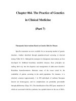
Fig. 10.1 Gottron’s sign. (a) Violaceous plaques over
the dorsal surface of the metacarpal phalangeal joints and
interphalangeal regions. (b) Gottron’s papules and plaques
over knee joint.
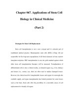
Fig. 10.2 Heliotrope rash of dermatomyositis
demonstrating violaceous erythema overlying the
upper eyelids with associated periorbital edema.
NICP_C10 03/05/2007 10:38 AM Page 171
172 S. CHRISTINE KOVACS
skin involvement. In the review by Gerami et al.
(2006), patients with CADM, as well as, patients with
classic DM have a similar association with malig-
nancy and interstitial lung disease. Anti-Jo-1 anti-
bodies are rarely found in CADM patients even if they
have interstitial lung disease. The other interesting
finding in this review is that in adult-onset CADM
calcinosis is very uncommon.
Juvenile dermatomyositis differs from the adult
form both histologically and clinically (Pachman
and Dooke, 1980; Reed and Mason, 2005). The his-
tological finding on muscle biopsy of perifascicular
atrophy is much more common in the pediatric
population. The skin lesions are similar in both popu-
lations, but there is a higher incidence of calcinosis
in the pediatric group. The calcific deposits tend to
occur over the elbows, knees, buttocks, or other pres-
sure point regions and have been shown to correlate
with disease activity and duration. The incidence
of calcinosis ranges between 30–70% in children
compared to adults where the occurrence of this
finding is less than 10% (Bowyer et al., 1986; Martini
et al., 1987). Vasculitis involving the gastrointest-
inal tract has a predilection for the pediatric popula-
tion. The Gower’s sign describes how a child with
proximal muscle weakness is able to get up off the
floor without relying on his or her lower extremity
strength.
Laboratory evaluation
Serum creatine kinase (CK) assay is a good initial
screening test in patients suspected of having an
inflammatory myopathy. Muscle enzymes used as
markers for muscle damage include CK, aldolase,
lactic dehydrogenase (LDH), aspartate aminotrans-
ferase (AST), and alanine aminotransferase (ALT).
The most sensitive marker is CK, which has three
isoforms: muscle type (CK-MM); brain type (CK-BB);
and hybrid type (CK-MB). The major source of CK is
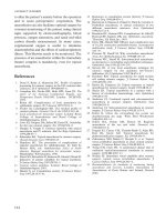
Fig. 10.3 Shawl sign. Erythematous macules distributed on the neck, upper back, shoulders, and arms.
NICP_C10 03/05/2007 10:38 AM Page 172
Polymyositis and dermatomyositis 173
skeletal muscle where the CK-MM predominates, but
CK-MB may be present in smaller concentrations.
Although disease states may be associated with ele-
vations in CK levels, exercise, intramuscular injec-
tions, and EMG testing may be responsible for such
elevations and they may remain elevated for up to
48 hours. Medications such as morphine, benzo-
diazepines, and barbiturates impair the excretion of
CK from circulation and may cause a mild elevation.
In addition, racial differences in CK levels exist; with
healthy African American men having higher levels
than Caucasians or Hispanics.
Autoantibodies
Antinuclear antibodies can be identified in 40–80%
of patients with an idiopathic inflammatory myopa-
thy. Attention has focused in particular on myositis-
specific antibodies (MSAs). The categories of MSAs
that have been identified include antibodies directed
against aminoacyl-transfer RNA synthetase (anti-
synthetases), antibodies directed against signal recog-
nition particle proteins (anti-SRP), and antibodies
directed against the nuclear protein complex Mi-2
(anti-Mi-2). The antisynthetases are the most com-
mon MSAs, with anti-SRP and anti-Mi-2 occurring
considerably less often. The presence of MSAs appears
to be associated with a particular clinical subset of
patients with myositis. Anti-Mi-2 antibodies are usu-
ally specific for dermatomyositis, whereas anti-SRP
antibodies are specific for polymyositis and portend a
poor prognosis (Targoff, 1990). The antisynthetases
are seen in half of the patients with dermatomyositis
or polymyositis but are neither sensitive nor specific
for either disease. Miller et al. (2002) described clin-
ical and histopathological features of a group of seven
anti-SRP positive patients. Interestingly, the results
did not show the SRP antibody to be associated
with PM, but with a rapidly progressive course. The
patients in this study tended to respond poorly to
steroids. Hengstman et al. (2006) collected samples
from 417 patients with myositis – 23 patients were
anti-SRP positive. This study confirmed the lack of
response to treatment and the rapidly progressive
course. Interstitial lung disease was present in one-
fourth of the patients. The muscle biopsy demonstrated
the presence of a necrotizing myopathy without the
typical features seen with an inflammatory myositis.
Several biopsies showed staining of necrotic fibers to
membrane-attack complex (MAC) (which is also seen
in the muscle in paraneoplastic myositis biopsies), but
only two patients in this group had neoplastic disease.
The most common antisynthetase is antihistidyl-
transfer RNA synthetase (anti-Jo-1); it is most specific
for the antisynthetase-myositis syndrome. The pres-
ence of anti-PM-Scl, a nonspecific antibody, helps
define a unique subset of patients with scleroderma
and dermatomyositis or polymyositis (Miller, 1993;
Oddis et al., 1992; Targoff, 1990, 1994).
Electromyographic (EMG) evaluation
EMG testing is useful in differentiating a neurogenic
from a myogenic process. EMG is useful in confirming
an active myopathic process, but is of no help in dis-
tinguishing between the inflammatory myopathies.
EMG of affected muscles in inflammatory myopathies
demonstrates short duration motor unit potentials;
polyphasic motor unit potentials, which may have
long duration, bizarre, high-frequency repetitive dis-
charges; and fibrillation potentials, positive sharp
waves and insertional irritability (Barkhaus et al.,
1990). There is full recruitment, despite weakness,
which some term early recruitment. It is typically
used to aid in the localization of an active site for
biopsy.
Noninvasive diagnostic modalities
MRI evaluation
Magnetic resonance imaging (MRI) offers a non-
invasive technique that can identify muscle edema,
edema in myofacial distribution, subcutaneous
change, muscle calcification, and fatty infiltration
(Adams et al., 1995). MRI may be helpful in identi-
fying “occult” muscle disease or to aid in localizing
an active site for potential biopsy to optimize the yield
of obtaining a meaningful biopsy result. It also may
be helpful in distinguishing active inflammation
from atrophy. The MRI techniques used include T1-
and T2-weighted images with T2 being sensitive
for detecting muscle edema in acute myositis.
Additionally, short tau inversion recovery (STIR),
and gadolinium may provide additional information
(Childs, 1997). Studies using MRI in patients with
dermatomyositis have shown prior to treatment
that the vastus lateralis muscle is the most severely
involved and that changes in the hamstrings occur
in only the extremely weak. Images early in the dis-
ease course show preservation of muscle architec-
ture without extensive fat replacement or atrophy,
but studies have shown that the inflammation
may resolve fairly rapidly over weeks to months
NICP_C10 03/05/2007 10:38 AM Page 173
174 S. CHRISTINE KOVACS
with fatty change usually occuring after three to
five months.
Ultrasound evaluation
Conventional ultrasound evaluation of muscle can
be helpful in detecting muscle edema in the acute
inflammatory stage of these disorders and shows
up as areas of increased echogenicity. This finding,
however, is not specific and can be found in other
conditions.
A recent study has demonstrated that contrast-
enhanced ultrasound is able to noninvasively detect
increased perfusion in the involved muscle groups
in patients with myositis. This study showed a high
correlation between increased perfusion on contrast-
enhanced ultrasound and increased signal intensity
on T2-weighted images. The results are preliminary
and suggest further analysis in the future (Weber
et al., 2005).
Muscle biopsy
Biopsy should be performed on a clinically affected
muscle, which is usually a proximal muscle group
such as the biceps or quadriceps. Because of the
chance of sampling error, MRI can be useful in local-
izing an affected area for biopsy. Care must be taken
to avoid muscles where electromyography has already
been done or where injectable anesthetics have been
used, both of which can cause alterations in histo-
pathological features. Arahata and Engel (1984)
reported inflammatory cells infiltrating non-necrotic
muscle fibers as the hallmark of polymyositis. Inter-
pretation of muscle biopsy results has been reviewed
(Dalakas, 2002). The main feature in PM is the pres-
ence of endomysial lymphocytic infiltration. Unfor-
tunately, similar findings can also be found in patients
with certain muscular dystrophies (Amato and Griggs,
2004). Thus screening for immunocytochemical
staining for proteins known to be associated with
muscular dystrophies is appropriate. The main finding
in DM is perivascular B-cell-predominant inflamma-
tion associated with microinfarcts and perifascicular
atrophy. Though perifascicular atrophy is specific
for DM, sometimes the diagnosis of DM or PM can-
not be made on the basis of histopathology alone.
Immunopathological markers such as the MHC-I/
CD8 complex may be more specific for polymyositis
in addition to inclusion myositis and may be helpful
in distinguishing antigen-driven inflammatory cells
that characterize PM and IBM from secondary
inflammation seen in other disorders, such as dystro-
phies (Dalakas, 2004). Also, the presence of the MAC
on these specimens may be helpful (Greenberg and
Amato, 2004). It has been identified in patients with
DM but also in necrotizing myopathies.
Association with malignancy
Literature reviews have demonstrated that certain
cancers (e.g. ovarian, stomach, and lymphoma) are
highly associated with DM and PM relative to the
normal population. The most common malignancies
in the general population (breast, lung, colorectal,
and prostate) are seen in these patients, but a strong
statistical association does not necessarily exist,
however, these reports are conflicting (Buchbinder
et al., 2001; Hill et al., 2001; Sigurgeirsson et al.,
1992). A continuing question in managing this
group of patients is what kind of evaluation should
be undertaken to screen for occult malignancy.
Unfortunately, large controlled prospective studies
are not available to adequately research this question.
At this point, age-appropriate cancer screening and
perhaps pelvic imaging to evaluate for ovarian cancer
in women is reasonable. A recent study looking at
tumor antigen markers (CEA, CA15-3, CA19-9, and
CA125) for detection of solid cancers did identify
both the CA125 and CA19-9 as useful markers in
determining a patient’s risk of developing tumors in
this population of patients (especially if they did not
have interstitial lung disease) (Amoura et al., 2005).
This risk was most notable in the first year following
the increased CA125 and CA19-9 levels.
Immunogenetics and pathogenesis
The exact cause of the idiopathic inflammatory
myopathies is unknown, but they are generally
accepted to be the result of an immune-mediated
process. Evidence supporting this hypothesis includes
the association of idiopathic inflammatory myopathies
with other autoimmune diseases such as thyroiditis,
vitiligo, myasthenia gravis, and other connective
tissue disease; the high prevalence of circulating
autoantibodies in patients with polymyositis and
dermatomyositis; and the pathological changes seen
in the muscle of affected patients. The myopathies
appear to have an environmental component as
suggested by geographic clustering of cases and the
seasonal onset of disease (Sarkar et al., 2005). Genetic
factors seem to play an important role, with the
association with certain histocompatability genes
NICP_C10 03/05/2007 10:38 AM Page 174
Polymyositis and dermatomyositis 175
(Chinoy et al., 2004). Viral infections like influenza,
mumps, cytomegalovirus, and Epstein–Barr have
been inconclusively implicated in the pathogenesis,
perhaps as a trigger for the immune response.
Dermatomyositis in general is felt to be a B-cell
mediated process with microangiopathy. Antibodies
are directed against the endothelium of the endo-
mysial capillaries leading to a prominent vascular
reaction. Eventually, it is hypothesized that the com-
plement system is activated and the MAC is formed
(Greenberg and Amato, 2004).
Polymyositis on the other hand appears to be sec-
ondary to cytotoxic T-cell response directed against
the muscle fibers and the histology shows invasion
of the muscle fibers by inflammatory cells.
Treatment
Regardless of the pharmacological treatment, all pati-
ents with myositis and/or calcinosis require extensive
physical therapy to prevent joint contractures and
disuse atrophy of the muscle tissue. This may
include gentle, passive stretching and splinting in
the initial stages of the disease and more aggressive
strength building once the muscle inflammation has
subsided (Dalakas, 1991).
In regard to dermatomyositis topical therapy con-
sists of the routine use of class I or II steroids for
the pruritis and inflammatory erythematous skin
changes. Hydroxychloroquine also has been used
in the treatment of the rash. Treatment of calcinosis
cutis is difficult and anecdotal reports of diltiazem
(Oliveri et al., 1996), probenicid (Skuterud et al.,
1981), warfarin (Berger et al., 1987), colchicine
(Taborn et al., 1978), aluminum hydroxide (Wang
et al., 1988), EDTA (Herd and Vaughn, 1964), and
more recently successful reports with the use of bis-
phonates have been published (Ambler et al., 2005).
Steroids have been the mainstay in the treat-
ment of myositis. Unfortunately, many patients have
an inadequate response and need more aggressive
therapy. Additionally, treatment tends to be long term
and the side effects of steroids are compounded over
time. A variety of steroid-sparing agents have been
tried including azathioprine, methotrexate, cyclos-
porine, cyclophosphamide, mycophenolate (Edge
et al., 2006; Majithia and Harisdangkul, 2005), and
tacrolimus (Mitsui et al., 2005). High-dose intraven-
ous immunoglobulin (IVIg) infusions as treatment
for dermatomyositis have been shown to be bene-
ficial in a placebo-controlled double-blind random-
ized trial (Dalakas et al., 1993). Other studies have
shown similar positive results, but cost and availab-
ility is an issue for some (Dalakas, 2005, 2006; Illa,
2005; Wetter et al., 2005). Rapamycin (sirolimus) has
been reported to be useful in the treatment of DM,
and appears to also have some antineoplastic activity
which makes it appealing in this patient population
that appears to have increased risk for the develop-
ment of malignancy (Nadiminti and Arbiser, 2005).
With the burgeoning field of biologics in the treat-
ment of inflammatory arthritis clinicians are now
interested in the use of biologics in the inflammatory
myopathies in terms of treatment response and bet-
ter understanding of the underlying pathogenesis.
It is generally well accepted that DM is humor-
ally mediated and B cells may play a significant role
in the pathogenesis. An open-labeled trial of seven
adult patients with DM with inadequate response to
current available therapies underwent four intraven-
ous infusions of rituximab (Levine, 2005). Rituximab
is a CD20+ B-cell depleting antibody. Six of the pati-
ents had clinical improvement in muscle strength
over baseline by 36–113%. The maximal improve-
ment occurred 12 weeks after the initial infusion.
CD20 B cells were effectively depleted by 12 weeks.
Other small studies and case reports have shown
that rituximab is effective (Chiappetta et al., 2005;
Noss et al., 2005).
Tumor necrosis factor (TNF) alpha is a pro-
inflammatory cytokine, which appears to play a
role in mediating many inflammatory conditions.
Anecdotal reports of its use in patients with refrac-
tory DM/PM have been promising (Efthimiou et al.,
2004). The largest report has been a retrospective
study of eight patients with refractory disease, six of
whom had a dramatic decline in the serum CK and
significant improvement in fatigue and muscular
strength (Norman et al., 2006). The patients that did
not respond did not have significant elevations in the
pretreatment CK levels. It has been suggested that
TNF blockers may be more effective in the initial
acute inflammatory phase (as induction therapy). A
theoretical concern with the use of these agents long
term would be the potential to enhance the develop-
ment of malignancy in a population of patients that
already appear to be at high risk.
Prognosis
It has been well recognized that the long-term out-
come in patients with PM and DM is rather dismal.
The mortality rate ranges from 4–45% of patients.
The morbidity is also quite high. Hopefully, with
NICP_C10 03/05/2007 10:38 AM Page 175
176 S. CHRISTINE KOVACS
newer agents available to treat these patients these
rates will decline. Several variables have been iden-
tified as being useful in predicting worse outcomes
including older age, male sex, dysphagia, longstand-
ing symptoms prior to diagnosis, pulmonary or cardiac
involvement, presence of antisynthetase or anti-SRP
antibodies, and type of myositis (Devere and Bradley,
1975). A more recent study has shown that these
patients have a more than 10% chance of dying of a
cause related to their disease, mostly cancer, especi-
ally in the first year to onset (Bronner et al., 2006).
The majority of the surviving patients have a chronic
course or polycyclic disease. At two years follow
up of the patients in the study 65% had normal
strength, 34% had slight or no disability, and only
16% had normal scores on the quality of life scale.
A retrospective cohort study of 53 medical records
of patients with PM, DM, CTD-associated myositis,
and malignancy-associated myositis demonstrated
that PM and CTD-associated myositis had a higher
rate of relapse rate compared to the other groups
(Agarwal et al., 2005). Seventeen patients had mul-
tiple relapses and tended to occur within the first
two years. Advanced age and duration of symptoms
seemed to be associated with a higher relapse rate.
In juvenile dermatomyositis the outcomes prior
to the introduction of corticosteroids were poor with
a mortality rate greater than 30%, and 50% were
left with serious disabilities. After steroids were used
in the 1960s, the mortality rate dropped to less than
10%. Huber and Feldman (2005) conducted a long-
term follow-up study and contacted 65 of 80 children
3–19 years after their diagnosis. Calcinosis was pre-
sent in 34% and contributed to reduced function.
Continuing disease activity was a problem: 40%
had ongoing rash, 10% reported weakness, 22% had
ongoing pain, and 35% still required medication to
control their disease.
Conclusion
With the many advancements in biological treat-
ments for a variety of immune-mediated disorders, in
addition to the ongoing identification of newer anti-
body profiles, immunohistopathological and genetic
testing, it is likely that we will gain further insight
into the pathogenesis and hence treatment of the
inflammatory myopathies. Future work will need
to focus on a universally accepted and validated
classification schema so that these newer therapies
are applied and tested to a homogeneous patient
population to obtain the most meaningful results.
References
Adams, E.M., Chow, C.K., Premkumar, A. and Plotz, P.H.
1995. The idiopathic inflammatory myopathies:
Spectrum of MR imaging findings. Radiographics, 15,
563–74.
Agarwal, S.K., Monach, P.A., Docken, W.P. and
Coblyn, J.S. 2005. Characterization of relapses
in adult idiopathic inflammatory myopathies. Clin
Rheumatol, 3, 1–6.
Amato, A.A. and Griggs, R.C. 2003. Unicorns, dragons,
polymyositis and other mythical beasts. Neurology,
61, 288–9.
Ambler, G.R., Chaitow, J., Rogers, M., McDonald, D.W.
and Ouvrier, R.A. 2005. Rapid improvement of calci-
nosis in juvenile dermatomyositis with alendronate
therapy. J Rheumatol, 32, 1837–9.
Amoura, Z., Duhaut, P., Houng du, L.T. et al. 2005.
Tumor antigen markers for the detection of solid can-
cers in inflammatory myopathies. Cancer Epidemiol
Biomarkers Prev, 14(5), 1279–82.
Arahata, K. and Engel, A.G. 1984. Monoclonal anti-
body analysis of mononuclear cells in myopathies.
I: Quantitation of subsets according to diagnosis
and sites of accumulation, and demonstration and
counts of muscle fibers invaded by T cells. Ann Neurol,
16, 193–208.
Barkhaus, P.E., Nandedkar, S.K. and Sanders, D.B.
1990. Quantitative EMG in inflammatory myopathy.
Muscle Nerve, 13, 247–53.
Berger, R.B., Featherstone, G.L., Raasch, R.H.,
McCartney, W.H. and Hadler, N.M. 1987. Treatment
of calcinosis universalis with low-dose warfarin.
AM J Med, 83, 72–5.
Bohan, A. and Peter, J.B. 1975. Polymyositis and
dermatomyositis (first of two parts). N Engl J Med,
292, 344–7.
Bowyer, S.L., Clark, R.A., Ragsdale, C.G., Hollister, J.R.
and Sullivan, D.B. 1986. Juvenile dermatomyositis:
Histological findings and pathogenetic hypothesis
for the associated skin changes. J Rheumatol, 13,
753–8.
Bronner, I.M., van der Meulen, M.F., de Visser, M. et al.
2006. Long-term outcome in polymyositis and der-
matomyositis. Ann Rheum Dis, in press.
Buchbinder, R., Forbes, A., Hall, S., Dennett, X. and
Giles, G. 2001. Incidence of malignant disease
in biopsy-proven myopathy. Ann Intern Med, 134,
1087–95.
Chiappetta, N., Steier, J. and Gruber, B. 2005. Rituximab
in the treatment of refractory dermatomyositis. J Clin
Rheumatol, 11, 264–6.
Childs, N.D. 1997. STIR-MRI goes past fat to detect
myopathies. Intern Med News, 1, 37.
Chinoy, H., Ollier, W.E. and Cooper, R.G. 2004. Have
recent immunogenetic investigations increased our
understanding of disease mechanisms in the idio-
patic inflammatory myopathies? Curr Opin Rheumatol,
16, 707–13.
NICP_C10 03/05/2007 10:38 AM Page 176
Polymyositis and dermatomyositis 177
Dalakas, M.C. 1991. Polymyositis, dermatomyositis,
and inclusion-body myositis. N Engl J Med, 325,
1487–98.
Dalakas M.C. 2002. Muscle biopsy findings in inflam-
matory myopathies. Rheum Dis Clin North Am, 28,
779–98.
Dalakas, M.C. 2004. Inflammatory disorders of muscle:
progress in polymyositis, dermatomyositis and inclu-
sion body myositis. Curr Opin Neurol, 17561–7.
Dalakas, M.C. 2005. Intravenous immunoglobulin in
patients with anti-GAD antibody-associated neuro-
logical diseases and patients with inflammatory myo-
pathies. Effects on clinicopathological features and
immunoregulatory genes. Clin Rev Allergy Immunol,
29, 255–68.
Dalakas, M.C. 2006. The role of high-dose immune
globulin intravenous in the treatment of dermato-
myositis. Int Immunopharmacol, 6, 550– 6.
Dalakas, M.C., Illa, I., Dambrosia, J.M. et al. 1993. A
controlled trial of high dose intravenous immuno-
globulin infusions as treatment for dermatomysotis.
N Engl J Med, 329, 1993–2000.
DeVere, R. and Bradley, W.G. 1975. Polymyositis:
Its presentation, morbidity and mortality. Brain, 98,
637–66.
Dickey, B.F. 1984. Pulmonary disease in polymyositis/
dermatomyositis. Semin Arthritis Rheum, 14, 60–
76.
Edge, J.C., Outland, J.D., Dempsey, J.R. and Callen,
J.P. 2006. Mycophenolate mofetil as an effective
corticosterid-sparing therapy for recalcitrant
dermatomyosits. Arch Dermatol, 142, 65– 9.
Efthimiou, P., Schwartzman, S. and Kagen, L.J. 2006.
Possible role for TNF-inhibitors in the treatment
of resisitant dermatomyosits and polymyositis.
A retrospective study of eight patients. Ann Rheum
Dis, in press.
Euwer, R.L. and Sontheimer, R.D. 1996. Dermato-
myositis. In R.D. Sontheimer and T.T. Provost (eds.),
Cutaneous Manifestations of Rheumatic Disease, Williams
& Wilkins, Baltimore, pp. 73–114.
Gerami, P., Schope, J.M., McDonald, L., Walling, H.W.
and Sontheimer, R.D. 2006. A systematic review
of adult-onset clinically amyopathic dermat-
omyositis (dermatomyositis sine myositis): A miss-
ing link within the spectrum of the idiopathic
inflammatory myopathies. J Am Acad Dermatol,
54(4), 597–613.
Greenberg, S.A. and Amato, A.A. 2004. Uncertainties
in the pathogenesis of adult dermatomyositis. Curr
Opin Neurol, 17, 359–64.
Hengstman, G.J., Ter Laak, H.J., Vree Egberts, W.T.
et al. 2006. Anti-SRP autoantibodies, marker of a
necrotizing myopathy. Ann Rheum Dis, in press.
Herd, J.K. and Vaughan, J.H. 1964. Calcinosis uni-
versalis complicating dermatomyositis. Arthritis
Rheum, 7, 259.
Hill, C.L., Zhang, Y., Sigurgeirsson, B. et al. 2001.
Frequency of specific cancer types in dermatomyo-
sitis and polymyositis: a population-based study.
Lancet, 357, 96–100.
Huber, A. and Feldman, F.M. 2005. Long-term out-
comes in juvenile dermatomyositis: How did we get
here and where are we going? Curr Rheum Reports, 7
,
441–6.
Illa, I. 2005. IVIg in myasthenia gravis, Lambert–Eaton
myasthenic syndrome and inflammatory myopathies:
Current status. J Neurol, 252(1), 14–18.
Jackson, H. 1887. A case of acute, infectious universal
myositis. Boston Med Surg J, 116, 498.
Kovacs, S.O. and Kovacs, S.C. 1998. Dermatomyositis.
J Am Acad Dermatol, 39, 899–922.
Levine, T. 2004. Rituximab in the treatment of
dermatomyositis. An open-label pilot study. Arthritis
Rheum, 52, 601–7.
Majithia, V. and Harisdangkul, V. 2005. Mycopheno-
late mofetil (CellCept): An alternative therapy for auto-
immune inflammatory myopathy. Rheumatology,
44, 386–9.
Martini, A., Ravelli, A., Viola, S., Sambugaro, R. and De
Benedetti, F. 1987. Calcinosis as the presenting sign
of juvenile dermatomyositis in a 14-month old boy.
Helv Paediatr Acta, 42, 181–4.
Miller, F.W. 1993. Myositis-specific autoantibodies:
Touchstones for understanding the inflammatory
myopathies. JAMA, 270, 1846– 9.
Miller, T., Al-Lozi, M.T., Lopate, G. and Pestronk, A.
2002. Myopathy with antibodies to the signal recog-
nition particle: clinical and pathological features.
J Neurol Neurosurg Psychiatry, 73, 420–8.
Mitsui, T., Kuroda, Y., Kunishige, M. and Matsumoto, T.
2005. Successful treatment with tacrolimus in a
case of refractory dermatomyositis. Internal Med, 44,
1197–9.
Nadiminti, U. and Arbiser, J.L. 2005. Rapamycin
(sirolimus) as a steroid-sparing agent in dermato-
myositis. J Am Acad Dermatol, 52, S17–19.
Norman, R., Breenber, R.G. and Jackson, J.M. 2006.
Case reports of etanercept in inflammatory der-
matoses. J Am Acad Dermatol, 54, S139–42.
Noss, E.H., Hausner-Sypek, D.L. and Weinblat, M.E.
2005. Rituximab as therapy for refractory poly-
myositis and dermatomyositis. J Rheumatol, 33,
1021–6.
Oddis, C.V., Okano, Y., Rudert, W.A., Trucco, M.,
Duquesnoy, R.J. and Medsger, T.A. Jr. 1992. Serum
autoantibody to the nucleolar antigen PM-Scl.
Arthritis Rheum, 35, 1211–17.
Oliveri, M.B., Palermo, R., Mautalen, C. and Hubscher,
O. 1996. Regression of calcinosis during diltiazem
treatement in juvenile dermatomyositis. J Rheumatol,
23, 2152–5.
Pachman, L.M. and Dooke, N. 1980. Juvenile dermato-
myositis: A clinical and immunologic study. J Pediatr,
96, 226–34.
Reed, A.M. and Mason, T. 2005. Recent advances
in juvenile dermatomyositis. Curr Rheum Reports, 7,
94–8.
NICP_C10 03/05/2007 10:38 AM Page 177
178 S. CHRISTINE KOVACS
Sarkar, K., Weinberg, C.R., Oddis, C.V. et al. 2005.
Seasonal influence on the onset of idiopathic
inflammatory myopathies in serologically defined
groups. Arthritis Rheum, 52, 2433–8.
Sigurgeirsson, B., Lindelof, B., Edhag, O. and Allander,
E. 1992. Risk of cancer in patients with dermato-
myositis or polymyositis. A population based study.
N Engl J Med, 326, 363–7.
Skuterud, E., Sydnes, O.A. and Haavik, T.K. 1981.
Calcinosis in dermatomyositis treated with pro-
benicid. Scand J Rheumatol, 10, 92– 4.
Taborn, J., Bole, G.G. and Thompson, G.R. 1978.
Colchicine suppression of local and systemic inflam-
mation due to calcinosis universalis in chronic
dermatomyositis. Ann Intern Med, 89, 648.
Targoff, I.N. 1990. Immune mechanisms in myositis.
Curr Opin Rheumatol, 2, 882–8.
Targoff, I.N. 1994. Immune manifestation of inflam-
matory muscle disease. Rheum Dis Clin North Am,
20, 857–80.
Tazelaar, H.D., Viggiano, R.W., Pickersgill, J. and
Colby, T.V. 1990. Interstitial lung disease in poly-
myositis and dermatomyositis. Am Rev Respir Dis,
141, 727.
Unverricht, H. 1887. Ueber eine eigenthumliche
form von acuter muskelentzundung mit einem der
trichinose ahnelnden krankheitsbilde. Munchener
Med Sochnschrf, 34, 488.
Wagner, E. 1887. Ein Fall von acuter polymyositis.
Dtsch Arch Med, 40, 241.
Wagner, E. 1863. Falleiner Seltnen Muskelkrankheit.
Dtsch Arch Heilk, 4, 282.
Wang, W.J., Lo, W.I. and Wong, C.K. 1988. Calcinosis
cutis in juvenile dermatomyositis: remarkable
response to aluminum hydroxide therapy (letter).
Arch Dermatol, 124, 1721.
Weber, M.A., Krix, M., Jappe, U. et al. 2005. Pathologic
skeletal muscle perfusion in patients with myositis:
detection with quantitative contrast-enhanced US-
initial results. Radiology, 238, 640–9.
Wetter, D.A., Davis, M.D., Yiannias, J.A. et al. 2005.
Effectiveness of intravenous immunoglobulin therapy
for skin disease other than toxic epidermal necrolysis:
A retrospective review of Mayo Clinic experience. Mayo
Clin Proc, 80, 41–7.
Yale, S.H., Adlakha, A. and Stanton, M.S. 1990. Dermat-
omyositis with pericardial tamponade and polymyo-
sitis with pericardial effusion. Am Heart J, 126, 997.
NICP_C10 03/05/2007 10:38 AM Page 178
Sjögren’s syndrome (SS) is an autoimmune disease
first described in female patients with arthritic prob-
lems, dry eyes and mouth by a Swedish physician,
Henrik Sjögren (1933). SS can present in any age
and gender, but it predominantly occurs in middle-
aged women (female:male ratio = 9:1). It may pre-
sent as an isolated syndrome (primary or P-SS) or
be associated with other connective tissue disorders
(secondary S-SS). P-SS is characterized by lympho-
cytic infiltration of exocrine glands and epithelia
at multiple sites. The characteristic involvement of
lachrymal and salivary glands results in xeroph-
thalmia and xerostomia. About a third of patients
also have extraglandular manifestations with polyar-
thralgia, Raynaud’s phenomenon, and interstitial
pulmonary fibrosis occurring most frequently. A
revised version of the European classification criteria
for SS was recently published by the American-
European Consensus Group (2002) (Box 11.1). The
proposed evaluation process facilitates establishing
the diagnosis with high specificity and sensitivity.
Clinical features of neuro-SS
Neurological complications may be seen in about
40% of patients with P-SS. Diagnostic difficulties may
arise when the neurological abnormalities precede
systemic signs of P-SS. The involvement of the
nervous system in P-SS is dominated by abnormalit-
ies in the peripheral nervous system (PNS). The PNS
dysfunction may include distal sensory or sensory-
motor, usually axonal, polyneuropathy. The abnor-
mality more often involves temperature and light
touch sensation than proprioception. In addition to
a painful sensory polyneuropathy, however, ataxic
polyneuropathy may also develop. Demyelinating
polyneuropathy can be seen in a smaller proportion
of patients, and bilateral carpal tunnel syndrome
is not uncommon. Dysautonomia with tonic pupils,
paralytic ileus, upper gastrointestinal dysfunction,
neurogenic bladder and hypohydrosis have also
been noted in relationship with the involvement of
autonomous nerves and antibodies to the ganglionic
acetylcholine receptor (Lafitte et al., 2001; Mori et al.,
2003). Sural nerve biopsy usually shows axonal
degeneration, demyelination, remyelination, and
variable degrees of large or small fiber depletion.
T-cell infiltration in the dorsal root ganglia may be
found in patients with ataxic sensory neuropathy.
Perivascular infiltration (vasculitis) is not a frequent
histological finding, but microangiopathy in the
endoneurium is believed to contribute to polyneuro-
pathy, particularly in older patients.
The involvement of the central nervous system
(CNS) can present with progressive multifocal
neurological symptoms, spinal cord dysfunction,
motoneuron disease (with the involvement of upper
and lower mononeurons), and cortical or subcort-
ical cognitive decline (Lafitte et al., 2001). The spinal
cord pathology may cause transverse myelitis with
paraparesis and sphincter abnormalities or progress-
ive myelopathy. The cognitive deficits frequently
involve frontal executive functions, and manifest
as dysinhibition and difficulties with attention and
abstraction. Memory deficit and visuospatial dys-
function are also often present. Combined CNS and
PNS abnormalities occur in a subgroup of patients
(Lafitte et al., 2001).
Primary progressive multiple sclerosis
(MS) and P-SS
There has been an ongoing debate about the occur-
rence of P-SS in patients presenting with primary
progressive (PP-) MS. de Seze et al. (2001) assessed
clinical and laboratory criteria for SS in 60 consecut-
ive patients with PP-MS. Patients were questioned
about xerostomia, xerophthalmia, and underwent a
minor salivary gland biopsy, Schirmer test, salivary
gland scintigraphy, and tests for SSA and SSB sero-
logy. Ten out of 60 patients with PP-MS met four
or more criteria for P-SS (Box 11.1). The authors
11
Neuro-Sjögren’s syndrome
Bernadette Kalman
NICP_C11 03/05/2007 10:39 AM Page 181
182 BERNADETTE KALMAN
suggest that P-SS can mimic PP-MS. While the
remarkable resemblance of P-SS-related myelopathy
to PP-MS is well documented (Alexander et al.,
1986; Noseworthy et al., 1989; Pericot et al., 2003),
the current diagnostic and classification criteria for
MS and P-SS (McDonald et al., 2001; Polman et al.,
2005; Thompson et al., 2000; Vitali et al., 2002)
(Box 11.1) should help to differentiate the two con-
ditions in most cases. Ambiguities are only expected
in a small subgroup of patients that meet both the
criteria of PP-MS and P-SS. In these cases the ques-
tion arises as to whether an association occurred
between two autoimmune conditions or P-SS caused
a myelopathic phenotype indistinguishable from
PP-MS (de Seze et al., 2001; Pericot et al., 2003).
Immunopathogenesis
The etiology of SS is unknown. Involvement of
exogeneous microbial agents (e.g. retroviruses)
in the initiation of an abnormal immune response
against self-antigens has been postulated but not
proven (Konttinen and Kasna-Ronkainen, 2002).
Immune abnormalities of epithelial cells include an
increased expression of HLA Class II and costimulat-
ory molecules, which facilitate antigen presention to
I Ocular symptoms: the response is
positive if the patient has at least one
of the following:
– Daily, persistent, troublesome dry
eyes for more than three months
– A recurrent sensation of sand or
gravel in the eyes
– Use tear substitutes more than
three times a day
II Oral symptoms: a response is positive
if the patient has at least one of the
following:
– A daily feeling of dry mouth for
more than three months
– Had recently or persistently
swollen salivary glands as
an adult
– Frequently drinks liquids to aid in
swallowing dry food
III Ocular signs: objective evidence
of ocular involvement defined as
a positive result for at least one of
the following tests:
– Schirmer’s I test without anesthesia
(ഛ5 mm in 5 min)
– Rose Bengal score or other ocular
dye score (= or >4 according to
van Bijsterveld’s scoring)
IV Histopathology: focal lymphocytic
sialoadenitis in minor salivary glands
with a focus score ജ1, defined as
a number of lymphocytic foci per
4 mm
2
of glandular tissue.
V Salivary gland pathology: objective
evidence of salivary gland involvement
defined by a positive result for at least
one of the following tests:
– Unstimulated salivary flow
(ഛ1.5 ml in 15 min)
– Parotid sialography revealing diffuse
sialectasias without obstruction in
major ducts
– Salivary scintigraphy demonstrating
delayed uptake, reduced
concentration, and/or delayed
excretion of tracer
VI Autoantibodies: The presence of
antibodies to SSA (Ro) or SSB (La)
antigens or to both in the serum
For P-SS: presence of any four of six items
is indicative of P-SS, if either IV or VI is
positive; presence of any three of items III,
IV, V, and VI.
For S-SS: in patients with connective tissue
disease, the presence of items I and II plus
two from items III, IV, and V may be
consistent with S-SS.
Exclusion criteria: past head and neck
irradiation; hepatitis C infection; AIDS;
lymphoma; sarcoidosis; graft versus host
disease; use of anti-cholinergics
Box 11.1 A revised version of the European classification criteria
for SS proposed by the American-European Consensus Group
(after Vitali et al., 2002).
NICP_C11 03/05/2007 10:39 AM Page 182
Neuro-Sjögren’s syndrome 183
and activation of the infiltrating, predominantly
CD4 lymphocytes. The presented antigens include
SSA (Ro), SSB (La), α-fodrin, β-fodrin, and muscarinic
cholinergic receptors. The helper T-cell activation
leads to B-cell stimulation and antibody production.
Anti-SSA may occur alone, while anti-SSB is usually
present in association with anti-SSA. An association
of P-SS with the HLA A1, B8, DR3, DQ2 haplotype
was suggested (Fei et al., 1991; Rischmueller et al.,
1998), but this association may be restricted to anti-
SSA and anti-SSB positive forms of P-SS (Gottenberg
et al., 2003).
The CSF can be completely normal, or have
mildly elevated proteins and lymphocytic pleocyto-
sis. Intrathecal IgG synthesis and oligoclonal bands
may occur, but are not typical in P-SS.
MRI imaging
T2-weighted and FLAIR MRI of the brain may reveal
punctate hyperintensities in the periventricular
white matter, deep gray matter, and the brainstem,
but normal imaging is not rare even in cognitively
affected patients. Spinal imaging in patients with
spinal cord syndromes usually reveals abnormalities
compatible with transverse myelitis, myelopathy, or
discrete MS-like lesions (Fig. 11.1).
Treatment
High-dose corticosteroids have been used in acute
exacerbations and low-dose steroids in chronic stages
of P-SS. In progressive cases, azathioprine, cyclo-
phosphamide, and other immune-suppressive regi-
mens have been tried. Most recently rituximab, a
chimeric human–mouse antibody to CD20 positive
B cells (90% of all B cells), has been successfully used
in P-SS and lymphoma (Somer et al., 2003).
Summary
Sjögren’s syndrome is a systemic autoimmune dis-
order that may occasionally overlap with other con-
nective tissue diseases. A great proportion of patients
with SS will develop neurological abnormalities most
commonly affecting the PNS. A progressive multi-
focal CNS involvement can also be seen in P-SS
which may mimic PP-MS. Recently developed diag-
nostic criteria for SS and MS provide important guid-
ance for the diagnostic evaluation which should
direct disease-specific therapeutic decisions. Patients
with P-SS require long-term treatment and labor-
atory monitoring with alertness for a potential
development of a lymphoproliferative condition.
References
Alexander, E.L., Malinow, K., Lejewski, J.E., Jerdan,
M.S., Provost, T.T. and Alexander, G.E. 1986.
Primary Sjögren’s syndrome with central nervous
system disease mimicking multiple sclerosis. Ann
Intern Med, 104, 323–30.
de Seze, J., Devos, D., Castelnovo, G. et al. 2001. The
prevalence of Sjögren’s syndrome in patients with
primary progressive multiple sclerosis. Neurology,
57, 1359–63.
Fei, H.M., Kang, H., Scharf, S., Erlich, H., Peebles, C. and
Fox, R. 1991. Specific HLA-DQA and HLA-DRB1
alleles confer susceptibility to Sjögren’s syndrome
and antibody production. J Clin Lab Anal, 5, 382–91.
Gottenberg, J.E., Busson, M., Loiseau, P., et al. 2003. In
primary Sjögren’s syndrome, HLA class II is associ-
ated exclusively with autoantibody production and
spreading of the autoimmune response. Arthritis
Rheum, 48, 2240–5.
Konttinen, Y.T. and Kasna-Ronkainen, L. 2002.
Sjögren’s syndrome: Viewpoint on pathogenesis.
One of the reasons I was never asked to write a
textbook chapter on it. Scan J Rheumatol Suppl, 116,
15–22.
Lafitte, C., Amoura, Z., Cacoub, P. et al. 2001.
Neurological complications of primary Sjögren’s
syndrome. J Neurol, 248, 577–84.
Fig. 11.1 MRI images of the spinal cord from a patient with
Sjögren’s syndrome. T2-weighted images of a cervical cord
lesion at the level of C2–C3. The patient presented with
progressive myelopathy imitating primary progressive MS.
Complications
The underlying lymphoproliferative process increases
the risk 44 times for a malignant lymphoma in
patients with P-SS. Therefore, patients with P-SS
require regular monitoring and laboratory testing.
NICP_C11 03/05/2007 10:39 AM Page 183
184 BERNADETTE KALMAN
McDonald, W.I., Compston, A., Edan, G. et al. 2001.
Recommended diagnostic criteria for multiple
sclerosis: Guidelines from the International Panel on
the diagnosis of multiple sclerosis. Ann Neurol, 50,
121–7.
Mori, K., Iijima, M., Sugiura, M. et al. 2003. Sjögren’s
syndrome associated painful sensory neuropathy
without sensory ataxia. J Neurol Neurosurg Psy-
chiatry, 74, 1320–2.
Noseworthy, J.H., Bass, B.H., Vandervoort, M.K., et al.
1989. The prevalence of primary Sjögren’s syn-
drome in a multiple sclerosis population. Ann Neurol,
25, 95–8.
Pericot, I., Brieva, L., Tintore, M. et al. 2003.
Myelopathy in seronegative Sjögren’s syndrome
and/or primary progressive multiple sclerosis. Mult
Scler, 9, 256–9.
Polman, C.H., Reingold, S.C., Edan, G. et al. 2005.
Diagnostic criteria for multiple sclerosis: 2005
revisions to the “McDonald Criteria”. Ann Neurol,
58, 840–6.
Rischmueller, M., Lester, S., Chen, Z. et al. 1998. HLA
class II phenotype controls diversification of the
autoantibody response in primary Sjögren’s syn-
drome. Clin Exp Immunol, 111, 365–71.
Sjögren, H. 1933. Zur kenntnis der keratoconjunctiv-
itis sicca. Acta Opthalmol, 11(2), 1–151.
Somer, B.G., Tsai, D.E., Downs, L., Weinstein, B.,
Schuster, S.J.; American College of Rheumatology ad
hoc Committee on Immunologic Testing Guidelines.
2003. Improvement in Sjögren’s syndrome fol-
lowing therapy with rituximab for marginal zone
lymphoma. Arthritis Rheum, 49, 394–8.
Thompson, A.J., Montalban, X., Barkhof, F., et al. 2000.
Diagnostic criteria for primary progressive multiple
sclerosis: A position paper. Ann Neurol, 47, 831–5.
Vitali, C., Bombardieri, S., Jonsson, R. et al.; European
Study Group on Classification Criteria for Sjögren’s
Syndrome. 2002. Classification criteria for Sjögren’s
syndrome: A revised version of the European criteria
proposed by the American-European Consensus
Group. Ann Rheum Dis, 61, 554– 8.
NICP_C11 03/05/2007 10:39 AM Page 184
Behçet’s disease is a multisystem disorder with oral
and genital ulcerations and uveitis first described
by a Turkish dermatologist, H. Behçet (1937). Addi-
tional pathology with cardiovascular, pulmonary
and gastrointestinal involvement, and erythema
nodosum, pustular skin lesions, or pseudofolliculitis
may occur. Oligoarthropathy affecting large joints is
also relatively common. Constitutional symptoms
include fatigue, fever, nausea, and weight loss. The
etiopathogenesis is unknown. Viral and bacterial
infectious origins have been investigated but not
proven. The postulated autoimmune etiology of the
episodic vasculitic process is supported by the asso-
ciation with the HLA B51 allele, neutrophil hyper-
activity, and increased CD8/CD4 cell ratio. However,
in contrast to the usual female predominance in
autoimmunity, males develop Behçet’s disease more
frequently. Behçet’s disease is most commonly seen
in the eastern Mediterranean countries, the Middle
East, and East Asia, but Caucasians are also affected
(Kidd et al., 1999). The prevalence is estimated to be
37/10
5
in rural and 8/10
5
in urban regions of Turkey
(Yurdakul et al., 1988). In contrast, 0.4/10
5
preval-
ence rate was estimated in the UK (Chamberlain,
1977). The first autopsy case with neurological
involvement was reported by Berlin (1944). A large
series of autopsy cases with Behçet’s disease revealed
histological evidence of neurological involvement in
20% of patients (Lakhanpal et al., 1985).
Diagnosis of Behçet’s disease
Based on the criteria defined by the International
Study Group for Behçet’s disease (1990), the pre-
requisite for diagnosis is the presence of recurrent
oral ulcerations plus any two of the following:
recurrent genital ulcerations, erythema nodosum,
pseudofolliculitis, papulopustular eruption, acnei-
form nodules, positive pathergy test, anterior or pos-
terior uveitis, and retinal vasculitis. In the pathergy
test, the forearm is pricked with a sterile needle. The
test is positive if a small red bump or pustule appears
1–2 days after the needle insertion.
Clinical presentation of neuro-Behçet
Early studies (Pallis and Fudge, 1956; Wadia and
Williams, 1957) classified three forms of neuro-
logical complications:
1 Subacute brainstem involvement with cranial
neuropathy, oculomotor abnormalities, nystag-
mus, gaze palsy, dysarthria, ataxia and bulbar
weakness, accompanied by systemic symptoms
of fever, skin lesions, and arthropathy.
2 Meningomyelitis with signs of meningeal inflam-
mation and spinal cord lesions.
3 A confusional syndrome caused by meningo-
encephalitis initially without focal neurological
signs, but with dementia, quadriparesis, pseudo-
bulbar palsy, and Parkinsonism in chronic stages.
Subsequent studies pointed to the involvement
of blood vessels, particularly with venous sinus
thrombosis and intracranial hypertension. Arterial
thrombosis and aneurism formation appeared less
frequently (Akman-Demir et al., 1996; Bienenstock
and Margulies, 1961; Bousser et al., 1980; Wechsler
et al., 1989). Idiosyncratic presentations including
tumefactive neuro-Behçet disease have been reported
(Bennett et al., 2004). While the involvement of
optic nerves is relatively rare, isolated optic neuritis
may occur (Kocer et al., 1999). Further, neuro-
Behçet’s disease may imitate multiple sclerosis (MS)
(Ashjazadeh et al., 2003). The involvement of peri-
pheral nervous system appears to be rare.
In a recent report, Akmar-Demir et al. (1999)
reviewed the records of 558 patients with neuro-
Behçet in Turkey. After the exclusion of patients who
did not fulfill the diagnostic criteria, the records of
200 predominantly male patients were analyzed. Of
these 200, 162 patients had parenchymal disease,
12
Neuro-Behçet’s syndrome
Bernadette Kalman
NICP_C12 03/05/2007 10:39 AM Page 185