Neuroimmunology in Clinical Practice - part 10 pps
Bạn đang xem bản rút gọn của tài liệu. Xem và tải ngay bản đầy đủ của tài liệu tại đây (280.43 KB, 22 trang )
242 ANDREW J. CHURCH AND GAVIN GIOVANNONI
Outbreaks of SC have recently been reported in
developed countries even among communities with
good access to healthcare, although this worldwide
increase could be coincidental, it could also suggest
the emergence of highly pathogenic or antibiotic-
resistant strains (Ayoub, 1992).
Poststreptococcal tic disorders and PANDAS
Interest in SC was reignited in the 1980s after the
recognition that sudden-onset tic disorders in chil-
dren appeared to follow an outbreak of streptococcal
infection. An outbreak of streptococcal tonsillitis
in Rhode Island, USA was associated with a 10-fold
increase in children presenting with a motor tic
disorder, without evidence for RHF or SC (Kiessling
et al., 1993). As the clinical phenotype was tics
and neuropsychiatric features, SC was proposed as a
model of these disorders, which were termed PAN-
DAS (pediatric neuropsychiatric disorders associated
with streptococcal infections) (Swedo et al., 1998).
As GABHS is a prevalent infectious agent in the com-
munity, two or more exacerbations of the tic disorder
following streptococcal infection were required to
make a diagnosis (Swedo et al., 1998) (Table 21.1).
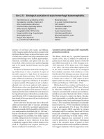
The PANDAS classification is defined as the pres-
ence of OCD and/or a tic disorder which meets DSM-
III-R or DSM-IV criteria with an acute, pediatric
(prepubescent) onset occurring after three years of
age, with a later episodic course of symptom exacer-
bations and recovery (Swedo et al., 1998). The asso-
ciation with streptococcal infection(s) was shown by
a positive GABHS throat culture with initial raised
streptococcal serology, which declined with clinical
recovery (Swedo et al., 1998). Patients with RHF,
SC, or other neurological disease were excluded from
the study in order to meet the clinical diagnosis of
PANDAS (Swedo et al., 1998). As a large number
of patients are excluded by the narrow definition of
the PANDAS classification, the phenotypic breadth
of neuropsychiatric and motor disorder symptoms
associated with streptococcal infections is currently
unknown.
However, PANDAS is phenotypically identical to
Tourette’s syndrome. The hypothesis that Tourette’s
may have an autoimmune eitology has proved to be
exceedingly controversial. Recently two adult cases
which conform to a wider definition of PANDAS have
been described, expanding the proposed syndrome
classification (Martinelli et al., 2002) to adult-onset
tic disorders.
Pathology of poststreptococcal disorders
Due to the nonfatal course of SC, pathological studies
of brain abnormalities have been rare, and those that
exist may only reflect severe or complicated cases, or
inadvertently include cases of encephalitis, metabolic
or genetic syndromes. The reports all found abnor-
malities mostly localized in the basal ganglia, which
included cellular infiltration and neuronal loss
with relative sparing of other brain areas (Colony
and Malamud, 1956; Marie and Tretiakoff, 1920),
(Table 21.2). These focal changes have also been
reported in the context of diffuse neuronal loss, which
was the predominant feature, and an encephalitic
pathogenesis was proposed (Greenfield and Wolfsohn,
1922) (Table 21.2). The consistent findings appeared
to be specific abnormalities of the basal ganglia with
conflicting evidence regarding disseminated brain
involvement (Table 21.2). The clinical similarities of
SC to HD may have influenced early reports of degener-
ative changes in the pathology of SC (Table 21.2).
However, the similarities of SC to HD and the recog-
nition of the basal ganglia as an area controlling
movement, led to the hypothesis that the basal gan-
glia were also the central area of pathogenesis
causing SC (Aron, 1965; Dale, 2003; Jummani and
Okun, 2001).
Neuroimaging
Brain imaging studies have been reported as normal
in most cases of SC and PANDAS, casting doubt as to
Table 21.1 The diagnostic criteria for PANDAS
devised by Swedo and colleagues (1998).
Number Criterion
1 Tics (chorea would be an exclusion
criteria)
2 Obsessive-compulsive disorder
3 Acute onset with an episodic course
4 Exacerbation following proven
GABHS infection
5 GABHS infection diagnosed by
throat culture and/or falling,
rising streptococcal serology
6 Other neuropsychiatric
manifestations and nonchoreic
movement disorders
NICP_C21 03/05/2007 10:50 AM Page 242
Poststreptococcal movement disorders 243
whether widespread neuronal loss is an important
feature of the disease, although this does not rule
out subtle alterations (Dale, 2003; Giedd et al., 1995;
Swedo et al., 1993). Only rarely have suspected
inflammatory changes seen on magnetic resonance
imaging (MRI) been associated with SC, and these
have been predominantly localized to the basal gan-
glia (Kienzle et al., 1991). The abnormalities in these
cases were reversible with disease remission, sug-
gesting that temporary neuronal disruption rather
than neuronal loss is one possible mechanism of
pathogenesis (Giedd et al., 1995). One study has also
found an increased association between MRI and
basal ganglia abnormalities in SC patients who had
repeated episodes of chorea during a one-year study
(Faustino et al., 2003).
However, MRI lesions in some cases of SC may be
linked to severe disease spectrum or a tendency of
SC to recur or become persistent in some patients.
Alternatively abnormal MRI may be associated with
a diffuse inflammatory disease with basal ganglia
features. For example, Dale et al. (2001) described
10 cases of acute disseminated encephalomyelitis
(ADEM) associated with GABHS infection. The clin-
ical phenotype was novel, with 50% having a dys-
tonic extrapyramidal movement disorder, and 70%
a behavioral syndrome. None of the patients had
RHF or SC. MRI studies showed hyperintense basal
ganglia in 80% of patients with poststreptococcal
ADEM, compared to 18% of patients with non-
streptococcal ADEM. These findings may support a
new subgroup of postinfectious autoimmune inflam-
matory disorders associated with GABHS, abnormal
basal ganglia imaging, and extrapyramidal move-
ment disorder.
Volumetric imaging studies have also found basal
ganglia (caudate nucleus and putamen) involvement
in SC. The basal ganglia have been reported to be
enlarged during acute SC compared to controls, which
may suggest inflammation (Giedd et al., 1995).
Further evidence for basal ganglia involvement in SC
has come from magnetic resonance spectroscopy
studies, which have shown increased glucose turn-
over and hypermetabolism, which could suggest
that alterations in local metabolism are important
(Weindl et al., 1993). It has been shown that these
metabolic changes can be reversible with disease
recovery, which may be important in light of the
reversible volumetric changes.
Autoimmune hypothesis
Rheumatic fever is considered to be an inflam-
matory or autoimmune disorder so SC and PANDAS
have been proposed to have a similar pathogenesis
(Church et al., 2002; Dale, 2003; Swedo et al.,
1998). While T cells have an important role in RHF
their role in SC has not been studied. However, one
study has reported a modest upregulation of cytokine
production in acute SC as a third of patients with
acute SC had elevated Th1 or Th2 serum cytokines
compared to controls. In cerebrospinal fluid (CSF)
both IL-4 and IL-10 (Th2 cytokines) were raised
while the Th1 cytokine, INF-γ, was undetectable in
acute SC. Additional evidence for the importance
of Th2 cytokines and perhaps antibody production
Table 21.2 Pathological reports in Sydenham’s chorea.
Reference Pathology Conclusions
Delcourt and Sand, 1908 Inflammatory Perivascular inflammation of basal ganglia
and cortex
Guizzetti and Camisa, 1911 Inflammatory and vascular Disseminated encephalitis
Harvier and Levanditi, 1920 Inflammatory Perivascular inflammation of mesencephalon
Marie and Treitiakoff 1920 Inflammatory Perivascular inflammation of basal ganglia
Greenfield and Wolfsohn, Inflammatory Perivascular inflammation of basal ganglia
1922 and cortex
Lewy et al., 1923 Inflammatory Widespread
Ziegler, 1927 Degenerative Basal ganglia
Lhermitte and Pagniez, 1930 Inflammatory/degenerative Basal ganglia
Von Santha, 1932 Vascular Encephalitic
Glaser, 1952 Vascular Inflammatory features
Colony and Malamud, 1956 ?Degenerative Cortex and thalamic involvement
NICP_C21 03/05/2007 10:50 AM Page 243
244 ANDREW J. CHURCH AND GAVIN GIOVANNONI
came from the persistent SC results, as 50% of these
cases had raised CSF IL-4 levels, whereas other serum
and CSF cytokines were within normal ranges.
Antibasal ganglia antibodies, ABGA
However, the majority of research has focussed on
the detection of antineuronal antibodies as an indic-
ator of an autoimmune pathology in SC and PAN-
DAS. Husby in 1976 first described IgG antineuronal
antibodies against neurons within the basal ganglia
using an indirect immunofluorescence method in 46%
of patients with acute SC (n=30) and only 1.8–4% of
controls (n=203); interestingly, a higher proportion,
14% (n=50) of patients with RHF without chorea, were
also positive. The staining pattern was described
as cytoplasmic binding to caudate and subthalamic
neurons with weaker staining in the cortex. This
antibody reactivity was removed by preincubating
positive samples with extracts of streptococcus (Husby
et al., 1976). This led to the hypothesis that “basal
ganglia antibodies” could be produced as a conse-
quence of molecular similarity (mimicry) between
streptococcal proteins and brain ones (Fig. 21.3).
Later reports suggested that ABGA detected by
indirect immunofluorescence (IF) were present in
100% of acute SC but less prevalent in persistent or
recovered cases (Church et al., 2002; Kotby et al.,
1998).
To expand upon this hypothesis western immuno-
blotting, a common methodology used in the iden-
tification of paraneoplastic antibodies, has been
used to detect basal ganglia antibodies. Rather than
polyspecific binding to basal ganglia proteins, react-
ivity to discrete antigens of 40, 45, and 60 kDa has
been described (Church et al., 2002) (Fig. 21.4).
A separate study using western immunoblotting
was less discriminating between SC and normal and
neurological disease controls, but found increased
antibody activity using a soluble supernatant frac-
tion from caudate nucleus (Morshed et al., 2001).
However, the presence of the same antineuronal
antibodies in PANDAS and particularly a small sub-
group of patients with Tourette’s syndrome have led
to significant controversy regarding both their role
and presence (Kurlan, 1998; Singer et al., 2005a).
As naturally occurring autoantibodies are known
to exist secondary to local damage the ABGA responses
reported in SC and PANDAS could be an epiphen-
omenon secondary to other disease mechanisms.
Alternatively different methodological approaches,
particularly in western immunoblotting detection
of antibody reactivity and sampling at different time
points in disease, may explain the differences in anti-
body results (Martino et al., 2005). However, what
is clear is that the significance of these antineuronal
antibodies must only be made in the context of the
clinical phenotype and the presence of documented
or laboratory supported streptococcus infection.
Without streptococcus evidence these antibodies are
of no obvious significance.
Fig. 21.3 Anti-neuronal antibodies against human
basal ganglia in Sydenham’s chorea, PANDAS, and normal
controls. (A) Second antibody diluted 1/30 and tested against
human basal ganglia section. No specific staining. (B) Normal
control sample diluted 1/50 and tested against human basal
ganglia section. Lipofuschin granules. (C) PANDAS sample
diluted 1/50 and tested against human basal ganglia tissue.
Staining of neuronal-like cells (arrow). (D) SC sample diluted
1/50 and tested against human basal ganglia tissue. Strong
staining of neuronal-like cells (arrow). Key: Bar = 5 µm.
Reproduced with permission from Church et al. (2002),
Neurology, published by Lippincott Williams & Wilkins.
Fig. 21.4 Western immunoblotting of human basal ganglia
showing IgG reactivity from SC patients.
NICP_C21 03/05/2007 10:50 AM Page 244
Poststreptococcal movement disorders 245
Potential prevalence
Focusing on TS and OCD only, the prevalence in
children may be of the order of 1%. In a community-
based study the prevalence of TS in children was 2%
(Hornse et al., 2001) and the prevalence of OCD was
between 2% and 4% (Douglass et al., 1995; Flament
et al., 1988; Valleni-Basile, 1994). We have shown
the ABGA positivity rate in children attending spe-
cialist clinics with TS and OCD to be 25% and 42%,
respectively (Church et al., 2003; Dale et al., 2005).
Potential functional effects of ABGA
ABGA recognize four main protein bands of 40,
45, 60, and 98 kDa in an antigen preparation from
human basal ganglia. The 45 and 98 kDa antigens
are the monomeric and dimeric forms of γ-enolase,
the 40 kDa antigen is aldolase C (neuron specific)
and the 60 kDa antigen is pyruvate kinase. γ-Enolase
or neuron-specific enolase-reactive ABGA cross-
react with α-enolase. All the antigens are glycolytic
enzymes and are involved in energy homeostasis
and as expected are found in the cytosol. These pro-
teins are also located on the neuronal surface (Lim et
al., 1983; Nakajima et al., 1994), where they appear
to have “moonlighting” or alternative functions; e.g.
enolase located on the surface of neurons acts as a
receptor for plasmin/plasminogen (Pancholi, 2001)
and has been shown to be a trophic factor for
neurons (Hattori et al., 1995). Plasminogen binding
to neuronal surface enolase also provides trophic
support to mesencephalic dopaminergic neurons
(Nakajima et al., 1994). Membrane neuronal aldolase
provides local membrane energy and is enzymatic-
ally active (Bulliard et al., 1997). It also forms an
oxidoreductase complex with enolase and other pro-
teins on the neuronal membrane and is thought to
monitor oxidative stress and induce an appropriate
cellular response (Bulliard et al., 1997). Aldolase binds
tightly with ATPase protein pumps on the plasma
membrane allowing direct coupling of glycolysis to
the proton pump (Lu et al., 2001). The monomer of
pyruvate kinase acts as thyroid hormone (T3) bind-
ing protein. Binding of T3 to pyruvate kinase inhibits
enzymatic activity, suggesting that this process may
be centrally involved in the control of some cellular
metabolic effects induced by thyroid hormones
(Kato et al., 1989). Interestingly, hyperthyroidism
is a well-described cause of chorea. Membrane gly-
colysis provides a preferential source of ATP in order
to maintain myocyte K
+
channels (Weiss and Lamp,
1987), ATPase and calcium uptake (Hardin et al.,
1992), and Na
+
K
+
pumps on intestinal cells (Dubinsky
et al., 1998). The maintenance of these pumps may
be directly linked to functionally compartmentalized
ATP to ADP ratios on the cell membrane (Dubinsky
et al., 1998). In summary therefore, membrane
glycolytic enzymes are involved in the energy pro-
vision and maintenance of ion channels on the
neuronal membrane, trophic support, and other func-
tions. Disrupting their activity may lead to neuronal
dysfunction.
Molecular mimicry
All three of the major candidate autoantigens
have protein homologs in Streptococci. Interestingly,
streptococcal enolase is also found on the surface of
the bacterium and appears to function as an efficient
plasmin(ogen) binding protein which influences
tissue invasiveness and pathogenicity (Pancholi and
Fischetti, 1998). The streptococcal surface enolase
antibodies appear to recognize a shared epitope with
neuronal surface and cytoplasmic enolase.
An important question is whether or not ABGA
are directly involved in the pathogenesis of these
disorders or simply a diagnostic marker. Two studies
investigating the effects of infusing serum immuno-
globulin from patients with PANDAS into rat stria-
tum found an increase in stereotypical movements
compared to control antibodies (Hallett et al., 2000;
Taylor et al., 2002). However, another group using
the same methods failed to reproduce the results
(Loiselle et al., 2004) and a study to replicate these
results was unsuccessful (Singer et al., 2005b). A
controlled trial of treatment with either plasma
exchange or intravenous immunoglobulin (IVIg) in
children with PANDAS demonstrated a significant
improvement in motor and psychiatric symptoms
for both therapies compared to placebo (Perlmutter
et al., 1999). These observations and insights from
the proposed treatment effects of IVIg suggest that
these autoantibodies may be pathogenic.
Phenotypic spread
As discussed above basal ganglia dysfunction has
various manifestations, all of which fall into a relat-
ively well-defined symptom complex or syndrome
(Ring and Serra-Mestres, 2002). It is difficult to make
an etiological diagnosis in disorders of basal ganglia
using clinical criteria alone. Although a particular
phenotype can be typically associated with “specific”
NICP_C21 03/05/2007 10:50 AM Page 245
246 ANDREW J. CHURCH AND GAVIN GIOVANNONI
disease entities, for example chorea or tics in SC and
TS, respectively, one would expect from applying
basic principles that immune-mediated basal gan-
glia dysfunction should result in the full spectrum of
movement and emotional disorders that have been
attributed to basal ganglia pathology. Huntington’s
disease and Wilson’s disease, well-defined genetic
disorders with a predilection for the basal ganglia,
are similarly associated with a wide spectrum of both
hyper- and hypokinetic movement disorders. There-
fore using a biomarker, such as ABGA, in addition to
specific clinical features, may be appropriate in defining
this emerging group of disorders. The apparent over-
lap between the clinical phenotype of SC, PANDAS,
TS and OCD, and the finding of serological evidence
of recent streptococcal infection and ABGA in these
disorders, suggests that they may represent one
disease entity. For example, patients with PANDAS
usually have psychiatric features and frequently
have choreiform movements. Patients with SC often
have tics and OCD and patients with OCD often have
tics and other subtle movement disorders. If PAN-
DAS, TS and OCD are the same disease as SC, why
don’t patients with these disorders have associated
RHF? A detailed cardiac evaluation of 60 subjects
with PANDAS did not reveal evidence of rheumatic
carditis (Snider et al., 2004). Whether or not sub-
jects with ABGA have subtle cardiac involvement
has yet to be investigated systematically. One could
speculate that the current strains of Streptococci that
induce neuropsychiatric disease are different from
those that are capable of inducing rheumatic car-
ditis. These issues will hopefully be resolved with
further research.
Treatment
The treatment of SC is well established and can be
divided in symptomatic or disease-modifying strat-
egies. Antibiotic prophylaxis in subjects who have had
RHF and/or SC is essential and standard clinical
practice ( />Recent studies suggest that antibiotic prophylaxis
may be effective in reducing symptomatic exacer-
bations in children with PANDAS. Once-weekly
500 mg of azithromycin was effective in reducing
both symptomatic streptococcal infections and
exacerbation of symptoms in patients with PANDAS
(Table 21.3) (Snider et al., 2005).
Disease-modifying therapies
There have been no well-controlled studies of IVIg or
plasma exchange in SC. In a small study of five sub-
jects treated with plasma exchange and four with
IVIg (Garvey et al., 1996), subjects in both treatment
arms improved although the plasma exchange group
improved more rapidly. Three of the four IVIg-treated
Table 21.3 Secondary continuous prophylaxis for recurrent rheumatic fever or rheumatic heart disease (from 3 years to
either 21 or 35 years of age).
Antibiotic
Benzathine penicillin
Or
Phenoxymethyl penicillin
Notes:
Intramuscular penicillin should be encouraged in all patients. It is more effective than oral penicillin and results
in better compliance.
Adherence is very important. Continue up to 21 years of age or with cases of confirmed rheumatic heart disease up
to 35 years of age.
If a subject has a history of penicillin allergy, give erythromycin (same dosage as oral penicillin).
Give one to two aspirins for migratory ployarthritis in acute rheumatic fever.
Bedrest determined by doctor.
Fluids and nourishment are very important in the recuperation period.
Source: Adapted from />Mode of administration
Intramuscular (keep child under
close observation for 30 minutes
after the injection)
Oral
Dose
Given every four weeks
1.2 MU for subjects weighing more than 30 kg
600,000–900,000 U for subjects weighing less
than 30 kg
250 mg twice daily
125 mg twice daily for subjects less than 30 kg
NICP_C21 03/05/2007 10:50 AM Page 246
Poststreptococcal movement disorders 247
children relapsed within four months of completing
treatment. Several small studies have examined the
effectiveness of corticosteroids in SC. A retrospective
study of eight subjects with SC showed rapid improve-
ments with corticosteroids (Green, 1978). In another
study five subjects with SC, refractory to standard
symptomatic therapy (valproate and neuroleptics),
were treated successfully with intravenous methyl-
prednisolone and then oral prednisolone.
The only placebo-controlled trial examining the
benefit of immunomodulation (plasma exchange and
IVIg) in PANDAS demonstrated improvements in
the patients treated with active agents compared to
patients treated with sham (saline) infusions. Import-
antly, the treatment improvements were maintained
at one year (Perlmutter et al., 1999). Interestingly,
the same finding was not reproduced in OCD patients
who did not have PANDAS, suggesting that the benefit
of immune modulation is restricted to the PANDAS
subgroup of neuropsychiatric disorders (Nicolson et al.,
2000). Currently, it is our recommendation that
immune treatments should not be given routinely to
SC or PANDAS patients until further controlled trials
confirm their benefit. Carbamazepine and sodium
valproate have been proposed to be useful sympto-
matic treatments of SC, and are preferable to neuro-
leptics (haloperidol and tetrabenazine), which can
cause unacceptable side effects (Pena et al., 2002).
Interestingly, in some countries antibiotic pro-
phylaxis for rheumatic fever, which by definition
includes Sydenham’s chorea, has now extended
to the age of 35 (South African recommendations,
because
of the observation of delayed exacerbations. In most
parts of the world GABHS remains sensitive to peni-
cillin, which is the antibiotic of choice. In subjects
who cannot tolerate penicillin, macrolides are recom-
mended although there is a risk of development of
antibiotic resistance.
Summary
The identification of putative antigens would help to
define the existence and role of antineuronal anti-
bodies in Sydenham’s chorea, PANDAS, and the con-
troversial finding in a small subgroup of Tourette’s
syndrome. A recent report suggested that brain-
specific glycolytic enzymes: neuron-specific enolase,
pyruvate kinase M1, and aldolase C might be putat-
ive autoantigens. These same enzymes are known to
be present in Streptococcus and neurons and might
support molecular mimicry (Dale et al., 2006).
However, in common with earlier reports using
brain tissue, these results have not been reproduced
(Singer et al., 2005b). An antibody response against
lysoganglioside has also been reported in SC (Kirvan
et al., 2003). The heterogeneity of antibody responses
in these disorders might suggest that a classic auto-
immune disorder is not supported. The antineuronal
responses might be related to GABHS infection,
transitory, of no functional role but useful for dif-
ferential diagnosis. Until methodological concerns
are addressed, an autoimmune hypothesis of putat-
ive poststreptococcal, extrapyramidal movement
disorders must be treated cautiously at present.
References
Alexander, G.E., DeLong, M.R. and Strick, P.L. 1986.
Parallel organization of functionally segregated
circuits linking basal ganglia and cortex. Ann Rev
Neurosci, 9, 357–81.
Aron, A.M., Freeman, J.M. and Carter, S. 1965. The
natural history of Sydenham’s chorea: Review of
the literature and long-term evaluation on cardiac
sequelae. Am J Med, 38, 89 – 93.
Asbahr, F.R., Ramos, R.T., Negrao, A.B. and Gentil, V.
1999. Case series: Increased vulnerability to obsessive-
compulsive symptoms with repeated episodes of
Sydenham chorea. J Am Acad Child Adolesc Psychi-
atry, 38, 1522–5.
Ayoub, E.M. 1992. Resurgence of rheumatic fever
in the United States: The changing picture of a
preventable illness. Postgrad Med, 92, 133–136,
139–42.
Ayoub, E.M. and Wannamaker, L.W. 1966. Streptococcal
antibody titers in Sydenham’s chorea. Pediatrics, 38,
946–56.
Bhatia, K.P. and Marsden, C.D. 1994. The behavioural
and motor consequences of focal lesions in the basal
ganglia in man. Brain, 117, 859–76.
Bouteile, E.M. 1810. Traite de la Choree, ou Danse de
St. Guy, Vincard, Paris.
Bronze, M.S. and Dale, J.B. 1993. Epitopes of strepto-
coccal M protein that evoke antibodies that cross-
react with Human brain. J Immunol, 151, 2820 –8.
Brown, J., Bullock, D. and Grossberg, S. 1999. How
the basal ganglia use parallel excitatory and inhib-
itory learning pathways to selectively respond
to unexpected rewarding cues. J Neurosci, 19,
10502–11.
Bulliard, C., Zurbriggen, R., Tornare, J., Faty, M.,
Dastoor, Z. and Dreyer, J.L. 1997. Purification of
a dichlorophenol-indophenol oxidoreductase from
rat and bovine synaptic membranes: Tight complex
association of a glyceraldehyde-3-phosphate dehyd-
rogenase isoform, TOAD64, enolase-gamma and
aldolase C. Biochem J, 324, 555–63.
NICP_C21 03/05/2007 10:50 AM Page 247
248 ANDREW J. CHURCH AND GAVIN GIOVANNONI
Cardoso, F. 2002. Chorea gravidarum. Arch Neurol, 59,
868–70.
Cardoso, F., Vargas, A.P., Oliveira, L.D., Guerra, A.A.
and Amaral, S.V. 1997. Persistent Sydenham’s
chorea. Mov Disord, 12, 701– 3.
Church, A.J., Cardoso, F., Dale, R.C., Lees, A.J.,
Thompson, E.J. and Giovannoni, G. 2002. Anti-
basal ganglia antibodies in acute and persistent
Sydenham’s chorea. Neurology, 59, 227–31.
Church, A.J., Dale, R.C., Lees, A.J., Giovannoni, G. and
Robertson, M.M. 2003. Tourette’s syndrome: A cross
sectional study to examine the PANDAS hypothesis.
J Neurol Neurosurg Psychiatry, 74, 602–7.
Colony, H.S. and Malamud, N. 1956. Sydenham’s
chorea. A clinicopathologic study. Neurology, 6,
672–6.
Dale, R.C. 2003. Autoimmunity and the basal ganglia:
New insights into old diseases. Q J Med, 96, 183–91.
Dale, R.C., Candler, P.M., Church, A.J., Wait, R.,
Pocock, J.M. and Giovannoni, G. 2006. Neuronal
surface glycolytic enzymes are autoantigen targets
in post-streptococcal autoimmune CNS disease. J
Neuroimmunol, 172, 187–97.
Dale, R.C., Church, A.J., Cardoso, F. et al. 2001.
Poststreptococcal acute disseminated encephalo-
myelitis with basal ganglia involvement and auto-
reactive antibasal ganglia antibodies. Ann Neurol,
50, 588–95.
Dale, R.C., Heyman, I., Giovannoni, G. and Church,
A.W. 2005. Incidence of anti-brain antibodies in
children with obsessive compulsive disorder. Br J
Psychiatry, 187, 314–19.
Delcourt and Sand. 1908. Archiv de Med des Enfants, 11,
826.
Douglass, H.M., Moffitt, T.E., Dar, R., McGee, R. and
Silva, P. 1995. Obsessive-compulsive disorder in
a birth cohort of 18-year-olds: prevalence and pre-
dictors. J Am Acad Child Adolesc Psychiatry, 34,
1424–31.
Dubinsky, W.P., Mayorga-Wark, O. and Schultz, S.G.
1998. Colocalization of glycolytic enzyme activity
and KATP channels in basolateral membrane of
Necturus enterocytes. Am J Physiol, 275, C1653–9.
Faustino, P.C., Terreri, M.T., da Rocha, A.J.,
Zappitelli, M.C., Lederman, H.M. and Hilario, M.O.
2003. Clinical, laboratory, psychiatric and magnetic
resonance findings in patients with Sydenham chorea.
Neuroradiology, 45, 456–62.
Flament, M.F., Whitaker, A., Rapoport, J.L. et al. 1988.
Obsessive compulsive disorder in adolescence: An
epidemiological study. J Am Acad Child Adolesc Psy-
chiatry, 27, 764–71.
Freeman, J.M., Aron, A.M., Collard, J.E. and MacKay,
M.C. 1965. The emotional correlates of Sydenham’s
chorea, Pedratrics, 35, 42 – 9.
Garvey, M.A., Swedo, S.E., Shapiro, M.B. et al. 1996.
Intravenous immunoglobulin and plasmapheresis as
effective treatments of Sydenham’s chorea. Neurology,
46, A147.
Gerfen, C.R. 1984. The neostriatal mosaic: Compart-
mentalisation of corticostriatal input and striatoni-
gral output systems. Nature, 311, 461–4.
Gibb, W.R., Lees, A.J. and Scadding, J.W. 1985. Persist-
ent rheumatic chorea. Neurology, 35(1), 101–2.
Glaser, G.H. 1952. Lesions of the central nervous
system in disseminated lupus erythematosus. Arch
Neurol Psych, 67, 745.
Green, L.N. 1978. Corticosteroids in the treatment of
Syednham’s chorea. Arch Neurol, 35(1), 53–4.
Greenfield, J.G. and Wolfsohn, J.M. 1922. The pathology
of Sydenham’s chorea. Lancet, 2, 603–6.
Giedd, J.N., Rapaport, J.L., Kruesi, M.J. et al. 1995.
Sydenham’s chorea: Magnetic resonance imaging
of the basal ganglia. Neurology, 45, 2199–202.
Guizzetti and Camisa. 1911. Riv Sper de Fernait. E di
Med Leg, 37, 266.
Haber, S.N., Kunishio, K., Mizobuchi, M. and Lynd-
Balta, E. 1995. The orbital and medial prefrontal
circuit through the primate basal ganglia. J Neurosci,
15, 4851–67.
Hallett, J.J., Harling-Berg, C.J., Knopf, P.M., Stopa, E.G.
and Kiessling, L.S. 2000. Anti-striatal antibodies
in Tourette syndrome cause neuronal dysfunction.
J Neuroimmunol, 111, 195–202.
Hardin, C.D., Raeymaekers, L. and Paul, R.J. 1992.
Comparison of endogenous and exogenous sources
of ATP in fueling Ca2+ uptake in smooth muscle
plasma membrane vesicles. J Gen Physiol, 99(1),
21–40.
Harvier and Levanditi. 1920. Bull et Mem de la Soc Med
des Hosp de Paris, xliv, t.iii:583.
Hattori, T., Takei, N., Mizuno, Y., Kato, K. and
Kohsaka, S. 1995. Neurotrophic and neuroprotect-
ive effects of neuron-specific enolase on cultured
neurons from embryonic rat brain. Neurosci Res, 21,
191–8.
Hornse, H., Banerjee, S., Zeitlin, H. and Robertson, M.
2001. The prevalence of Tourette syndrome in
13–14-year-olds in mainstream schools. J Child
Psychol Psychiatry, 42, 1035–9.
Husby, G., van de Rijn, I., Zabriskie, J.B., Abdin, Z.H.
and Williams, R.C. Jr. 1976. Antibodies reacting
with cytoplasm of subthalamic and caudate nuclei
neurons in chorea and acute rheumatic fever. J Exp
Med, 144, 1094–110.
Jones, T.D. and Bland, E.F. 1935. Clinical significance
of chorea as a manifestation of rheumatic fever: a
study in prognosis. JAMA, 105, 571–7.
Jummani, R. and Okun, M. 2001. Sydenham chorea.
Arch Neurol, 58, 311–13.
Kato, H., Fukuda, T., Parkison, C., McPhie, P.
and Cheng, S.Y. 1989. Cytosolic thyroid hormone-
binding protein is a monomer of pyruvate kinase.
Proc Natl Acad Sci USA, 86, 7861–65.
Kienzle, G.D., Breger, R.K., Chun, R.W., Zupanc, M.L.
and Sackett, J.F. 1991. Sydenham chorea: MR
manifestations in two cases. AJNR Am J Neuroradiol,
12, 73–6.
NICP_C21 03/05/2007 10:50 AM Page 248
Poststreptococcal movement disorders 249
Kiessling, L.S., Marcotte, A.C. and Culpepper, L. 1993.
Anti-neuronal antibodies in movement disorders.
Pediatrics, 92, 39–43.
Kirvan, C.A., Swedo, S.E., Heuser, J.S. and
Cunningham, M.W. 2003. Mimicry and autoantibody-
mediated neuronal cell signaling in Sydenham chorea.
Nat Med, 9, 914–20.
Kotby, A.A., El Badawy, N., El Sokkary, S., Moawad, H.
and El Shawarby, M. 1998. Antineuronal antibodies
in rheumatic chorea. Clin Diagn Lab Immunol, 5,
836–9.
Kurlan, R. 1998. Tourette’s syndrome and “PANDAS”:
Will the relationship bear out? Pediatric auto-
immune neuropsychiatric disorders associated with
streptococcal infection. Neurology, 50, 1530–4.
Laplane, D., Levasseur, M., Pillon, B. et al. 1989.
Obsessive-compulsive and other behavioural changes
with bilateral basal ganglia lesions. A neuropsycho-
logical, magnetic imaging and positon tomography
study. Brain, 112, 699 – 725.
Lewy, F.H. 1923. Die histopathologie der choreatis-
chen Erkrankungen. Ztschr. Neurol.u.Pschiat, 85,
632.
Lhermitte, J. and Pagniez, P. 1930. Anatomie et Physio-
logie Pathologiques de la choree de Sydenham.
Enceph, 25, 24.
Lim, L., Hall, C., Leung, T., Mahadevan, L. and
Whatley, S. 1983. Neurone-specific enolase and
creatine phosphokinase are protein components of
rat brain synaptic plasma membranes. J Neurochem,
41, 1177–82.
Loiselle, C.R., Lee, O., Moran, T.H. and Singer, H.S.
2004. Striatal microinfusion of Tourette syndrome
and PANDAS sera: Failure to induce behavioural
changes. Mov Disord, 19, 390 – 6.
Lu, M., Holliday, L.S., Zhang, L., Dunn, W.A. Jr. and
Gluck, S.L. 2001. Interaction between aldolase and
vacuolar H+-ATPase: evidence for direct coupling
of glycolysis to the ATP-hydrolyzing proton pump.
J Biol Chem, 276, 30407–13.
Marie, P. and Tretiakoff, C. 1920. Examen histologique
de centres nerveux dans un cas de choree aigue de
Sydenham. Rev Neurol, 36, 603 – 6.
Martinelli, P., Ambrosetto, G., Minguzzi, E., Battaglia, S.,
Rizzo, G. and Scaglione, C. 2002. Late-onset PANDAS
syndrome with abdominal muscle involvement. Eur
Neurol, 48, 49–51.
Martino, D., Church, A.J., Dale, R.C. and Giovannoni, G.
2005. Antibasal ganglia antibodies and PANDAS.
Mov Disord, 20, 116–17.
Mercadante, M.T., Busatto, G.F., Lombroso, P.J. et al.
2000. The psychiatric symptoms of rheumatic fever.
Am J Psychiatry, 157, 2036–8.
Moore, D.P. 1996. Neuropsychiatric aspects of
Sydenham’s chorea: A comprehensive review. J Clin
Psych, 57, 407–14.
Morshed, S.A., Parveen, S., Leckman, J.F. et al. 2001.
Antibodies against neural, nuclear, cytoskeletal, and
streptococcal epitopes in children and adults with
Tourette’s syndrome, Sydenham’s chorea, and auto-
immune disorders. Biol Psychiatry, 50, 566–77.
Nakajima, K, et al., 1994. Plasminogen binds speci-
fically to alpha-enolase on rat neuronal plasma
membrane. J Neurochem, 63, 2048 – 57.
Nakano, K. 2000. Neural circuits and topographic
organisation of the basal ganglia and related regions.
Brain Dev, 22(Suppl 1), S5–S16.
Nausieda, P.A., Bieliauskas, L.A., Bacon, L.D., Hagerty,
M., Koller, W.C. and Glantz, R.N. 1983. Chronic
dopaminergic sensitivity after Sydenham’s chorea.
Neurology, 33, 750–4.
Nicolson, R., Swedo, S.E., Lenane, M. et al. 2000. An
open trial of plasma exchange in childhood-onset
obsessive-compulsive disorder without poststreptococ-
cal exacerbations. J Am Acad Child Adolesc Psychiatry,
39, 1313–15.
Pancholi, V. 2001. Multifunctional alpha-enolase: Its
role in diseases. Cell Mol Life Sci, 58, 902–20.
Pancholi, V. and Fischetti, V.A. 1998. Alpha-enolase, a
novel strong plasmin(ogen) binding protein on the
surface of pathogenic streptococci. J Biol Chem, 273,
14503–15.
Pena, J., Mora, E., Cardozo, J., Molina, O. and Montiel, C.
2002. Comparison of the efficacy of carbamazepine,
haloperidol and valproic acid in the treatment of
children with Sydenham’s chorea: Clinical follow-up
of 18 patients. Arq Neuropsiquiatr, 60, 374–7.
Perlmutter, S.J., Leitman, S.F., Garvey, M.A. et al. 1999.
Therapeutic plasma exchange and intravenous
immunoglobulin for obsessive-compulsive disorder
and tic disorders in childhood. Lancet, 354, 1153–8.
Ring, H.A. and Serra-Mestres, J. 2002. Neuropsychiatry
of the basal ganglia. J Neurol Neurosurg Psychiatry,
72, 12–21.
Rolls, E.T. 1994. Neurophysiology and cognitive
functions of the striatum. Rev Neurol (Paris), 150,
648–60.
Rose, N.R. and Bona, C. 1993. Defining criteria for auto-
immune disease (Witebsky’s postulates revisted).
Immunol Today, 114, 426–30.
Singer, H.S., Hong, J.J., Yoon, D.Y. and Williams, P.N.
2005a. Serum autoantibodies do not differentiate
PANDAS and Tourette syndrome from controls.
Neurology, 65, 1701–7.
Singer, H.S., Mink, J.W., Loiselle, C.R. et al. 2005b.
Microinfusion of antineuronal antibodies into rodent
striatum: failure to differentiate between elevated
and low titers. J Neuroimmunol, 163, 8–14.
Snider, L.A., Sachdev, V., MaCkaronis, J.E., St Peter, M.
and Swedo, S.E. 2004. Echocardiographic findings
in the PANDAS subgroup. Pediatrics, 114, 748–51.
Snider, L.A., Lougee, L., Slattery, M., Grant, P. and
Swedo, S.E. 2005. Antibiotic prophylaxis with
azithromycin or penicillin for childhood-onset
neuropsychiatric disorders. Biol Psychiatry, 57,
788–92.
Special Writing Group of Committee of Rheumatic
Fever, Endocarditis and Kawasaki Disease of the
NICP_C21 03/05/2007 10:50 AM Page 249
250 ANDREW J. CHURCH AND GAVIN GIOVANNONI
Council on Cardiovascular Disease of the Young of
the American Heart Association. 1992. Guidelines
for the diagnosis of rheumatic fever. JAMA, 268,
2069–73.
Swedo, S.E., Leonard, H.L., Garvey, M. et al. 1998.
Pediatric autoimmune neuropsychiatric disorders
associated with streptococcal infections: clinical
description of the first 50 cases. Am J Psychiatry, 155,
264–71. Erratum in: Am J Psychiatry, 155, 578.
Swedo, S.E., Leonard, H.L., Schapiro, M.B. et al.
1993. Sydenham’s chorea: Physical and psycho-
logical symptoms of St. Vitus dance. Pediatrics, 91,
706–13.
Sydenham, T. 1848. The Entire Works of Thomas
Sydenham. Vols. 1 and 2, Sydenham Society, London;
Reprinted by ALA Classics in Medicine Society,
Birmingham, 1979.
Taranta, A. and Stollerman, G.H. 1956. The relation-
ship of Sydenham’s chorea to infection with Group A
Streptococci. Am J Med, 20, 170.
Taylor, J.R., Morshed, S.A., Parveen, S. et al. 2002. An
animal model of Tourette’s syndrome. Am J Psychiatry,
159, 657–60.
Valleni-Basile, L.A., Garrison, C.Z., Jackson, K.L. et al.
1994. Frequency of obsessive-compulsive disorder in
a community sample of young adolescents. J Am Acad
Child Adolesc Psychiatry, 33, 782–91. Erratum in:
J Am Acad Child Adolesc Psychiatry, 1995, 34, 128–9.
Von Santha, K. 1932. Ueber Gefassveranderungen
im Zentralnervensystem bei Chorea Rheumatica.
Arch Path Anat, 287, 405.
Weindl, A., Kuwert, T., Leenders, K.L. et al. 1993.
Increased striatal glucose consumption in Sydenham’s
chorea. Mov Disord, 8, 437– 44.
Weiss, J.N. and Lamp, S.T. 1987. Glycolysis preferen-
tially inhibits ATP-sensitive K+ channels in isolated
guinea pig cardiac myocytes. Science, 238, 67–9.
Yamashiro, K., Tomiyama, N., Ishida, A. et al. 1997.
Characteristics of neurons with high frequency
discharge in the central nervous system and their
relationship to chronic pain. Experimental and
clinical investigations. Stereotact Funct Neurosurg,
68, 149–54.
Ziegler, Z.H. 1927. The neuropathology findings in
case of Sydenham’s chorea. J Nerv and Ment Dis, 65,
273.
NICP_C21 03/05/2007 10:50 AM Page 250
Celiac disease (CD) was first described by Aretaeus
the Cappadocian, one of the most distinguished
ancient Greek doctors of the first century AD. In a
chapter entitled “on the celiac diathesis” from his
book on chronic diseases he named this disease
entity κοιλιακη, the Greek word for abdominal.
Aretaeus’ books were first published in Latin in 1500
and the new Latin word coeliac was used to trans-
late κοιλιακη.
CD remained obscure until 1887 when Samuel Gee
gave a lecture entitled “On the celiac affection” at
the Hospital for Sick Children, Great Ormond Street,
London. In it he acknowledged Areteaus’ contribution
and went on to give an accurate description of CD in
children based on his own clinical observations.
With clinical manifestations primarily confined
to the gastrointestinal tract or attributable to malab-
sorption, it was logical to assume that the target
organ and hence the key to the pathogenesis of this
disease was the gut. The first report of neurological
manifestations associated with CD was by Carnegie
Brown in 1908. In his book entitled Sprue and its
Treatment he mentioned two of his patients who
developed “peripheral neuritis”. Elders reported the
association between “sprue” and ataxia in 1925. The
validity of these and other such reports prior to 1960
remains doubtful given that a precise diagnosis of CD
was not possible prior to the introduction of small-
bowel biopsies.
The treatment of CD remained empirical until the
Dutch pediatrician Willem Dicke noted the deleteri-
ous effect of wheat flour on children with CD (Dicke
et al., 1953). Removal of dietary products containing
wheat was shown to result in complete resolution
of the gastrointestinal symptoms and a resumption
of normal health.
The introduction of the small-bowel biopsy by
Paulley in 1954 confirmed the gut as a target organ.
The characteristic features of villus atrophy, crypt
hyperplasia, and increase in intraepithelial lym-
phocytes with subsequent improvement while on
a gluten-free diet became the mainstays of the
diagnosis of CD.
In 1961 Taylor published an immunological study
of CD. In his paper he commented that “. . . an
obstacle to the acceptance of the immunological theory
of causation has been the lack of satisfactory demon-
stration of antibodies to the protein concerned.” He
went on to demonstrate the presence of circulating
antibodies against gliadin, the protein responsible
for CD (Taylor et al., 1961). These antibodies became
known as antigliadin antibodies. This provided further
evidence that CD is immunologically mediated and that
the immune response is not confined to the mucosa
of the small bowel. Antigliadin antibodies became a
useful screening tool for the diagnosis of CD.
In 1966, Marks and her colleagues demonstrated
an enteropathy in nine of 12 patients with dermatitis
herpetiformis, an itchy vesicular skin rash mainly
occurring over the extensor aspect of elbows and
knees. The enteropathy had a striking similarity to
that seen in CD (Marks et al., 1966). It was later
shown that the enteropathy and the skin rash were
gluten dependent but skin involvement could occur
even without histological evidence of gut involve-
ment. This was the first evidence that the gut may
not be the sole protagonist in this disease.
During the same year a landmark paper on 16
patients with neurological disorders associated with
adult CD was published (Cooke et al., 1966). This
was the first systematic review of the subject follow-
ing the introduction of diagnostic criteria for CD. Ten
of these patients had severe progressive neuropathy.
All patients had gait ataxia and some had limb ataxia.
Neuropathological data from postmortem examina-
tions showed extensive perivascular inflammatory
changes affecting both central and peripheral nervous
systems. A striking feature was the loss of Purkinje
cells with atrophy and gliosis of the cerebellum. All
16 patients had evidence of severe malabsorption as
evident by anemia and vitamin deficiencies as well as
profound weight loss.
22
Neurological manifestations of gluten sensitivity
Marios Hadjivassiliou
NICP_C22 04/05/2007 12:25PM Page 251
252 MARIOS HADJIVASSILIOU
A number of case reports followed primarily based
on patients with established CD often with persisting
troublesome gastrointestinal symptoms who then
developed neurological dysfunction. A review of all
such reports (with biopsy-proven CD) from 1964 to
date reveals that ataxia and peripheral neuropathy
are the commonest neurological manifestations
seen in patients with established CD (Hadjivassiliou
et al., 2002a). Less common manifestations included
myopathy, myoclonic ataxia, and myelopathy. A
number of patients with epilepsy associated with
occipital calcifications on CT and CD have also been
described (Gobbi et al., 1992), mainly in Italy. The
vast majority of these cases present with epilepsy in
childhood. Based on data from patients with estab-
lished CD followed up in a gastrointestinal clinic it
has been estimated that neurological dysfunction
complicates 6% of cases (Holmes, 1997).
The immunology of celiac disease has been pur-
sued with renewed interest in the last 20 years or so,
thanks to the work of a number of visionaries who
were prepared to abandon dogma and suggest that
the definition of gluten sensitivity based solely on
bowel involvement was outmoded. This is what led
to the modern definition of gluten sensitivity as “a
state of heightened immunological responsiveness
in genetically susceptible individuals,” a definition
that does not imply gut involvement is a prerequisite
for the diagnosis (Marsh, 1995). By altering the
gluten load Marsh demonstrated a range of bowel
mucosal abnormalities in patients with gluten sens-
itivity, ranging from histologically normal to the flat
destructive lesion. This observation suggests that
some patients with gluten sensitivity can have a his-
tologically normal mucosa with the only marker of
the disease being the presence of circulating anti-
bodies against the etiological agent or based on more
recent work the presence of IgA deposits against
tissue transglutaminases in the small-bowel biopsies
(Hadjivassiliou et al., 2006). The term celiac disease
is best reserved for those patients with evidence of
enteropathy on small-bowel biopsy.
The realization that gluten sensitivity, as defined
by the presence of antibodies against the etiological
agent (antigliadin antibodies), is a systemic disease
with diverse manifestations of which enteropathy
(celiac disease) and dermatopathy (dermatitis her-
petiformis) are examples, together with the fact that
prevalence studies suggested that for every one
patient with CD presenting with gastrointestinal
symptoms there are eight patients without such
symptoms (silent CD), led to the identification of
neurological dysfunction as another manifesta-
tion belonging to the spectrum of gluten sensitivity
(Hadjivassiliou et al., 1996). Such neurological
manifestations can occur with or without the pres-
ence of enteropathy (Hadjivassiliou et al., 1998;
Hadjivassiliou et al., 2002a).
Clinical phenotypes/prevalence/diagnosis
The commonest neurological manifestations of gluten
sensitivity perhaps have been shown to be ataxia
(gluten ataxia) and peripheral neuropathy (gluten
neuropathy). Additional manifestations are listed in
Table 22.1 and include myopathies (Hadjivassiliou
et al., 1997), headache with white-matter abnor-
malities on magnetic resonance imaging (MRI)
(Hadjivassiliou et al., 2001), myelopathies and
chorea (Pereira et al., 2004). The interesting rela-
tionship of gluten sensitivity with stiff-person syn-
drome will be discussed further in Chapter 23. The
data in Table 22.1 are based on probably the largest
series of 300 patients who have been diagnosed with
gluten sensitivity as a result of their neurological
presentation and have been seen and followed up
in a specialist gluten sensitivity/neurology clinic at
The Royal Hallamshire Hospital, Sheffield, UK over
the last 12 years.
Prevalence studies suggest that gluten ataxia
accounts for a substantial number of cases of idio-
pathic sporadic ataxias ranging from 11 to 41%
(Bürk, 2001; Hadjivassiliou et al., 2003b). Variation
in these figures may relate to the antigliadin assays
used, the prevalence of these antibodies in the healthy
population, and the number of patients screened (a
number of studies have looked at very small numbers
of patients and controls making meaningful conclu-
sions impossible). As antigliadin antibodies can also
be found in the healthy population (5–12%) it is pos-
sible that in a proportion of patients with ataxia and
positive antigliadin antibodies these antibodies are
coincidental rather than being etiologically linked
to the ataxia. At present there is no marker that is
100% specific to the neurological manifestations
of gluten sensitivity. Antigliadin antibodies, how-
ever (particularly of the IgG class), remain the most
sensitive marker of the whole spectrum of gluten
sensitivity. Antiendomysium antibodies are specific
to the presence of enteropathy. Anti-tissue trans-
glutaminase antibodies are also said to be specific for
the presence of enteropathy but in our extensive
experience they are found to be positive in more than
50% of patients with gluten ataxia at various titers
NICP_C22 04/05/2007 12:25PM Page 252
Neurological manifestations of gluten sensitivity 253
despite the fact that the prevalence of enteropathy
in patients with gluten ataxia is about 30%. In prac-
tice it is best to perform all of the gluten-related
antibodies (antigliadin IgG and IgA, endomysium,
transglutaminase) as well as the HLA typing. The
results will give information as to the presence or not
of enteropathy. The HLA DQ2 or DQ8 have a strong
association with CD and has been found to be present
in 70% of patients with neurological manifestations.
Small-bowel biopsy is advisable in all patients with
one or more of these antibodies being positive. We
have several patients with gluten ataxia who were
found to have an enteropathy with only IgG anti-
gliadin antibody positivity. In our experience it is
extremely rare to have endomysium and/or trans-
glutaminase antibody positivity without positive
antigliadin antibodies. In the absence of an altern-
ative cause for the ataxia or the neuropathy a strict
gluten-free diet should be introduced with regular
monitoring both clinically and with the use of repeat
serological markers. The antibodies should become
negative usually within six months and definitely
within a year of strict adherence to the diet.
The clinical characteristics of patients with gluten
ataxia are as follows: The age at onset of the ataxia is
54 years, with equal male to female ratio. All patients
have gait ataxia and the majority (70–80%) will
also have limb ataxia and ocular signs. Up to 60%
will have evidence of cerebellar atrophy on brain
MRI scans and 30% will have evidence of entero-
pathy despite the absence of gastrointestinal symp-
toms (Hadjivassiliou et al., 2004). There is often
evidence of a coexistence of additional autoimmune
diseases such as hypothyroidism, pernicious anemia,
and type 1 diabetes mellitus.
Gluten neuropathy has been found to account for
up to 34% of patients with sporadic idiopathic axonal
neuropathy. The commonest type is symmetrical
axonal peripheral neuropathy but other types of
neuropathies have also been reported (mononeuro-
pathy multiplex, small-fiber neuropathy, sensory
neuronopathy, and pure motor neuropathy). The
age at onset is similar to that of gluten ataxia. It is
a slowly progressive disease. The mean duration of
the neuropathy in 100 patients with gluten neuro-
pathy attending our clinic was found to be nine
years. It has been shown that up to 23% of patients
with established CD who are on a diet have evidence
of a neuropathy (Luostarinen et al., 2003). This
emphasizes the importance of strict adherence to
the diet and the need for monitoring with the use of
serological markers such as antigliadin antibodies.
Pathogenesis
Gluten sensitivity is an autoimmune disease with
a known trigger factor (gluten). The etiology of the
neurological manifestations has an immunological
basis and is not related to vitamin deficiencies. Up to
50% of patients with gluten ataxia have been shown
to have positive oligoclonal bands on examination
of the cerebrospinal fluid (CSF). Pathological data
obtained from sural nerve biopsies as well as post-
mortem examinations suggest an inflammatory patho-
genesis with evidence of perivascular inflammation
with predilection for the cerebellum, brainstem,
Table 22.1 300 patients seen in the gluten sensitivity/neurology clinic (1994 to January 2006).
Manifestation Number of cases
Ataxia (three patients with myoclonus, two with palatal tremor) 142
Peripheral neuropathy
Sensorimotor axonal neuropathy 112
Mononeuropathy multiplex 24
Motor neuropathy 10
Small-fiber neuropathy 5
Encephalopathy 28
Myopathies 13
Myelopathy 8
Stiff-person syndrome 6
Chorea 3
Neuromyotonia 2
Epilepsy and occipital calcifications 1
NICP_C22 04/05/2007 12:25PM Page 253
254 MARIOS HADJIVASSILIOU
and/or the peripheral nerves. Other parts of the
nervous system can also be affected less commonly
(muscle, dorsal root ganglia, spinal cord, cerebrum).
Patients with gluten ataxia have evidence of IgA
deposits against tissue transglutaminase within the
small-bowel mucosa (Hadjivassiliou et al., 2006).
This finding has been shown to be a reliable marker
of the whole spectrum of gluten sensitivity and has
been described in patients with dermatitis herpeti-
formis and patients with celiac disease prior to the
development of enteropathy. Of interest is also the
finding of such deposits on vessels within the cerebel-
lum and brainstem in a patient with gluten ataxia.
This suggests that transglutaminase may play an
important role in the pathogenesis of a vascular-
based inflammation that may contribute to the
breakdown of the blood–brain barrier allowing the
entry of gluten sensitivity-related antibodies. It has
already been shown that there is cross-reactivity
between antigliadin antibodies and Purkinje cell
antigens (Hadjivassiliou et al., 2002b). Intraven-
tricular injection of serum from a patient with gluten
ataxia in mice induces ataxia. This is accompanied
by intense staining of the Purkinje cells following
examination of the mice brain.
Treatment
The neurological manifestations of gluten sensitivity
have been shown to improve with strict gluten-free
diet. Symptomatic improvement is not seen until
about a year after the introduction of the diet and six
months after the elimination of the antibodies. Such
improvement has been demonstrated in a controlled
study of patients with gluten ataxia (Hadjivassiliou
et al., 2003a). This study also demonstrated that the
improvement was independent of the presence or
not of enteropathy. Intravenous immunoglobulins
have also been shown to be beneficial in a small
uncontrolled trial. In cases where neurological pro-
gression is seen despite what appears to be a strict
gluten-free diet, we recommend a wheat-free diet to
ensure elimination of the antibodies. If the progres-
sion continues despite a strict gluten-free diet we use
immunosuppressive treatment in the form of cyclo-
sporine, mycophenolate, or intravenous immunoglo-
bulins. It is important to emphasize, however, that
such cases are rare and that the majority of patients
will respond to a strict gluten-free diet.
The largest controlled study of the effect of a
gluten-free diet on gluten neuropathy over a period
of one year has shown significant improvement of
the primary end point, which was the change in
the sural sensory action potential in those patients
on a gluten-free diet by comparison to those patients
that did not go on the diet (Hadjivassiliou et al.,
2006).
Summary
Gluten sensitivity is an autoimmune systemic dis-
ease with diverse manifestations. Neurological mani-
festations can be seen in the absence of an enteropathy
and are common. They account for a substantial
number of patients with sporadic idiopathic ataxias
and sporadic idiopathic axonal neuropathies but
can also involve other parts of the peripheral and
central nervous systems. The pathophysiology has
an immunological basis. Treatment in the form of
a strict gluten-free diet can be beneficial and should
be considered early after diagnosis before permanent
damage can be established. Immunosuppressive
treatment should only be used if adherence to a
strict gluten-free diet with evidence of elimination
of gluten-related antibodies is not associated with
stabilization or improvement of the neurological
dysfunction. The search for a specific marker for
the neurological spectrum is ongoing but until this
becomes available antigliadin antibodies are the
most sensitive immunological marker for the whole
spectrum of gluten sensitivity.
References
Aretaeus. 1956. Liber IV περι κοιλιακης διαθεσιος.
In Corpus Medicorum Graecorum, Akademie-Verlag,
Berlin, 74.
Brown, C.W. 1908. Sprue and its Treatment, Ed J Bale,
Sons and Danielson, London.
Bürk, K., Bösch, S., Müller, C.A. et al. 2001. Sporadic
cerebellar ataxia associated with gluten sensitivity.
Brain, 124, 1013–19.
Cooke, W.T. and Thomas-Smith, W. 1966. Neurolo-
gical disorders associated with adult celiac disease.
Brain, 89, 683–722.
Dicke, W.K., Weijers, H.A. and Van De Kamer, J.H.
1953. Celiac disease II: The presence in wheat of
a factor having a deleterious effect in cases of celiac
disease. Acta Paediatrica, 42, 34 – 42.
Elders, C. 1925. Tropical sprue and pernicious anaemia,
aetiology and treatment. Lancet, 1, 75–7.
Gee, S. 1888. On the celiac affection. St Bartholomew’s
Hospital Reports, 24, 17–20.
Gobbi, G., Bouquet, F., Greco, L. et al. 1992. Coeliac
disease, epilepsy, and cerebral calcifications. Lancet,
340, 439–43.
NICP_C22 04/05/2007 12:25PM Page 254
Neurological manifestations of gluten sensitivity 255
Hadjivassiliou, M., Boscolo, S., Davies-Jones, A. et al.
2002b. The humoral response in the pathogenesis
of gluten ataxia. Neurology, 58, 1221–6.
Hadjivassiliou, M., Chattopadhyay, A.K., Davies-
Jones, G.A.B., Gibson, A., Grünewald, R.A. and
Lobo, A.J. 1997. Neuromuscular disorder as a pre-
senting feature of celiac disease. J Neurol Neurosurg
Psychiatry, 63, 770–5.
Hadjivassiliou, M., Davies-Jones, G.A.B., Sanders, D.S.
and Grünewald, R.A.G. 2003a. Dietary treatment
of gluten ataxia. J Neurol Neurosurg Psychiatry, 74,
1221–4.
Hadjivassiliou, M., Gibson, A., Davies-Jones, G.A.B.,
Lobo, A., Stephenson, T.J. and Milford-Ward, A.
1996. Is cryptic gluten sensitivity an important
cause of neurological illness? Lancet, 347, 369–71.
Hadjivassiliou, M., Grünewald, R.A., Chattopadhyay,
A.K. et al. 1998. Clinical, radiological, neurophysio-
logical and neuropathological characteristics of
gluten ataxia. Lancet, 352, 1582–5.
Hadjivassiliou, M., Grünewald, R.A. and Davies-Jones,
G.A.B. 2002a. Gluten sensitivity as a neurological
illness. J Neurol Neurosurg Psychiatry, 72, 560 – 3.
Hadjivassiliou, M., Grünewald, R.A.G., Lawden, M.,
Davies-Jones, G.A.B., Powell, T. and Smith, C.M.L.
2001. Headache and CNS white matter abnormalit-
ies associated with gluten sensitivity. Neurology, 56,
385–8.
Hadjivassiliou, M., Grünewald, R.A., Sharrack, B. et al.
2003b. Gluten ataxia in perspective: Epidemiology,
genetic susceptibility and clinical characteristics.
Brain, 126, 685–91.
Hadjivassiliou, M., Kundler, R.H. Chattopadhyay, A.K.
et al. 2006. Dietary treatment of gluten neuropathy.
Muscle and Nerve, 34, 762–6.
Hadjivassiliou, M., Maki, M., Sanders, D.S. et al.
2006. Autoantibody targeting of brain and intestinal
transglutaminase in gluten ataxia. Neurology, 66,
373–7.
Hadjivassiliou, M., Williamson, C.A. and Woodroofe,
N. 2004. The immunology of gluten sensitivity:
Beyond the gut. Trends Immunol, 25, 278–82.
Holmes, G.K.T. 1997. Neurological and psychiatric
complications in celiac disease. In G. Gobbi,
F. Anderman, S. Naccarato and G. Banchini (eds.),
Epilepsy and Other Neurological Disorders in Celiac
Disease, John Libbey, London.
Luostarinen, L., Himanen, S.L., Luostarinen, M., Collin,
P. and Pirttila, T. 2003. Neuromuscular and sensory
disturbances in patients with well treated celiac
disease. J Neurol Neurosurg Psychiatry, 74, 490 – 4.
Marks, J., Shuster, S. and Watson, A.J. 1966. Small
bowel changes in dermatitis herpetiformis. Lancet,
ii, 1280–2.
Marsh, M.N. 1995. The natural history of gluten
sensitivity: Defining, refining and re-defining. Q J
Med, 85, 9–13.
Pereira, A.C., Edwards, M.J., Buttery, P.C. et al. 2004.
Choreic syndrome and celiac disease: A hitherto un-
recognised association. Mov Disord, 19(4), 478–82.
Taylor, K.B., Truelove, S.C., Thomson, D.L. and
Wright, R. 1961. An immunological study of celiac
disease and idiopathic steatorrhoea. B Med J,
2,
1727–31.
NICP_C22 04/05/2007 12:25PM Page 255
Glutamic acid decarboxylase (GAD) is the rate-limiting
enzyme in the synthesis of the inhibitory neurotrans-
mitter gamma-aminobutyric acid (GABA). GAD is
found in both the central and peripheral nervous
systems (including the enteric nervous system) as
well as in the pancreatic beta cells (Kerr et al., 1995).
Antibodies against pancreatic islet cell proteins were
first detected in children with newly diagnosed
insulin-dependent diabetes mellitus (IDDM) and were
subsequently characterized as anti-GAD antibodies
(Baekkeskov et al., 1982, 1990). The first neurological
disease to be associated with anti-GAD antibodies
was stiff-person syndrome (Solimena et al., 1988a).
The term “stiff-man syndrome” was introduced by
Moersch and Woltman in 1956. This was a previ-
ously unrecognized disorder characterized by pro-
gressive fluctuating muscular rigidity and spasms
without other neurological signs. Many cases have
since been described. Increasing awareness of the
condition has led to the recognition of different pat-
terns of presentation such as focal and jerking stiff-
man syndrome and progressive encephalomyelitis
with rigidity and myoclonus. Stiff-person syndrome
(SPS) is perhaps a preferred terminology given that
this condition can affect both men and women. SPS
is said to be a rare condition but this is at least partly
due to underdiagnosis.
The rigidity and muscle stiffness are usually sym-
metrical and most prominent in axial and proximal
limb muscles. The lumbar paraspinal rigidity gives
rise to a characteristic exaggerated lumbar lordosis
causing limitation of truncal mobility, particularly
truncal flexion. The rigidity often fluctuates in sever-
ity. Severe episodic spasms can be triggered spon-
taneously or in response to a variety of stimuli such
as sudden noise, touch, and movement. Such attacks
can last for few seconds to several hours and are
often associated with intense pain. They can be
severe enough to result in fractures. The author has
seen a patient with SPS admitted acutely with spon-
taneous fracture of the neck of the femur occurring
during one of these spasms. Many patients exhibit
autonomic signs such as tachycardia, sweating,
hyperthermia, hypertension, and pupil dilatation.
Acute autonomic failure causing death has also been
described but is rare.
Patients with SPS tend to have exaggerated reflexes
including extensor plantar responses. Extensor postur-
ing can often be seen in the legs and at times can be
confused with dystonic posturing. Examination of
the abdominal and paraspinal muscle shows marked
rigidity erroneously suggesting abdominal pathology
due to a rigid abdomen. Mobility tends to be impaired
as a result of the stiffness and some patients are sig-
nificantly troubled by the episodic spasms during
walking that can result in falls. Imaging of the spinal
cord and brain tends to be normal but should be done
to exclude other pathology. More than half of patients
with SPS have been shown to have oligoclonal bands
in their cerebrospinal fluid (CSF). Neurophysiological
assessment can demonstrate continuous motor unit
activity in affected muscles (e.g. paraspinal muscles,
rectus abdominis). Treatment of the symptoms of
SPS can be achieved with the use of diazepam. Other
antispasmodic medication can be used but is less
effective. Plasmapheresis, intravenous immunoglo-
bulins, and steroids have been used with some success.
A placebo-controlled cross-over trial using intraven-
ous immunoglobulins over three months resulted in
a decrease of stiffness in the treatment group but not
in the placebo group (Dalakas et al., 2001).
A small proportion of patients with SPS have
recently been shown to have antibodies to amphi-
physin or gephyrin that have been associated with
malignancies. Thus SPS can rarely be a paraneo-
plastic manifestation.
The discovery of anti-GAD antibodies in patients
with SPS in 1988 raised the possibility that this con-
dition has an autoimmune pathogenesis. Anti-GAD
antibodies have been shown to be present in the sera
of 60 to 80% of patients with SPS and in the sera
of 50 to 80% of patients with IDDM (Meinck et al.,
23
Anti-GAD associated neurological diseases
Marios Hadjivassiliou
NICP_C23 04/05/2007 12:25PM Page 256
Anti-GAD associated neurological diseases 257
2002; Leslie et al., 1999). These antibodies have also
been described in patients with polyendocrine auto-
immune syndromes (Bjork et al., 1994). It has been
suggested that anti-GAD antibodies are not simply
an epiphenomenon of cell damage. While the pos-
sibility that these antibodies may be pathogenic has
been promoted by some, the fact that up to 40% of
patients with SPS do not have anti-GAD antibodies
and that they are also found in other diseases (IDDM,
polyendocrine syndromes) argues against this hypo-
thesis. It is more likely that they are markers of con-
current existence of multiple autoimmune diseases
in the same patient. This is based on the observation
that one or more additional autoimmune disorders
are present in 60% of anti-GAD positive patients with
SPS versus 6% in anti-GAD negative patients (Ellis
et al., 1996).A similar observation has recently been
reported in patients with IDDM where anti-GAD
positive patients have a higher prevalence of auto-
immune thyroiditis than anti-GAD negative ones
(Barova et al., 2004). This contention is also supported
by the presence of anti-GAD in autoimmune poly-
(multiple) endocrine syndromes.
Anti-GAD antibodies have consistently been re-
ported in some cases of sporadic idiopathic ataxias
(Meinck et al., 2001). In fact the first such report
dates from 1988, the same year that these antibodies
were described in the context of SPS by Solimena
(Solimena et al., 1988b). A number of case reports have
been published since, culminating in the publication
of a study of a collection of 14 patients with anti-GAD
antibodies and ataxia (Honnorat et al., 2001).
Gluten sensitivity is also an immune-mediated
disease with diverse manifestations including neuro-
logical dysfunction (see Chapter 22). The common-
est neurological manifestations are ataxia and/or
neuropathy. We have found a high prevalence of
antigliadin antibodies in patients with stiff-person
syndrome (six out of seven patients tested). This may
simply reflect multiple autoimmune diseases in the
same patient but given that SPS is such a rare
condition this observation led us to investigate the
prevalence of anti-GAD antibodies in patients with
gluten ataxia. We found this prevalence to be 64%
(Hadjivassiliou et al., 2004). The finding of such a
high prevalence of anti-GAD antibodies in patients
with gluten ataxia thus blurs the distinction between
anti-GAD associated ataxia and gluten ataxia. Both
conditions are immune mediated and are associated
with other autoimmune diseases and both are char-
acterized by insidious onset of progressive gait ataxia
with a similar age at onset. The presence of oligoclonal
bands has been reported in 10 of 14 patients with
anti-GAD ataxia and in 12 of 28 patients with gluten
ataxia (Hadjivassiliou et al., 2003). It is very likely
that gluten ataxia and anti-GAD associated ataxia
are one and the same disease. There is nonetheless a
subgroup of patients with sporadic idiopathic ataxia
and anti-GAD antibodies who have no evidence of
gluten sensitivity on serological testing (negative anti-
gliadin, antiendomysium and antitransglutaminase
antibodies). Of interest is the fact that the HLA
haplotype of these patients is in keeping with the one
found in patients with gluten sensitivity (HLA DQ2).
We have encountered a patient with ataxia and anti-
GAD antibodies but no gluten sensitivity-related
antibodies who underwent duodenal biopsy because
of anemia and was found to have gluten-sensitive
enteropathy responding to a gluten-free diet. This
suggests that anti-GAD antibodies can even be the
sole marker of gluten sensitivity.
In addition to the above diseases anti-GAD anti-
bodies have also been reported in patients with
therapy-resistant localization-related epilepsy (Peltola
et al., 2000) and in a patient with epilepsy and palatal
myoclonus (Nemni et al., 1994). This diversity
among diseases associated with anti-GAD antibodies
may be explained by different epitope recognition by
these antibodies. Thus in IDDM, stiff-person syndrome
and autoimmune polyendocrine syndrome anti-GAD
antibodies display marked differences in epitope
recognition and indicate that during the development
of these diseases, the autoantigen is being presented
to the immune system through separate pathogenic
mechanisms (Bjork et al., 1994). There are two types
of GAD proteins against which antibodies are formed,
GAD65 and GAD67. They are 65% identical but
the expression of autoimmunity against each is of
considerable interest with regards to disease patho-
genesis. In SPS and polyendocrine autoimmune
syndrome autoantibodies are present which are able
to block the enzymatic activity of GAD65 but not of
GAD67. In IDDM GAD65 antibodies are present at
lower titer but they do not seem to interfere with the
enzymatic function of GAD65 (Lernmark, 1996).
It remains to be seen which GAD protein may be of
importance in the pathogenesis of anti-GAD asso-
ciated ataxia and/or gluten sensitivity.
Summary
GAD antibodies remain enigmatic. The only thing
that is clear at present is that they appear to be an
early marker of multiple autoimmunity. They may
NICP_C23 04/05/2007 12:25PM Page 257
258 MARIOS HADJIVASSILIOU
well hold the key to understanding and possibly
also treating or preventing autoimmunity by the
development of specific immunotherapies.
References
Baekkeskov, S., Nielsen, J.H., Marner, B., Bilde, T.,
Ludvigsson, J. and Lernmark, A. 1982. Autoanti-
bodies in newly diagnosed diabetic children immuno-
precipitate human pancreatic islet cell proteins.
Nature, 298, 167–9.
Baekkeskov, S., Aanstoot, H.J., Christgau, S. et al.
1990. Identification of the 64K autoantigen in
insulin-dependent diabetes as the GABA-synthesizing
enzyme glutamic acid decarboxylase. Nature, 347,
151–6.
Barova, H., Perusicova, J., Hill, M., Sterzl, I., Vondra, K.
and Masek, Z. 2004. Anti-GAD positive patients with
Type 1 diabetes mellitus have higher prevalence
of autoimmune thyroiditis than anti-GAD negative
patients with Type 1 and Type 2 diabetes mellitus.
Physiol Res, 53, 279–86.
Bjork, E., Velloso, L.A., Kampe, O. and Karlsson, A.
1994. GAD autoantibodies in IDDM, stiff man syn-
drome and autoimmune polyendocrine syndrome
Type 1 recognise different epitopes. Diabetes, 43,
161–5.
Dalakas, M.C., Fujii, M., Li, M., Lufti, B., Kyhos, J. and
McElroy, B. 2001. High dose intravenous immuno-
globulin for stiff person syndrome. N Engl J Med, 345,
1870–6.
Ellis, T.M. and Atkinson, M.A. 1996. The clinical
significance of an autoimmune response against
glutamic acid decarboxylase. Nature Med, 2, 148–
53.
Hadjivassiliou, M., Williamson, C.A. and Woodroofe, N.M.
2003. The humoral response in the pathogenesis
of gluten ataxia: Reply from authors. Neurology, 60,
1397–9.
Hadjivassiliou, M., Williamson, C.A. and Woodroofe, N.
2004. The immunology of gluten sensitivity: Beyond
the gut. Trends Immunol, 25, 578–82
Honnorat, J., Saiz, A., Giometto, B. et al. 2001. Cere-
bellar ataxia with anti-glutamic acid decarboxylase
antibodies. Arch Neurol, 58, 225 – 30.
Kerr, D.I.B. and Ong, J. 1995. GABA receptors. Pharmac
Ther, 67, 187–246.
Lernmark, A. 1996. Glutamic acid decarboxylase-Gene
to antigen to disease. J Intern Med, 240, 259–77.
Leslie, R.D.G., Atkinson, M.A. and Notkins, A.L.
1999. Autoantigens IA-2 and GAD in Type I (insulin
dependent) diabetes. Diabetologia, 42, 3–14.
Meinck, H.M., Faber, L., Morgethaler, N. et al. 2001.
Antibodies against glutamic acid decarboxylase: pre-
valence in neurological diseases. J Neurol Neurosurg
Psychiatry, 71, 100–3.
Meinck, H.M. and Thompson, D. 2002. Stiff man syn-
drome and related conditions. Movement Disorders, 5,
853–66.
Moersch, F.P. and Woltman, H.W. 1956. Progressive
fluctuating muscular rigidity and spasm (stiff man
syndrome): Report of a case and some observations
in 13 other cases. Mayo Clin Proc, 31, 421–7.
Nemni, R., Braghi, S., Natali-Sora, M.G. et al. 1994.
Autoantibodies to glutamic acid decarboxylase in
palatal myoclonus and epilepsy. Ann Neurol, 36,
665–7.
Peltola, J., Kulmala, P., Isojarvi, J. et al. 2000. Autoanti-
bodies to glutamic acid decarboxylase in patients with
therapy-resistant epilepsy. Neurology, 55, 46–50.
Solimena, M., Folli, F., Denis-Donini, S. et al. 1988b.
Autoantibodies to glutamic acid decarboxylase in a
patient with stiff-man syndrome, epilepsy, and type 1
diabetes melitus. N Engl J Med, 318, 1012–20.
Solimena, M., Piccolo, G. and Martino, G. 1988a. Auto-
antibodies directed against gabaminergic nerve
terminals in a patient with idiopathic late-onset
cerebellar ataxia and type 1 diabetes mellitus. Clin
Neuropathol, 7, 211.
NICP_C23 04/05/2007 12:25PM Page 258
Index
Page numbers in italics represent figures,
those in bold represent tables.
action potential 12
acute autonomic neuropathy 143–5
acute cholinergic neuropathy 144
acute cholinergic pandysautonomia
140
acute disseminated encephalomyelitis
88–100
in childhood 93
clinical aspects 91–4
clinical subtypes 94
comparison with MS 88
CSF immune profile tests 90
diagnostic groupings 109–10
differential diagnosis 90
first disease bout 93
hyper-recurrent 99–100
imaging studies 95–6
infections associated with 91
laboratory tests 94–5, 94
possible forms of 108
prognosis 97–8
recurrent 98–9, 99
related complications 108–10
risk modification 99
syndromes 89
treatment 96–7
without prodrome 100
acute hemorrhagic leukoencephalitis
102–3
acute inflammatory demyelinating
polyradiculoneuropathy see
Guillain–Barré syndrome
acute motor axonal neuropathy
117–18
acute necrotic encephalitis 107–8
acute transverse myelitis 105–7
causes 106
ADEM see acute disseminated
encephalomyelitis
albumin 54
Alexander’s disease 49
antibasal ganglia antibodies 244, 244
antibodies 3–4, 3
antibasal ganglia 244, 244
anti-glutamic acid decarboxylase
256–8
antineuronal nuclear 146, 213
monoclonal 4
antigen-presenting cells 5, 8
anti-glutamic acid decarboxylase
antibodies 256– 8
anti-MAG neuropathy 130
antineuronal nuclear antibody 146,
213
antiphospholipid antibody syndromes
49
ataxia, in MS 70
autoimmunity 7–8
autonomic testing 140–1
axons 11
azathioprine
Lambert–Eaton myasthenic syndrome
159–60
vasculitis 237
Balo’s disease 40
basal ganglia disorders 240
basophils 4
Behçet’s syndrome 49, 185–8,
228
cerebrospinal fluid 186–7
clinical presentation 185–6
diagnosis 185
imaging 186
pathology 187
treatment 187
beta interferon
adverse effects 65
in MS 64–5
blood–brain barrier 54
B lymphocytes 3
Borrelia burgdorferi 201, 229
brainstem symptoms in MS 45
branch retinal artery occlusion 197,
198
Brown–Séquard syndrome 106
CADASIL 49
Campylobacter jejuni 117
CD4+ 6, 8
CD8+ 6
CD9 13
CD28 5
celiac disease see gluten sensitivity
cell-mediated immunity 3, 4– 6, 5
cerebrospinal fluid
acute disseminated encephalomyelitis
94
Behçet’s syndrome 186–7
Dévic’s disease 84
Guillain–Barré syndrome 118–19
immune profile tests 90, 94
diseases provoking abnormalities in
91
multiple sclerosis 54– 6, 54, 55
Rasmussen’s encephalitis 194
Charcot–Marie–Tooth disease 132
Charcot’s triad 46
chemokines 6
cholinesterase inhibitors
Lambert–Eaton myasthenic syndrome
164
myasthenia gravis 158
chronic inflammatory demyelinating
polyneuropathy 143
chronic inflammatory demyelinating
polyradiculoneuropathy
123–38
associated diseases 131–2
Charcot–Marie–Tooth disease
132
diabetes mellitus 132
subacute demyelinating
polyneuropathy 132
clinical presentation 124
diagnosis 125–7, 126 –7
differential diagnosis 130–1
anti-MAG neuropathy 130
multifocal motor neuropathy
130–1
peripheral neuropathy of
osteosclerotic myeloma 131
electrophysiology 124–5
epidemiology 123
immunogenetics 124
immunopathogenesis 123–4
laboratory tests 124
pathology 125
regional variants 128–30
distal CIDP 129
Lewis–Sumner syndrome 129
sensory CIDP 129–30
treatment 128
Churg–Strauss syndrome 220
NICP_D01 03/05/2007 10:54 AM Page 259
260 INDEX
CIDP see chronic inflammatory
demyelinating
polyradiculoneuropathy
claudin 11 17–18
claudin 19 18
Cogan’s syndrome 200–2
clinical characteristics 200
diagnostic criteria 200–1
paraclinical findings 201
therapy 201
collagen vascular diseases 225–8
concentric sclerosis, Balò type 103–4
connective tissue diseases 147–8
contactin 18
contactin-associated protein 18
corticosteroids
ADEM 96–7
Dévic’s disease 86
multiple sclerosis 63
myasthenia gravis 159
neurosarcoidosis 205
vasculitis 236
cryoglobulinemia 222
cyclophosphamide 236–7
cyclosporin A 159–60
cysticercosis 49
cytokines 6
cytotoxic T lymphocytes 6
dendritic cells 5
dermatomyositis see polymyositis/
dermatomyositis
Dévic’s disease 83–7, 106, 107
cerebrospinal fluid 84
clinical phenotype 83–4
diagnostic criteria 86
genetics 85
MRI 84, 84
NMO-IgG biomarker 85–6
pathology 84–5
treatment 86
diabetes mellitus 132
3, 4-diaminopyridine 164
diffusion imaging 62
disease-modifying agents
multiple sclerosis 64– 8
beta interferon 64–5
glatiramer acetate 64–5
mitoxantrone 66–7
natalizumab 67–8
poststreptococcal movement disorders
246–7
drug-related vasculitis 221
dysautonomia 148–9
ELISA 4
ELISPOT 4
encephalitis
acute necrotic 107–8
herpes-related 92
idiopathic limbic 207–9, 208
meningoencephalitic 108–10
Miller–Fisher/Bickerstaff 107
periaxialis concentrica 103–4
encephalography 193
encephalomyelitis, acute disseminated
see acute disseminated
encephalomyelitis
encephalomyeloradiculoneuropathy
107
encephalopathy
steroid-responsive 189–90
in Susac’s syndrome 197
enteric neuropathy 146–7
experimental autoimmune neuritis 118
fatigue, in MS 48
treatment 68–9
functional MRI 62–3
giant cell arteritis 224–5
glatiramer acetate
adverse effects 66
in multiple sclerosis 65–6
glutamic acid decarboxylase 256
gluten sensitivity 251–5
clinical phenotypes 252–3
diagnosis 252–3
pathogenesis 253–4
prevalence 252–3, 253
treatment 254
glycosphingolipids 131
Gottron’s sign 171
granulomatous angiitis of nervous
system 225
guanidine hydrochloride 164
Guillain–Barré syndrome 13, 117–22,
142–3
clinical presentation 118
diagnosis 118
differential diagnosis 119–20
epidemiology 117
immunopathogenesis 117–18
laboratory tests and CSF 118–19
neurophysiology 119
prognosis 120–1
supportive, preventive and intensive
care 120
treatment 120
variants of 118
Hashimoto’s thyroiditis 189–90
hearing loss in Susac’s syndrome 197
hemorrhagic brain purpura 102–3
Henoch–Schönlein purpura 221
herpes encephalitis 92
Holmes–Adie syndrome 140
humoral immunity 3–4, 3
hypersensitivity vasculitis 220–2
hypocomplementemic vasculitis 221–2
imaging
acute disseminated encephalomyelitis
95–6
Behçet’s syndrome 186
myasthenia gravis 157
poststreptococcal movement disorders
242–3
Rasmussen’s encephalitis 193–4,
193
see also magnetic resonance imaging
immune-mediated autonomic
neuropathies 139–52
acute autonomic neuropathy 143–5
autonomic testing 140–1
chronic inflammatory demyelinating
polyneuropathy 143
connective tissue diseases 147–8
dysautonomia 148–9
enteric neuropathy 146–7
Guillain–Barré syndrome see
Guillain–Barré syndrome
immune-mediated paraneoplastic
syndromes 145
Lambert–Eaton myasthenic syndrome
see Lambert–Eaton myasthenic
syndrome
myasthenia gravis see myasthenia
gravis
orthostatic intolerance and postural
orthostatic tachycardia
syndrome 148
peripheral autonomic neuropathies
142
symptoms 139
immune privilege 7–8
immune system 3
immunogenetics 6–7
CIDP 124
polymyositis/dermatomyositis
174–5
immunoglobulins 4
immunotherapy
Lambert–Eaton myasthenic syndrome
164–5
multiple sclerosis 68
myasthenia gravis 158– 60
vasculitis 236–7
infections
and ADEM 91
and vasculitis 228–9
inflammatory diabetic vasculopathy
230–1
innate immunity 3
interferon-gamma 5
internodal proteins 13–17
CD9 13
myelin-associated glycoprotein
13–14
myelin-associated oligodendrocyte
basic protein 14–15
myelin basic protein 14
myelin oligodendrocyte glycoprotein
15
myelin protein zero 15
Nogo 15–16
NICP_D01 03/05/2007 10:54 AM Page 260
Index 261
oligodendrocyte-myelin glycoprotein
16
peripheral myelin protein (PMP22)
16
proteolipid protein (PLP1) 16–17
T-lymphocyte maturation-associated
protein/myelin and T-
lymphocyte protein (MAL) 17
internuclear ophthalmoplegia 44
Isaacs’ syndrome 140, 207
isotype switching 4
Lambert–Eaton myasthenic syndrome
140, 145–6, 160–5
clinical features 161
diagnosis 163
differential diagnosis 164
epidemiology 162
history 160–1
natural history 161–2
pathophysiology 162–3
treatment and management 164–5
immunotherapy 164–5
symptomatic treatment 164
LD mapping in MS 33–4, 34
lethal midline granuloma 224
Lewis–Sumner syndrome 129
limbic encephalitis, idiopathic 207–9,
208
linkage studies, multiple sclerosis 32–3
Link index 54
Lyme disease 49
lymphocyte maturation 6–7
lymphocytes see B lymphocytes;
T lymphocytes
lymphomatoid granulomatosis 223
magnetic resonance imaging
Dévic’s disease 84, 84
multiple sclerosis 40–2, 41, 42,
56–63
conventional 57–8, 57
diffusion imaging 62
functional MRI 62–3
gadolinium enhancement 58–9,
59
magnetic resonance spectroscopy
61
magnetization transfer imaging 61
non-conventional 59–61, 60
polymyositis/dermatomyositis
173–4
sarcoidosis 204
Sjögren’s syndrome 183, 183
magnetic resonance spectroscopy 61
magnetization transfer imaging 61
major histocompatibility complex 4–5
multiple sclerosis 32
mast cells 4
meningoencephalitic encephalitis
108–10
microneurography 141
microscopic polyangiitis 219–20
Miller–Fisher/Bickerstaff encephalitis
107
mitochondrial DNA in multiple sclerosis
34–5
mitoxantrone
adverse effects 66–7
in multiple sclerosis 66–7
mixed connective tissue disease 228
molecular mimicry 8, 245
monoclonal antibodies 4
Morvan’s syndrome 140, 207
MRI see magnetic resonance imaging
MRZ reaction 55
MS see multiple sclerosis
multifocal motor neuropathy 130–1
multiple sclerosis 3, 29–82
acute 100–1
Balo’s disease 40
candidate gene association studies
31–2, 31, 32
cerebrospinal fluid 54– 6, 54, 55
clinical features 42–50
autonomic dysfunction 48
brainstem symptoms 45
cerebellar symptoms 46–7
cognitive and mood disturbances
48–9
fatigue 48
motor symptoms 46
myelitis and myelopathy 47–8
oculomotor abnormalities 44–5
optic neuritis 43–4
pain 46
paroxysmal symptoms 48
sensory symptoms 45–6
uveitis 44
course of 38– 40, 39
diagnosis 40–2, 41, 42
differential diagnosis 90
epidemiology 29–31, 30
fulminant form 39–40
immunopathogenesis 35–8
autoreactive immune response
36–7
extent of inflammatory changes 38
inflammatory response 37
lesion characteristics and model
systems 35–6, 36
recruitment of inflammatory cells
37–8
linkage studies 32–3
magnetic resonance imaging 56– 63
conventional 57–8, 57
diffusion imaging 62
functional MRI 62–3
gadolinium enhancement 58–9,
59
magnetic resonance spectroscopy
61
magnetization transfer imaging 61
non-conventional 59–61, 60
mimics 49–50
mitochondrial DNA 34–5
pathology 50–3, 50
axonal 52–3
cortical lesions 53
subtypes 51–2
postgenomics 35
pregnancy 50
primary progressive 181–2
risk modification 99
treatment 63–71
corticosteroid therapy 63
disease-modifying agents 64– 8
future directions 70–1, 71
immunosuppressants 68
plasmapheresis 63–4
symptomatic treatment 68–70
timing of 70
tumefactive form 40
variants of 49, 89
muscle biopsy
polymyositis/dermatomyositis
174
vasculitis 234
myasthenia gravis 146, 153–60
clinical features 154
differential diagnosis 157
electrophysiological testing 157
epidemiology 155
history 153–4
imaging 157
natural history 154–5
pathophysiology 155–6
role of thymus in 156
serology 156–7
Tensilon test 157
treatment 157–60
immunotherapy 158–60
symptomatic 158
thymectomy 159–60
Mycoplasma pneumoniae 117
myelin 11–25
diseases of 13
functions of 11–13
as immune-privileged compartment
20–1
transcriptional regulation of myelin
genes 19–20
myelin-associated glycoprotein 13–14
myelin-associated oligodendrocyte basic
protein 14–15
myelin basic protein 14
myelin lipids 19
myelin oligodendrocyte glycoprotein
15
myelin protein zero 15
myelin sheath 11, 12
myelin and T-lymphocyte protein (MAL)
17
myelitis 47–8
acute transverse 105–7
myelopathy 47–8
NICP_D01 03/05/2007 10:54 AM Page 261
262 INDEX
natalizumab
adverse effects 67–8
in MS 67–8
neurodegenerative diseases 13
neurofascin 18–19
neuromyelitis optica see Dévic’s disease
neuropathy
acute autonomic 143–5
acute cholinergic 144
acute motor axonal 117–18
anti-MAG 130
enteric 146–7
immune-mediated see immune-
mediated autonomic
neuropathies
multifocal motor 130–1
nonsystemic vasculitis 228
peripheral
autonomic 142
of osteosclerotic myeloma 131
porphyric 119
Nkx6-2 20
NMO-IgG 85–6
nodes of Ranvier 11, 12
Nogo 15–16
nonsystemic vasculitis neuropathy 228
nystagmus 44–5
Olig1 20
Olig2 20
oligodendrocyte-myelin glycoprotein
16
oligodendrocytes 11
ophthalmoplegia 44
optic neuritis
adult 105
childhood 104–5
in MS 43–4
orthostatic intolerance 148
osteosclerotic myeloma 131
pain in MS 46
treatment 69–70
PANDAS 242, 242
paraneoplastic neurological
autoimmunity 210–17
definition 210
diagnosis 214–15, 214
pathophysiology 213–14
presentation 210–11, 211
serological markers 211–13, 212,
213
therapy 215
paraneoplastic syndromes 145
paraneoplastic vasculitis 230
paranodal and juxtaparanodal proteins
17–19
claudin 11 17–18
claudin 19 18
contactin 18
contactin-associated protein 18
neurofascin 18–19
peripheral autonomic neuropathies 142
peripheral myelin protein (PMP22) 16
plasmapheresis
Guillain–Barré syndrome 120, 143
multiple sclerosis 63–4
myasthenia gravis 160
polyarteritis nodosa 218–19, 220
polymyositis/dermatomyositis 169–78
association with malignancy 174
autoantibodies 173
clinical phenotype 169–72
diagnostic criteria
dermatomyositis 169
polymyositis 170
electromyography 173
Gottron’s sign 171
immunogenetics 174–5
laboratory evaluation 172–3
magnetic resonance imaging 173–4
muscle biopsy 174
noncutaneous manifestations 170
pathogenesis 174–5
prognosis 175–6
shawl sign 172
treatment 175
ultrasound 174
porphyric neuropathy 119
postgenomics 35
poststreptococcal movement disorders
240–50
antibasal ganglia antibodies 244,
244
autoimmune hypothesis 243–4
basal ganglia disorders 240
disease-modifying therapies 246–7
molecular mimicry 245
neuroimaging 242–3
PANDAS 242, 242
pathology 242, 243
phenotypic spread 245–6
potential functional effects 245
potential prevalence 245
Sydenham’s chorea 240–2, 241
treatment 246, 246
postural orthostatic tachycardia
syndrome 148
pregnancy in MS 50
proteolipid protein (PLP1) 16–17
Rasmussen’s encephalitis 191–6
cerebrospinal fluid 194
clinical characteristics 191
diagnostic criteria 194–5
encephalogram 193
imaging 193–4, 193
immune abnormalities 191–2
pathology 192–3
rheumatoid arthritis 140, 228
sarcoidosis 49, 203–6
clinical characteristics 203–4
diagnostic criteria 205
magnetic resonance imaging 204
paraclinical findings 204–5
treatment 205
Schilder disease 101–2
Schwann cells 11
scleroderma 227–8
self-tolerance 8
semiautomatic brain region extraction
60
serum sickness 221
shawl sign 172
Sjögren’s syndrome 49, 140, 148,
181–4, 228
clinical features 181
complications 183
European classification criteria
182
immunopathogenesis 182–3
magnetic resonance imaging 183,
183
treatment 183
spasticity, in MS 69
steroid mediated inflammatory
leukoencephalopathy
99–100
steroid-responsive encephalopathy
189–90
subacute demyelinating polyneuropathy
132
substance abuse 229–30
Susac’s syndrome 49, 197–9
clinical characteristics 197
paraclinical studies 197–9
therapy 199
Sydenham’s chorea 240–2, 241
systemic granulomatous vasculitis
222–4
systemic lupus erythematosus 140,
147–8, 225–7
systemic necrotizing arteritis 218–20
Takayasu arteritis 224–5
temporal arteritis 224–5
Tensilon test 157
thermoregulatory sweat test 141
thymectomy 159–60
thymus in myasthenia gravis 156
tic disorders, poststreptotoccal 242
T-lymphocyte maturation-associated
protein 17
T lymphocytes 3, 4– 6, 5
recruitment into CNS 36
see also cytotoxic T lymphocytes
tolerance 7–8
toll-like receptors 6
tremor 70
Treponema pallidum 229
tumor necrosis factor alpha 175
ultrasound, in polymyositis/
dermatomyositis 174
uveitis, in MS 44
NICP_D01 03/05/2007 10:54 AM Page 262
Index 263
vasculitis 218–39
classification 218–31
collagen vascular diseases 225–8
giant cell arteritis 224–5
granulomatous angiitis of nervous
system 225
hypersensitivity vasculitis 220–2
infection-associated vasculitis
228–9
inflammatory diabetic
vasculopathy 230–1
nonsystemic vasculitis neuropathy
228
paraneoplastic vasculitis 230
substance abuse 229–30
systemic granulomatous vasculitis
222–4
systemic necrotizing arteritis
218–20
immunopathogenesis 234– 6
laboratory diagnosis 231–4
pathological spectrum 218
treatment 236– 8
immunomodulating therapy 237
immunosuppressants 236–7
supportive therapy 237–8
see also individual conditions
vasoactive endothelial growth factor
131
Wegener granulomatosis 223
Weston–Hurst disease 102–3
white-matter plaques 35
NICP_D01 03/05/2007 10:54 AM Page 263