Opthalmic microsurgical suturing techniques - part 5 docx
Bạn đang xem bản rút gọn của tài liệu. Xem và tải ngay bản đầy đủ của tài liệu tại đây (1.26 MB, 15 trang )
53
to lower subsequent risk of infection. In order to mini-
mize discomfort and promote re-epithelialization,
overnight patching with antibiotic ointment can be ad-
ministered when many sutures are removed at once.
e interrupted suture technique has been associa-
ted with a wide range of postoperative astigmatism.
is technique can be associated with a high degree of
astigmatism early in the postoperative course prior to
selective suture removal. However, long-term kerato-
metric astigmatism is quite acceptable, as reported in
various clinical studies (Table 6.1).
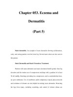
Table 6.1 Comparison of nal mean keratometric astigmatism in various suture techniques
Author(s) No. of eyes Suture technique Final average keratometric
astigmatism (D)
Murta et al. [35] 61 IS 2.77±1.34
Busin et al. [8] 15 IS 4.89±3.16
Troutman et al. [50] 74 IS/CICS 4.4–5.1
Heidemann et al. [26] 156 IS 6.36
Binder [4] 204 CICS 2.6
Karabatsas et al. [27] 51 CICS 2.66±1.70
Gross et al. [25] 63 (group 1)
103 (group 2)
CICS
CICS
2.94
3.27
Van Meter et al. [53] 31 CICS 3.2±1.9
Binder [6] 188 CICS 3.5
Filatov et al. [22] 20 CICS 3.9±2.5
Assil et al. [2] 19 CICS 4.07
Dursun et al. [18] 92 CICS 4.19±2.94
Van Meter et al. [53] 26 SCS 1.5±1.1
Serdarevic et al. [43] 25 SCS 1.75±1.04
Filatov et al. [22] 18 SCS 2.7±2.2
Ramirez et al. [39] 44 SCS 3.00±2.20
Karabatsas et al. [27] 44 SCS 3.12±2.62
Murta et al. [35] 14 SCS 3.90±1.70
Van Meter [51] 43 SCS 4.4±2.5
Assil et al. [2] 14 DCS 1.54
Clinch et al. [11] 30 DCS 2.66±0.24
Heidemann et al. [26] 57 DCS 3.75
Dolorico et al. [17] 91 DCS 3.98
Busin et al. [8] 22 DCS 3.98±3.69
Ramirez et al. [39] 48 DCS 4.2±2.1
Davison et al. [14] 33 DCS 4.5
Wi en et al. [54] 313 DCS 4.6
IS interrupted suture technique, CICS combined interrupted and continuous suture technique, SCS single con-
tinuous suture technique, DCS double continuous suture technique, D Diopter
Chapter 6 Corneal Suturing Techniques
54
6.4.2
Combined Interrupted and
Single Continuous Suture
A combination of interrupted sutures and a continu-
ous running suture (CICS) may be used to provide ap-
propriate wound apposition and closure [2, 4–6, 25,
45]. One of the most commonly utilized suture pat-
terns includes 12 interrupted sutures and a 12-bite
continuous running suture (CS), although eight inter-
rupted sutures and a 16-bite continuous running su-
ture is also commonly employed (Fig. 6.4a, b). A er
placement of the four cardinal interrupted sutures,
eight additional interrupted sutures are placed with
10-0 nylon suture. A er the sutures are trimmed and
the knots are buried, the CS is completed. Some sur-
geons employ qualitative keratoscopy to adjust or re-
place interrupted sutures before placement of the CS
component. ese surgeons generally repeat keratos-
copy a er CS placement as well. e CS is typically
completed in a clockwise fashion, using 10-0 or 11-0
nylon suture, with the rst bite midway between the 12
and 1 o’clock interrupted sutures. e CICS technique
can be performed with radial bites equidistant between
each of the interrupted sutures or by using an anti-
torque technique in which the apex of each bite in the
donor cornea forms an isosceles triangle with each in-
terrupted suture (Fig. 6.5). Several authors have sug-
gested that the antitorque CR reduces the torquing ef-
fects and pressure distortion induced from each bite
compared with radial running bites which may cause
pressure distortion and torque when the running por-
tion overlies the radial interrupted sutures [3, 19, 41,
47–49]. e CS depth is generally more super cial
than are the deeper interrupted sutures, creating better
approximation of Bowman’s layer. e CS is run the
entire 360°, with placement of a temporary knot at 12
o’clock. Additional slack is removed along the entire
length of the CS to square the apical points within the
corneal gra , and the suture is tied permanently at 12
o’clock with a 3-1-1 tying technique. e CS is then
rotated with two tying forceps, and the knot is buried.
Alternatively, the continuous running suture can be
started within the wound, and nished at the same
clock hour within the wound. A slipknot can be used
to secure the suture and adjusted a er the slack is re-
lieved. Once tied permanently, the knot can be le in
place a er the ends are trimmed, and further manipu-
lation of the CS to bury the knot is unnecessary.
e combination of interrupted and continuous
running sutures allows for earlier removal of inter-
rupted sutures to reduce postoperative astigmatism. If
astigmatism is acceptable (generally less than 3 diop-
ters [D]), sutures may be le alone until breakage,
loosening, scarring, or vascularization develops,
though the patient must be warned to call immediately
if they develop a foreign-body sensation. Interrupted
sutures can be removed as early as 4 weeks postopera-
tively to reduce corneal distortion and astigmatism as
measured by keratometry, photokeratoscopy, or com-
puterized corneal topography. Selective interrupted
suture removal can be performed until only the run-
ning suture is le in place. e CS can be le in place
Fig. 6.4 a Schematic diagram demonstrating appropriate
suture placement using the combined interrupted and single
continuous running keratoplasty technique with 12 inter-
rupted sutures and a 12-bite running suture. b A slit-lamp
photograph of the combined interrupted and continuous 12-
bite running suture.
Fig. 6.5 Schematic diagram of the combined interrupted
and single continuous running pattern using an antitorque
running technique.
W. Barry Lee and Mark J. Mannis
b
a
55
inde nitely, but is generally removed 12–18 months
following surgery. Astigmatism with this technique
varies from 2.16 to 4.19 D, with selective removal of
interrupted sutures followed by removal of the CS (Ta-
ble 6.1; [6, 18, 20, 27, 42]). If signi cant astigmatism
exists following removal of all interrupted sutures, cor-
neal astigmatism correction with surgical methods
will need to wait until the wound is stable enough to
have the continuous running suture removed. is
technique should not be performed in pediatric kera-
toplasty, tectonic keratoplasty, vascularized host cor-
neas from previous in ammation or infection, multi-
ple gra rejections, or conditions in which a risk of
melting is present such as in autoimmune conditions
like rheumatoid arthritis.
6.4.3
Single CRS Technique
e single continuous running suture technique was
rst described by Roper-Hall and popularized by
McNeill in the United States [33, 40]. is technique
carries the advantages of faster surgical time, one-time
suture removal, and potential for suture adjustment
intraoperatively and postoperatively. e disadvan-
tages of this technique include increased technical dif-
culty, the increased risk of needle dullness, impaired
wound integrity with only one improper bite, and dif-
culties of suture manipulation if the continuous su-
ture breaks intraoperatively. e technique is typically
performed with a 24-bite SCS of 10-0 nylon, although
some surgeons have performed this with a 16-bite
SCS. A er the four cardinal sutures are placed as
des cribed above, the surgeon starts the SCS between
12 and 1 o’clock, and the suture is run clockwise until
it is temporarily secured at 12 o’clock a er completion.
e surgeon places six bites per quadrant. e four
cardinal sutures are removed, and the anterior cham-
ber is lled to physiologic level before permanently
tightening the knot to avoid a topographically at do-
nor cornea. Tightening of the is achieved by using
tying forceps to release excess tension from each bite
in a clockwise manner until the desired tension is
achieved. Any excess tension from the lid speculum or
a scleral xation ring, if used, should be alleviated be-
fore the knot is tied permanently. Once the suture is
tied permanently, it is trimmed to the knot, and the
knot is buried. However, if the SCS is initiated within
the wound, once tied and cut ush, the knot will be
buried without further manipulation. e SCS can
then be adjusted for appropriate sphericity with typ-
ing forceps using intraoperative qualitative keratos-
copy (Fig. 6.6a, b).
When using the SCS technique, the surgeon should
pay careful attention to care of the needle point, place-
ment of continuous suture bites in a radial orientation,
placement of evenly spaced, symmetrical bites at 95%
depth, and prevention of suture breakage. If the SCS
does inadvertently break, it should be removed if the
pattern is in the rst quadrant and restarted to prevent
time delays. If the suture is broken in the nal quad-
rants, a new suture can be spliced to proximal end with
continuation, using the new needle (see Chap. 3 for
suture-splicing technique description). e rst spliced
knot can be buried at the end of suturing, and the SCS
can be tightened from the buried knot toward the su-
ture completion at 12 o’clock. In some situations, if the
initial suture end was le long, the SCS can be ad-
vanced to beyond the 12 o’clock position, and the
spliced section can subsequently be removed, leaving
only one knot to tie, as described earlier. When two
knots are buried a er splicing, suture adjustment
should be performed in two separate portions to mini-
mize astigmatism, but without exposure of either knot
or repeat breakage.
While the SCS technique represents an e cient and
e ective method for keratoplasty wound closure, it can
be problematic, as early suture removal may result in
wound instability and unacceptably high astigmatism.
Several clinical studies evaluating the single continu-
Fig. 6.6 a Schematic diagram of the single continuous run-
ning keratoplasty suturing technique using 24 bites with
10-0 nylon suture. b A slit-lamp photograph of the single
continuous running suture (CS) technique.
Chapter 6 Corneal Suturing Techniques
b
a
56
ous running suture describe low levels of astigmatism
and more rapid visual recovery as compared with oth-
er suturing techniques when early postoperative suture
adjustment techniques are implemented [22, 33, 42,
43, 45, 51–53]). Van Meter and colleagues compared
the SCS, and CICS techniques and found that the for-
mer was associated with signi cantly less astigmatism
(1.5±1.1 D as compared with 3.2±1.9 D), fewer post-
operative suture adjustments (0.9 as compared with
3.8), and earlier refractive stability (7 months earlier)
(see Table 6.1 [53]. Patients undergoing intraoperative
suture adjustment are reported to have signi cantly
decreased astigmatism, more regular corneas, and bet-
ter spectacle-corrected visual acuity until the running
suture is eventually removed [42, 43, 51]. ese data
must be weighed against a 7.2% risk of spontaneous
wound dehiscence following suture removal associated
with this technique [1].
e ideal time for postoperative suture adjustment
is 3–6 weeks following keratoplasty, since this provides
adequate time for gra re-epithelialization and ade-
quate measurement of astigmatism with corneal to-
pography or keratoscopy. is timeframe also allows
for easier manipulation of the SCS, with microsurgical
instruments reducing risk of suture breakage, a risk
that increases with later postoperat ive suture adjust-
ments a er more complete wound healing. SCS ad-
justment is performed with sterile tying forceps at the
slit-lamp, following administration of a drop of topical
antibiotic or topical povidone iodine and topical anes-
thetic. Keratometry, photokeratoscopy, or corneal to-
pography should be reviewed prior to suture adjust-
ment in order to establish the proper adjustment plan.
Prior to suture adjustment, one tip of the typing for-
ceps is placed through the epithelium and Bowman’s
layer along the gra –host junction at the steep merid-
ian. A er severing these anterior layers, the tip of the
forceps is used to li the suture, which is carefully ad-
vanced along the suture direction. is maneuver is
performed at the steep meridian to serve as a relaxing
incision in conjunction with suture adjustment. Once
the suture moves, it is advanced loop by loop from the
area of the attest meridian and distributed to the area
of the steepest meridian as measured by topography or
keratometry until the tension has been evenly dis-
persed around the entire circumference of the wound.
If a suture is too tight, adjustment plans should be
aborted since attempting to adjust a very tight suture
may lead to breakage. Avoidance of twisting the suture
over the tips of the tying forceps and careful advance-
ment of the suture along the line of suture placement
can reduce the risk of the SCS breaking during adjust-
ment. A er completion of suture adjustment, the kera-
tometric astigmatism should be measured a er stabili-
zation has occurred (typically in 2–3 weeks) to
determine the e ectiveness of the adjustment. Several
adjustments may be required to arrive at an acceptable
level of keratometric astigmatism and subsequent vi-
sual acuity.
6.4.4
Double Continuous Suture Technique
e double continuous running suture technique was
rst described in 1977 [32]. e DCS can be performed
with two 10-0 nylon sutures, a 10-0 nylon suture and
11-0 nylon suture, or a 10-0 nylon suture and an 11-0
Mersilene suture. is technique provides the bene ts
of a SCS, with the added safety and security of a sec-
ond SCS.
A er placement of the four cardinal interrupted su-
tures, a 12-bite running 10-0 nylon CRS is placed. e
CRS is run clockwise for 360° and tied temporarily at
12 o’clock. Each suture pass should be placed at 80% of
the depth of the donor cornea and recipient cornea.
e slack is removed and the knot is tied permanently
at 12 o’clock and buried. e four cardinal sutures are
removed. A second suture (10-0 or 11-0 nylon or 11-0
Mersiline) is placed between each of the previous bites
and run clockwise for 360°. e second CRS is placed
at 50–60% of the corneal depth to approximate Bow-
man’s layer on both sides of the wound. e knot is
tied temporarily at 12 o’clock, and the slack is removed,
with a permanent tie completed at 12 o’clock (Fig. 6.7).
e tension of the second suture should allow for only
enough tension to take up slack in the suture. e sec-
ond running suture permits early removal of the rst
CRS in 2–3 months, depending of the level of astigma-
tism. e second CRS may be le inde nitely, depend-
ing on the level of astigmatism, or it may be removed
at 12–18 months.
Fig. 6.7 Slit-lamp photograph depicting the double running
suture technique. (Photo courtesy of Woodford Van Meter,
M.D.)
W. Barry Lee and Mark J. Mannis
57
e disadvantage of this technique is the time re-
quired to perform two continuous running sutures
and requirement of signi cant expertise. is tech-
nique can potentially cause premature breakage or
severing of the rst suture with an improper pass of
the second continuous running suture. In addition,
care must be taken to avoid bending the needle during
each pass or dulling the tips of the needle with each
instrument grasp. Also, each suture bite must be regu-
lar and symmetrical in order to close the wound in an
adequate fashion. Any irregular or improperly placed
bite can lead to wound instability and inappropriate
wound healing.
Several studies report excellent long-term stability
with an acceptable range of postoperative astigmatism,
and some authors consider it the most stable and se-
cure suture technique [7, 14, 17, 32]. Rapid visual re-
covery and low levels of nal astigmatism occur with
early postoperative adjustment of the 10-0 nylon deep
[11, 14, 32]. Marked variability exists in the literature
regarding the e ect of vision and postoperative astig-
matism following suture removal with this technique,
since some studies have shown an increase in astigma-
tism, whereas others have found no change or a de-
crease in the amount of astigmatism [7, 17, 32, 36, 46,
47, 50]. e deep 10-0 nylon CRS is typically removed
rst, followed by removal of the more anterior suture
at 12–24 months. A retrospective study of 91 patients
undergoing the DCS technique found an average post-
operative keratometric astigmatism of 3.73 D a er su-
tures were removed at an average follow-up of 13.7
months with 94% having best-correctable vision of
20/60 or better [17]. Average sutures-out keratometric
astigmatism with this technique can vary widely as
with all suture patterns in keratoplasty (see Table 6.1;
[36, 54]).
6.5
Pediatric Keratoplasty
Pediatric keratoplasty deserves special mention, as
these cases present a variety of challenges that are not
routinely encountered in adult cases. Challenges in pe-
diatric keratoplasty include smaller working space, de-
creased corneal dimensions, smaller ocular structures
and shallow anterior chambers, more signi cant poste-
rior pressure, and more scleral and corneal tissue elas-
ticity. ese factors provide heightened risks for intra-
operative and postoperative complications, with a
greater potential for iris prolapse or expulsion of ocular
contents. A scleral xation ring should be placed, with
xation to the episclera during the initial stages of the
case to provide better globe stabilization and assist with
management of increased posterior pressure and tissue
elasticity.
In regard to suture placement, children have more
elastic recipient corneas as compared with adults, more
posterior pressure, more elastic donor corneas due to
younger donor tissue, and a tendency for suture loos-
ening sooner than adults, all of which account for
greater suture pattern variability. is variability makes
the single interrupted suture technique the ideal tech-
nique in pediatric cases, because it provides for better
wound apposition and a more stable wound as sutures
began to loosen over time. Continuous suture patterns
are not recommended for pediatric cases. In pediatric
keratoplasty, suture removal is o en initiated as early
as 2 weeks postoperatively, depending on the state of
corneal healing. Frequent examinations under anes-
thesia are commonly encountered in pediatric cases
for suture removal and adequate viewing of corneal
gra s in cases where children are too young to cooper-
ate with a slit-lamp examination. A team approach is
o en needed in these cases, with coordination of care
between a pediatric ophthalmologist, a glaucoma spe-
cialist, and the corneal surgeon as amblyopia, glauco-
ma, and gra failure are very common occurrences in
pediatric keratoplasty cases.
6.6
Suture-Related Complications
Complications from corneal suturing techniques in
keratoplasty can be divided into intraoperative and
postoperative complications. Intraoperative complica-
tions may include forward movement of the lens–iris
diaphragm, disrupting suturing by iris prolapse and
creating a potential for lens damage or expulsion. e
most dreaded complication creating this forward shi
is a suprachoroidal hemorrhage, a complication that
can progress to an expulsive choroidal hemorrhage
with expulsion of intraocular contents. Other intraop-
erative complications may include violation of, or con-
tact with, the anterior lens capsule, leading to a trau-
matic cataract; inadvertent iridectomy when excising
the diseased cornea; and damage to the donor endo-
thelium from tissue manipulation or poor handling
techniques. Improper suture placement can lead to iris
incarceration, lens violation, and a higher risk of su-
ture abscess or endophthalmitis in the postoperative
course. Improper suture tension can create undesir-
able astigmatism or donor–recipient mismatch, which
can lead to di culty in creating a watertight wound
once suturing is completed, as well as signi cant astig-
matic refractive error postoperatively.
While intraoperative complications for an experi-
enced corneal surgeon typically remain limited, post-
operative complications are numerous and are com-
monly encountered. Postoperative complications
following suture techniques in keratoplasty include
Chapter 6 Corneal Suturing Techniques
58
wound leak with a at anterior chamber, hyphema,
traumatic cataract, iris prolapse and peripheral iris
synechiae to the gra –host junction, secondary glau-
coma, and retrocorneal membranes [15]. Loose su-
tures in the immediate or late postoperative course can
lead to suture vascularization or wound dehiscence
[10]. In particular, late postoperative wound dehis-
cence has been reported in one study with the 24-bite
SCS in 7.2% of patients, with the majority of cases oc-
curring within 2 weeks of suture removal [1]. While
wound dehiscence typically occurs soon a er removal
of sutures, late postoperative wound dehiscence has
also been reported 10–19 years a er suture removal
[38]. Infections such as endophthalmitis, suture ab-
scesses, and gra ulceration may also occur in associa-
tion with loose sutures. Postkeratoplasty surface kera-
topathy is one of the most common postoperative
complications a er astigmatism. It can present in
many forms including hurricane keratopathy, lamen-
tary keratitis, keratitis medicamentosa, persistent epi-
thelial defects, and super cial hypertrophic dendri-
form epitheliopathy (SHDE) [19, 28]. ese surface
complications can indirectly a ect sutures and require
observation for potential suture melting or in ltration
when present.
Astigmatism is the most common postoperative su-
ture-related complication in keratoplasty. Factors felt
to increase the risk of high amounts of astigmatism in-
clude increased external pressure exerted on the globe
such as a tight lid speculum or improperly sutured
scleral xation ring. Other factors related to astigma-
tism include inappropriate trephination procedures,
donor–recipient mismatch, sutures with inappropriate
tension, inconsistent suture depth, lack of suture radi-
ality, asymmetrical suture placement, and/or malposi-
tioned cardinal sutures.
6.7
Future Challenges
Despite the many advances made in corneal surgery
over the last decade, the ideal suturing technique re-
mains to be identi ed. Regardless of the various ad-
vances in instrumentation, surgical technique, and our
knowledge of immunobiology, perfect and reproduc-
ible results in corneal surgery and keratoplasty in par-
ticular do not exist. Although the success of penetrat-
ing keratoplasty is commonly over 90% in routine
cases, intraoperative and postoperative complications
will always remain a risk with corneal surgery [7]. De-
spite the increased success of contemporary kerato-
plasty, suture-related complications continue to exist.
Regardless of these inherent risks, postoperative com-
plications such as astigmatism, wound dehiscence, and
suture-related infections can be diminished with care-
ful attention to appropriate suture technique and care-
ful and close follow-up of patients a er corneal sur-
gery techniques. Nonetheless, the fundamentals of
corneal wound closure and appropriate tissue apposi-
tion represent the core foundation of knowledge for
the corneal surgeon.
References
1. Abou-Jaoude ES, Brooks M, Katz DG et al (2002) Spon-
taneous wound dehiscence a er removal of single con-
tinuous penetrating keratoplasty suture. Ophthalmology
109:1291–1296
2. Assil KK, Zarnegar SR, Schanzlin DJ (1992) Visual out-
come a er penetrating keratoplasty with double con-
tinuous or combined interrupted and continuous suture
wound closure. Am J Ophthalmol 114:63–71
3. Au Y-K, Mahjoub SB, Hart JC (1990) Suture patterns
and corneal gra rotation in the cadaver eye. Ophthal-
mic Surg 21:472–474
4. Binder PS (1985) Reduction of postkeratoplasty astig-
matism by selective suture removal. Dev Ophthalmol
11:86–90
5. Binder PS (1985) Selective suture removal can reduce
postkeratoplasty astigmatism. Ophthalmology 92:1412–
1416
6. Binder PS (1988) e e ect of suture removal on post-
keratoplasty astigmatism. Am J Ophthalmol 105:637–
645
7. Bourne WM (1981) Current techniques for improved
visual results a er penetrating keratoplasty. Ophthalmic
Surg 12:321–327
8. Busin M, Monks T, Al-Nawaiseh I (1998) Di erent su-
turing techniques variously a ect the regularity of post-
keratoplasty astigmatism. Ophthalmology 105:1200–
1205
9. Brady SE, Rapuano CJ, Arensten JJ et al (1989) Clinical
indications for and procedures associated with penetrat-
ing keratoplasty 108:118–122
10. Christo CG, Rooij J, Geerards AJM et al (2001) Suture-
related complications following keratoplasty. Cornea
20:816–819
11. Clinch TE, ompson HW, Gardner BP et al (1993) An
adjustable double running suture technique for kerato-
plasty. Am J Ophthalmol 116:201–206
12. Cosar CB, Sridhar MS, Cohen EJ (2002) Indications for
penetrating keratoplasty and associated procedures,
1996–2000. Cornea 21:148–151
13. Cottingham AJ (1980) Residual astigmatism following
keratoplasty. Ophthalmology 87(S):113
14. Davison J, Bourne WM (1980 Results of penetrating
keratoplasty using a double running suture technique.
Arch Ophthalmol 99:1591–1595
15. Dhanda RP, Kalevar V (1972) Corneal surgery. Int Oph-
thalmol Clin 12:3–420
16. Dobbins KRB, Price FW, Whitson WE (2000) Trends in
the indications for penetrating keratoplasty in the Mid-
western United States. Cornea 19:813–816
17. Dolorico AMT, Tayyani, Ong HV et al (2003) Shortterm
and longterm visual and astigmatic results of an oppos-
ing 10-0 nylon double running suture technique for
penetrating keratoplasty. J Am Coll Surg 197:991–999
18. Durson D, Forster RK, Feuer WJ (2002) Suturing tech-
nique for control of postkeratoplasty astigmatism and
myopia. Trans Am Ophthalmol 100:51–60
19. Eisner G (1980) Eye Surgery: An Introduction to Opera-
tive Technique. Springer, Berlin Heidelberg New York,
pp38–40
W. Barry Lee and Mark J. Mannis
59
20. Eliason JA, McCulley JP (1990) A comparison between
interrupted and continuous suturing techniques in kera-
toplasty. Cornea 9:10–16
21. Feiz V, Mannis MJ, Kandavel G et al (2001) Surface kera-
topathy a er penetrating keratoplasty. Trans Am Oph-
thalmol Soc 99:159–168
22. Filatov V, Steinert RF, Talamo JH (1993) Postkeratoplas-
ty astigmatism with single running suture or interrupted
sutures. Am J Ophthalmol 115:715–721
23. Fine M (1962) Technique of penetrating keratoplasty.
Symposium on the Cornea. Trans New Orleans Acad
Ophthalmol. Mosby, St. Louis, pp 132–142
24. Fine M (1970) Techniques of keratoplasty. Int Ophthal-
mol Clin 10:271–296
25. Gross RH, Poulsen EJ, Davitt S et al (1997) Comparison
of astigmatism a er penetrating keratoplasty by experi-
enced cornea surgeons and cornea fellows. Am J Oph-
thalmol 123:636–643
26. Heidemann DG, Sugar A, Meyer RF et al (1985) Over-
sized donor gra s in penetrating keratoplasty. Arch
Ophthalmol 103:1807–1811
27. Karabatsas CH, Cook SD, Figueiredo FC et al (1998)
Combined interrupted and continuous versus single
continuous adjustable suturing in penetrating kerato-
plasty. Ophthalmology 105:1991–1998
28. Lee WB, Mannis MJ, Mehra N et al (2005) Super cial
hypertrophic dendriform epitheliopathy: A follow-up
series. Cornea 25:273–276
29. Lois N, Kowal VO, Cohen EJ et al (1997) Indications for
penetrating keratoplasty and associated procedures,
1989–1995 16:623–629
30. Mannis MJ, Tran L, A Panorama, 1789–1999 (1999) In:
Mannis MJ, Mannis AA (eds) Corneal transplantation: a
history in pro le. Wayenborgh, Belgium
31. Mannis MJ, Krachmer JH (1981) Keratoplasty: a histori-
cal perspective. Surv Ophthalmol 25:333–338
32. McNeill JI, Kaufman HE (1977) A double running su-
ture technique for keratoplasty: earlier visual rehabilita-
tion. Ophthalmic Surg 8:58–61
33. McNeill JI, Wessels F (1989) Adjustment of single con-
tinuous suture to control astigmatism a er penetrating
keratoplasty. Refract Corneal Surg 5:216–223
34. Melles GRH, Binder PS (1990) A comparison of wound
healing in sutured and unsutured corneal wounds. Arch
Ophthalmol 108;546–548
35. Murta JN, Amaro L, Tavares C et al (1994) Astigmatism
a er penetrating keratoplasty. Doc Ophthalmol 87:331–
336
36. Musch DC, Meyer RF, Sugar A (1988) e e ect of re-
moving running sutures on astigmatism a er penetrat-
ing keratoplasty. Arch Ophthalmol 106:488–492
37. Olson RJ (1988) Prevention of astigmatism in corneal
transplant surgery. Int Ophthalmol Clin 28:37–45
38. Pettinelli DJ, Starr CE, Stark WJ (2005) Late traumatic
corneal wound dehiscence a er penetrating keratoplas-
ty. Arch Ophthalmol 123:853–856
39. Ramirez M, Hodge DO, Bourne WM (2001) Keratomet-
ric results during the rst year a er keratoplasty: Adjust-
able single running suture technique versus double run-
ning technique. Ophthalmic Surg Lasers 32:370–374
40. Roper-Hall M (1985) Control of postoperative astigma-
tism. Br J Ophthalmol 69:348–351
41. Rowsey JJ (1987) Prevention and correction of corneal
transplant astigmatism. Trans New Orleans Acad Oph-
thalmol 35:35–51
42. Serdarevic ON (1994) Refractive corneal transplanta-
tion: control of astigmatism and ametropia during pen-
etrating keratoplasty. Int Ophthalmol Clin 34:13–33
43. Sedarevic ON, Rneard GJ, Pouliquen Y (1995) Random-
ized clinical trial of penetrating keratoplasty. Ophthal-
mology 102:1497–1503
44. Stainer GA, Perl T, Binder PS (1982) Controlled reduc-
tion of postkeratoplasty astigmatism. Ophthalmology
89:668–676
45. Temnycky GO, Lindahl KJ, Aquavella JV (1991) Early
visual rehabilitation following keratoplasty using a sin-
gle continuous adjustable suture technique. Ophthalmic
Surg 22:208–212
46. Troutman RC (1974) Microsurgery of the anterior seg-
ment of the eye, vo1. 1. Mosby, St. Louis, pp 187–195
47. Troutman RC (1977) Microsurgery of the anterior seg-
ment of the eye, vol. 2. Mosby, St. Louis, pp 40–41
48. Troutman RC Willard DE (1965) Management of the
aphakic patient. Symposium on cataracts. Trans New
Orleans Academy of Ophthalmology. Mosby, St. Louis,
pp 261–279
49. Troutman RC, Meltzer M (1972) Astigmatism and myo-
pia in keratoconus. Trans Am Ophthalmol Soc 70:265–
277
50. Troutman RC, Gaster RN (1980) Surgical advances and
results of keratoconus. Ophthalmology 90:131–136
51. Van Meter W (1996) e e cacy of a single continuous
nylon suture for control of post keratoplasty astigma-
tism. Tr Am Ophth Soc 44:1157–1180
52. Van Meter W, Katz DG (2004) Keratoplasty suturing
techniques. In: Krachmer JH, Mannis MJ, Holland EJ
(eds) Cornea, 2nd edn. Mosby, St. Louis
53. Van Meter WS, Gussler JR, Soloman KD et al (1991)
Postkeratoplasty astigmatism control. Ophthalmology
98:177–183
54. Wi en SJ, Maguire LJ, Bourne WM (1997) Keratometric
results of penetrating keratoplasty with the Hessburg-
Barron and Hanna trephine systems using a standard
double-running suture technique. Cornea 16:306–313
Chapter 6 Corneal Suturing Techniques
Chapter 7
Trauma Suturing
Techniques
Marian S. Macsai and Bruno Machado Fontes
7
Key Points
• Assess the presence of life-threatening inju-
ries.
• Vision at the time of presentation and the
presence or absence of a erent pupillary de-
fect are important prognostic factors in the
Ocular Trauma Classi cation System [1].
• Surgical goals include:
– Watertight wound closure
– Restoration of normal anatomic relation-
ships
– Restoration of optimal visual function
– Prevention of possible future complica-
tions
• Surgical indications:
– Any perforating injury
– Any wound with tissue loss
– Any clinical suspicion of globe rupture re-
quires exploration and possible repair
• Instrumentation:
– Complete ophthalmic microsurgical tray
– Phacoemulsi cation, vitrectomy and irri-
gation and aspiration machines
– Variety of microsurgical sutures
• Surgical techniques:
– Self-sealing wounds or lacerations <2 mm
may not require surgical repair.
– Close perpendicular aspect of the wound
rst, the oblique aspect second.
– Avoid wound override.
– In the zigzag laceration a mattress suture
may be needed.
– In a stellate laceration a purse string may
su ce.
– Extruded vitreous is a strong risk factor
for retinal detachment.
• An ideal initial surgical repair may eliminate
the need for future reconstruction.
• Monitor patient for postoperative complica-
tions.
• Long-term follow up indicated.
7.1
Introduction
Ocular trauma is an important cause of unilateral vi-
sion loss worldwide, especially in young people, and
surgical repair is almost always challenging [1–7]. A
patient with an eye injury may need immediate inter-
vention, and all ophthalmologists who cover emergen-
cy patients must have the knowledge and skills to deal
with di cult and complex surgeries, as these initial ac-
tions and interventions may be determinants for the
nal visual prognosis [7–15]. One must keep in the
mind that the result of the rst surgery will determine
the need for future reconstruction.
e epidemiology of ocular trauma varies according
to the region studied. In the World Trade Center disas-
ter, ocular trauma was found to be the second most
common type of injury among survivors [16]. e
most common causes of eye injuries include automo-
tive, domestic, and occupational accidents, together
with violence. Risk factors most commonly described
for eye injuries are male gender (approximately 80% of
open-globe injuries), race (Hispanics and African-
Americans have higher risk), professional activity (e. g.,
military personnel), younger age (third decade), low
education, contact sports, and failure to comply with
safety devices and equipment [1–3, 5, 6, 9, 16–19]. An-
terior corneoscleral lacerations, in sites of previous
ocular surgery, and posterior ruptures are more com-
mon in the elderly as a result of frequent falls.
Any potentially life-threatening injury takes prece-
dence over ocular injuries. e patient should undergo
a careful evaluation by quali ed emergency medical
personnel and severe pain or nausea should be treated
to decrease lid squeezing and Valsalva maneuver ef-
fects [4, 20–23]. e initial ophthalmologic evaluation
is critical. Trauma mechanism and injury characteris-
tics according to the Ocular Trauma Classi cation Sys-
tem [1, 7] can predict the prognosis and nal visual
outcomes (Fig. 7.1).
e evaluation of initial visual function is the most
important measurement by the initial as visual func-
tion is directly related to visual prognosis, and is also
important from a medicolegal perspective. e exam-
iner should assess visual acuity with whatever equip-
62
ment is available and this information must be docu-
mented in the patient’s chart. In addition, the examiner
should assess the pupillary re ex with attention to the
presence or absence of an a erent defect. A slit-lamp
assessment of the extent of the injury should deter-
mine if the cornea is lacerated and whether the lens is
clear or opaci ed. Any opaci cation may indicate rup-
ture of the lens capsule. Visualization of the posterior
pole should be attempted, as the rst examiner may be
the only one able to obtain a clear view of the posterior
segment and their ndings must be documented.
THE OCULAR TRAUMA CLASSIFICATION
SYSTEM
1
FOR OPEN-GLOBE INJURIES
Type
A Rupture
B Penetrating
C Intraocular foreign
body
D Perforating
E Mixed
Grade
Visual acuity *
1. ≥ 20/40
2. 20/50 to 20/100
3. 19/100 to 5/200
4. 4/200 to light
perception
5. No light perception†
Pupil
Positive: relative a erent
pupillary defect present
in a ected eye
Negative: relative
a erent pupillary defect
absent in a ected eye
Zone
I. Isolated to cornea
(including the
corneoscleral
limbus)
II.
Corneoscleral limbus
to 5 mm posterior to
the sclera
III.
Posterior to the
anterior 5 mm of
sclera
* Measured at distance (20 , 6m) using Snellen chart or
Rosenbaum near card, with correction and pinhole
when appropriate.
† Con rmed with bright light source and fellow eye well
occluded.
7.2
Surgical Indications
Surgery is indicated when there is a risk of loss of nor-
mal anatomic structure or function of the eye. Indica-
tions include partial and/or full-thickness lacerations
with aqueous leakage and intraocular tissue extrusion
or prolapse. Surgery may be delayed by the patient’s
medical condition, but is best performed as soon as
possible to reduce the risk of complications (such as
endophthalmitis, tissue necrosis, and expulsive hemor-
rhage). A rigid shield is indicated to protect the globe
from external pressure in all patients with open-globe
injuries. Topical ocular medications should be avoided
because of the risk of intraocular toxicity, and systemic
antibiotic prophylaxis should be started immediately. If
indicated, tetanus prophylaxis must be updated.
Simple self-sealing wounds or short lacerations (<2
mm) with good tissue approximation, minimal gape,
no evidence of intraocular penetration, and no sign of
infection or necrosis can be managed with a bandage
contact lens and/or tissue adhesive (Fig. 7.2), in addi-
tion to topical broad-spectrum antibiotics, cycloplegic,
and hypotensive drugs. With this approach the need
for sutures is diminished, but patients must be closely
followed. An eye shield must be placed, and the patient
must refrain from any activity that results in a Valsalva
maneuver. is is not a reasonable approach for chil-
dren and mentally disabled patients.
As a general principle, a surgical plan should be
made before surgery, including tissue conservation
and iatrogenic damage minimization. However, unex-
pected intraoperative situations can require a broad
spectrum of surgical techniques. To avoid delays dur-
ing the surgical procedure, the need for special equip-
ment, such as a vitrectomy and/or phacoemulsi cation
machine, should be determined previously. General
anesthesia is preferred, as retrobulbar or peribulbar
anesthesia may increase the intraocular pressure (IOP)
and risk extrusion of intraocular contents. Surgical
closure should proceed in a timely manner to decrease
the risk of endophthalmitis, avoid tissue necrosis and
decrease patient diskomfort [23].
Overall surgical goals in ocular lacerations include
(1) watertight wound closure, (2) restoration of nor-
mal anatomic relationships, (3) restoration of optimal
visual function, and (4) prevention of possible future
complications (e. g., glaucoma).
e overall goal is to restore the native corneal con-
tour with minimal scarring. Corneal tissue should be
conserved as much as possible to avoid wound distor-
tion or misalignment resulting in irregular astigma-
Fig. 7.1 Ocular Trauma Classi cation System
Fig. 7.2 Technique to apply tissue adhesive to small lacera-
tions. A broken wooden applicator with a small cu of oint-
ment is adherent to a small polyethylene disk. e tissue glue
is on the opposite side of the polyethylene disk and is applied
to the area of the laceration. e wound must be dry and free
of epithelium for the adhesive to stick. Tissue glue will not
adhere to a wet or epithelialized surface
Marian S. Macsai and Bruno Machado Fontes
63
tism. If an avulsed piece of viable corneal tissue is pres-
ent, it should be sutured back into place. Any
anatomic landmark (such as pigmentation lines, scars,
laceration edges, or the limbus) can help the surgeon
identify and restore the eye’s normal anatomy. Lacera-
tions should be carefully explored to identify and re-
move any foreign materials. Infection should be as-
sumed, and the wound and any intraocular samples
should be submitted for culture and sensitivity.
7.3
Instrumentation and Equipment
• Lid speculum
• Microsurgical 0.12-mm forceps
• Microsurgical tying forceps (two)
• Nonlocking needle holder
• Vannas scissors
• Iris hooks
• Cyclodialysis spatula
• Muscle retractors
• Viscoelastic
• Cellulose sponges
• Tissue glue (when applicable)
• Phacoemulsi cation, irrigation, and aspiration,
and automated vitrectomy units should be imme-
diately available
7.4
Surgical Technique
For proper healing, the wound edges should be exactly
apposed. Regardless of design, sutures seek (when
tightened) their most stable geometric con guration.
erefore, correct passage of a suture is necessary to
achieve good wound apposition.
Perpendicular parts of the wound will open under
normal IOP, so initial closure of these areas will en-
hance anterior chamber formation as the shelved areas
of the incision are o en self-sealing (Fig. 7.3). Tempo-
rary sutures may be needed to obtain a watertight clo-
sure, and once the shelved areas are closed, the initial
sutures may be replaced with more astigmatically neu-
tral sutures. If at all possible, suture bites through the
visual axis should be avoided.
Management of prolapsed tissue is one of the initial
step as the wound is closed, it is imperative that intra-
ocular contents not be incarcerated in the wound or
sutures. Extruded vitreous or lens fragments should be
excised at the eye’s surface. Retinal and uveal tissue
should be gently repositioned if the tissue shows no
sign of infection or necrosis. is can be done with a
viscoelastic and smooth instruments to avoid addi-
tional damage.
7.4.1
Suturing the Cornea
A mono lament suture (nylon or polypropylene)
works well in the cornea, because of its low tissue reac-
tivity. Spatulated needles are preferred for maintenance
of suture depth in partial-thickness lacerations. e
most stable con guration of interrupted sutures is a
planar loop, so the tissue contained within the suture
can be warped and distorted with inadequate suture
tension. For proper placement, the tip of the needle
For proper placement, the placed perpendicular to the
corneal surface, and the needle is rotated through the
wound along its curve, exiting perpendicular to the
cut surface. Corneal sutures should be 90% deep in the
stroma and of equal depth on both sides of the wound.
Full-thickness sutures may allow the suture material to
act as a conduit for microbial invasion. Suture passes
should be approximately 1.5 to 2.0 mm in total length,
and the needle pass through the opposite side should
mirror the initial needle pass in depth and length. is
can be di cult in macerated and edematous tissue,
and one must keep in mind the need to incorporate
healthy tissue in each suture pass, or else the sutures
will pull through the tissue when tied.
Sutures result in wound apposition by compressing
the tissues within the loop. Interrupted sutures gener-
ate a plane of compression in the tissue contained with-
in the suture loop and a zone of compression extending
away from the suture itself. e compression zones
have a roughly triangular con guration extending ap-
proximately one half the suture total length in either
direction along the wound. Wound closure is achieved
when compression zones abut. Wound leakage occurs
when there is insu cient overlap of compression zones
so as to permit wound gape and leakage (see Chap. 1).
Opens
Closes
Fig. 7.3 e drawing illustrates the relationship between the
perpendicular areas of the laceration and the shelved areas.
If the perpendicular areas are closed initially, then the shelved
areas are self-sealing and require fewer sutures under less
tension
Chapter 7 Trauma Suturing Techniques
64
All knots should be trimmed short and super cially
buried in the tissue, on the side away from the visual
axis [23]. e ends of the buried knot should be di-
rected away from the surface to facilitate subsequent
removal. e suture should be tied using the smallest
possible knot to facilitate burying of the surgical knot
in the tissue. A granny-style slipknot allows for con-
trolled closure of the wound and is small enough to be
buried easily [23] (see Chap. 3).
Tissue compression leads to attening of the overly-
ing surface, and this fact is most important when su-
turing the cornea. e goal of cornea suturing is to
make the wound watertight with minimal scarring and
astigmatism. e Rowsey-Hays technique (Fig. 7.4)
was developed with this aim [24] as the normal cornea
attens over any vertical or sutured incision, but steep-
ens adjacent to tight limbal sutures. erefore, corneal
lacerations be closed with long, tight sutures in the
corneal periphery, and shorter, minimally compressive
sutures in corneal center (thus causing peripheral at-
tening and central steepening) (Fig. 7.5). Long suture
bites allow a greater distance between sutures, and
smaller bites require more closely spaced sutures, to
overlap the zones of compression. But excessive over-
lap of compression zones can lead to excessive scarring
and tissue attening [20].
To avoid wound override (Fig. 7.6), the entry and
exit of suture bites must be of equal tissue depth. Also,
the bites on either side of the perpendicular laceration
must be of equal depth from the anterior perspective,
and the passage of the suture of equal lengths as gauged
from the posterior aspect of the shelved wound not
from the anterior view. As a result, the suture place-
ment in a perpendicular laceration will appear very
di erent from the suture placement in a shelved inci-
sion. Suture placement is critical to avoid tissue over-
ride and the inducement of irregular astigmatism.
Running sutures have more complex e ects on con-
tained tissue: a single running suture will cause hori-
zontal wound slippage equal to approximately one half
the average suture bite. Running sutures tend to atten
the overlying corneal surface throughout the length of
the suture and to straighten curvilinear wounds be-
cause of the continuous nature of the compressive ef-
fects of running sutures. In addition, closure with run-
ning sutures places the integrity of the entire wound
on a single suture, which may pose a safety risk. For
these reasons, running sutures avoided in traumatic
corneal wounds.
Normal
A
BB
C
C
D
Problem
Solution
Fig. 7.5 Photograph of an astigmatically neutral closure of a
large corneal laceration. e patient recovered 20/40 vision
a er suture removal
Marian S. Macsai and Bruno Machado Fontes
Fig. 7.4 (A) Small, short bites will atten the central cornea
and create a bend (B) in the paracentral cornea. Placing larg-
er bites in the periphery will steepen the peripheral cornea
(C), while the small central short bites atten the central cor-
nea (D), resulting in a more normal cornea curvature.
65
7.4.2
Suturing the Zigzag Incision
Each linear aspect of the incision should be closed indi-
vidually to allowself-sealing of the wound apices and
avoiding additional trauma. In repairing these lacera-
tions, the use of slipknots is helpful. e straight aspects
of the zigzag incision are closed rst with interrupted
sutures. e apical portion of the incision may then self-
seal (Fig. 7.7). If the apical portions require suture clo-
sure, a mattress suture technique [21, 25] (Fig. 7.8) may
be useful.
Correct
Incorrect
Tissue override
Tissue override
<
Incorrect
Correct
<
d
c
b
a
B
A=
B
A
D=
C
B
A=
B
A
Chapter 7 Trauma Suturing Techniques
Fig. 7.6 a Correct closure of perpendicular incision. e
distance from the point of entry of the suture to the wound
(A) is equal to the distance from the wound to the point of
exit (B), and the sutures are passed at equal depths. b Incor-
rect closure of a perpendicular wound. e distance from the
point of entry to the wound (A) is not equal to the distance
from the wound to the point of exit (B). is results in wound
override. c Incorrect closure of an oblique wound. If the
same technique is followed for an oblique wound as is fol-
lowed for a perpendicular wound (A = B), tissue override
will result. d Correct closure of an oblique wound to ensure
proper tissue apposition. In this technique, the distance from
the point of entry of the suture to the point of exit through
the wound (C) should be measured from the posterior as-
pect of the cornea, and should be equal to the distance from
the wound to the point of exit (D) as measured from the pos-
terior aspect of the cornea. As a result, C = D and A ≠ B as
they are measured from the anterior aspect of the cornea
66
a b c
Fig. 7.7 e linear aspects
of the zigzag laceration are
closed initially, as the apical
portions may be shelved
and self sealing
Fig. 7.8 a A trephine is used to mark the area around the
ulcerated area. Trephination is not performed, as the intra-
ocular contents may extrude from the external pressure. b
An exceptionally sharp blade is used to cut down to a 50–
60% depth. c A lamellar dissection is performed to remove
the necrotic tissue. e trephine is then moved to another
peripheral area of the same cornea and used to trephine a
50% depth in the healthy peripheral corneal tissue. Lamellar
dissection is used to harvest this donor lenticule. d e do-
nor lenticule is secured in position with interrupted 10-0
nylon sutures to close the perforated area. e exposed stro-
ma where the donor lenticle was harvested is allowed to heal
by secondary intention, with either patching or a bandage
contact lens
Marian S. Macsai and Bruno Machado Fontes
67
7.4.3
Stellate Laceration Closure
In the stellate laceration, the straight arms of the lac-
eration are closed initially with interrupted sutures.
e stellate portion is closed last. Two di erent tech-
niques may be used including the Eisner method purse
string (Fig. 7.9 and 7.10) [25] or the Akkin method
(Fig. 7.11) [26].
Full-thickness or penetrating gra s at the time of
initial surgery are rarely required. e surgeon should
notify the eye bank of the possible need for corneal
tissue before surgery, as donor corneas are not univer-
sally available. Gra survival when performed as part
of the primary repair is guarded, because of the post
operative in ammatory response. However, a partial
thickness lamellar patch may be needed in areas of tis-
sue loss (Fig. 7.12). If the surgeon discovers an area of
Diamond knife
½ thickness
stromal incision
1
2
3
4
5
Eisner Method
1
4
2
3
Akkin Method
Fig. 7.9 In the Eisner
method, a partial thick-
ness incision is made be-
tween the arms of the
laceration and a purse-
string suture is passed
through these grooves
and tightened to approxi-
mate the apices of the
wound. Overtightening
of the purse-string suture
will result in forward dis-
placement of the apices
and wound leakage. e
suture is buried when it is
tied, and it is le in placed
inde nitely
Fig. 7.10 Photograph of an Eisner-style purse-string suture.
(Photography courtesy of Dr. Steve Koenig)
Fig. 7.11 With the Akkin
method, no partial thick-
ness groove is made. e
suture is passed through
the tissue and over the
apices of the wound to
appose the tissue
Fig. 7.12 e area around the wound is marked with a tre-
phine, and a partial thickness bed is created with lamellar
dissection. An autologous same-size lamellar piece of tissue
is harvested from a separate area of the same eye and secured
into position with interrupted sutures
Chapter 7 Trauma Suturing Techniques
68
tissue loss and donor tissue is not available, and autol-
ogous tissue patch gra can be used. A partial thick-
ness trephination is performed over the area of tissue
loss to create a bed for the autologous patch gra . A
second partial thickness trephination is performed in
an area separate from the area of tissue loss and out of
the visual axis. Lamellar dissection of the autologous
gra is performed, and the tissue is moved to the area
of tissue loss and secured into position with interrupt-
ed 10-0 nylon sutures. e knots are buried, and the
area of tissue loss is sealed. e donor site of the lamel-
lar dissection heals by primary intention.
7.4.4
Suturing the Sclera
Scleral lacerations are a special problem as the complete
extension of the laceration is not always visible, and
careful exploration of the wound is advised. Headlamps
and loupes are useful in some situations as the sclera
curves away from the horizontal plane and using the
microscope is extremely di cult. If possible, the limbus
is reapproximated rst to restore normal anatomic rela-
tionships, using 8-0 or 9-0 nylon interrupted sutures.
To prevent prolapse of intraocular contents, the
sclera should be closed in a step-wise fashion—with a
limited anterior/posterior dissection and exposure of a
small portion of the defect. Closure should be per-
formed at the exposed site with repositioning of the
intraocular contents, before further posterior dissec-
tion (hand-over-hand technique) is performed.
When dealing with separated edges, closure is fa-
cilitated by regrasping the needle a er passage through
the proximal wound edge. Extruded intraocular con-
tents may be repositioned by the assistant with a spat-
ula while the surgeon regrasps the needle and passes it
through the distal wound edge. As the suture is tied,
the intraocular contents are held in place by the assis-
tant with the use of a blunt spatula. If the wound inter-
sects a muscle insertion, the muscle may be disinserted
to continue closure of the defect and reattached once
the wound is closed. A small malleable retractor can be
used to improve visibility.
Scleral lacerations are best repaired with polyglactin
( Vicryl) sutures. Uveal tissue must be handled with ex-
tra care, and every e ort made to preserve it. Prolapsed
vitreous should be excised to minimize traction to the
vitreous base and retina. In one study [3], extruded vit-
reous from scleral lacerations was found to be a strong
risk factor for retinal detachment. ese patients need
close postoperative serial peripheral fundus examina-
tions to rule out tractional detachments.
Injuries with tissue loss sometimes require replace-
ment with either fresh or preserved donor scleral tissue.
It is necessary to excise necrotic or infected tissue be-
fore gra placement to achieve success. e same tech-
nique can be used as described for the autologous cor-
neal patch gra ; however, donor sclera should be used.
Scleral lacerations that extend far posteriorly (near
the optic nerve) are best managed by observation, as
the surgical approach may increase tissue prolapse and
cause additional damage. e orbital so tissue serves
to tamponade the wound as it heals. e prognosis in
these cases is guarded.
7.5
Complications
7.5.1
Iris Damage
Iris wounds can lead to several complications, such as
excess light scatter (leading to refractive issues), syn-
echiae formation, secondary glaucoma, cystoid macular
edema, prolonged in ammation, bleeding, and an un-
desirable cosmetic appearance. Prolapsed iris should be
repositioned and the wound freed of incarcerated iris
strands. Viscodissection may facilitate iris repositioning
and future reconstruction of the iris diaphragm.
ere is no “magical” time at which iris can safely
be repositioned, but general recommendations indi-
cate that 24 to 36 h is the maximal safe period. Ne-
crotic, infected, and/or macerated iris tissue should be
excised during surgery, no matter how long the tissue
has been extruded from the eye. Signs of surface epi-
thelialization indicate the need for excision.
Pharmacologic manipulation of the iris (dilating or
myotic agents) may be useful, as well as indirect pres-
sure with viscodissection in the anterior chamber to
cause further deepening. If direct mechanical ( surgical
instruments) manipulation of the iris is needed, one
must try to work from the center toward the periphery
in order to minimize tension on iris root, thereby re-
ducing the risk of bleeding (major arterial circle of the
iris) and iridodialysis. Grasping the iris with a xation
forceps produces force in two directions, one traction-
al force moves toward the iris root, and the other ex-
tends to the pupil margin [27].
When excision is necessary, the iris tissue must be
carefully inspected to ensure that the ciliary body is not
involved. Trauma to the ciliary body can lead to serious
bleeding. A surgical peripheral iridectomy in the area
in which peripheral iris incarceration was relieved may
minimize the risk of peripheral anterior synechiae for-
mation [20]. e location of the surgical iridectomy
should be considered. If created superiorly, the lid may
cover the iridectomy and eliminate any resultant glare
however, in the inferior aspect of the eye a surgical iri-
dectomy may result in visually signi cant glare.
Marian S. Macsai and Bruno Machado Fontes