A Practical Guide to Clinical Virology Second Edition - part 3 ppt
Bạn đang xem bản rút gọn của tài liệu. Xem và tải ngay bản đầy đủ của tài liệu tại đây (692.32 KB, 29 trang )
Table 5.1 SOME CURRENTLY AVAILABLE VIRUS VACCINES
Vaccine Nature Route** Timing
Polio Live attenuated
Inactivated
Oral
s/c or i/m
Infancy/childhood
Similar (or when live
vaccine is contra-
indicated)
Measles* Live attenuated s/c or i/m Infancy/childhood
Mumps* Live attenuated s/c or i/m Infancy/childhood
Rubella* Live attenuated s/c or i/m Infancy/childhood/
adolescent girls,
susceptible women
post-partum
Influenza Inactivated s/c or i/m The elderly and those
with certain chronic
diseases
Hepatitis A Inactivated i/m Travellers, occupational
exposure
Hepatitis B Inactivated i/m High-risk groups,
occupational exposure
Rabies Inactivated s/c, i/m or i/d Occupational exposure,
post-exposure
treatment
Yellow fever Live attenuated s/c Travellers
Japanese
encephalitis
Inactivated s/c Travellers
Tick-borne
encephalitis
Inactivated i/m Travellers
Varicella Live attenuated s/c Immunocompromised
Vaccinia Live i/d Laboratory workers
handling smallpox
virus
*Available in combination as MMR vaccine.
**s/c, i/m, i/d stand for subcutaneous, intramuscular and intradermal, respectively.
43
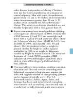
ENTEROVIRUS TRUNK ROUTES
A Practical Guide to Clinical Virology. Edited by L. R. Haaheim, J. R. Pattison and R. J. Whitley
Copyright
2002 John Wiley & Sons, Ltd.
ISBNs: 0-470-84429-9 (HB); 0-471-95097-1 (PB)
6. ENTEROVIRUSES: POLIOVIRUSES,
COXSACKIEVIRUSES, ECHOVIRUSES
AND NEWER ENTEROVIRUSES
Gr. enteron ¼ small intestine, the main replication site for most enteroviruses.
A L. Bruu
The Enterovirus genus of the picornavirus family is a large group of viruses
associated with a spectrum of diseases ranging from paralytic poliomyelitis to
mild, non-specific febrile illness and rarely associated with disease of the
gastrointestinal tract. They are worldwide in distribution but more than 90%
of infections with enteroviruses are subclinical.
The Enterovirus genus comprises several subgroups of which the following
may cause disease in humans:
. Polioviruses (types 1–3). Gr. polios ¼ gray, myelos ¼ marrow.
. Coxsackieviruses, Group A (types 1–22, 24) and Group B (types 1–6).
Coxsackie is the village in the USA where the patients from whom these
viruses were first isolated lived.
. Echoviruses (types 1–9, 11–27). Enteric cytopathogenic human orphan viruses,
originally considered not to be associated (‘orphan’) with human disease.
. Newer enteroviruses (types 29–34, 68–72). Human enterovirus 72 is hepatitis
A virus, see Chapter 24.
The enteroviruses have a diameter of 24–30 nm, an icosahedral structure and
consist of 60 subunits, each containing one set of the structural proteins VP1–4.
The single-stranded RNA has positive sense (mRNA function). The complete
nucleotide sequence has been determined for the polioviruses and some other
enterovirus types. Some enteroviruses may cross-react to a certain degree,
mainly due to determinants on VP1.
Clinical syndromes frequently associated with specific types of enteroviruses
include the following:
. Paralytic disease: polioviruses.
. Herpangina: coxsackie A viruses.
. Hand, foot and mouth disease: coxsackie A virus (A16).
. Epidemic myalgia/pleurodynia: coxsackie B viruses.
. Generalized disease in the newborn: coxsackie B viruses.
. Myocarditis/pericarditis: coxsackie B viruses.
. Conjunctivitis: enterovirus 70.
. Fever and rash: echoviruses especially.
. Meningitis: many enteroviruses.
45
1400BC EGYPTIAN STELE SHOWING PRIEST WITH ‘HORSE-FOOT’.
POLIOMYELITIS? (Courtesy of Ny Carlsberg Glyptotek, Copenhagen)
A Practical Guide to Clinical Virology. Edited by L. R. Haaheim, J. R. Pattison and R. J. Whitley
Copyright
2002 John Wiley & Sons, Ltd.
ISBNs: 0-470-84429-9 (HB); 0-471-95097-1 (PB)
7. POLIOVIRUSES
Infantile paralysis; acute anterior poliomyelitis; Ger. Kinderla
¨
hmung.
A L. Bruu
Poliomyelitis is an acute infectious disease with or without signs of CNS
involvement.
TRANSMISSION/INCUBATION PERIOD/CLINICAL FEATURES
The infection is spread by the faecal–oral route. The incubation period is
usually 1–2 weeks. The patient can infect susceptible persons from some
days before illness and for one to several weeks after the illness. Children
are infectious for a longer period than adults.
SYMPTOMS AND SIGNS
Systemic: Fever, Headache, Myalgia, Nausea, Vomiting
Local: Signs of Meningitis, Pareses
About 95% of infections run a subclinical course. Patients suffering from
aseptic meningitis will recover in 1–2 weeks. Paralysis often results in
persistent lameness.
COMPLICATIONS
Respiratory failure, obstruction of airways, involvement of the
autonomic nervous system.
THERAPY AND PROPHYLAXIS
No specific therapy, immunoglobulin is of no practical value. Vaccine,
either attenuated or inactivated, gives more than 90% protection.
LABORATORY DIAGNOSIS
Demonstration of poliovirus in throat swab or in faecal sample collected
in the acute phase of the disease, viral RNA in faecal sample, and
poliovirus IgM antibodies or IgG antibody rise in paired sera.
47
48
Figure 7.1 POLIOVIRUS (PARALYTIC POLIOMYELITIS)
CLINICAL FEATURES
SYMPTOMS AND SIGNS
The incubation time of poliomyelitis is usually 7–14 days, but may vary from
4 to more than 30 days. The disease typically starts with a prodromal phase
of a few days’ dur ation. The patient has fever and comp lains of myalgia.
Constipation is a common feature. This phase (‘minor disease’) is usually
followed by an interval of a few days when temperature becomes normal and
the patient seems to recover. The temperature then increases again with the
development of paralysis and frequently also aseptic meningitis. Such a
biphasic course is especially common in children. The second phase is initially
characterized by hyperirritability and increased tendon reflexes. This may last
from several hours to a few days, leading to the paralytic stage with loss of
tendon reflexes. The paralysis is flaccid and most frequently affects the
extremities, but any voluntary muscle (group) may be involved. The
development of paralysis may take some hours or a few days. Duri ng the
initial phase of the paralytic stage the patient may also exhibit sensory
disturbances. Strenuous exercise, injections (vaccination), operations (tonsil-
lectomy) and possibly also pregnancy may increase the incidenc e, severity and
site of paralytic disease. Bulbar poliomyelitis may occur alone (in about 10%
of all patients with paralysis) or as a mixed bulbospinal form. This
localization may lead to involvement of cranial nerves wi th paralysis of
pharyngeal muscles and dysphagia, and of respiratory muscles followed by
dyspnoea. Bulbar involvement is often accompanied by lesions of the
respiratory and circulatory centres leading to respiratory failure, fall in blood
pressure and circulatory shock. The lethality of this condition varies between
20 and 60%. The CSF often shows normal values, but an increase in cell
count up to a few hundred/ml is sometimes seen. Polymorphonuclear cells
may be prevalent in the very beginning of the disease, but are soon
outnumbered by lymphocytes. Slight increase in the protein content may be
seen. Immunity after poliovirus infection, whether asymptomatic or paralytic,
is type-specific and lifelong.
The most important differential diagnosis is polyradiculitis (Guillain–
Barre
´
syndrome), where the pareses are ascending and symmetrical
combined with a variety of sensory disturbances. The CSF shows a rather
high protein content with no or only slight increase in cell count. Other
diseases which may mimic poliomyelitis are acute transverse myelitis, tick-
borne encephalitis and reduced mobility due to arthritis and osteomyelitis.
The diagnosis of poliomyelitis is based upon the development of
asymmetrical pareses in the course of some hours to a few days with little
or no sensory loss.
49
CLINICAL COURSE
Fever and general symptoms last for 1–2 weeks. The paralysis reaches a
maximum within 2–3 days. More than 50% of cases recover during the
subsequent weeks or months. The remaining patients will suffer from residual
deficits in one or more muscles. The overall lethality of poliomyelitis has been 5
to 10%, but is substantially reduced by maintaining patients in respirators.
COMPLICATIONS
Encephalitis and myocarditis may occur during the acute stage (see bulbar
poliomyelitis). A post-poliomyelitis syndrome is observed in some 25% of
survivors of paralytic poliomyelitis. After several decades with no changes in
their clinical condition, they develop new weakness, pain and fatigue. This may
be due to a denervation of initially reinnervated muscle fibres, but the aetiology
is not clear. These patients are not excreting poliovirus and are not contagious.
THE VIRUS
Poliomyelitis is caused by one of the three types of poliovirus (Figure 7.2). The
virion is naked and has a diameter of 28 nm. It contains single-stranded RNA
of positive polarity (mRNA) within a protein shell (capsid) composed of 60
capsomeres. The capsid is built up of four proteins, VP1–4. Virus replication is
initiated by RNA transcription into nega-
tive strands to act as templates for new
viral RNAs. From the viral RNA a large
polyprotein is made, which later is cleaved
to generate the capsid proteins VP1–4 and
a range of other proteins. Final assembly
of new virions takes place in the cyto-
plasm. There are some minor antigenic
cross-reactions between some entero-
viruses. Even though the three serotypes
of poliovirus share some antigenic proper-
ties, in particular between types 1 and 2,
they are characterized by marked inter-
typic differences. The epitopes responsible
for inducing neutralizing antibodies are
located on the three structural proteins
VP1, VP2 and VP3 of the viral capsid,
VP1 being the major immunogen. For
differentiation between the three types, type-specific antisera prepared by
cross-adsorption with heterologous types, or suitable monoclonal antibodies,
are used. However, the capsid proteins induce a mainly specific immune
response during an infection and after vaccination. All three polioviruses are
highly cytopathic to many primary cell cultures and permanent cell lines,
50
Figure 7.2 POLIOVIRUS.
Bar, 50nm (Electron micro-
graph courtesy of E. Kjelds-
berg)
causing cell death without changes in cell morphology typical of entero-
viruses. Polioviruses are stable at pH values between 3 and 9, resistant to lipid
solvents and rather slowly inactivated at room temperature. Because of this,
the virus may remain infectious for several days in water, milk, food, faeces
and sewage.
EPIDEMIOLOGY
Poliomyelitis has probably been with us for centuries. However, it was not until
the later part of the nineteenth century that the disease was described as a
separate clinical entity. During the first half of the twentieth century several
large epidemics of poliomyelitis were observed in Europe and North America.
The disease was then most frequent among young children, but in the later part
of the period it became more common among older children and adolescents.
This was most probably due to improved hygienic conditions reducing the
possibilities for faecal–oral spread. In countries with a temperate climate, the
disease is mainly seen during summer and autumn months, whereas in tropical
and subtropical climates poliomyelitis is prevalent throughout the year and
most often occurs in small children. The introduction of polio vaccines in the
1950s has led to more or less complete eradication of poliomyelitis in several
countries, especially in Europe and North America. Due to vaccination
programmes of small children, most clinical cases are now found among
unvaccinated infants, older children and adults. It is therefore important to
maintain a high vaccination coverage rate (490%) to accomplish a sufficiently
high degree of herd immunity. Complacency in adhering to vaccination
programmes invariably leads to cluster outbreaks of poliomyelitis from
imported cases when herd immunity in certain regions or communities comes
under a critical limit. In 1988 the 41st World Health Assembly committed the
World Health Organization and had set to target the year 2000 as the year of
global eradication of poliomyelitis. This goal will probably be reached within
the next few years.
THERAPY AND PROPHYLAXIS
There is no specific treatme nt for poliomyelitis. Impairment of respiratory
function may necessitate artificial respiration. Physiot herapy as early as
possible is important in preventing or reducing lasting sequelae. Although
improved sanitati on and hygiene help to limit the spread of poliovirus, the only
efficient means of preventing paralytic polio is through widespread immuniza-
tion. Two types of vaccine are available against poliomyelitis, inactivated
vaccine (IPV, Salk) and live attenuated oral vaccine (OPV, Sabin). Both
vaccine formulations contain all three pol io types.
OPV is the most widely used vaccine for prevention of poliomyelitis. It is
composed of attenuated strains of the three poliovirus types, and is
administered orally. At least two or three doses are considered necessary to
51
ensure adequate immunity, in some countries even five to six or more doses are
given in the primary course. Revaccinat ion is used to a varying degree. A full
primary course induces an antibody response against all three types in more
than 90% of vaccinees and gives a high degree of protection against disease.
OPV also induces intestinal immunity due to production of secretory IgA
antibodies. This is important for inhibition of virus replication in the gut,
diminishing the possible spread of virus to susceptible contacts. OPV is almost
non-reactogenic, and is very safe. However, in a few cases an attenuated
vaccine strain may induce paralytic disease. This occurs in about one case per
1–10 million vaccine doses administered.
IPV was the first vaccine used against poliomyelitis. It contains the three
types of poliovirus inactivated by formaldehyde and is administered
parenterally. The use of IPV in the late 1950s was followed by a 90%
reduction of poliomyelitis cases when it was replaced in many countries by the
more easily administered OPV around 1960. Newer IPVs have higher
immunogenic potency which has led to a reintroduction of IPV in many
developed and developing countries. The primary vaccination course with IPV
consists of two or three doses, usually followed by revaccination after intervals
of about 5–10 years during childhood and adolescence. Some countries are
using a combination of OPV and IPV.
LABORATORY DIAGNOSIS
Recommended methods for laboratory diagnosis of poliovirus infection are:
. Virus isolation from faeces and throat washings by inoculation into cell
cultures. The presence of poliovirus is shown by degeneration of cultured
cells within a few days. The result of conventional typing by neutralization
will require another couple of days. Alternatively immunofluorescence using
monoclonal antibodies can be used, allowing the distinction between wild-
type virus and vaccine strains.
. Detection of poliovirus RNA in faecal samples by PCR . This method will also
distinguish between wild strains and vaccine strains.
. Antibody investigations. The method of choice is m-chain capture (IgM)
ELISA, which is specific for each poliovirus type. Other antibody tests are
neutralization and CFT on paired serum samples.
The samples should be collected as early as possible in the course of the disease.
Children usually excrete virus for 1–2 weeks, adults for a shorter time. As the
excretion may be intermittent during the later phases of the disease, repeated
samples should be collected. A negative culture or no poliovirus RNA detected
may not exclude infection, especially if the material is taken late in the disease.
In such cases, antibody investigations will be useful.
52
AN ORPHAN VIRUS LOOKING FOR PARENTAL DISEASES
A Practical Guide to Clinical Virology. Edited by L. R. Haaheim, J. R. Pattison and R. J. Whitley
Copyright
2002 John Wiley & Sons, Ltd.
ISBNs: 0-470-84429-9 (HB); 0-471-95097-1 (PB)
8. COXSACKIEVIRUSES,
ECHOVIRUSES AND ENTEROVIRUSES
29–34 AND 68–71
A L. Bruu
These enteroviruses may cause febrile diseases, in some cases with signs of
infection of CNS, muscle, heart, skin, eye and respiratory tract.
TRANSMISSION/INCUBATION PERIOD/CLINICAL FEATURES
Enteroviruses spread from person to person mainly by the faecal–oral
route, and to a lesser degree by the respiratory route. Some types
associated with conjunctivitis spread by direct contact. The incubation
period is 5–14 (2–25) days. Enterovirus conjunctivitis has an incubation
period of 12–24 hours.
SYMPTOMS AND SIGNS
General: Fever, Headache, Malaise
Neurological: Meningitis, rarel y Encephalitis and Transient
Paralysis
Other: Epidemic Myalgia/Pleurodynia (Bornholm
Disease), Myocarditis, Pericarditis, Generalized
Disease in the Newborn, Vesicular and
Maculopapular Exanthems, Haemorrhagic
Conjunctivitis
Usual duration is a few days to about 1 week.
COMPLICATIONS
Occasionally neurological sequelae.
THERAPY AND PROPHYLAXIS
There is no specific therapy, and no immunoglobulin or vaccine against
these enteroviruses.
55
LABORATORY DIAGNOSIS
Virus may be isolated from the pharynx early in the disease, from faeces
for at least 1 week and in some cases from other sites of infection.
Enterovirus nucleic acid may be detected by PCR in faecal sample,
throat swab, vesicle fluid, myocardial tissue, pericardial fluid or in
cerebrospinal fluid (CSF) for aseptic meningitis. For antibody investiga-
tions the m-capture ELISA is the method of choice.
56
Figure 8.1 ENTEROVIRUS (SEROUS MENINGITIS)
CLINICAL FEATURES
SYMPTOMS AND SIGNS
Like poliovirus the coxsackieviruses and echoviruses multiply primarily in
lymphoid tissue in the pharynx and the small intestine. In about 5% of cases
virus may spread to other target organs, the main ones being the meninges, the
brain and spinal cord, myocardium and pericardium , striated muscles and skin.
Infection leads to lasting type-specific immunity. Fever of short duration and
sometimes a rash or mild upper respiratory symptoms are the most frequent
clinical diseases. A few cases progress to one of the following syndromes:
Aseptic meningitis. In typical cases a biphasic course is seen. After an interval of
1–2 days with few or no symptoms, the temperature rises again to 38–398C,
accompanied by headache, neck stiffness and vomiting. A non-specific
maculopapular rash, sometimes with petechial elements, may be seen. The
CSF is clear with slight or moderate elevation of cell count (up to 500610
6
/
litre, mainly lymphocytes) and protein content, but with normal sugar content.
The illness may last for 2–10 days, sometimes followed by a convalescent phase
of rather long duration. The prognosis is good as most patients recover
completely. Meningoencephalitis or encephalitis may occur in some cases. In
differential diagnosis, meningitis caused by other viruses and early or
inadequately treated bacterial meningitis which may mimic aseptic meningitis
should be considered. Note a petechial rash is also seen in meningococcal
disease. Lymphocytes are seen in the CSF (tuberculous, listerial and
cryptococcal mening itis), but usually the glucose content is lowered.
Complications are transient paralysis and polio-like disease.
Epidemic myalgia/pleurodynia (Bornholm disease). This is a painful inflamma-
tion of the muscles, most pronounced in the intercostal muscles or abdominal
muscles, accompanied by pain that may be severe (devil’s grip) and resemble
ischaemic heart disease or ‘acute abdomen’. The pain is often intermittent for
periods of 2–10 hours, combined with rise in temperatur e. The illness lasts for
4–6 days, but relapses in the following weeks are not infrequent. Complete
recovery is the rule.
Myocarditis/pericarditis. This is observed in 5% of patients with coxsackie B
virus infections. Typical features are fever, chest pain and dyspnoea. Other
signs are pericardial rub, heart dilatation and arrhythmias. Heart failure may
occur. The illness usually lasts for 1–2 weeks. Relapse may occur during the
following weeks and months in 20% of patients. The most important
differential diagnoses are cardiac ischaemia, infarction and myopericarditis of
other aetiology.
Neonatal myocarditis. Some enteroviruses, mostly coxsackie B3 and 4, may
cause a severe, often fatal disease in infants characterized by sudden onset,
lethargy, tachycardia, dy spnoea and cyanosis. It is a systemic infection as many
organs (heart, brain, liver, pancreas) are involved. The virus is transmitted
from mother to child just before or at birth.
57
Herpangina. The illness is seen mainly in children. Some 8–10 vesicles or small
ulcers, 1–3 mm in diameter, are seen on the posterior pharyngeal wall. There is
pain on swallowing and usually slight fever of a few days’ duration. Differential
diagnoses are herpes simplex, varicella, aphthous stomatitis.
Hand, foot and mouth disease. This occurs most often in children. Moderate
fever of 38–398C may be seen. Vesicles up to 5 mm in diameter are localized on
the buccal mucosa and tongue as well as on the hands and feet.
Rashes. Maculopapu lar rashes (‘rubelliform’ or non-specific) are seen quite
frequently in coxsackie A and echovirus infections, accompanied by
pharyngitis and fever. A rash is sometimes seen in the course of meningitis.
Differential diagnoses are erythema infectiosum, rubella, measles and rashes
seen in meningococcal disease.
Acute haemorrhagic conjunctivitis. This eye disease is characterized by pain,
swelling of the eyelids and subconjunctival haemorrhages of a few days’
duration, usually healing spontaneously in less than a week. It is highly
contagious, with an incubation time of 12–24 hours, and spreads by direct
contact. Extensive epidemics have been observed in the Far East (caused by
coxsackie A type 24) and in Africa, Japan and India (enterovirus type 70).
Spread is favoured by poor hygienic conditions as in refugee camps. Associated
neurological disease (radiculomyelopathy, cranial nerve involvement) occurs
rarely and may lead to residual paralysis.
Coxsackie B has also been associated with idiopathic dilated cardiomyo-
pathy. Some studies have shown evidence for a connection between juvenile
diabetes type 1 and coxsackie B virus infection.
THE VIRUS
The enterovirus group (Figure 8.2) is one genus in the Picornaviridae family.
They are small (28 nm), rough ly spherical and contain a single-stranded RNA
molecule of pos itive polarity, which func-
tions as mRNA. The RNA is surrounded
by a protein shell (capsid) with icosahedral
symmetry. All picornaviruses contain four
polypeptides, VP1–4, VP1 being the major
immunogen. There is a certain degree of
serological cross-reactivity between entero-
virus types, especially between types within
the same subgroup, due to shared epitopes
not exposed at the surface of the virus, as
seen when using the complement fixation
test. Enteroviruses retain infectivity at
pH 3–9 and are resistant to several proteo-
lytic enzymes and lipid solvents. They are
stable for days at room temperature. In the
laboratory, the coxsackie B and e choviruses
will grow in several different cell cultures.
58
Figure 8.2 ECHOVIRUS
WITH ANTIBODY (Electron
micrograph courtesy of
E. Kjeldsberg)
The coxsackie B viruses will also infect newborn mice. Coxsackie A viruses
replicate in mice, but only a few will do so in cell cultures.
EPIDEMIOLOGY
Man is the only natural host for human enteroviruses. The virus replicates in
the upper and lower alimentary tract and is excreted from these sites.
Enteroviruses spread mainly by the faecal–oral route, and during the acute
stage also by the respiratory route. They have a worldwide distribution. In the
temperate zones spread takes place in the summer a nd autumn months, in
tropical and subtropical zones throughout the year. Children are infected more
frequently than adults, and males somewhat more frequently than females.
Poor sanitary conditions will favour spread of these viruses.
THERAPY AND PROPHYLAXIS
There is at present no known specific therapy, nor is there any vaccine against
enteroviruses other than the polioviruses. Only symptomatic treatment is
available.
LABORATORY DIAGNOSIS
Isolation of virus from stools, rectal swabs, nasopharynx samples, CSF,
vesicular fluid and eye secretions has until recently been the most reliable
method for laboratory diagnosis of an enterovirus infection. Several types of
cell cultures may be used for isolation. Appearance of cytopathic effect (CPE)
is observed after a few days, and neutralization tests are used for virus
identification. Inoculation of coxsackie viruses into newborn mice will lead to
disease and death.
During the last years molecular virological methods such as PCR for the
detection of enterovirus nucleic acid (RNA or cDNA) have been developed,
and nested PCR is considered to be more sensitive than virus isolation,
particularly since some enterovirus strains do not grow or fail to show CPE in
cell culture.
Samples should be taken in the early phase of the disease since patients will
usually excrete virus in the faeces for about 1 week (several weeks for children).
Presence of virus is a strong indication of a causal relationship to disease. As
virus shedding may be intermittent during the later phases of illness, a negative
result does not exclude recent infection.
Antibody investigations. A test for specific IgM is used in some laboratories
and is considered to be the method of choice for coxsackievirus B infections.
The CFT is easy to perform, but because of the occurrence of cross-reactions
the CFT is of limited value for enterovirus diagnosis.
59
MIGHT AS WELL TAKE SOMETHING ENJOYABLE
A Practical Guide to Clinical Virology. Edited by L. R. Haaheim, J. R. Pattison and R. J. Whitley
Copyright
2002 John Wiley & Sons, Ltd.
ISBNs: 0-470-84429-9 (HB); 0-471-95097-1 (PB)
A Practical Guide to Clinical Virology. Edited by L. R. Haaheim, J. R. Pattison and R. J. Whitley
Copyright
2002 John Wiley & Sons, Ltd.
ISBNs: 0-470-84429-9 (HB); 0-471-95097-1 (PB)
A Practical Guide to Clinical Virology. Edited by L. R. Haaheim, J. R. Pattison and R. J. Whitley
Copyright
2002 John Wiley & Sons, Ltd.
ISBNs: 0-470-84429-9 (HB); 0-471-95097-1 (PB)
A Practical Guide to Clinical Virology. Edited by L. R. Haaheim, J. R. Pattison and R. J. Whitley
Copyright
2002 John Wiley & Sons, Ltd.
ISBNs: 0-470-84429-9 (HB); 0-471-95097-1 (PB)
A Practical Guide to Clinical Virology. Edited by L. R. Haaheim, J. R. Pattison and R. J. Whitley
Copyright
2002 John Wiley & Sons, Ltd.
ISBNs: 0-470-84429-9 (HB); 0-471-95097-1 (PB)
A Practical Guide to Clinical Virology. Edited by L. R. Haaheim, J. R. Pattison and R. J. Whitley
Copyright
2002 John Wiley & Sons, Ltd.
ISBNs: 0-470-84429-9 (HB); 0-471-95097-1 (PB)
9. RHINOVIRUSES AND
CORONAVIRUSES
Lat. rhinus ¼ nose; Ger. Erka
¨
ltung; Fr. rhuˆme ¼ common cold.
I. Ørstavik
Rhinoviruses are the most frequent cause of common colds. All age groups are
affected. Infections are endemic with higher frequencies during autumn and
spring in temperate climates.
TRANSMISSION/INCUBATION PERIOD/CLINICAL FEATURES
The common cold is spread by close contact and by inhalation of virus-
containing droplets. The incubation period is 2–4 days, and a person is
probably infectious from 1 day postinfection and as long as there are
clinical symptoms.
SYMPTOMS AND SIGNS
Systemic: None or Low-Grade Fever, Headache
Local: Coryza, Sneezing, Sore Throat, Cough,
Hoarseness
In uncomplicated cases the illness usually lasts for 1 week, with maximal
symptoms on days 2 and 3.
COMPLICATIONS
Secondary bacterial infections may occur (sinusitis, otitis media).
Rhinovirus infections may precipitate acute asthma in predisposed
children, and may aggravate chronic bronchitis in adults.
THERAPY AND PROPHYLAXIS
No specific therapy or prophylaxis is available.
61
LABORATORY DIAGNOSIS
During acute illness the virus can be isolated from the nose, the throat
and sputum. Special cell culture techniques are needed for virus isolation
and these are performed in very few virus laboratories. Serological
diagnosis is not routinely used either, because of the many serotypes.
62
Figure 9.1 RHINOVIRUS (THE COMMON COLD)
CLINICAL FEATURES
SYMPTOMS AND SIGNS
After an incubation period of 2–4 days, the illness starts with symptoms of
nasal congestion/blockage and irritation, sneezing and a sore throat. Excess
nasal secretion follows which is serous at first and later becomes purulent if
secondary bacterial infection ensues. Cough is a frequent symptom, as is
headache during the first days of illness. Fever occurs seldom, and if so, it is
moderate. Rhinovirus infection causes the same symptoms in all age groups.
The infection is limited to the respiratory tract. It has been suggested that
rhinoviruses may cause a more serious infection of the lower respiratory tract
in small children. Rhinovirus infection has also been shown to precipitate
attacks of asthma in children and aggravate chronic bronchitis in adults.
Asymptomatic infections are reported to occur in about 25% of individuals
infected with rhinovirus.
Differential diagnosis. Symptoms of common cold, particularly in children,
may be due to other virus infections, e.g. influenzavirus, parainfluenzavirus,
adenovirus, RSV and coronaviruses. Coronaviruses are now considered to be
second to rhinoviruses as a cau se of common cold, but the symptoms are
usually milder in coronavirus infections. Influenzavirus infections occur in
epidemics, and general symptoms such as fever and malaise are more severe. In
parainfluenza and adenovirus infections pharyng itis is more pronounced.
During epidemics of RSV some of the patients, children as well as adults, may
have the same symptoms as in rhinovirus infections. Pharyngitis and tonsillitis
will dominate infections with Streptococcus pyogenes. However, it is usually
not possible to determine the aetiology on the basis of the clinical findings
alone in upper respiratory infections.
CLINICAL COURSE
As a rule, the illness will last for 1 week, but 25% of the patients will need 2
weeks to recover completely. The illness tends to last longer in smokers than in
non-smokers.
COMPLICATIONS
Bacterial sinusitis and otitis media are the most common complications.
Occasionally a bacterial bronchopneumonia is seen.
THE VIRUS
Rhinovirus and Enterovirus are two genera in the family Picornaviridae. They
are small (28–32 nm) single-stranded RNA viruses (Figure 9.2).
63
Rhinovirus now comprises more than 100
different serotypes, and new types are still
being identified. As with other picorna-
viruses the virion capsid consists of a naked
icosahedron of 60 capsomers, each made up
of four proteins. Depressions in the virus
capsid represent the sites on the virus where
the cellular receptors bind. These depres-
sions (‘sockets’) are the targets for
experimental studies of synthet ic anti-
rhinoviral agents. A fifth protein is
associated with the single-stranded RNA.
Due to the lack of a lipid envelope, the virus
is resistant to inactivation by organic
solvents. Rhinoviruses are more acid-labile
than enteroviruses.
EPIDEMIOLOGY
The rhinoviruses probably cause about half of all cases of common cold and
are considered to be one of the most frequent causes of infections in man.
Studies in the USA have revealed an infection rate of at least 0.6 per individual
per year. The rate is highest among infants and decreases with age.
Schoolchildren are considered to be important transmitters of rhinovirus
infections. Parents with children in kindergarten or in primary school may have
more common cold episodes than single adults. Rhinovirus infections are
endemic, but occur most frequently during autumn and spring in temperate
climates. Several serotypes can circulate simultaneously in the same
population, and it is possible that new serotypes emerge over the years.
There is no evidence that some serotypes cause more serious illness or occur
more frequently than others.
THERAPY AND PROPHYLAXIS
Specific chemotherapy is not available, and treatment with immunoglobulin is
without effect. Experiments in volunteers have found a-andb-interferon
given intranasally to be effective in preventing rhinovirus infection, whereas
studies using g-interferon have been unsuccessful. The suggestion that large
quantities of vitamin C (ascorbic acid) taken prophylactically or during illness
influences the course of the disease, has not been proven. Symptomatic
treatment includes mild analgesics and nasal drops. Prophylactic use of
antibiotics against bacterial superinfections is not recommended in otherwise
healthy individuals.
No vaccine is available. The high and uncertain number of serotypes and
their relative impor tance and distribution during various outbreaks are
64
Figure 9.2 RHINOVIRUS
(Electron micrograph courtesy
of E. Kjeldsberg)
obstacles to vaccine development. In addition to inhalation of droplets, spread
of infection by contact is considered to play a significant role. Measures should
be taken to avoid infection from virus-contaminated hands. Persons suffering
from asthma and from chronic bronchitis should avoid close contact with
common cold patients.
LABORATORY DIAGNOSIS
Cultivation of rhinoviruses requires special cell cultures which are incubated at
338C (the temperature in the nasal mucosa). Also, since many serotype s are
difficult to cultivate, rhinovirus isolation is performed only by very few virus
laboratories. Serological diagnosis is complicated by the large number of
serotypes and is therefore not routinely performed.
CORONAVIRUS
Coronaviruses are the second most frequent cause of the common cold (15–
20%). They are single-stranded RNA viruses belonging to the Coronaviridae
family. The virions vary in diameter from 80 to 160 nm. They have club-shaped
spikes on the surface which give a crown (corona)-like picture by electron
microscopy (Figure 9.3). At least four different proteins are known , and the
S (spike)-protein induces virus-neutralizing antibodies contributing to
immunity. The coronaviruses are divided into three serological groups, the
human coronaviruses have been allocated to two of these serological groups,
and the two human prototypes are OC43 and 229E. The coronaviruses are
believed to spread as the rhinoviruses, and the incubation period is about 2
days. The symptoms are similar to those following rhinovirus infections,
lasting for about 1 week. As many as 50% of coronavirus infections may be
asymptomatic. Serological studies suggest that the infection occurs in all age
groups. Reinfection viruses have been
observed, suggesting that protective
immunity is not long-lasting. Corona-
virus infections occur most frequently in
late winter/early spring. Coronavirus
may be isolated from the nose and
throat during the acute phase of illness
if organ cultures of human fetal trachea
are used. Only a small number of
coronavirus strains have been identified,
and most knowledge about this virus
infection has been obtained by sero-
logical studies on paired sera from
patients. Very few laboratories diagnose
coronavirus infections as part of their
routine work.
65
Figure 9.3 CORONAVIRUS.
Bar, 100 nm (Electron micrograph
courtesy of E. Kjeldsberg)
A REAL KNOCK-OUT
A Practical Guide to Clinical Virology. Edited by L. R. Haaheim, J. R. Pattison and R. J. Whitley
Copyright
2002 John Wiley & Sons, Ltd.
ISBNs: 0-470-84429-9 (HB); 0-471-95097-1 (PB)
10. INFLUENZAVIRUSES
Influenza; influenced by cosmic events (medieval Italy). Ger. Grippe;
Fr. grippe.
L. R. Haaheim
Influenzavirus causes illness in all age groups. During epidemics a large
number of individuals may fall ill within a span of a few weeks.
TRANSMISSION/INCUBATION PERIOD/CLINICAL FEATURES
Virus is transmitted by aerosols, mean incubation time is 2 days (1–4).
The patient is contagious during the first 3–5 days of illness.
SYMPTOMS AND SIGNS
Systemic: Sudden Fever (38–408C), Myalgia, Headache
Local: Coryza, Dry Cough, Sore Throat, Hoarseness
Systemic symptoms dominate initially with fever for the first 3–4 days.
Full recovery within 7–10 days. Occasionally long convalesence.
COMPLICATIONS
Secondary bacterial pneumonia. More rarely primary viral pneumonia,
myocarditis, encephalitis.
THERAPY AND PROPHYLAXIS
No specific treatment. Amantadine chemoprophylaxis. Vaccination is
recommended for high-risk groups and key personnel within the health
services.
LABORATORY DIAGNOSIS
Virus can be isolated from/demonstrated in nasopharyngeal specimens
taken in the acute phase of illness (days 1–3). An antibody rise can be
demonstrated in paired sera by HI or CFT.
67
68
Figure 10.1 INFLUENZAVIRUS (INFLUENZA)