A Practical Guide to Clinical Virology Second Edition - part 6 pot
Bạn đang xem bản rút gọn của tài liệu. Xem và tải ngay bản đầy đủ của tài liệu tại đây (523.89 KB, 29 trang )
THERAPY AND PROPHYLAXIS
When given within 24 hours after the eruption antiviral therapy with
aciclovir is effective in shortening the duration of varicella. Famciclovir
and valaciclovir are also effective and have better bioavailability.
Foscarnet is used in the seldom aciclovir-resistant cases. Antiviral
therapy is recommended in infants, adults and immunocompromised
patients. Specific VZ-immunoglobulin (VZIG) given up to 3 days after
VZV exposure is usually protective, but is reserved for use mainly in
high-risk patients. High-risk patients can also be protected by
vaccination.
LABORATORY DIAGNOSIS
The diagnosis is clinical given by the typical vesicular rash, and
laboratory confirmation is seldom necessary. If the rash is atypical
direct fluorescent antibody staining of cell scrapings or identification of
virus antigen by PCR technique is most useful. PCR analysis of
cerebrospinal fluid is valuable if neurological complications occur. A
rising antibody titre in paired serum samples is diagnostic. Serologic
testing may be unreliable in immunocompromised patients.
138
Figure 19.1 VARICELLA-ZOSTER VIRUS (VARICELLA)
CLINICAL FEATURES
SYMPTOMS AND SIGNS
The incubation pe riod is usually 14–16 days, but may vary from 10 to 21
days, and up to 28 days after prophylactic treatment with VZIG. Especially
in teenagers and adults, prodromal symptoms with malaise and low-grade
fever may occur 1–2 days before eruption of the vesicular rash. The typical
crops of varicella lesions initially observed on the face, scalp or trunk develop
during hours from pruritic macules to oval 2–3 mm vesicles with clear fluid
becoming cloudy and crusty after 1–2 days. During the first 2–4 days skin
lesions at different developmental stages coexist. Usually 100–300 lesions are
found, and the active disease lasts for 1 week. The diagnosis may be missed
when only a few lesions occur, and rarely the infection may be subclinical .
Mucosal vesicles in the mouth, pharynx, conjunctiva and the external
genitalia rupture easily and may therefore be overlooked. Itching is common
and usually mild constitutional symptoms and fever occur during the first
days of the rash. Higher fever and more symptoms generally accompany an
extensive eruption.
Differential diagnosis. Mild cases of varicella may go unnoticed or be mistaken
for impetigo. In hand, foot and mouth disease due to coxsackie virus, vesicles
up to 5 mm occur on hands, feet and mouth mucosa, but not on the trunk
(Chapter 8). Seldom will herpes sim plex viral infection in children with atopic
eczema become varicella-like (Chapter 18). Very rarely a varicelliform rash is
caused by Rickettsia akari transmitted by a mouse mite. These vesicles are
smaller, more deeply seated on a firm papule, and lack the typical crusting seen
in varicella.
CLINICAL COURSE
In healthy children varicella usually has a benign course. Secondary family
contacts often have a more severe disease due to a higher viral load.
Extensive eruptions and a more serious course may be seen in adults. If the
mother gets varicella during the perinatal period, especi ally 5 days before or
2 days after delivery, the infant may develop serious varicella, often fatal if
untreated, due to lack of maternal antibody protection. Prematures born
before 30 weeks’ gestation also lack maternal antibodies. Other high-risk
groups for potential fatal VZV infec tion are patients with compromised
cellular immunity such as leukaemia, lymphoproliferative diseases, HIV
infection and individuals treated, for whatever reason, with corticosteroids
and other immunosuppressive or cytotoxic drugs. VZV infection usually
results in lifelong immunity against reinfection, but not against reactivation
(zoster).
139
COMPLICATIONS
Common complications are superinfection with Staphylococcus aureus or
group A Streptococcus pyogenes causing impetigo and erysipelas or less
commonly extensive cellulitis, necrotic or bullous skin infection. Bullous
varicella is caused by epidermolytic toxin-producing staphylococci. Invasive
infection with septicaemia, arthritis, osteomyelitis or bacterial pneumonia
may occur.
Visceral spread of VZV can affect the lungs, brain, liver, pancreas, kidneys
or heart. Fatal cases are most often due to interstitial varicella pneumonia
(pneumonitis) which is 25 times more common in adults than in children.
Smokers, pregnant women and patients with chronic lung diseases are at
increased risk for developing serious pneumonitis that may be fatal. Usually
rapid clinical recovery takes place, though radiographic changes may persist
for weeks, sometimes leaving calcifications.
Neurologic complications include meningoencephalitis due to direct VZV
infection during the first week with high fever and deterioration of
consciousness. VZV may also play some direct part in cerebellar ataxia,
thought to be mainly immunological. Ataxia usually starts 1 week after
appearance of the rash and is a benign condition lasting up to 1–2 weeks in
children. Rare cases of limb paresis due to transverse myelopathy or brain
arteritis have been reported. Reye syndrome (fatty liver and encephalopathy)
has become very rare since the use of aspirin has declined. VZV hepatitis,
mostly subclinical, still occurs as a separate entity.
Rarely thrombocytopenia, haemorrhagic varicella, fulminant purpura and
leucopenia may occur, especially in patients with immunodeficiency. A
congenital varicella syndrome with limb atrophy and scarring of the skin
occurs after VZV infection in 1–2% of those contracting varicella during the
first 20 weeks of pregnancy.
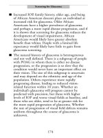
THE VIRUS
Varicella-zoster virus (Figure 19.2) is one of eight herpesviruses. It is a
double-stranded DNA virus 150–200 nm in diameter with a lipid envelope
where glycoprotein spikes surround an inner icosahedral nucleocaps id. After
penetration of the infected cell, the virion is uncoated, and the capsid
penetrates the cell nucleus where replication occurs. Viral DNA is integrated
in the host cells thereby avoiding immune surveillance and eradication by
antiviral drugs. VZV is quickly inactivated outside host cells. Haemato-
genous spread by mononuclear cells, secondary viraemia, occurs 4–5 days
before and 1–2 days after onset of symptoms. Man is the only natural host.
Only one antigenic VZV type has been identified. Attenuated viral strains
have been developed through serial passages in cell cultures, and the Oka
strain is used in the live-virus vaccine now available. Mutant VZV strains
140
resistant to aciclovir have been isolated,
especially from AIDS patients repeatedly
treated with antiviral drugs.
EPIDEMIOLOGY
Varicella is very contagious. Thus 90% of
susceptible household contacts contract the
disease. Varicella is therefore predomi-
nantly a childhood disease in temperate
areas where 90% of cases occur below 10
years. In the USA 96% of adults are
immune, while adults often remain suscep-
tible to varicella in tropical countries.
Epidemics are seen in temperate climates
most frequently during late winter and early
spring. Infection is usually spread by droplet or direct contact, but may be
airborne in institutions. The infectivity is maximal 1–2 days before and 3–4
days after the eruption, but may be extended if new crops of vesicles occur.
Nosocomial infection is a serious problem, especially in units treating
malignancies and immu nodeficient patients and performing transplantations.
THERAPY AND PROPHYLAXIS
The antiviral drug aciclovir has improved the prognosis of serious VZV
infections. VZV is, however, less sensitive than HSV, and therefore a 4-fold
higher dose is needed against VZV. Antiviral therapy is not recommended for
use in children without chronic disease, except secondary household and
teenage cases. Risk groups such as adults and immunocompromised
individuals should be offered treatment, preferably intravenously. Antiviral
treatment should also be given when complications such as varicella
pneumonia and encephalitis occur.
Aciclovir and penciclovir with their respective oral prodrugs valciclovir and
famciclovir reduce clinical symptoms and shorten the course of VZV
infection when started within 48 hours after skin eruption. Early treatment
gives the best results. Hopefully the occurrence of complications is reduced,
though this has not been proved due to their rarity. These antiviral drugs are
all dependent on the virus-encoded thymidine kinase for intracellular
activation. Cross-resistance to these drugs has been reported for viral
strains isolated from AIDS patients having had repeated treatment courses
with aciclovir. When VZV resistance is suspected, treatment with foscarnet
should be given.
Specific immunoglobulin has no proven therapeutic effect. In uncompli-
cated varicella symptomatic treatment of pruritus is recomm ended to prevent
141
Figure 19.2 INNER NUCLEO-
CAPSID OF VARICELLA-
ZOSTER VIRUS. Bar, 50 nm
(Electron micrograph courtesy
of G. Haukenes)
impetigo. Antibiotics are given against secondary bacterial infections. Specific
zoster immunoglobulin (VZIG) given up to 3 days after exposure may
prevent or modify clinical disease. Because of the scarcity of VZIG, this
preparation must be reserved for use in high-risk patients. Pooled normal
immunoglobulin preparations contain small amounts of specific immuno-
globulin, insufficient to prevent disease in ordinary doses. Probably high-dose
immunoglobulin given intravenously exerts a prophylactic effect. VZIG
treatment does not prevent the development of immunity unless the patient
has an immunopathy.
In hospitals strict isolation (in negative-pressure rooms) of infectious
patients is necessary, or preferably, they should be discharged as soon as
possible and treated as outpatients.
Varicella vaccine with live attenuated VZV has proven effective in protecting
healthy individuals as well as high-risk patients against varicella. Seroconver-
sion rates after one vaccine dose are at least 95% in children younger than 12
years, whereas older persons require two doses for equivalent protection. Non-
immune individuals scheduled for transplantation should be vaccinated at least
3 months before operation. In most countries vaccination is recommended for
use in non-immune teenagers and adults, whereas widespread vaccination of
healthy young childr en is not recommended, though the vaccine may be
approved for administration to this group. Vaccination during ongoing
cytostatic treatment is less effective, and usually not recommended 6 months
after postponing such treatment.
LABORATORY DIAGNOSIS
Usually the clinical diagnosis is accurate with no need for laboratory tests. For
diagnostic help in the acute stage the sensitive PCR technique can detect VZV
DNA in vesicles, blood and spinal fluid. VZV is abundant in vesicle fluid, but
the electron microscopic picture cannot be distinguished from HSV and CMV.
Immunofluorescent staining of vesicle fluid with monoclonal antibodies can
identify VZV. The cytopathic effect of VZV in cell culture is characteristic, but
takes some time, and VZV is not always readily cultured. A rise in antibody
titre or demonstration of specific IgM usually confirms the diagnosis. However,
during VZV infection a simultaneous antibody rise against HSV may occur,
and vice versa. Specific CF antibodies are found 6–7 days after the onset of the
rash. CF-antibody titres may, however, be below detectable level 3 years after
the infection. Then latex agglutination assay, indirect immunofluorescence or
ELISA techniques are necessary to detect VZV antibodies. Because of the
potential severe course of VZV infection, and the possibility of giving
prophylaxis by exposure, sensitive techniques are needed for identification of
susceptible persons.
142
HELL’S FIRE—CHICKENPOX REVISITED
A Practical Guide to Clinical Virology. Edited by L. R. Haaheim, J. R. Pattison and R. J. Whitley
Copyright 2002 John Wiley & Sons, Ltd.
ISBNs: 0-470-84429-9 (HB); 0-471-95097-1 (PB)
20. VARICELLA-ZOSTER VIRUS
(VZV)—ZOSTER
Shingles. Gr. zoster ¼ belt; Ger. Gu
¨
rtelflechte, Gu
¨
rtelrose;Fr.zona.
A. Winsnes and R. Winsnes
Zoster, ‘herpes zoster’, is usually a disease of adults, especially elderly people,
caused by reactivation of latent varicella-zoster virus (VZV) in dorsal root
ganglion cells. In AIDS and other immunocompromised patients zoster is both
a frequent and dreaded disease.
REACTIVATION/TRANSMISSION
VZV persists in a latent form in sensory nerve cells (dorsal roots of the spinal
medulla or cranial nerve ganglia) for decades after varicella infection. Though
re-exposure to VZV may be a factor in reactivation of virus in some
circumstances, it is generally poorly understood why VZV starts replicating
and spreading down sensory nerve fibres. VZV appears in vesicles on the skin
area corresponding to the dermatome innervated by the nerve in question.
Although zoster is less contagious than varicella, it may cause varicella in
susceptible contacts.
CLINICAL FEATURES
Neuralgic pain and tenderness in the affected area frequently start several days
before eruption of the rash. The zoster vesicles are usually somewhat larger
than those of varicella. The development to crusting is slower (7–10 days), and
the occurrence of new crops of vesicles is seen less often than in varicella.
Pigmentary changes and scarring may be seen following the loss of crusts after
3–4 weeks. The rash is usually unilateral and local ized to the area (dermatome)
innervated by one or two sensory nerves. Localization is most frequent on the
thorax, neck or face. With involvement of cranial nerves vesicles may occur on
the eyes, in the external ear canal and in the mouth. Regional lymph nodes are
regularly enlarged and tender. General symptoms with malaise and fever are
usually not very prominent. The uncomplicated clinical course is 1–3 weeks.
COMPLICATIONS
Complications are especially seen when zoster is located in cranial nerve areas
or when the host resistance is compromised. Involvement of the ophthalmic
branch of the trigeminal nerve (zoster ophthalmicus) may result in dendritic
145
keratitis that may cause scarring of cornea and reduced vision. VZV may also
cause retinitis with poor visual prognosis. Immediate ophthalmological
examination is recommended. Involvement of the seventh cranial nerve may
cause facial nerve palsy (Ramsay Hunt syndrome), where prognosis for
recovery is not so good as in the common Bells palsy. Pareses are due to spread
of the virus to the motor neurons in the medulla or cranial nerve ganglia. By
EMG it has been sho wn that motor involvement occurs in 35% of the thoracic
zoster cases.
Other neurologic complications such as encephalitis, myelitis and poly-
neuropathy may be caused by immunological inflammatory processes, but also
by direct spread of VZV, even without the presence of the typical zoster rash.
PCR methods have in several cases shown VZV DNA to be present in
mononuclear blood cells, blood vessels and spinal fluid. Thus VZV may cause
neuronal damage because of direct destruction of neurons and compromised
blood flow because of arteritis in small or large vessels.
In zoster patients who are 50 years and older postherpetic neuralgia (pain
persisting more than 6 weeks after appearance of the rash) is a common
complication, occurring in 40% of zoster patients above 60 years. VZV DNA
has been detected in mononuclear blood cells of some patients with
postherpetic neuralgia, and speculation of a possible higher viral load in
patients with neuralgia would argue for aggressive antiviral treatment. Once
postherpetic neuralgia disappears, it does not recur.
In immunocompromised patients, particularly transplant recipients, cancer
and AIDS patients, zoster may become generalized and life-threatening.
Haematogenous dissemination to internal organs may occur as in varicella
(Chapter 19). As in varicella secondary bacterial infections of the rash may
occur, sometimes bec oming invasive.
EPIDEMIOLOGY
With increasing age cellular immunity becomes weake r, explaining why zoster
is 10 times more frequent in persons over 70 years of age than in teenagers. The
disease is also more prevalent and serious among imm unocompromised
patients. It has been calculated that by the age of 80 about 50% will have had
one attack of zoster, and 1% in this age group will have had two attacks.
Increased risk of contracting zoster is seen in children who have had varicella
infection during fetal life or early infancy, probably due to lower specific
immunity. Adults with frequent re-exposure to varicella through contact with
children have a lower incidence of zoster.
THERAPY AND PROPHYLAXIS
When given within the first 3–5 days after eruption of the rash, aciclovir has
proved effective for treatment of zoster both in otherwise healthy and
immunocompromised patients. In the latter group intravenous antiviral
146
treatment shuld be given as soon as possible. In acute zoster the standard
treatment is 7–10 days of oral aciclovir treatment. For ophthalmic zoster,
topical treatment with aciclovir is given in addition to systemic treatment. A
variety of treatment regimens (even combined treatment with aciclovir and
prednisolone) against postherpetic neuralgia have had limited success. Possibly
more aggressive antiviral treatment at the start of zoster will diminish the
occurrence of complications, but this is not settled so far.
LABORATORY DIAGNOSIS
As zoster is caused by reactivation of VZV, the IgG response in serum is
quicker and more pronounced than that seen in varicella. Specific IgM is found
in small amounts. Viral DNA may be identified by PCR methods used with
vesicular fluid or scrapings, as well as with blood and spinal fluid. VZV
infection of the nervous system may be protracted, especially in immunocom-
promised patients. In dermatomal pain without rash (preherpe tic zoster and
‘zoster sine herpete’), and in cases of acute pareses or meningoencephalitis or
myelitis, PCR analyses may be important.
147
WE HAVE ENOUGH PROBLEMS HERE WITHOUT YOU
A Practical Guide to Clinical Virology. Edited by L. R. Haaheim, J. R. Pattison and R. J. Whitley
Copyright
2002 John Wiley & Sons, Ltd.
ISBNs: 0-470-84429-9 (HB); 0-471-95097-1 (PB)
21. CYTOMEGALOVIRUS (CMV)
The name of the virus refers to the size of the infected cells, which
contain large intranuclear inclusions.
A. B. Dalen
The cytomegaloviruses (CMV) belong to the herpesvirus family (subfamily
Betaherpesvirinae). They are widely distributed in man and other mammals,
but possess a high degree of species specificity. Human infections may be
asymptomatic, or cause severe, generalized disease. Following primary
infection, CMV establishes a latent infection of lymphocytes and possibly
endothelial cells from which they may be reactivated.
TRANSMISSION/INCUBATION PERIOD/CLINICAL FEATURES
Fetal infection can foll ow reactivation or prim ary maternal infection.
Perinatal infection occurs through infected cervical secretions and milk.
Postnatal infection is acquired by the respiratory route, infected semen
and through blood transfusions and organ transplants. The incubation
period is about 3–6 weeks.
SYMPTOMS AND SIGNS
Postnatal: Infectious Mononucleosis-like
Congenital and immuno- Extensive Organ Damage in Severe
compromised patients: Cases
Most infections are asymptomatic. After an insidious start symptoms
may last 1–5 weeks. Immunocompromised patients: Extensive organ
damage in severe cases.
COMPLICATIONS
Interstitial pneumonia, hepatitis and occasionally Guillain–Barre
´
syndrome. Retinitis, gastrointestinal infection.
149
THERAPY AND PROPHYLAXIS
Foscarnet and ganciclovir. CMV vaccine s are still at the developmental
stage.
LABORATORY DIAGNOSIS
Virus can be cultured from urine, saliva, blood, milk, cervical discharges,
semen, biopsies and autopsied orga ns. Laboratories usually receive
urine, blood or bronchoalveolar lavage samples. Immunocytochemical
assays for CMV may be performed on the same materials. The PCR
technique is widely used in detecting CMV infections. A quantitative
PCR or additional tests for CMV are usually required to establish an
aetiological diagnosis. Tests are available for CMV IgM and IgG
antibodies. A latex agglutination test is available for rapid IgG antibody
screening of blood donors.
150
Figure 21.1 CYTOMEGALOVIRUS (MONONUCLEOSIS-LIKE ILLNESS)
CLINICAL FEATURES
SYMPTOMS AND SIGNS
CMV is an opportunistic agent which may seriously damage a developing
fetus, while disease is otherwise rare unless the host has lowered resistance to
infection. Reactivation of latent CMV is also often related to immune
deficiencies.
Congenital infection. CMV can be transmitted in utero in both primary and
reactivated maternal infections. Gestational age at the time of maternal
infection does not seem to affect transmission in utero or expression of disease
in the fetus. Infants have a generalized infection at birth, but only 5–10% of
them have clinical symptoms at birth. Generalized cytomegalic inclusion
disease of the newborn results mostly from primary maternal infection. The
most common signs are in decreasing order of frequency: petechiae,
hepatosplenomegaly, jaundice, microcephaly and chorioretinitis. Multiple
organ involvement is frequent. The fetal infection may show minimal
manifestations at birth and still result in significant damage in later life,
especially to the central nervous system. Infants with subcli nical infections at
birth may develop sensorineural deafness within the first years of life. Minimal
brain dysfunction syndromes have been reported in children with a congenital,
subclinical CMV infection not apparent at birth.
Perinatal infection. Newborn infants become infected from exposure to CMV
in cervical secretions of the mother at delivery or in breast milk 2–4 months
post partum. Perinatal CMV infections are subclinical with the exception of
rare pneumonias.
Postnatal infection. The incubation period is 3–6 weeks following transfusion,
and may be longer after naturally acquired infection. Salivary spread is
common and sexual transmission may occur. The infections are usually
subclinical, but infectious mononucleosis may occur. The disease is
characterized by malaise, myalgia, protracted fever and liver function
abnormalities. Atypical, peripheral lymphocytes may resem ble those of EBV
mononucleosis. Lymphadenopathy is usually not prominent, and heterophile
antibodies are not present. Reactivation of CMV is consistently seen in
seropositive patients following renal transplantation. Clinical symptoms in
primary infections through transfusions or latently infected donated organs are
seen in about 85% of transplant recipients with primary infection and in
20–40% of those with a recurrent infection. The most common sites of
involvement are: adrenals, lungs, gastro intestinal tract, CNS and eyes
(retinitis). In acquired imm unodeficiency due to infections (AIDS) or
immunosuppressive regimens both recurrent and primary infections with a
high morbidity occur with high frequency.
Differential diagnosis. Congenital CMV infections must be distinguished
from congenital rubella and toxoplasmosis by laboratory means. CMV
151
mononucleosis closely resembles EBV mononucleosis clinically, but can be
differentiated by the lack of heterophile antibodies and the application of
CMV antibody tests. CMV infections in immunocompromised individuals
are often complicated by other infections which make the clinical diagnosis
difficult.
COMPLICATIONS
Signs of hepatitis are often seen, but it is usually mild and never becomes
chronic. The interstitial pneumonia that may develop in immunocompromised
patients is severe and life-threatening. CMV infections occasionally seem to be
associated with the Guillain–Barre
´
syndrome.
THE VIRUS
CMV (Figure 21.2), belonging to the Betaherpesvirinae subfamily, is the largest
of the members of the human herpesvirus family (200 nm in diameter). The
morphology is similar to that of other
members of the group with a 64 nm
core containing double-stranded viral
DNA, enclosed by a 110 nm icosa-
hedral capsid and an outer envelope.
Human CMV is strictly species-
specific and infects cell cultures of
fibroblasts and to a lesser extent
certain epithelial cells and B- and T-
lymphocytes. Latent infections in vivo
are found in leucocytes and possibly
endothelial cells. A great number of
genetic variants of CMV have been
demonstrated by the use of restriction
endonuclease assays. There is at
present no generally accepted immu-
nological system for classification of
CMV. The virus contains 33 struc-
tural proteins and codes for an
unknown number of non-structural proteins. The glycoproteins of the envelope
are important antigens. The cytopathic effect in tissue culture characteristically
consists of islands of enlarged cells with nuclei filled up with large inclusion
bodies.
EPIDEMIOLOGY
Human CMV is ubiquitous and humans are the only reservoir. The virus is
readily inactivated, and close contact is required for horizontal spread. The
152
Figure 21.2 CYTOMEGALOVIRUS.
Bar, 100 nm (Electron micrograph
courtesy of A. B. Dalen)
sources of virus are oropharyngeal, vaginal and cervical secretions, semen,
urine, breast milk and blood. Infection rates vary greatly, and socioeconomic
factors affect both intrauterine and extrauterine transmission. By puberty,
40–80% of children have been infected, and the prevalence increases to
70–90% in adults. Cervical shedding of CMV is common in pregnancy, 1–2%
in the first trimester, 5–10% in the second and 10–15% in the third. The risk of
congenital infections varies between 0.4 and 2.6% (average 1%) of live births.
Of seropositive mothers, 30% or more excrete CMV intermittently into breast
milk, most commonl y 2–4 months post partum. Intrauterine, perinatal and
early postnatal acquisition of CMV is followed by a prolonged excretion of
virus (5 years or more in the urine, 2–4 years in the nasopharynx).
THERAPY AND PROPHYLAXIS
Foscarnet, a pyrophosphate analogue and ganciclovir, a nucleoside analogue
with a modified pentose, have anti-CMV activity and are in clinica l use both in
prophylaxis (transplantations) and in suppressive treatment in established
infections. Foscarnet has a survival benefit compared to ganciclovir for CMV
retinitis in AIDS patients, but may be less well tolerated. The prophylactic use
of human leucocyte interferon and human hyperimmune gammaglobulin have
had limited success. CMV vaccines are still at the developmental stage.
Practical measures such as the use of CMV seronegative blood for
immunocompromised individuals requiring frequent transfusions (especially
prematures) reduce the incidence of severe CMV infections.
LABORATORY DIAGNOSIS
Virus isolation, which is the preferred method for diagnosing productive CMV
infections, is performed on urine and/or throat or genital secretions, milk and
blood. The sample should be mixed with a suitable transport medium, brought
to the laboratory as soon as possible and never be frozen. CMV is grown in
human lung fibroblasts, and the use of monoclonal antibodies allows an early
detection of virus-infected cells (24–48 hours). Monoclonal antibodies may be
used for the detection of CMV in biopsy material, blood leuco cytes or cells
obtained by bronchoalveolar lavage. Tissues should be brought to the
laboratory in transport medium or fixed in ethanol. Blood leucocytes may be
obtained from buffy coats. Positive findings are highly indicative of productive
infections. The PCR method is extremely sensitive and a positive reaction may
only reflect a latent infection. A (semi)quantitative PCR is therefore highly
desirable. Sensitive ELISA allows the detection of specific IgG and IgM
antibodies. IgM antibodies peak early in the infection and are usually
undetectable 12–16 weeks after the onset of subclinical infections. IgM persists
for longer periods in symptomatic infections and especially in congenital
infections. Low levels of IgM antibodies may be detected in recurrent CMV
infections. Specific CMV serum IgG antibodies last for decades. A variable rise
153
in the IgG titre is seen in recurrent CMV infec tions. It is difficult to distinguish
primary from recurrent CMV infection in immunocompromised patients. A
pretreatment serological status is of great value in transplant patients and
cancer patients receiving chemotherapy.
154
BEWARE YOUR SUITORS
A Practical Guide to Clinical Virology. Edited by L. R. Haaheim, J. R. Pattison and R. J. Whitley
Copyright
2002 John Wiley & Sons, Ltd.
ISBNs: 0-470-84429-9 (HB); 0-471-95097-1 (PB)
22. EPSTEIN–BARR VIRUS (EBV)
M. A. Epstein and Y. M. Barr: Scientists who discovered the virus in 1964.
Infectious mononucleosis (IM); glandular fever; kissing disease.
E. Tjøtta
Epstein–Barr virus belongs to the herpesvirus group. After the primary
infection the virus establishes a lifelong latency. Nearly all adults have been
infected with EBV.
TRANSMISSION/INCUBATION PERIOD/CLINICAL FEATURES
Infectious virus can be isolated from saliva for mon ths after primary
infection and can be transferred by kissing. The incubation period has
been estimated to be between 30 and 5 0 days, sometimes longer.
SYMPTOMS AND SIGNS
Local: Tonsillitis
Systemic: Fever, Fatigue, Lympho cytosis with Atypical
Lymphocytes, Lymphadenopathy, Splenomegaly
Other: See Complications
Symptoms last for about 10 days (1–4 weeks). Most infections in children
are asymptomatic. Pronounced fatigue and protracted convalescence
sometimes occur in adolescents and adults.
COMPLICATIONS
Allergic rash following use of antibiotics, especially ampicillin, is
common. Rare complications are rupture of the spleen, airway
obstruction, fatal myocarditis, meningoencephalitis, icterus, nephritis,
pneumonia, thrombocytopenic purpura and Guillain–Barre
´
syndrome.
In some patients a chronic fatigue syndrome has been linked to EBV
infection. EBV may cause special problems in immunodeficient persons.
EBV may be associated wi th certain malignant tumours and also with
erythrophagocytosis.
157
THERAPY AND PROPHYLAXIS
Usually no specific treatment or prophylaxis.
LABORATORY DIAGNOSIS
Leucocytosis with atypical mononuclear cells occurs in the second week
of illness. The presence of heterophile antibodies with a particular
absorption pattern is diagnostic of glandular fever (Paul–Bunnell test).
Specific EBV antibodies appear early and some persist for years.
158
Figure 22.1 EPSTEIN–BARR VIRUS (INFECTIOUS MONONUCLEOSIS)
CLINICAL FEATURES
The virus is mainly shed from oropharynx, but may be excreted in the uterine
cervix. Replication has been found mainly in B-lymphocytes, but also in
epithelial cells in the pharynx, parotis and cervix, as well as in epidermal skin of
immunocompromised patients. The typical primary illness is infectious
mononucleosis (IM). Atypical illnesses are meningoencephalitis, myocar ditis,
and hepatitis. EBV has also been linked to hairy leukoplakia of the tongue in
patients with AIDS and in malignancies such as Burkitt’s lymphoma,
nasopharyngeal carcinoma, Hodgkin’s lymphoma, lethal midline granuloma,
thymoma and malignant lymphoepithelial lesions of the salivary gland.
Primary infection Reactivated EBV infection
Asymptomatic Chronic active EBV infection
Infectious mononucleosis (IM) Lymphoproliferative disorders
Primary atypical EBV infection Burkitt’s lymphoma
X-linked lymphoproliferative syndrome Nasopharyngeal carcinoma
Meningoencephalitis Other tumours
Myocarditis Interstitial pneumonia
Hepatitis Uveitis
Skin or additional blood symptoms Hairy leukoplakia in AIDS
Erythrophagocytosis
SYMPTOMS AND SIGNS
Primary Infection
The incubation period is reported to range from 30 to 50 days. Infection is
usually asymptomatic in children, but characteristic IM develops in adolescents
and young adults. In typical cases the patients have mild symptoms during the
first 3–5 days such as oe dema of the eyelids and meningism, especially in the
evening. Later, after 7–20 days, the clinical picture is dominated by tonsillitis
and general enlargement of lymph nodes, first recognized in submandibular,
nuchal and axillary regions. The tonsils may be greatly enlarged, causing a
variable degree of airway obstruction. Thick membranes and necro tic ulcers
may be observed on the tonsils, often combined with foetor ex ore. Liver
enzymes are usually elevated and sometimes hepatitis develops. Splenomegaly
is evident in about 50% of patients. In about 5% of cases a maculopapular skin
rash is seen on the body or the extremities.
In addition, ampicillin generates a rash in a high proportion of patients
treated with the drug, and should therefore be avoided. Recently, antibodies to
manganese superoxide dismutase (MnSOD) were found in mononucleosis
patients. These autoantibodies inhibited the dismutation of superoxide
159
radicals; their rise and fall coincided with the clinical symptoms and they are
suspected to be of pathogenetic importance.
Differential diagnosis. Initially leukopenia may be found. An increase in white
cell count, especially mononuclear cells including lymphocytes and atypical
mononuclear cells. The atypi cal cells, enlarged lymphocy tes with basophilic/
vacuolated cytopl asm, make up 10–20% of the leukocytes. Many of those are
atypical T-lymphocytes, probably killer cells, being able to lyse EBV-
containing lymphocytes, a process considered vital for the control of the
disease. A cytomegalovirus infection may give similar white blood cell findings,
but there is no tonsillitis or splenomegaly and no heterophile antibodies. A
bacterial tonsillitis can be misdiagnosed as mononucleosis, but lacks the typical
blood picture and heterophile antibodies. Minor involvement of the liver with
raised transaminase levels in serum is regula rly seen. Icterus sometimes appears
as the first symptom, and hepatitis caused by other agents must be excluded.
Mesenteric EBV adenitis can be misdiagnosed as appendicitis.
CLINICAL COURSE
The primary infection is usually asymptomatic in children. However, in
adolescents and young adults mononucleosis develops. A typical disease lasts
for about 10 days (1–4 weeks) with fever and sore throat. There may be a
prolonged convalescence with tiredness, fatigue and low-grade fever lasting for
weeks or months. A more serious course is often seen in immunodeficiencies
and certain malignancies, and some may develop a chronic EBV infection or
tumours.
COMPLICATIONS
Serious complications are splenic rupture, severe airway obstruction, encephalitis
or cardiac arrest. Other complications are meningoencephalitis, thrombo-
cytopenia, haemolytic anaemia, haemophagocytic syndrome, pneumonitis and
hepatitis. Some patients develop a prolonged, relapsing disease, frequently
associated with pneumonitis, hepatitis, or abnormal haematological findings,
which can be lethal.
Chronic fatigue syndrome has been related to EBV infection. Extreme
fatigue, muscle weakness, decreased memory combined with other symptoms
resembling EBV infection: sore throat, low-grade fever and painful lymph
nodes constitute the syndrome which lasts for at least 6 months. However, the
findings of parameters indicating reactivated or active EBV infection are not
routinely reported. In AIDS or AIDS-related complex EBV may be
reactivated. EBV antibodies are elevated and the proportion of EBV-positive
B-lymphocytes increases together with increased oral shedding of EBV. Also
lymphadenopathy and hyper-IgG production in some HIV-infected persons
may be caused by EBV.
160
Oral hairy leukoplakia in symptom-free HIV carriers indicates a poor
prognosis. About 30% of HIV-infected persons get this complication which
consists of 5–30 mm large skin wart-like lesions with ‘hairy’ surface laterally on
the tongue. EBV is repli cating in the upper epithelial layers.
Kawasaki’s disease (mucocutaneous lymph node syndrome of children) is
associated with potential lethal coronary artery aneurysms. A relatively high
incidence of EBV DNA sequences in peripheral blood mononuclear cells
indicates an association to EBV.
EBV-ASSOCIATED TUMOURS
EBV has been associated with Burkitt’s lymphoma, nasopharyngeal
carcinoma, Hodgkin’s disease, lethal midline granuloma, X-linked lympho-
proliferative syndrome and T-cell lymphoma.
Burkitt’s lymphoma was discovered in East Africa, especially among children
of 5–10 years of age, and especially in regions with malaria. EBV is postulated
as being the aetiological agent, since:
. EBV DNA copies are found in high numbers in the tumour cells.
. EBNA antigen is expressed in nearly all tumours.
. Antibodies to EBV antigens are elevated in patients compared with matched
controls.
Nasopharyngeal carcinoma is common in southern parts of China, Taiwan,
Hong Kong, Singapore and Malaysia where it is the most common tumour
among men. The disease is rare in other countries. Inheritance or special
environmental conditions have been suggested as precipitating factors. Usually
antibody levels against the early antigen of EBV (EA) and viral capsid antigen
(VCA) are high, especially IgA. This has also been found in patients with other
tumours: thymus, parotid, palatine tonsil and supraglottal larynx. EBV
genome is found at higher frequencies in cells of Hodgkin’s lymphoma or lethal
midline granuloma than in non-Hodgkin’s lymphoma, indicating a possible
aetiological relationship.
In X-linked lymphoproliferative syndrome EBV develops a life-threatening
disease with fatal IM and malign ant lymphoma in about 75% of cases. The
first patient successfully treated with allogeneic bone marrow transplantation
was reported in 1994.
The numbers of lymphomas in HIV-infected individuals are several times
higher than the number occurring in control populations. Among these, 25%
are histologically Burkitt’s lymphomas, where a majority showed the EBV
genome. The dominating lymphoma in HIV-infected patients, however, is non-
Hodgkin’s lymphoma where the EBV genome is found in less than 50%. These
tumours are often extranodal, sometimes intracerebral.
Immunosuppressive treatment may be complicated with the development of
malignant lymphomas in 1–2% of cases. Especially children with primary EBV
161
infection are susceptible, and may develop B-cell lymphomas with demon-
strable EBV genome. The tumours often start as a polyclonal hyperplasia of B-
lymphocytes that ends as monoclonal tumours. Organ transplantation has
become increasingly associated with a post-transplant lymphoproliferative
syndrome (PTLS), implicating up to 1% of liver and heart recipients.
THE VIRUS
The Epstein–Barr virus is a DNA virus belonging to the subfamily
Gammaherpesvirinae of human herpesviruses. It is a 172 kb linear, double-
stranded molecule with GC content of 59%. Two types can be detected by
nested PCR of the EBNA-2 region of the genome. The antigens produced are:
. Early antigens that initiate, but are not dependent on, replication.
. Late antigens, VCA, and membrane antigen (MA) that are struc tural
components of the viral particle. Antibodies to MA may neutralize the virus.
. Latent phase antigens, the EB nuclear antigen (EBNA) and the latent
membrane protein (LMP), probably a component of the lymphocyte detected
membrane antigen (LYDMA) that help killer cells detect lymphocytes
immortalized by EBV. There are several EBNAs. EBNA-2 is required for
initiation of immortalization. The HR-1 and Daudi strains of EBV are
examples of viruses with deletions affecting the ability to gen erate immortal
cells. Other strains, such as B95–8, have deletions with unknown effects.
Epstein–Barr virus enters the cell using the same receptor as the complement
factor C3d. It goes directly into latency without complete replication.
B-lymphocytes that contain the EBV genome become immortalized or
transformed. Upon cultivation such B-lymphocytes will demonstrate a
capability to grow continuously. The EBV antigens can be demonstrated and
the EBV genome by PCR or hybridization. Most of the EBV genomes of the
immortalized cells are episomal, double-stranded, circular DNA. However,
some may be integrated in the cellular genome, a process being enhanced by B-
cell mitogens. Only one of these intracellular forms of EBV is necessary for
immortalization. The process of immortalization is complex and involves a
number of host cell and viral gene products.
Upon cultivation of EBV-infected B-lymphocytes, less than 10% form
continuous cell lines. Immortalized lymphocytes retain their differentiation,
such as immunoglobulin production. In addition they may produce virus or
viral antigens.
Epithelial cells of oropharynx, parotis and uterine cervix support replication
in vivo. However, the viral receptors disappear when cultivated. Epithelial cells
of nasopharyngeal carcino ma produce virus in culture.
162
EPIDEMIOLOGY
EBV is found all over the world and shows no seasonal variation. IM is a
moderately contagious disease with an attack rate of 10–38% among
susceptible family contacts. At 5 years of age about 50% have antibodies
against EBV. In children the infection is usually asymptomatic. A new wave of
infection comes in the second decade, particularly so in the higher socio-
economic classes, probably because fewer of them have been infected during
childhood. Among adults 90–95% have antibodies against EBV.
THERAPY AND PROPHYLAXIS
Since more than 95% of the IM patients recover without specific therapy, and
specific therapy does not show significant clinical benefit, the treatment is
usually symptomatic. The duration of fever is reduced to 2 to 5 days using
prednisolone (40 mg/day decreased to 5 mg/day by the 12th day). However, this
treatment has gained no general acceptance because of the fear of indu cing
myocarditis or meningoencephalitis. Corticosteroids may be used in airway
obstruction, severe thrombocytopenia, haemolytic anaemia or selected cases of
prolonged prostration. Some also use similarly administered corticoids in
involvement of the central nervous system, myocarditis, or pericarditis, starting
at a higher dose (prednisone, 60–80 mg/day). Metroni dazole has been claimed
to reduce the pharyngeal symptoms, but neither this drug nor chloroquine,
which has also been tried, showed any significant effect on general health. In
randomized trials aciclovir showed no significant clinical effect. Oropharyngeal
EBV replication, however, was temporarily inhibited. Phosphonoacetic acid
(PAA), adenine arabinoside (ara-A), desciclovir, sorovudine (BV-ara-U) and
interferon-a and -g have demonstrated an ability to inhibit oropharyngeal
shedding, but significant clinical benefit was not observed. Oral hairy
leukoplakia, however, showed a significant response to aciclovir therapy.
Aciclovir is also included in treatment of erythrophagocytosis, but needs to be
combined with immunoglobulin and a-interferon. Cytotoxic drug therapy,
sometimes combined with irradiation, is dominating therapy for EBV-related or
unrelated lymphomas in immunodeficiencies, or HIV infection. Surgical
removal combined with cytotoxic drugs may be required as treatment of
lymphomas in transplanted patients if the tumours are monoclonal and/or show
extranodal localization. However, discontinuation of the immunosuppressive
treatment may be the way to stop the tumour growth, but may require removal
of a transplant. Ex vivo generated EBV-specific CTLs may prevent or treat these
lymphomas. Burkitt’s lympho ma is highly sensitive to cytotoxic drugs.
LABORATORY DIAGNOSIS
The aetiological diagnosis of IM must be based on serology. A slide
agglutination test for heterophile antibodies is easiest to perform. The
163
heterophile antibodies of mononucleosis are not directed against viral antigens
but will react with red blood cells from sheep, horse, goat and camel, and they
have a distinct absorption pattern with other tissues of animal origin. The
heterophile antibodies usually appear in the first week of the illness and may
last for 8 weeks. The heterophile antibodies are pathognomonic for the disease
with a sensitivity of about 95% in teenagers and adults. However, in children
these antibodies may be absent. Specific serology for EBV includes detection of
viral capsid antigen (VCA) and nuclear antigens EBNA-1 and EBNA-2. Acute
infection is characterized by early detection of VCA antibodies. Antibodies to
the nuclear antigens appear weeks to months after acute infection. For
posttransplant lymphomas specific EBV serology is required.
The EBV may be detected in 80–90% of patients with IM by culturing
oropharyngeal washings. However, diagnostic EBV cultures are laborious, and
the interpretation is not clear since virus is excret ed for weeks or months after
IM. Hybridization of viral DNA and amplification (PCR) can detect EBV
DNA with a maximal sensitivity of about 10 genomes. Detected genomes may
also be typed using specific probes (EBV1 and EBV2). The quality of these
reactions should be controlled by using cell lines infected with different EBV
types, or by using plasmids containing the actual DNA fragment of EBV. A
mononuclear lymphocytosis of 60–70% of total white cell count is found in the
second week of IM, and peaks in the second or third week, usually with about
30% atypical lymphocytes.
164