Gastrointestinal microbiology - part 6 ppsx
Bạn đang xem bản rút gọn của tài liệu. Xem và tải ngay bản đầy đủ của tài liệu tại đây (454.49 KB, 43 trang )
immunological deviancies that could result in impaired recognition of specific bacterial
groups and thus allow them to flourish. These defects include compromised expression of
Toll-like receptor (TLR) 4 and its soluble co-receptor CD14 (sCD14), albeit the results
regarding sCD14 are conflicting (59–64). However, also low breast-milk levels of sCD14
have been associated with subsequent development of eczema in children irrespective of
atopy (65). TLR4 and sCD14 are pattern recognition receptors of innate immune systems
that are important in detection of components in both Gram-positive and Gram-negative
bacteria but especially the cell-wall lipopolysaccharides (LPS) in the latter (66,67).
Notably, CD14-independent recognition of LPS would seem to be defective during the
neonatal period (68). Compromised recognition may facilitate colonization by bacteria
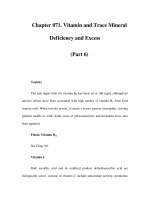
Figure 2 Mechanisms by which specific components of intestinal microbiota may protect from
allergic sensitization and/or alleviate symptoms. “Adequate” microbial composition may reduce
allergen uptake by providing maturational stimulus for gut barrier function, enhancing allergen
degradation by production of digestiveenzymes (this may also reduce allergen allergenicity), improving
mucosal integrity by direct exclusion of pathogens that may cause epithelial damage or by enhancing
secretory IgA (sIgA) production (possibly via inducing TGF-b secretion) and by inducing secretion of
anti-inflammatory cytokines, which may break a vicious circle where inflammation increases gut
permeability allowing invasion of pathogens and allergens, which then results in further inflammation.
Danger signals caused by epithelial damage and inflammation promote the maturation of dendritic cells,
which influence the differentiationof naı
¨
ve Th cells. Presentation of allergeninabsenceof danger signals
may promote formation of regulatory T cells (Treg) and thus formation of tolerance to the allergen. The
fate of Th cells in the presence of danger signals depends on additional stimulus: presence of TGF-b
(produced, e.g., by epithelial cells) may promote development of Treg population and again tolerance to
the allergen, presence of IL-12 and IFN-g (produced, e.g., by macrophages or dendritic cells) promotes
development of Th1 population and non-allergic type immune responses, whereas presence of IL-10
may promote formation of allergen specific Th2 cells. In the symptomatic phase induction of anti-
inflammatory cytokines may also directly alleviate the allergic inflammation by active suppression.
Abbreviations: sIgA, secretory IgA; M, M-cell; iDC, immature dendritic cell; mDC, mature dendritic
cell; IL, interleukin; TGF, transforming growth factor; Th, T-helper; Treg, regulatory T-cell; MF,
macrophage.
The Infant Intestinal Microbiota in Allergy 195
which would otherwise be cleared or reduced in numbers due to immune responses mounted
against them. This could partly explain why relatively a high prevalence and numbers of
potentially pathogenic Gram-negative bacteria but low numbers of Gram-positive bacteria
appear to accompany atopic eczema and high levels of IgE (18,39,42–45,50).
From another perspective, microbial compositional differences may reflect their
influence on allergic sensitization and disease development. If the recognition of gut
colonizers is compromised, then so may be the interactions that drive the normal
immunological maturation (10,32,60,69,70). Recognition of peptidoglycan, a major
component of Gram-positive cell-wall, is less dependent on CD14 and TLR4 but rather on
co-operation between TLRs 2 and 6 (71–73). Thereby, an atopic host, with deficient TLR4
and CD14 recognition, may have better chances to interact with Gram-positive than Gram-
negative bacteria. This interaction may, on one hand, limit the ability of Gram-positive
bacteria to colonize the gut, but on the other, provide maturational stimulus for the
developing immune system (44,69).
Whereas the recognition of one specific bacterial component occurs primarily via one
or two different pattern recognition receptors, the recognition of whole bacterium is likely to
involve a set of different receptors such as TLR9 recognizing unmethylated bacterial CpG
DNA and TLR5 recognizing flagella (74). Accordingly, a quantitatively strong enough
exposure may compensate the poor recognition of Gram-negative bacteria, especially dueto
ligation of TLR9. This would be in agreement with the observation that postnatal
administration of exogenous Gram-negative bacteria, namely non-enteropathogenic E. coli
strain, was associated with reduced risk of developing allergic diseases later in life (14,15).
Reflection of Effects on Th1, Th2, and Treg Differentiation
The effects of intestinal bacteria on cytokine production, epithelia-damaging action or
proinflammatory action may have a major influence on naive T-cell differentiation to Th1,
Th2 or Treg cells (Fig. 2). A study in mice with compromised Toll-mediated signaling
capacity indicated that antigen specific Th1 responses to food allergens are dependent on
simultaneously induced Toll-mediated activities, whilst similar dependency was not
observed in Th2 responses. Re-exposing the mice to the allergen enhanced the production
of IL-13 by T-cells, a cytokine capable of inducing isotype class-switching of B-cells to
produce IgE (75).
Th differentiation is directed by dendritic cells, which monitor the antigenic
environment and presence of danger signals in the gut. Danger signals may include
epithelial damage and inflammation. In the absence of maturational/inflammatory stimuli,
dendritic cells aim to tolerize the immune system to what they assume to be harmless
antigens. It is noteworthy that the immunological stimulus initiated may vary depending on
which TLR or combination of TLRs are ligated (76). This may provide a mechanistic basis
for consistent data from in vitro studies, which indicate that cytokine responses mounted by
mononuclear cells in response to whole Gram-negative and whole Gram-positive bacteria
are different. The induction of IL-12 is greater for Gram-positive bacteria and IL-10 for
Gram-negative bacteria (77–79). IL-12 is produced by dendritic cells and macrophages and
is a key cytokine promoting the Th cell differentiation into Th1 cells. IL-10 may contribute
in maintaining a Th2 bias, but it may also induce tolerance by promoting the formation of
Tregs and anergic T-cells (80–82).
In a study by He and co-workers (2002) bifidobacteria isolated from the feces of
allergic infants tended to induce murine macrophage-like cells to produce more of IL-12,
but less IL-10 than bifidobacteria from the feces of healthy infants (83). In their earlier,
aforementioned, study B. adolescentis was associated with allergic and B. bifidum with
Kirjavainen and Reid196
healthy infants (47). Accordingly, in a recent study, Young and co-workers showed that
B. bifidum enhanced IL-10 production by dendritic cells isolated from cord blood (84).
However, B. adolescentis, or any other bifidobacterial strain, did not induce IL-12
production. Moderate differences were observed in the effects of bifidobacterial strains on
the expression of dendritic cell activation markers. The basis for speculation on the possible
significance of these findings is weak until more detailed characterization is performed.
Arguably, the findings could collectively indicate that bifidobacteria in allergic infants may
promote formation of tolerogenic responses but this remains to be confirmed (Fig. 2).
Also Lactobacillus strains have been shown to confer differential effects on cytokine
production and expression of surface markers on murine dendritic cells (85). Furthermore,
lactobacilli induced in vitro, in a strain dependent manner, Treg-like low proliferating Th
population producing TGF-b and IL-10 (86). TGF-b is the key cytokine in induction of
T-cell differentiation towards Tregs (Fig. 2) (87). In a clinical study, improvement in
atopic eczema symptoms following oral administration of lactobacilli was accompanied
by increased serum concentrations of TGF-b (17). Interestingly, oral supplementation of
lactobacilli in breast-feeding mothers was followed by increased TGF-b concentrations in
breast-milk (88). This increase may have contributed to subsequently lower prevalence of
atopic eczema in children. It should be noted, however, that allergic sensitization was not
affected and allergic rhinitis and asthma may have increased in frequency (89).
Nevertheless, these studies are not only indicative of the influence of infant microbiota
on allergy development but also of the possible influence of maternal microbiota during
pregnancy and via breast-milk.
Reflection of Effects on Allergen Uptake, Processing, and Presentation
The original hygiene hypothesis implicated pathogens in an allergy-preventing role.
However, their role may be two-sided (90). Whereas the host immune system may become
tolerant towards commensal microbes, this should and will not happen with pathogens
(91,92). Therefore, pathogens may have a greater potential to stimulate the neonatal
immunity away from the allergic type responsiveness than the commensal microbes
towards which tolerance has been formed (90). Conversely, potential pathogens may
induce and sustain inflammation and compromise the gut barrier (18,93). This may allow
greater numbers of allergens to pass the barrier and alter their presentation to lymphocytes
due to the presence of danger signals. Consequently, allergic sensitization may be more
likely to occur, and may be aggravated in already sensitized subjects with allergic disease
(94–96). E. coli and Bacteroides bacterial groups colonizing these subjects may include
strains with such detrimental properties (97–100). Such bacteria were implicated with
higher serum total IgE concentrations and sensitivity to cow’s milk proteins in studies
referred to above (18,44). Some non-pathogenic bacteria, such as lactobacilli and
bifidobacteria, may have the opposite effects by reducing gut inflammation either via
excluding colonization by pathogens or inducing secretion of anti-inflammatory
cytokines, reducing gut permeability, allergen antigenicity, and fortifying gut defense
barrier e.g., by stimulating IgA production (101–110). Intestinal microbes are likely to
affect the allergen uptake also by promoting the maturation and integrity of gut barrier but
there is little information on how this ability may vary between different bacteria (111).
Reflection of Allergic Symptoms
The possibility that allergic symptoms either affect, or are affected by, the microbiota is
supported by an observation that alleviation in atopic eczema and allergic inflammation
The Infant Intestinal Microbiota in Allergy 197
following oral administration of bifidobacteria was accompanied by modified dynamics in
the microbiota (i.e., restriction in the growth of E. coli and Bacteroides) (18). Also, earlier
findings attest to this possibility implicating direct correlation between numbers of
Enterobacteriaceae family bacteria and severity of atopic eczema symptoms (39). The
compositional characteristics associated with the severity of symptoms may be caused by
intestinal inflammation exacerbated in some allergic conditions (95,112–115).
Reflection of Environmental Factors
Amongst the best examples of factors which have been clearly shown to influence the
development of the gut microbiota and have also been implicated in allergic diseases
include the mode of delivery and breast-feeding (116–123). Indeed, it is plausible that the
characteristics of fecal microbiota associated with atopic eczema and allergic sensitization
may partly reflect dietary factors. It is well known that changes in diet may dramatically
affect the microbial composition of the gut. Then again, in allergic infants the diet can reflect
the child’s health status due to food restrictions. In 39–63% of all infants and young
children, atopic eczema is triggered by one or more challenge-confirmed food allergies
(124–126). Moreover, the development of manifestations of allergic diseases in children
correlates with differences in the composition and immunological characteristics of breast-
milk, which on the other hand are affected by maternal gut microbiota and atopy (127–133).
For example, the polyunsaturated fatty acid composition in breast-milk has been shown to
correlate with the development of allergic disease in children (131,132). In vitro these
compounds have been shown to selectively affect microbial growth and adhesion to
intestinal cells (134). Recently, lactobacilli in breast-milk were shown to have properties
in vitro that could promote the development and maintenance of gutbarrier in neonates, thus
warranting further studies on this area (135). Albeit the effect of caesarean delivery in
promoting allergy is disputable, it is notable that colonization by Lactobacillus- and
Bifidobacterium-like bacteria, the high numbers of which have mainly been associated with
non-allergic phenotype, may be delayed for up to 10 days and 1 month, respectively, as
compared to vaginally delivered infants (136).
Regarding our earlier discussion on pathogens and E. coli, it is noteworthy that in
developing countries with low prevalence of allergies, the establishment of intestinal
microbiota is characterized by rapid initial colonization, formation of enterobacterial
microbiota predominated by E. coli, and frequent colonization by pathogens such as
salmonellae. The E. coli population is characterized by a wide spectrum of strains and
instability (137,138). Whether such rapid colonization and strongly variable exposure has
special influence on immunological maturation and gut barrier formation and maintenance
remain to be established.
CONCLUSION
It has been well established that allergic sensitization and the development of allergic
disease are associated, at least in some infants, with characteristic developmental patterns
in fecal microbiota composition that are atypical to healthy infants. With relative
consistency these characteristics include low numbers of bifidobacteria and anaerobes in
total and high numbers of clostridia, S. aureus and certain coliforms such as Klebsiellae.
Data on lactobacilli, Bacteroides and E. coli are somewhat variable. How this aberrancy in
fecal microbiota depicts the situation in the intestine and how it is clinically significant,
remains to be known. The possibility that the characteristics are secondary to the disease
Kirjavainen and Reid198
cannot be excluded, but it is also feasible that they reflect their significance in the aetiology
of allergy. Extensive experimental data implies that the development of atopic type
immunoreactivity could be promoted by the establishment of an early gut microbiota that
(1) is incapable of directing the immune system towards tolerogenic responses to, what
should be, harmless environmental antigens and/or (2) induces inflammatory responses
against itself, thereby increasing mucosal permeability to potential allergens.
It has been convincingly demonstrated that microbial exposure is likely to be the
primary exogenous stimulus directing the immunological maturation away from allergic
type immunoresponsiveness early in life. However, it is still not clear what are the
qualitative or quantitative characteristics of the indigenous microbiota or other sources of
microbial exposure that could protect from, or conversely promote (“allow”), the
expression of allergies. Future studies should assess whether specific microbial species
have particular importance in this respect or whether the “adequate” stimulus is only a
matter of quantitatively high enough exposure or strongly variable exposure. More efforts
should be directed to characterizing microbial composition of nasal and oral cavities and
different compartments in the intestinal tract of children as well as the gut of pregnant
women and the gut and breast-milk of breast-feeding mothers.
ACKNOWLEDGMENTS
Pirkka Kirjavainen gratefully acknowledges financial support from the Academy
of Finland.
REFERENCES
1. Arruda LK, Sole D, Baena-Cagnani CE, Naspitz CK. Risk factors for asthma and atopy. Curr
Opin Allergy Clin Immunol 2005; 5:153–159.
2. Steinke JW, Borish L, Rosenwasser LJ 5. Genetics of hypersensitivity. J Allergy Clin
Immunol 2003; 111:S495–S501.
3. von Mutius E. Influences in allergy: epidemiology and the environment. J Allergy Clin
Immunol 2004; 113:373–379; quiz 380.
4. von Mutius E. The environmental predictors of allergic disease. J Allergy Clin Immunol 2000;
105:9–19.
5. O’Connell EJ. Pediatric allergy: a brief review of risk factors associated with developing
allergic disease in childhood. Ann Allergy Asthma Immunol 2003; 90:53–58.
6. Strachan DP. Hay fever, hygiene, and household size. BMJ 1989; 299:1259–1260.
7. Noverr MC, Huffnagle GB. Does the microbiota regulate immune responses outside the gut?
Trends Microbiol 2004; 12:562–568.
8. Moreau MC, Corthier G. Effect of the gastrointestinal microflora on induction and
maintenance of oral tolerance to ovalbumin in C3H/HeJ mice. Infect Immun 1988;
56:2766–2768.
9. Moreau MC, Gaboriau-Routhiau V. The absence of gut flora, the doses of antigen ingested
and aging affect the long-term peripheral tolerance induced by ovalbumin feeding in mice.
Res Immunol 1996; 147:49–59.
10. Sudo N, Sawamura S, Tanaka K, Aiba Y, Kubo C, Koga Y. The requirement of intestinal
bacterial flora for the development of an IgE production system fully susceptible to oral
tolerance induction. J Immunol 1997; 159:1739–1745.
11. Gaboriau-Routhiau V, Moreau MC. Gut flora allows recovery of oral tolerance to ovalbumin
in mice after transient breakdown mediated by cholera toxin or Escherichia coli heat-labile
enterotoxin. Pediatr Res 1996; 39:625–629.
The Infant Intestinal Microbiota in Allergy 199
12. Hooper LV, Gordon JI. Commensal host-bacterial relationships in the gut. Science 2001;
292:1115–1118.
13. Kallioma
¨
ki M, Salminen S, Arvilommi H, Kero P, Koskinen P, Isolauri E. Probiotics in
primary prevention of atopic disease: a randomised placebo-controlled trial. Lancet 2001;
357:1076–1079.
14. Lodinova
´
-Z
ˇ
a
´
dnı
´
kova
´
R, Cukrowska
´
B. Influence of oral colonization of the intestine with a
non-enteropathogenic E. coli strain after birth on the frequency of infectious and allergic
diseases after 10 and 20 years. Immunol Lett 1999; 69:64.
15. Lodinova
´
-Z
ˇ
a
´
dnı
´
kova
´
R, Cukrowska B, Tlaskalova-Hogenova H. Oral administration of
probiotic Escherichia coli after birth reduces frequency of allergies and repeated infections
later in life (after 10 and 20 years). Int Arch Allergy Immunol 2003; 131:209–211.
16. Majamaa H, Isolauri E. Probiotics: a novel approach in the management of food allergy.
J Allergy Clin Immunol 1997; 99:179–185.
17. Isolauri E, Arvola T, Sutas Y, Moilanen E, Salminen S. Probiotics in the management of
atopic eczema. Clin Exp Allergy 2000; 30:1604–1610.
18. Kirjavainen PV, Arvola T, Salminen SJ, Isolauri E. Aberrant composition of gut microbiota of
allergic infants: a target of bifidobacterial therapy at weaning? Gut 2002; 51:51–55.
19. Celedon JC, Weiss ST. Use of antibacterials in infancy: clinical implications for childhood
asthma and allergies. Treat Respir Med 2004; 3:291–294.
20. Voor T, Julge K, Bottcher MF, Jenmalm MC, Duchen K, Bjorksten B. Atopic sensitization
and atopic dermatitis in estonian and Swedish infants. Clin Exp Allergy 2005; 35:153–159.
21. Johansson SG, Bieber T, Dahl R, et al. Revised nomenclature for allergy for global use: report
of the nomenclature review committee of the world allergy organization, October 2003.
J Allergy Clin Immunol 2004; 113:832–836.
22. O’Connell EJ. The burden of atopy and asthma in children. Allergy 2004; 78:7–11.
23. Blaiss MS. Important aspects in management of allergic rhinitis: compliance, cost, and quality
of life. Allergy Asthma Proc 2003; 24:231–238.
24. Cisternas M, Blanc P, Yen I, et al. A comprehensive study of the direct and indirect costs of
adult asthma. J Allergy Clin Immunol 2003; 111:1212–1218.
25. Jarvis D, Burney P. ABC of allergies. The epidemiology of allergic disease. BMJ 1998;
316:607–610.
26. Wickman M, Lilja G. Today, one child in four has an ongoing allergic disease in Europe.
What will the situation be tomorrow? Allergy 2003; 58:570–571.
27. Johansson SG, Hourihane JO, Bousquet J, et al. A revised nomenclature for allergy. An
EAACI position statement from the EAACI nomenclature task force. Allergy 2001;
56:813–824.
28. Kay AB. Allergy and allergic diseases. First of two parts. N Engl J Med 2001; 344:30–37.
29. Ebner C, Schenk S, Najafian N, et al. Nonallergic individuals recognize the same T cell
epitopes of Bet v 1, the major birch pollen allergen, as atopic patients. J Immunol 1995;
154:1932–1940.
30. Prescott SL. Allergy: when does it begin and where will it end?. Allergy 2003; 58:864–867.
31. Prescott SL, Macaubas C, Holt BJ, et al. Transplacental priming of the human immune system
to environmental allergens: universal skewing of initial T cell responses toward the Th2
cytokine profile. J Immunol 1998; 160:4730–4737.
32. Prescott SL, Macaubas C, Smallacombe T, Holt BJ, Sly PD, Holt PG. Development of
allergen-specific T-cell memory in atopic and normal children. Lancet 1999; 353:196–200.
33. Robinson DS, Larche M, Durham SR. Tregs and allergic disease. J Clin Invest 2004;
114:1389–1397.
34. Nakamura K, Kitani A, Fuss I, et al. TGF-beta 1 plays an important role in the mechanism of
CD4CCD25C regulatory T cell activity in both humans and mice. J Immunol 2004;
172:834–842.
35. Nakamura K, Kitani A, Strober W. Cell contact-dependent immunosuppression by
CD4(C)CD25(C) regulatory T cells is mediated by cell surface-bound transforming
growth factor beta. J Exp Med 2001; 194:629–644.
Kirjavainen and Reid200
36. Ling EM, Smith T, Nguyen XD, et al. Relation of CD4CCD25Cregulatory T-cell
suppression of allergen-driven T-cell activation to atopic status and expression of allergic
disease. Lancet 2004; 363:608–615.
37. Akdis M, Verhagen J, Taylor A, et al. Immune responses in healthy and allergic individuals
are characterized by a fine balance between allergen-specific T regulatory 1 and T helper 2
cells. J Exp Med 2004; 199:1567–1575.
38. Kuvaeva IB, Zakharova NV, Orlova NG, Veselova OL. Functional state of the immunological
system and of the gastrointestinal tract in children with a food allergy. Vopr Pitan
1980;33–40.
39. Kuvaeva IB, Orlova NG, Veselova OL, Kuznezova GG, Borovik TE. Microecology of the
gastrointestinal tract and the immunological status under food allergy. Nahrung 1984;
28:689–693.
40. Shaternikov VA, Kuvaeva ID, Ladodo KS, Orlova NG, Veselova OL. General and local
humoral immunity and intestinal microflora in children with skin manifestations of food
allergy. Vopr Pitan 1982;51–56.
41. Ionescu G, Radovicic D, Schuler R, et al. Changes in fecal microflora and malabsorption
phenomena suggesting a contaminated small bowel syndrome in atopic eczema patients.
Microecol Ther 1986; 16:273.
42. Ionescu G, Kiehl R, Ona L, Schuler R. Abnormal fecal microflora and malabsorption
phenomena in atopic eczema paitents. J Adv Med 1990; 3:71–91.
43. Ionescu G, Kiehl R, Wichmann-Kunz F, Leimbeck R. Immunobiological significance of
fungal and bacterial infections in atopic eczema. J Adv Med 1990; 3:47–58.
44. Kirjavainen PV, Apostolou E, Arvola T, Salminen SJ, Gibson GR, Isolauri E. Characterizing
the composition of intestinal microflora as a prospective treatment target in infant allergic
disease. FEMS Immunol Med Microbiol 2001; 32:1–7.
45. Bjo
¨
rkste
´
n B, Naaber P, Sepp E, Mikelsaar M. The intestinal microflora in allergic estonian
and Swedish 2-year-old children. Clin Exp Allergy 1999; 29:342–346.
46. Bjo
¨
rkste
´
n B, Sepp E, Julge K, Voor T, Mikelsaar M. Allergy development and the intestinal
microflora during the first year of life. J Allergy Clin Immunol 2001; 108:516–520.
47. Ouwehand AC, Isolauri E, He F, Hashimoto H, Benno Y, Salminen S. Differences in
Bifidobacterium flora composition in allergic and healthy infants. J Allergy Clin Immunol
2001; 108:144–145.
48. Kallioma
¨
ki M, Kirjavainen P, Eerola E, Kero P, Salminen S, Isolauri E. Distinct patterns of
neonatal gut microflora in infants in whom atopy was and was not developing. J Allergy Clin
Immunol 2001; 107:129–134.
49. Bo
¨
ttcher MF, Nordin EK, Sandin A, Midtvedt T, Bjo
¨
rkste
´
n B. Microflora-associated
characteristics in faeces from allergic and nonallergic infants. Clin Exp Allergy 2000;
30:1590–1596.
50. Kirjavainen P. The Intestinal Microbiota—A Target for Treatment in Infant Atopic
Eczema, in Department of Biochemistry and Food Chemistry. Turku: University of
Turku, 2003: 79.
51. Howard TD, Meyers DA, Bleecker ER. Mapping susceptibility genes for allergic diseases.
Chest 2003; 123:363S–368S.
52. Mikelsaar M, Ma
¨
ndar R, Sepp E. Lactic acid microflora in the human microbial ecosystem
and its development. In: Salminen S, Von Wright A, eds. Lactic Acid Bacteria: Microecology
and Functional Aspects. New York: Marcel Dekker Inc., 1998:278–342.
53. Marteau P, Pochart P, Dore J, Bera-Maillet C, Bernalier A, Corthier G. Comparative study of
bacterial groups within the human cecal and fecal microbiota. Appl Environ Microbiol 2001;
67:4939–4942.
54. Zoetendal EG, von Wright A, Vilpponen-Salmela T, Ben-Amor K, Akkermans AD, de
Vos WM. Mucosa-associated bacteria in the human gastrointestinal tract are uniformly
distributed along the colon and differ from the community recovered from feces. Appl
Environ Microbiol 2002; 68:3401–3407.
The Infant Intestinal Microbiota in Allergy 201
55. Kirjavainen PV, ElNezami HS, Salminen SJ, Ahokas JT, Wright PF. Effects of orally
administered viable Lactobacillus rhamnosus GG and Propionibacterium freudenreichii
subsp. shermanii JS on mouse lymphocyte proliferation. Clin Diagn Lab Immunol 1999;
6:799–802.
56. Kirjavainen PV, El-Nezami HS, Salminen SJ, Ahokas JT, Wright PF. The effect of orally
administered viable probiotic and dairy lactobacilli on mouse lymphocyte proliferation.
FEMS Immunol Med Microbiol 1999; 26:131–135.
57. Wollenberg A, Bieber T. Atopic dermatitis: from the genes to skin lesions. Allergy 2000;
55:205–213.
58. Feijen M, Gerritsen J, Postma DS. Genetics of allergic disease. Br Med Bull 2000;
56:894–907.
59. Koppelman GH, Reijmerink NE, Colin Stine O, et al. Association of a promoter
polymorphism of the CD14 gene and atopy. Am J Respir Crit Care Med 2001; 163:965–969.
60. Baldini M, Lohman IC, Halonen M, Erickson RP, Holt PG, Martinez FD. A Polymorphism*
in the 5
0
flanking region of the CD14 gene is associated with circulating soluble CD14 levels
and with total serum immunoglobulin E. Am J Respir Cell Mol Biol 1999; 20:976–983.
61. Zdolsek HA, Jenmalm MC. Reduced levels of soluble CD14 in atopic children. Clin Exp
Allergy 2004; 34:532–539.
62. Kabesch M, Hasemann K, Schickinger V, et al. A promoter polymorphism in the CD14 gene
is associated with elevated levels of soluble CD14 but not with IgE or atopic diseases. Allergy
2004; 59:520–525.
63. Kedda MA, Lose F, Duffy D, Bell E, Thompson PJ, Upham J. The CD14 C-159T
polymorphism is not associated with asthma or asthma severity in an Australian adult
population. Thorax 2005; 60:211–214.
64. Fageras Bottcher M, Hmani-Aifa M, Lindstrom A, et al. A TLR4 polymorphism is associated
with asthma and reduced lipopolysaccharide-induced interleukin-12(p70) responses in
Swedish children. J Allergy Clin Immunol 2004; 114:561–567.
65. Jones CA, Holloway JA, Popplewell EJ, et al. Reduced soluble CD14 levels in amniotic fluid
and breast milk are associated with the subsequent development of atopy, eczema, or both.
J Allergy Clin Immunol 2002; 109:858–866.
66. Haziot A, Ferrero E, Kontgen F, et al. Resistance to endotoxin shock and reduced
dissemination of gram-negative bacteria in CD14-deficient mice. Immunity 1996; 4:407–414.
67. Miller SI, Ernst RK, Bader MW. LPS, TLR4 and infectious disease diversity. Nat Rev
Microbiol 2005; 3:36–46.
68. Cohen L, Haziot A, Shen DR, et al. CD14-independent responses to LPS require a serum
factor that is absent from neonates. J Immunol 1995; 155:5337–5342.
69. Kirjavainen PV. Exposure to gram-positive bacteria: the key in natural defence against atopic
sensitisation?. Microecol Ther 2002;109–114.
70. Sudo N, Yu XN, Aiba Y, et al. An oral introduction of intestinal bacteria prevents the
development of a long-term Th2-skewed immunological memory induced by neonatal
antibiotic treatment in mice. Clin Exp Allergy 2002; 32:1112–1116.
71. Ozinsky A, Underhill DM, Fontenot JD, et al. The repertoire for pattern recognition of
pathogens by the innate immune system is defined by cooperation between toll-like receptors.
Proc Natl Acad Sci USA 2000; 97:13766–13771.
72. Ozinsky A, Smith KD, Hume D, Underhill DM. Co-operative induction of pro-inflammatory
signaling by Toll-like receptors. J Endotoxin Res 2000; 6:393–396.
73. Dziarski R, Ulmer AJ, Gupta D. Interactions of CD14 with components of gram-positive
bacteria. Chem Immunol 2000; 74:83–107.
74. Takeda K, Akira S. Toll-like receptors in innate immunity. Int Immunol 2005; 17:1–14.
75. Schnare M, Barton GM, Holt AC, Takeda K, Akira S, Medzhitov R. Toll-like receptors
control activation of adaptive immune responses. Nat Immunol 2001; 2:947–950.
76. Aderem A, Ulevitch RJ. Toll-like receptors in the induction of the innate immune response.
Nature 2000; 406:782–787.
Kirjavainen and Reid202
77. Cross ML, Ganner A, Teilab D, Fray LM. Patterns of cytokine induction by gram-
positive and gram-negative probiotic bacteria. FEMS Immunol Med Microbiol 2004;
42:173–180.
78. Karlsson H, Hessle C, Rudin A. Innate immune responses of human neonatal cells to bacteria
from the normal gastrointestinal flora. Infect Immun 2002; 70:6688–6696.
79. Hessle C, Andersson B, Wold AE. Gram-positive bacteria are potent inducers of monocytic
interleukin-12 (IL-12) while gram-negative bacteria preferentially stimulate IL-10
production. Infect Immun 2000; 68:3581–3586.
80. Raghupathy R. Pregnancy: success and failure within the Th1/Th2/Th3 paradigm. Semin
Immunol 2001; 13:219–227.
81. Akdis CA, Blaser K. Mechanisms of interleukin-10-mediated immune suppression.
Immunology 2001; 103:131–136.
82. Levings MK, Gregori S, Tresoldi E, Cazzaniga S, Bonini C, Roncarolo MG. Differentiation of
Tr1 cells by immature dendritic cells requires IL-10 but not CD25CCD4C Tr cells. Blood
2005; 105:1162–1169.
83. He F, Morita H, Hashimoto H, et al. Intestinal Bifidobacterium species induce varying
cytokine production. J Allergy Clin Immunol 2002; 109:1035–1036.
84. Young SL, Simon MA, Baird MA, et al. Bifidobacterial species differentially affect
expression of cell surface markers and cytokines of dendritic cells harvested from cord blood.
Clin Diagn Lab Immunol 2004; 11:686–690.
85. Christensen HR, Frokiaer H, Pestka JJ. Lactobacilli differentially modulate expression of
cytokines and maturation surface markers in murine dendritic cells. J Immunol 2002;
168:171–178.
86. von der Weid T, Bulliard C, Schiffrin EJ. Induction by a lactic acid bacterium of a population
of CD4(C) T cells with low proliferative capacity that produce transforming growth factor
beta and interleukin-10. Clin Diagn Lab Immunol 2001; 8:695–701.
87. Huber S, Schramm C, Lehr HA, et al. Cutting edge: TGF-beta signaling is required for the
in vivo expansion and immunosuppressive capacity of regulatory CD4CCD25CT cells.
J Immunol 2004; 173:6526–6531.
88. Rautava S, Kallioma
¨
ki M, Isolauri E. Probiotics during pregnancy and breast-feeding might
confer immunomodulatory protection against atopic disease in the infant. J Allergy Clin
Immunol 2002; 109:119–121.
89. Kallioma
¨
ki M, Salminen S, Poussa T, Arvilommi H, Isolauri E. Probiotics and prevention of
atopic disease: 4-year follow-up of a randomised placebo-controlled trial. Lancet 2003;
361:1869–1871.
90. Kirjavainen PV. In: Mattila-Sandholm T, Saarela M, eds. Probiotics and the management of
food allergy, in functional dairy products. Cambridge, U.K.: Woodhead Publishing,
2003:108–131.
91. Neish AS, Gewirtz AT, Zeng H, et al. Prokaryotic regulation of epithelial responses by
inhibition of IkappaB-alpha ubiquitination. Science 2000; 289:1560–1563.
92. Nagler-Anderson C. Man the barrier! Strategic defences in the intestinal mucosa. Nat Rev
Immunol 2001; 1:59–67.
93. Kirjavainen PV, Apostolou E, Salminen SJ, Isolauri E. New aspects of probiotics–a novel
approach in the management of food allergy. Allergy 1999; 54:909–915.
94. Batt RM, Rutgers HC, Sancak AA. Enteric bacteria: friend or foe?. J Small Anim Pract 1996;
37:261–267.
95. Berin MC, Yang PC, Ciok L, Waserman S, Perdue MH. Role for IL-4 in macromolecular
transport across human intestinal epithelium. Am J Physiol 1999; 276:C1046–C1052.
96. Gee JM, Wal JM, Miller K, et al. Effect of saponin on the transmucosal passage of beta-
lactoglobulin across the proximal small intestine of normal and beta-lactoglobulin-sensitised
rats. Toxicology 1997; 117:219–228.
97. Dahlgren UI, Wold AE, Hanson LA, Midtvedt T. Expression of a dietary protein in E. coli
renders it strongly antigenic to gut lymphoid tissue. Immunology 1991; 73:394–397.
The Infant Intestinal Microbiota in Allergy 203
98. Deitch EA, Specian RD, Berg RD. Endotoxin-induced bacterial translocation and mucosal
permeability: role of xanthine oxidase, complement activation, and macrophage products. Crit
Care Med 1991; 19:785–791.
99. Obiso RJ, Jr., Lyerly DM, Van Tassell RL, Wilkins TD. Proteolytic activity of the Bacteroides
fragilis enterotoxin causes fluid secretion and intestinal damage in vivo. Infect Immun 1995;
63:3820–3826.
100. Duchmann R, Kaiser I, Hermann E, Mayet W, Ewe K, Meyer zum KH. Buschenfelde,
Tolerance exists towards resident intestinal flora but is broken in active inflammatory bowel
disease (IBD). Clin Exp Immunol 1995; 102:448–455.
101. Moreau MC, Ducluzeau R, Guy-Grand D, Muller MC. Increase in the population of duodenal
immunoglobulin A plasmocytes in axenic mice associated with different living or dead
bacterial strains of intestinal origin. Infect Immun 1978; 21:532–539.
102. De Simone C, Ciardi A, Grassi A, et al. Effect of Bifidobacterium bifidum and Lactobacillus
acidophilus on gut mucosa and peripheral blood B lymphocytes. Immunopharmacol
Immunotoxicol 1992; 14:331–340.
103. Kaila M, Isolauri E, Soppi E, Virtanen E, Laine S, Arvilommi H. Enhancement of the
circulating antibody secreting cell response in human diarrhea by a human Lactobacillus
strain. Pediatr Res 1992; 32:141–144.
104. Yasui H, Nagaoka N, Mike K, Hayakawa K, Ohwaki M. Detection of Bifidobacterium strains
that induce large quantities of IgA. Microb Ecol Health Dis 1992; 5:155–162.
105. Majamaa H, Isolauri E, Saxelin M, Vesikari T. Lactic acid bacteria in the treatment of acute
rotavirus gastroenteritis. J Pediatr Gastroenterol Nutr 1995; 20:333–338.
106. Isolauri E, Majamaa H, Arvola T, Rantala I, Virtanen E, Arvilommi H. Lactobacillus casei
strain GG reverses increased intestinal permeability induced by cow milk in suckling rats.
Gastroenterology 1993; 105:1643–1650.
107. Matsuzaki T, Yamazaki R, Hashimoto S, Yokokura T. The effect of oral feeding of
Lactobacillus casei strain Shirota on immunoglobulin E production in mice. J Dairy Sci 1998;
81:48–53.
108. Su
¨
tas Y, Soppi E, Korhonen H, et al. Suppression of lymphocyte proliferation in vitro by
bovine caseins hydrolyzed with Lactobacillus casei GG-derived enzymes. J Allergy Clin
Immunol 1996; 98:216–224.
109. Su
¨
tas Y, Hurme M, Isolauri E. Down-regulation of anti-CD3 antibody-induced IL-4
production by bovine caseins hydrolysed with Lactobacillus GG-derived enzymes. Scand
J Immunol 1996; 43:687–689.
110. Pessi T, Isolauri E, Su
¨
tas Y, Kankaanranta H, Moilanen E, Hurme M. Suppression of T-cell
activation by Lactobacillus rhamnosus GG-degraded bovine casein. Int Immunopharmacol
2001; 1:211–218.
111. Hooper LV, Falk PG, Gordon JI. Analyzing the molecular foundations of commensalism in
the mouse intestine. Curr Opin Microbiol 2000; 3:79–85.
112. Ogawa H, Yoshiike T. A speculative view of atopic dermatitis: barrier dysfunction in
pathogenesis. J Dermatol Sci 1993; 5:197–204.
113. Majamaa H, Laine S, Miettinen A. Eosinophil protein X and eosinophil cationic protein as
indicators of intestinal inflammation in infants with atopic eczema and food allergy. Clin Exp
Allergy 1999; 29:1502–1506.
114. Majamaa H, Miettinen A, Laine S, Isolauri E. Intestinal inflammation in children with atopic
eczema: faecal eosinophil cationic protein and tumour necrosis factor-alpha as non-invasive
indicators of food allergy. Clin Exp Allergy 1996; 26:181–187.
115. Majamaa H, Aittoniemi J, Miettinen A. Increased concentration of fecal alpha1-antitrypsin is
associated with cow’s milk allergy in infants with atopic eczema. Clin Exp Allergy 2001;
31:590–592.
116. Xu B, Pekkanen J, Hartikainen AL, Jarvelin MR. Caesarean section and risk of asthma and
allergy in adulthood. J Allergy Clin Immunol 2001; 107:732–733.
117. Kero J, Gissler M, Gronlund MM, et al. Mode of delivery and asthma—is there a connection?.
Pediatr Res 2002; 52:6–11.
Kirjavainen and Reid204
118. Saarinen UM, Kajosaari M, Backman A, Siimes MA. Prolonged breast-feeding as prophylaxis
for atopic disease. Lancet 1979; 2:163–166.
119. Gdalevich M, Mimouni D, Mimouni M. Breast-feeding and the risk of bronchial asthma in
childhood: a systematic review with meta-analysis of prospective studies. J Pediatr 2001;
139:261–266.
120. Gdalevich M, Mimouni D, David M, Mimouni M. Breast-feeding and the onset of atopic
dermatitis in childhood: a systematic review and meta-analysis of prospective studies. J Am
Acad Dermatol 2001; 45:520–527.
121. Schoetzau A, Filipiak-Pittroff B, Franke K, et al. Effect of exclusive breast-feeding and early
solid food avoidance on the incidence of atopic dermatitis in high-risk infants at 1 year of age.
Pediatr Allergy Immunol 2002; 13:234–242.
122. Kull I, Wickman M, Lilja G, Nordvall SL, Pershagen G. Breast feeding and allergic diseases
in infants-a prospective birth cohort study. Arch Dis Child 2002; 87:478–481.
123. Kirjavainen PV, Gibson GR. Healthy gut microflora and allergy: factors influencing
development of the microbiota. Ann Med 1999; 31:288–292.
124. Sampson HA. The immunopathogenic role of food hypersensitivity in atopic dermatitis. Acta
Derm Venereol Suppl (Stockh) 1992; 176:34–37.
125. Isolauri E, Turjanmaa K. Combined skin prick and patch testing enhances identification of
food allergy in infants with atopic dermatitis. J Allergy Clin Immunol 1996; 97:9–15.
126. Burks AW, James JM, Hiegel A, et al. Atopic dermatitis and food hypersensitivity reactions.
J Pediatr 1998; 132:132–136.
127. Kallioma
¨
ki M, Ouwehand A, Arvilommi H, Kero P, Isolauri E. Transforming growth factor-
beta in breast milk: a potential regulator of atopic disease at an early age. J Allergy Clin
Immunol 1999; 104:1251–1257.
128. Ja
¨
rvinen KM, Laine S, Suomalainen H. Defective tumour necrosis factor-alpha production in
mother’s milk is related to cow’s milk allergy in suckling infants. Clin Exp Allergy 2000;
30:637–643.
129. Ja
¨
rvinen KM, Laine ST, Jarvenpaa AL, Suomalainen HK. Does low IgA in human milk
predispose the infant to development of cow’s milk allergy? Pediatr Res 2000; 48:457–462.
130. Ja
¨
rvinen KM, Suomalainen H. Leucocytes in human milk and lymphocyte subsets in cow’s
milk-allergic infants. Pediatr Allergy Immunol 2002; 13:243–254.
131. Duchen K, Casas R, Fageras-Bottcher M, Yu G, Bjorksten B. Human milk polyunsaturated
long-chain fatty acids and secretory immunoglobulin A antibodies and early childhood
allergy. Pediatr Allergy Immunol 2000; 11:29–39.
132. Duchen K, Bjorksten B. Polyunsaturated n-3 fatty acids and the development of atopic
disease. Lipids 2001; 36:1033–1042.
133. Thijs C, Houwelingen A, Poorterman I, Mordant A, van den Brandt P. Essential fatty acids in
breast milk of atopic mothers: comparison with non-atopic mothers, and effect of borage oil
supplementation. Eur J Clin Nutr 2000; 54:234–238.
134. Kankaanpa
¨
a
¨
P, Nurmela K, Erkkila A, et al. Polyunsaturated fatty acids in maternal diet,
breast milk, and serum lipid fatty acids of infants in relation to atopy. Allergy 2001;
56:633–638.
135. Martin R, Olivares M, Marin ML, Fernandez L, Xaus J, Rodriguez JM. Probiotic potential of 3
Lactobacilli strains isolated from breast milk. J Hum Lact 2005; 21:8–17 quiz 18–21, 41.
136. Gro
¨
nlund MM, Lehtonen OP, Eerola E, Kero P. Fecal microflora in healthy infants born by
different methods of delivery: permanent changes in intestinal flora after cesarean delivery.
J Pediatr Gastroenterol Nutr 1999; 28:19–25.
137. Adlerberth I. In: Hanson LA
˚
, Yolken RH, eds. Establishment of Normal Intestinal Microflora
in the Newborn Infant., in Probiotics, Other Nutritional Factors, and the Intestinal Flora.
Philadelphia: Vevey/Lippicott-Raven Publishers, 1999.
138. Mata LJ, Urrutia JJ. Intestinal colonization of breastfed children in a rural area of low socio-
economic level. Ann NY Acad Sci 1971; 176:93–109.
The Infant Intestinal Microbiota in Allergy 205
11
Probiotics: A Role in Therapy for
Inflammatory Bowel Disease
Barbara Sheil, Jane McCarthy, Liam O’Mahony, and Malik M. Anwar
Alimentary Pharmabiotic Centre, Departments of Medicine and Surgery, Microbiology,
National Food Biotechnology Centre, National University of Ireland, Cork, Ireland
Fergus Shanahan
Alimentary Pharmabiotic Centre, Departments of Medicine and Surgery, National
University of Ireland, Cork, Ireland
INTRODUCTION
Hippocrates is credited with saying: “Let food be thy medicine and medicine be thy food”
(1). The term “functional food” includes “any food or food ingredient that may provide a
health benefit beyond the traditional nutrients it contains” (2). Probiotic bacteria are forms
of functional food that are of particular relevance to gastroenterologists, with evidence for
their role in the treatment of infectious and antibiotic-associated diarrhea. Their putative
therapeutic role in inflammatory bowel disease (IBD) is receiving growing interest;
however, it remains unproven. The Noble laureate, Elie Metchnikoff, suggested that
bacteria could be of some benefit to the health of man (3). He suggested that the
consumption of copious amounts of fermented dairy products, which served to introduce
“beneficial” bacteria to the gastrointestinal tract, was responsible for the longevity of
Bulgarian peasants. This marked the birth of probiotics, which are live microorganisms
that, when consumed in an adequate amount, confer a health effect on the host (4).
The last decade has seen a resurgence of interest in probiotic research. This renewal
of interest in enteric (intestinal) microbiota and gut host-microbe interactions has been
generated for a number of reasons. Firstly, the gut contains a complex microbial
community, the composition of which has remained elusive due to limited bacteriological
culturing techniques. Molecular techniques have now been applied to accurately profile
intestinal bacterial groups. Secondly, cross-talk between the gut epithelium and bacteria
has been demonstrated. The mechanisms underlying this interaction, and the role of the
microbiota in the development and function of the gastrointestinal tract needs further
investigation. A breakdown in immune tolerance to enteric microbiota has also been
implicated in the pathogenesis of inflammatory disorders, such as inflammatory bowel
disease. While evidence suggests that inflammatory bowel disease is characterized by an
aggressive immune response to luminal antigens, including members of the commensal
207
microbiota, the precise role of the luminal microbiota in the pathogenesis of disease has
yet to be elucidated. Finally, there is evidence suggesting a role for probiotic bacteria in
ameliorating inflammatory disease. This has led to the suggestion that probiotics may be
an option in the therapy of inflammatory bowel disease, the rationale being that these
bacteria without proinflammatory potential might alter the intestinal microbiota balance
and modulate the immune response (5–8).
Inflammatory bowel disease encompasses two major diseases, ulcerative colitis
(UC) and Crohn’s disease (CD). These two syndromes, while sharing similar features of
gut mucosal inflammation, are distinct entities. Their pathogenesis remains incompletely
understood. Both diseases are commonest in the Western, developed world, with highest
incidence in northern climates (9,10).
Genetic factors are known to play a role in the pathogenesis of inflammatory bowel
disease. This is demonstrated by concordance in monozygous twin studies. Also, 10–25%
of affected patients have a first-degree relative with the disease. However, the incomplete
concordance seen in twin studies (concordance rates are 40–50% for CD and !10% for
ulcerative colitis) suggests that environmental factors also contribute to the pathogenesis
of the disease. In addition, there has been a marked rise in the frequency of CD in the
developed world in the past fifty years, with a prevalence of approximately 100 per
100,000 population in North America and northern Europe. This rise in incidence in CD
underscores the importance of environmental factors in its etiology. The increase in the
incidence of CD has occurred as countries become more developed and industrialized.
With changes in lifestyle and environment, improving levels of sanitation have altered the
microbial environment. This means altered patterns of exposure to microbes and
infections during childhood (11). Inflammatory bowel disease may be a disorder of
mucosal immune responsiveness due to lack of stimulation and education of the immune
responses (12). It is interesting that parallel to an increase in CD, other chronic
inflammatory disorders, including allergies, asthma, multiple sclerosis and insulin-
dependent diabetes mellitus have also increased in incidence. Environmental changes
associated with industrialization may alter immune system development and pose a risk
factor for inflammatory bowel disease in the genetically susceptible individual (12).
THE ROLE OF THE ENTERIC MICROBIOTA IN THE NORMAL GUT
Underpinning the probiotic concept is the importance of the normal intestinal microbiota
in health and disease (12). Establishment of gut microbiota begins within minutes of
delivery of the newborn (13,14). During delivery the infant is exposed to bacteria in the
birth canal, the environment, maternal fecal microbiota, and other sources (15). The gut is
initially colonized by facultative anaerobes such as Escherichia coli and Enterococcus
species, possibly due to the absence of anaerobic conditions in the intestine (16).
Colonization with bifidobacteria follows, particularly in breast-fed infants, and as the
environment becomes more anaerobic, Bacteroides and Clostridia.
The importance of the intestinal microbiota is suggested by the fact that the healthy
adult gastrointestinal tract is home to a gut microbiota comprising over 400 different
species with more bacterial cells in the gut than eucaryotic cells in the human body and with
the average mass of bacteria being 1–2 kg. Commensal bacteria are present at a number of
10
4–6
per gram of intestinal content in the small bowel, up to 10
8
per gram of ileal content in
the distal ileum and up to 10
13
cells per gram of colonic content (17).
Sheil et al.208
The collective metabolic activity of the normal microbiota, of which little is known,
is estimated to rival that of the liver (18–21). Up to 99% of the microbiota is comprised of
30 to 40 strains, with the most abundant populations being strict anaerobes (22,23).
Bacterial members of the genus Bacteroides are amongst the most prominent species
found in human feces. Other species include bifidobacteria, clostridia, streptococci,
enterococci, lactobacilli, ruminococci, and eubacteria (4,22). Information regarding the
microbiota has been restricted by the limitations of bacteriological culture methodology
with only 40% of bacterial communities being cultivated on non-selective media in the
laboratory (24).
Effects of Enteric Microbiota in the Healthy Intestine
Experiments with germ-free and re-colonized animals demonstrate beneficial effects of the
resident microbiota (20). The commensal bacteria act as a defense against infection using
several mechanisms, including competition for nutrients, the production of antimicrobial
factors against pathogens, such as lactic acid and bacteriocins, and blockage or antagonism
of adhesion sites.
In addition, the integrity of the mucosa requires cell signaling between the
microbiota, epithelium, and mucosal immune system (7). Without the microbiota, mucosal
associated lymphoid tissue is underdeveloped and cell mediated immunity is defective.
The enteric microbiota plays an important role in immune system education by fine-tuning
T-cell repertoires and Th1/Th2 cytokine profiles (11). Compared with conventional
animals, germ-free animals have reduced mucosal cell turnover, cytokine production,
mucosal associated lymphoid tissue and lamina propria cellularity leading to an ineffective
cell mediated immunity, decreased vascularity and less muscle wall thickness (25–27).
There are also differences in intraepithelial lymphocytes (28,29). The intestinal microbiota
primes the mucosal immune response and keeps it in a state of “controlled physiological
inflammation” (26). Induction and/or maintenance of oral tolerance to ingested antigens
also require microbial colonization of the gastrointestinal tract in early life.
Understanding the influence of the gastrointestinal microbiota has prompted interest
in the therapeutic modification of the enteric microbiota with probiotics or prebiotics.
THE IMPORTANCE OF THE ENTERIC MICROBIOTA
IN INFLAMMATORY BOWEL DISEASE
Considerable evidence implicates the enteric microbiota in the pathogenesis of
inflammatory bowel disease (Table 1) (7,8,30,31). Firstly, mucosal inflammation occurs
in areas of the gut with highest bacterial numbers. Secondly, surgical diversion of the fecal
stream has been associated with clinical improvement in the distal bowel, but relapse is
predictable following surgical restoration. Thirdly, putative therapeutic efficacy is seen
with the use of antibiotics in colonic disease. Fourthly, immune reactivity to intestinal
bacteria is detectable in patients with inflammatory bowel disease suggesting a loss of
immune tolerance to components of the microbiota (32,33). Fifthly, there are reports of
increased numbers of bacteria within the mucosa of patients with inflammatory bowel
disease compared with controls (34,35). The highest bacterial numbers have been seen in
CD patients and numbers increase with severity of disease. Finally, the description of the
first susceptibility gene for CD, CARD15/NOD2, has provided a basis for explaining the
interaction between bacteria and the immune response. CARD15/NOD2 encodes a protein
Probiotics: A Role in Therapy for Inflammatory Bowel Disease 209
that is involved in the recognition of bacterial products and initiates the inflammatory
cascade via activation of the transcription factor Nuclear Factor kappaB (NFkB) (36,37).
Compelling evidence for the interactive role of genes, bacteria, and immunity has
been derived from experimental animal models of both Crohn’s-like and colitis-like
disease (38,39). There are now about 30 different spontaneously occurring or genetically
engineered (knockout or transgenic) animal models for inflammatory bowel disease
(40–42). Colonization with normal enteric microbiota is required for full expression of
disease. Thus, the normal microbiota is a common factor driving the inflammatory process
irrespective of the genetic underlying predisposition and immunological effector
mechanism (43,44). Several different microorganisms have been demonstrated to induce
colitis in animal models. These include Enterococcus faecalis, causing colitis in the anti-
inflammatory interleukin-10 (IL-10) knockout mice, and Bacteroides vulgatus, which
induced inflammation in the HLA-B27 rat model (45,46). This evidence has prompted the
therapeutic modification of the enteric microbiota in inflammatory bowel disease.
In patients with ulcerative colitis, the construction of an ileal pouch following
a colectomy represents a human “model” showing the contribution of genes, bacteria,
and immune mechanisms to its pathogenesis. A genetic contribution is consistent with
the relative frequency of pouchitis in patients undergoing surgery for colitis compared
with those having a pouch created surgically for familial polyposis coli. The contribution
of bacteria to the pathogenesis of pouchitis is shown by the efficacy of both antibiotic
and probiotic therapy in treating the disease (47). The immune system mediates the
tissue damage and pouchitis appears to be a colitis-like process occurring in the
colonized ileum.
Specific Microorganisms in Inflammatory Bowel Disease
Despite the importance of bacteria in the pathogenesis of colitis and CD, no specific micro-
organism has been implicated in causing the intestinal inflammation. The roles of
Mycobacterium paratuberculosis, measles virus, Listeria monocytogenes and adherent
E. coli in the pathogenesis have been examined. Strains of adherent-invasive E. coli have
been isolated in the mucosa of patients with CD (48). M. paratuberculosis has been
cultured from the intestine of patients with CD and detected by molecular methods in the
granulomas of resected tissue from patients (49). Possible disease modifying mechanisms
Table 1 Evidence Implicating the Enteric Microbiota in the Pathogenesis of IBD
The distribution of the lesions is greatest in areas of highest numbers of luminal bacteria
Interruption of the fecal stream has been associated with clinical improvement but relapse is
predictable following surgical restoration
Evidence for loss of immunological tolerance to components of the commensal microbiota
Serology and cellular immune reactivity to enteric microbiota that has formed the basis of putative
diagnostic tests
Efficacy of antibiotics in patients
Description of first susceptibility gene for Crohn’s disease (CARD15/NOD2)
Colonization with normal enteric microbiota is required for expression of disease in animal models
of colitis irrespective of the underlying defect
Attenuation of inflammation in animal models of enterocolitis
Efficacy of probiotics in animal models of colitis
Effect of probiotics in human studies of IBD
Abbreviation: IBD, inflammatory bowel disease.
Sheil et al.210
of transient pathogens include the disruption of the mucosal barrier (allowing increased
uptake of luminal antigens), mimicry of self-antigens and activation of the mucosal
immune system via modulation of transcription factors such as NFkB. However, a direct
cause and effect relationship has not been established for any of these organisms. Indeed,
conditions favoring transmission of infection (low socio-economic status, overcrowding,
poor sanitation) appear to protect against inflammatory bowel disease, arguing against an
infectious aetiology (50).
Since there is evidence for the role of luminal microbiota in the pathogenesis of
inflammatory bowel disease, the alteration of the microbiota by the introduction of
probiotic bacteria may result in clinical improvement of the condition. Conventional drug
therapy for inflammatory bowel disease involves suppression of the immune system or
modulation of the inflammatory response. Probiotics offer an alternative without the risk of
side effects associated with conventional therapy.
PROBIOTICS
Probiotic Definition
Probiotics may be defined as “Live microorganisms which when administered in adequate
amounts confer a health benefit on the host” (4,51). Probiotics are non-pathogenic
microbial organisms which survive passage through the gastrointestinal tract and are
believed to have potential beneficial health effects. The desirable properties of probiotic
bacteria include having generally regarded as safe status, acid, and bile stability,
adherence to intestinal cells, persistence for some time in the gut, antagonism against
pathogenic bacteria and modulation of the immune response (52). Bacteria of human
origin were originally required for safety reasons and because probiotic efficacy appeared
to be host-specific. This stipulation may now be unnecessary as potential probiotics are
fully identified and characterized by phenotypic and genotypic methods and tested for
safety before use. Probiotic activity has been associated most commonly with lactobacilli
and bifidobacteria, but other non-pathogenic bacteria including species of streptococci and
enterococci, non-pathogenic E. coli Nissle 1917, and the yeast Saccharomyces boulardii
have been used (53).
However, the current definition of a probiotic may now be too limited. Whilst the
definition is one of live microorganisms, studies have demonstrated that bacterial DNA or
bacterial components could themselves be responsible for any observed probiotic effects
(54). Genetically modified bacteria have also been tested and a genetically engineered
lactobacillus secreting the anti-inflammatory cytokine IL-10 has attenuated colitis in
animals (55). Therefore, future use of the functional microbes may be outside the definition
of probiotics. The definition of probiotics is likely to undergo continuing modification,
and the term “pharmabiotics” may be more appropriate [(56), www.apc.ucc.ie]. This
umbrella term includes live and dead organisms and constituents thereof, and
encompasses genetically engineered microbes.
How Probiotics May Exert an Effect in Inflammatory Bowel Disease
The mechanisms of action of probiotic bacteria in the setting of inflammation are not
completely elucidated and are likely to involve a number of factors and be strain specific.
Proposed mechanisms focus on how probiotics influence the immune response. Commensal
microbiota are known to contribute to immune homeostasis (7,26). There are several
Probiotics: A Role in Therapy for Inflammatory Bowel Disease 211
molecular pathways which are suggested as candidates for the site of probiotic immune
effects. In the context of IBD, anti-inflammatory activity may involve signaling with the
gastrointestinal epithelium and perhaps mucosal regulatory T-cells (7).
Gut Epithelium and Dendritic Cells
Within the gut, intestinal epithelial cells are the first point of contact for bacteria and play
an important role in bacteria-host communication (57). The epithelial cells act as sensors
of commensal and pathogenic bacteria, with discriminatory capacity to activate signaling
pathways (8,58,59). Interactions with Toll-like receptors and dendritic cells in the gut are
believed to be involved in this communication between host and bacteria (8,60). Dendritic
cells in the gut mucosa are responsible for the stimulation of T cells and seem to have an
important role in the balance between inducing TH1, TH2, and TH3 cytokine profiles (61).
Gut dendritic cells are mostly immature and potentially prone to modulation by the
environment, containing microorganisms. TH1/TH2/TH3 cytokine profiles induced by gut
dendritic cells have been modulated by the administration of lactobacilli (62). In a further
study, the probiotic bacteria Bifidobacterium infantis and Lactobacillus salivarius have
induced dendritic cells to produce the anti-inflammatory cytokine IL-10 rather than pro-
inflammatory IL-12 (63). In addition, intestinal dendritic cells have been shown to retain
small numbers of commensal bacteria. This allows induction of protective IgA by the
dendritic cells, preventing mucosal penetration by bacteria (64).
Modulation of the Cytokine Response
The ability of probiotic bacteria to induce an anti-inflammatory or regulatory cytokine
profile by in vitro immunocompetent cells has been confirmed (65). In vitro studies
examined the effect of probiotics on cytokine production by human intestinal mucosa.
Both Lactobacillus casei and Lactobacillus bulgaricus down-regulated the production of
TNF-a from normal and inflamed mucosa (66,67). The effects of various lactic acid
bacteria on the cytokine profile produced by peripheral blood mononuclear cells in vitro
have been studied (57,68–71). Alterations in cytokine production have been observed in
the IL-10 knockout mouse model which develops colitis similar to human inflammatory
bowel disease. The anti-inflammatory effects of Lactobacillus salivarius UCC118, and
Bifidobacterium infantis 35624, when administered both orally and subcutaneously to
IL-10 knockout mice, were accompanied by a reduction in pro-inflammatory cytokines
IFN-g, TNF-a and IL-12 from splenocytes, while levels of the regulatory cytokine TGF-b
were maintained (72,73).
It is suggested that live bacteria may not be necessary for the immune responses seen
with probiotics. Indeed bacterial DNA has been shown to have potent immunostimulatory
effects and has reduced colitis in a number of murine models (54). The DNA sequences
used are termed immunostimulatory sequences or CpG motifs. CpG DNA can activate
dendritic cells and its effects are mediated via Toll-like receptors (74,75).
Nuclear Factor kappaB Pathway
The NFkB pathway, a nuclear factor involved in the transcriptional regulation of
inflammatory genes, mediates responses to invasive pathogenic bacteria. Certain non-
pathogenic organisms have been shown to counterbalance epithelial responses to invasive
bacteria via an effect on the inhibitor kappaB / NFkB pathway (76). A recent study has
demonstrated that a commensal bacterium, Bacteroides thetaiotaomicron, also acted on
NFkB to attenuate pro-inflammatory cytokine expression, but via a unique mechanism.
The mechanism involved limiting the duration of action of NFkB by promoting its nuclear
Sheil et al.212
export through a peroxisome proliferator activated receptor-g-dependent (PPAR-g)
pathway (77).
Intestinal Permeability
Apart from immune mechanisms, it is also suggested that probiotic bacteria may have
a beneficial effect on permeability of the gut barrier. There is evidence to suggest that the
epithelial barrier function is reduced in inflammatory bowel disease (78).
Probiotic strains have demonstrated an ability to enhance the epithelial barrier
function, based on measurements of intestinal permeability in excised mucosal tissue from
animal models and humans (79,80). Probiotics given to IL-10 knockout mice normalized
colonic physiological function and barrier integrity, along with a reduction in severity
of colitis.
EFFICACY OF PROBIOTICS IN INFLAMMATORY BOWEL DISEASE
Probiotics in Animal Models of IBD
The efficacy of probiotics in attenuating colitis has been demonstrated in experimental
animal models (Table 2). These models include the interleukin-10 knockout murine model
(81–84), methotrexate induced colitis (85), HLA-B27 transgenic rats (86), and the
CD45Rbhi transfer model (87).
The model of IL-10 knockout mice develop colitis when colonized with normal
enteric microbiota but remain disease-free if kept in germ-free conditions. In a study of
IL-10
K/K
mice colonization with Lactobacillus plantarum 299v was performed 2 weeks
before transferring from a germ-free environment to a specific pathogen-free
environment (84). This treatment led to a reduction in disease activity and a significant
decrease in mesenteric lymph node IL-12 and IFN-g production. A role for Lactobacillus
reuteri in prevention of colitis in IL-10
K/K
mice was also demonstrated (81). In this
study, the oral administration of the prebiotic lactulose (shown to increase the levels of
Lactobacillus species) and rectal swabbing with L. reuteri restored Lactobacillus levels
to normal in neonatal mice, originally found to have low levels of lactobacilli species.
This effect was associated with the attenuation of colitis. In a placebo controlled trial,
orally administered Lactobacillus salivarius UCC118 reduced the incidence of colon
cancer and the severity of mucosal inflammation in IL-10
K/K
mice (82). L. salivarius
was also shown to modify the gut microbiota in these animals as Clostridium
perfringens, enterococci and coliform levels were significantly reduced in the probiotic
group. A further trial confirmed the efficacy of L. salivarius UCC118 and demonstrated
efficacy for Bifidobacterium infantis 35624 in attenuation of colitis in the IL-10
K/K
mouse model (83). The amelioration of disease activity in this study was associated with
modulation of the gut microbiota as investigated by culture-independent 16S ribosomal
RNA targeted PCR-direct gradient gel electrophoresis. In addition, mucosal pro-
inflammatory cytokine production was significantly reduced. Indeed, the oral route of
administration may not be essential for certain probiotic effects. Reduced inflammatory
scores and reduced production of pro-inflammatory cytokines have been observed in
IL-10
K/K
mice which had been injected subcutaneously with L. salivarius UCC118 (73).
Modified Probiotics in Animal Models
Combinations of probiotic treatment with prebiotics or antibiotics have been used to
increase the beneficial effect. The combination of the prebiotic inulin, and the probiotic
Probiotics: A Role in Therapy for Inflammatory Bowel Disease 213
organisms Lactobacillus acidophilus La-5, Lactobacillus delbrueckii subsp. bulgaricus,
Bifidobacterium lactis Bb-12, and Streptococcus thermophilus significantly decreased
inflammation in HLA-B27 rats (Schultz, unpublished data). Furthermore, genetically
modified probiotics have been developed. Lactococcus lactis was engineered to secrete
Table 2 Summary of Probiotic Efficacy in Animal Models of Enterocolitis
Probiotic microorganism Type of study Trial outcome Reference
Lactobacillus reuteri IL-10
K/K
mice. NZ4–8
per group. Placebo
controlled trial
Prebiotic lactulose and
probiotic L. reuteri
attenuated colitis
and improved
mucosal barrier
function.
Madsen et al.
1999 (81)
Lactobacillus salivarius
UCC118
IL-10
K/K
mice. NZ10
per group. Placebo
controlled
Reduced incidence of
colon cancer and
mucosal inflam-
mation. Modulation
of fecal microbiota.
O’Mahony et al.
2001(82)
Lactobacillus salivarius
UCC118 and Bifido-
bacterium infantis
35624
IL-10
K/K
mice. NZ10
per group. Placebo
controlled
Attenuation of disease.
Modulation of gut
microbiota.
Reduction in in vitro
production of IFN-
g, TNF-a and IL-12.
TGF-b levels main-
tained.
McCarthy et al.
2003 (83)
Lactobacillus salivarius
UCC118
L-10
K/K
mice. CIA
model NZ10 per
group. Placebo
controlled
Attenuation of colitis
and arthritis
following subcu-
taneous adminis-
tration of probiotic.
Reduction in proin-
flammatory cyto-
kine production.
Sheil et al. (73)
Lactobacillus plantarum
299v
IL-10
K/K
mice. Placebo
controlled
Attenuation of colitis.
Reduction in IL-12
and IFN-g produced
by stimulated
mesenteric lymph
node cells.
Schultz et al.
2002 (84)
Lactobacillus rhamnosus
GG
HLA-B27 transgenic
rats
Prevented recurrence
of colitis.
Dieleman et al.
2001 (86)
Combination of Lacto-
bacillus acidophilus
La-5, L. delbru
¨
ckii
subsp. bulgaricus,
Bifidobacterium
Bb-12, and Strepto-
coccus thermophilus
HLA-B27 transgenic
rats
Attenuated colitis
following treatment
with the prebiotic
inulin and a combi-
nation of probiotic
organisms.
Schultz et al.
unpublished
data
Abbreviations: HLA, human leukocyte antigen; IFN, interferon; IL, interleukin; N, number of animals; TGF,
transforming growth factor; TNF, tumor necrosis factor.
Sheil et al.214
biologically active IL-10 and a significant reduction in inflammation was observed in both
IL-10
K/K
and dextran sodium sulfate-induced murine colitis models (55). The investigators
concluded that genetically engineered bacteria for local administration of a therapeutic
agent, such as IL-10, may be a useful strategy in the treatment and prevention of IBD.
Live versus Dead Bacteria
It may not be necessary to administer live bacteria to achieve benefit. Bacterial DNA has
been shown to have potent immuno-stimulatory effects. In a trial by Rachmilewitz et al. (54)
bacterial DNA was used to attenuate colitis in a number of murine models suggesting an
anti-inflammatory effect for bacterial DNA that warrants further study. A more recent study
investigated the role of Toll-like receptors in mediating these effects of bacterial DNA (88).
Human Trials of Probiotics in Patients with Inflammatory Bowel Disease
Evidence that the enteric microbiota play a role in the pathogenesis of IBD and results
from models of IBD which have demonstrated beneficial effects for probiotics has
prompted clinical studies examining the effect of these organisms in patients with
inflammatory bowel disease.
Trials in Ulcerative Colitis
A number of studies have examined the use of a non-pathogenic E. coli strain Nissle
1917, in the setting of ulcerative colitis. Kruis et al. (89) first performed in 1997 a
randomized, double-blind clinical trial where 120 patients with inactive ulcerative colitis
were randomized to receive oral E. coli strain Nissle 1917 or mesalazine. They reported
that there was no difference in relapse rates in the probiotic treated group compared to
patients on mesalazine. Relapse rates were 11.3% for the mesalazine treated group and
16.0% for the E. coli group. Life table analysis showed a relapse free time of 103G
4 days for mesalazine and 106G5 days for E. coli. From the results of this preliminary
study, probiotic treatment appeared to offer another option for maintenance therapy of
ulcerative colitis (89). Further beneficial results were described by Rembacken et al. (90)
in a study where a total of 116 patients with active ulcerative colitis were recruited.
Seventy-five percent and 68% of the mesalamine and E. coli groups achieved remission,
respectively. In the second maintenance part of this study, the relapse rate in both groups
was markedly higher than the investigators anticipated, 73% for the mesalamine group
and 67% for the E. coli group. The time to relapse was not significantly different
between the groups (90). These results suggested that the non-pathogenic E. coli was
equivalent to mesalazine in maintaining remission, however these relapse rates are
similar to those of placebo-treated patients. In a larger, 1-year multi-center, randomized,
double-blind, remission maintenance study of 327 patients, E. coli was shown to be as
effective as mesalazine in maintaining remission with relapse rates of 45% for the E. coli
group and 36% in the mesalazine group, therefore offering an alternative to mesalazine
in maintenance of remission in ulcerative colitis patients (Table 3) (92).
The probiotic cocktail VSL#3, a mixture of four lactobacilli (Lactobacillus
plantarum, Lactobacillus casei, Lactobacillus acidophilus, Lactobacillus delbrueckii ssp.
Bulgaricus), three bifidobacteria strains (Bifidobacterium infantis, Bifidobacterium breve,
Bifidobacterium longum), and one strain of Streptococcus salivarius ssp. thermophilus,
has been studied in ulcerative colitis. There is a high concentration of bacteria in this
mixture with potential synergistic relationships to enhance suppression of potential
pathogens. The effect of VSL#3 on maintenance of remission in UC patients was
Probiotics: A Role in Therapy for Inflammatory Bowel Disease 215
evaluated using an open label design (91). In this pilot study, 20 patients in remission
were treated for 12 months. At the end of the trial 15 out of 20 patients (75%) remained
in remission.
A recent study has investigated the use of Saccharomyces boulardii in the setting of
ulcerative colitis. In an open, non-placebo controlled study, 25 patients with a relapse of
ulcerative colitis were treated with mesalazine in combination with S. boulardii.
Seventeen patients achieved remission (93).
Trials in Pouchitis
Convincing evidence for beneficial probiotic effects in inflammatory bowel disease is
seen in the treatment of pouchitis. In an open labeled study, patients with pouchitis were
treated with Lactobacillus GG and fructooligosaccharide (94). The patients reported
a beneficial effect when the probiotic-prebiotic mix was administered as an adjuvant to
antibiotic therapy. Remission was documented by suppression of symptom scores and
reversal of endoscopic findings (94). Gionchetti et al. (95) have studied VSL#3 in the
setting of pouchitis and have demonstrated the efficacy of this probiotic mix in
maintenance of remission in patients with chronic pouchitis. In a randomized, double-
blind, placebo-controlled trial, 40 patients with pouchitis received one month of
antibiotic treatment and were in clinical and endoscopic remission. Patients were then
randomized to receive VSL#3 or placebo for 9 months. At the end of the study three
patients (15%) had relapsed in the VSL#3 group compared to 20 (100%) in the placebo
group. In a follow-up study, this group has also used VSL#3 as prophylaxis in patients
after ileo-anal pouch formation surgery to prevent pouchitis. Forty patients were
randomized to receive VSL#3 or placebo. At 1-year follow-up, 10% of probiotic treated
patients had developed pouchitis, compared with 40% of the placebo treated group (96).
A recent study has again examined the role of VSL#3 in maintaining remission
following treatment of refractory or recurrent pouchitis. Thirty-six patients with
recurrent pouchitis (at least twice in the past year) or requiring continuous antibiotics, in
whom remission was induced by 4 weeks of antibiotics, were randomized to receive
Table 3 Summary of Human Trials of Probiotic Therapy in Ulcerative Colitis
Study type Organism used Trial outcome Reference
Randomized
controlled
trial
E. coli strain Nissle
1917. NZ120
Patients with active colitis
demonstrated similar relapse rates
compared to patients on mesalazine
Kruis et al.
1997 (89)
Randomized,
controlled
trial
E. coli strain Nissle
1917. NZ116
Confirmed result from Kruis
et al. 1997
Rembacken et al.
1999 (90)
Open labeled
trial
VSL#3. NZ20 Maintenance of remission in patients Venturi et al.
1999 (91)
Randomized
controlled
trial
E. coli strain Nissle
1917. NZ327
Remission maintained in patients
receiving probiotic
Kruis et al.
2001 (92)
Open labeled
trial
Saccharomyces
boulardii.
NZ25
Treatment given in combination with
mesalamine for relapse of ulcerative
colitis. Remission achieved in 17
patients
Guslandi et al.
2003 (93)
Abbreviation: N, number of subjects in trial.
Sheil et al.216
6 gram of VSL#3 or placebo daily for one year or until relapse. Eighty-five percent
of the VSL#3 treated group remained in remission at one year compared with 6%
(one patient) in the placebo group (Table 4) (97).
Trials in Crohn’s Disease
In CD, an early study involved the use of Sacccharomyces boulardii (98). In a double-
blind study, 20 patients with moderately active CD were randomized to treatment with this
organism or placebo for 7 weeks. The probiotic treated patients had a significant decrease
in CD activity index (CDAI) compared with the control group. More recently, a double-
blind trial randomized 32 CD patients in clinical remission to receive either mesalamine
alone or mesalamine plus S. boulardii. Clinical relapse was observed in only 6.25% of
patients receiving mesalamine plus S. boulardii, while 37.5% relapse rate was observed in
the group receiving mesalamine alone (Table 5) (103).
The efficacy of Lactobacillus rhamnosus GG in the treatment of CD has been
studied (99). Malin et al. (99) reported that in pediatric CD, consumption of Lactobacillus
GG was associated with increased gut IgA levels which could promote the gut
immunological barrier. Gupta et al. (101) also reported improved clinical scores and
improved intestinal permeability in an open labeled pilot study in a small study involving
four pediatric CD patients.
A double-blind study investigated the use of the E. coliNissle 1917 strain in CD (100).
Malchow et al. randomized 28 patients in remission to receive either E. coli or placebo. At
1-year follow-up, the relapse rates were significantly reduced in the group that received
E. coli (30%) compared with 70% in the placebo group. In a large double-blind, randomized
study the efficacy of VSL#3 combined with antibiotic treatment on the post-operative
recurrence of CD was compared to treatment with mesalamine alone (102). Forty patients
Table 4 Summary of Human Trials of Probiotic Therapy in Pouchitis
Study type Organism used Trial outcome Reference
Open labeled
trial
Prebiotic fructooli-
gosaccharide and
probiotic.
NZ10
Effective in inducing remission in
combination with antibiotic
Friedman et al.
2000 (94)
Randomized
controlled
trial
VSL#3. NZ40 Maintenance of remission in chronic
pouchitis after antibiotic induced
remission. 15% relapse rate
compared with 100% in control
group
Gionchetti et al.
2000 (95)
Randomized
controlled
trial
VSL#3. NZ40 Prevention of acute pouchitis in
patients after ileo-anal pouch sur-
gery. 10% pouchitis rate in probiotic
group compared with 40% in control
group
Gionchetti et al.
2003 (96)
Randomized
controlled
trial
VSL#3 (6 g).
NZ36
Maintenance of remission in recurrent
or refractory pouchitis after anti-
biotic induced remission. 85%
remained in remission at one year,
compared with 6% in placebo group
Mimura et al.
2004 (97)
Abbreviation: N, number of subjects in trial.
Probiotics: A Role in Therapy for Inflammatory Bowel Disease 217
were randomized to receive rifaximin for 3 months followed by VSL#3 for 9 months or
mesalamine for 12 months. At the end of the trial 20% of the patients had recurrent CDin the
probiotic/antibiotic group while 40% of patients in the mesalamine group relapsed (102). In
an open study of patients with mildly active CD despite 5-ASA therapy, patients were
offered either steroids or a trial of Lactobacillus salivarius subsp. salivarius UCC118 for
6 weeks (104). Of the 25 patients enrolled, 19 successfully completed the study and avoided
steroids for a 3-month follow-up period. The mean CDAI at enrolment was 217, falling to
150 at the end of the study period (104). Finally, in a recent study of 45 CD patients who
underwent curative surgery, the recurrence rate 1 year after surgery in patients treated with
Lactobacillus rhamnosus GG or placebo was compared. No difference was seen between
the patients receiving probiotic (16% recurrence rate) and the placebo group (10%) (105).
In conclusion, while the trials for probiotics in treatment of IBD to date are
promising, results have been mixed; consequently, better-designed trials are needed.
DISCUSSION
Although preliminary studies are promising, large placebo-controlled, randomized,
double-blinded clinical trials are needed to clarify the role of probiotic bacteria in the
treatment of inflammatory bowel disease. Studies of probiotics in inflammatory bowel
Table 5 Summary of Human Trials of Probiotic Therapy in Crohn’s Disease
Study type Organism used Trial outcome Reference
Randomized
controlled
trial
Saccharomy-
ces boular-
dii.NZ20
Decrease in CDAI in probiotic group Plein et al.
1993 (98)
Open labeled
trial
Lactobacillus
rhamnosus
GG. NZ14
Increase in gut IgA response Malin et al.
1996 (99)
Randomized
controlled
trial
E. coli strain
Nissle 1917.
NZ28
Remission achieved in patients on probiotics
and steroids greater than with steroids alone
Malchow et al.
1997 (100)
Open labeled
trial
Lactobacillus
rhamnosus
GG in chil-
dren. NZ4
Improved intestinal permeability and CDAI Gupta et al.
2000 (101)
Randomized
controlled
trial
VSL#3 with
antibiotic.
NZ40
Patients with CD had 20% remission when
given antibiotic and VSL#3 compared to
40% in mesalamine treated group
Campieri et al.
2000 (102)
Randomized
controlled
trial
Saccharomy-
ces boular-
dii.NZ32
Maintenance of remission in treatment group
superior as relapse observed in 6.25% of
patients receiving probiotic plus mesalasine
compared to 37.5% on mesalamine alone
Guslandi et al.
2000 (103)
Open labeled
trial
Lactobacillus
salivarius
118. NZ25
Reduction of mean CDAI and induction of
IgA in patients with relapse
McCarthy et al.
2001 (104)
Randomized
controlled
trial
Lactobacillus
rhamnosus
GG
No difference seen in rate of recurrence 1 year
after surgery between group given probiotic
or control
Prantrera et al.
2002 (105)
Abbreviations: N, number of subjects in trial; CD, Crohn’s disease; CDAI, Crohn’s disease activity index.
Sheil et al.218
disease in the future will also need to increase our knowledge of how probiotics exert their
effect. Optimal dosing schedules need to be determined. Detailed comparisons of probiotic
performance amongst different bacterial strains have not yet been performed, in vivo or
under clinical trial conditions, and the level of scientific characterization of individual
organisms has been variable. The route of administration also requires more study, in
particular to determine whether the oral route is always essential. The issue of live versus
dead bacteria remains unclear. The beneficial effect of bacterial DNA and other
metabolites or constituents versus whole organisms needs comprehensive study.
Irrespective of the mechanism of action, however, there are reasons which might
favor therapeutic usage of live over dead bacteria. Live bacteria may be more reliable for
enteric transit and occupation of microbial niche. Secondly, live bacteria offer the
advantage of elaborating biological molecules other than immunomodulatory DNA.
Detailed strain characterization is also required for all potential probiotic strains
before the use of combinations can be recommended. The potential exists for synergistic
or antagonistic effects amongst bacterial strains and this requires further study. Finally,
disease-specific probiotic organisms designed to target particular patients, (the “designer
probiotic”), may become a possibility as we increase our understanding of molecular
mechanisms behind the anti-inflammatory effects of individual probiotics. What is already
clear, is that there will be an increasing role for bacteria or bacterial products in a
therapeutic setting along with conventional treatments for inflammatory bowel disease.
The concept of a food influencing the health of the gastrointestinal tract is appealing to
many people. Therapeutic modification of the microbiota with functional foods such as
probiotics empowers patients with an enhanced sense of control in the management of
their illness. Microbial therapeutics is an expanding field inviting further investigation,
and we should not allow ourselves to become captive of the definition of probiotics.
REFERENCES
1. Milner JA. Functional foods and health promotion. J Nutr 1999; 129:1395S–1397S.
2. McCarthy J, Shanahan F. Functional food and probiotics: time for gastroenterologists to
embrace the concept. Curr Gastroenterol Rep 2000; 2:345–346.
3. Metchnikoff E. Prolongation of life. London: William Heinmann, 1907.
4. Fooks LJ, Fuller R, Gibson GR. Prebiotics, probiotics and human gut microbiology. Int Dairy
J 1999; 9:53–61.
5. Dunne C. Adaption of bacteria to the intestinal niche: probiotics and gut disorders. Inflamm
Bowel Dis 2001; 7:136–145.
6. Schultz M, Sartor RB. Probiotics and inflammatory bowel disease. Am J Gastroenterol 2000;
95:519–521.
7. Shanahan F. Probiotics and inflammatory bowel disease: is there a scientific rationale.
Inflamm Bowel Dis 2000; 6:107–115.
8. Shanahan F. Therapeutic manipulation of the gut flora. Science 2000; 289:1311–1312.
9. Shanahan F. Crohn’s disease. Lancet 2002; 359:62–69.
10. Ekbom A. The changing faces of Crohn’s disease and ulcerative colitis. In: Targan S,
Shanahan F, Karp L, eds. Inflammatory Bowel Disease. From Bench to Bedside. 2nd ed.
Dordrecht: Kluwer Academic Publishers, 2003:5–20.
11. Rook GAW, Stanford JL. Give us this day our daily germs. Immunol Today 1998;
19:113–116.
12. Shanahan F. The host-microbe interface within the gut. Best Pract Res Clin Gastroenterol
2002; 19:915–931.
13. Fuller R. Probiotics in man and animals. J Appl Bacteriol 1989; 66:365–378.
Probiotics: A Role in Therapy for Inflammatory Bowel Disease 219