Adaptive Motion of Animals and Machines - Hiroshi Kimura et al (Eds) part 10 pps
Bạn đang xem bản rút gọn của tài liệu. Xem và tải ngay bản đầy đủ của tài liệu tại đây (1.55 MB, 20 trang )
180 Auke Jan Ijspeert and Jean-Marie Cabelguen
4 Locomotion controller
4.1 Nonlinear oscillator
We construct our models of the CPGs by using the following nonlinear oscil-
lator to represent a local oscillatory center:
τ ˙v = −α
x
2
+ v
2
− E
E
v −x
τ ˙x = v
where τ,α,andE are positive constants. This oscillator has the interesting
property that its limit cycle behavior is a sinusoidal signal with amplitude
√
E and period 2πτ (x(t) indeed converges to ˜x(t)=
√
E sin(t/τ + φ), where
φ depends on the initial conditions, see also Figure 2, right).
We assume that the different oscillators of the CPG are coupled together
by projecting to each other signals proportional to their x and v states in the
following manner
τ ˙v
i
= −α
x
2
i
+ v
2
i
− E
i
E
i
v
i
− x
i
+
j
(a
ij
x
j
+ b
ij
v
j
)+
j
c
ij
s
j
τ ˙x
i
= v
i
where a
ij
and b
ij
are constants (positive or negative) determining how
oscillator j influences oscillator i. In these equations, the influence from sen-
sory inputs s
j
weighted by a constant c
ij
is also added, see next sections for
further explanations.
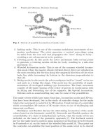
4.2 Body CPG
We assume that the body CPG is composed of a double chain of oscillators
all along the 40 segments of spinal cord. The type of connections investigated
in this article are illustrated in Figure 3 (left). For simplicity, we assume that
only nearest neighbor connections exist between oscillators. In our first in-
vestigation, the oscillators are assumed to be identical along the chain (with
identical projections), as well as between each side of the body. The connec-
tivity of the chain is therefore defined by 6 parameters, two (the a
ij
and b
ij
parameters) for each projection from one oscillator to the other (i.e. the ros-
tral, caudal, and contralateral projections). Of these 6 parameters, we fixed
the couplings between contralateral oscillators to a
ij
=0andb
ij
= −0.5in
order to force them to oscillate in anti-phase. We systematically investigated
the different combinations of the four remaining parameters (the rostral and
caudal projections) with values ranging from -1.0 to 1.0, with a 0.1 step.
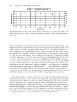
Gait Transition from Swimming to Walking 181
0 2 4 6 8 10 12
0
5
10
15
20
25
30
35
40
Signal X in segment i
Time [s]
Fig. 3. Left: Configuration of the body CPG. Right: oscillations in a 40-segment
chain (only the activity in a single side is shown).
Traveling wave Experiments on isolated spinal cords of the salamander sug-
gest that, similarly to the lamprey, the body CPG tends to propagate rostro-
caudal (from head to tail) traveling waves of neural activity. During (in-
tact) swimming, the wavelength of the wave corresponds approximately to
a bodylength. We therefore systematically investigated the parameter space
of the body CPG configuration to identify sets of parameters leading to sta-
ble oscillations with phase lags between consecutive segments approximately
equal to 2.5% of the period (in order to obtain a 100% phase lag between head
and tail). The goal is to obtain traveling waves which are due to asymmetries
of interoscillator coupling, while maintaining the same intrinsic period (the
same τ) for all oscillators.
We found that several coupling schemes could lead to such traveling waves.
The coupling schemes can qualitatively be grouped in three different cate-
gories: dominantly caudal couplings, balanced caudal and rostral couplings,
and dominantly rostral couplings.
1
By dominant, we mean that the sum of
the absolute values of the weights in one direction are significantly larger
than in the other direction. While all groups can produce traveling waves
corresponding to salamander swimming, solutions which have balanced cau-
dal and rostral couplings need significantly more cycles to stabilize into the
traveling wave (starting from random initial conditions) than the solutions
in which one type of coupling is dominant. It is therefore likely that the sala-
mander has one type of coupling which is dominant compared to the other.
A very similar conclusion has been made concerning the lamprey swimming
controller [9].
Figure 3 (right) illustrates the traveling waves generated by one of the
dominantly caudal chains. As can be observed, starting from random initial
1
Dominantly caudal and rostral couplings are essentially equivalent since each
coupling type which is dominant in one direction has an equivalent in the other
direction by inverting the sign of some weights. However, that equivalence is lost
when the intrinsic frequencies of some oscillators are varied, see the “Piece-wise
constant wavelength” paragraph.
182 Auke Jan Ijspeert and Jean-Marie Cabelguen
states, the oscillations rapidly evolve to a traveling wave. Since the period of
the oscillations explicitly depend on the parameter τ , the period can be mod-
ified independently of the wavelength. The wavelength of one-body length is
therefore maintained for any period, when all oscillators have the same value
of τ (i.e. the same intrinsic period). This allows one to modify the speed of
swimming by only changing the period of oscillation, as observed in normal
lamprey and salamander swimming.
Interestingly, while the connectivity of the oscillators favors a one-body
length wavelength, it is possible to vary the wavelength by modifying the
intrinsic period of some oscillators, the oscillators closest to the head, for
instance. Reducing the period of these oscillators leads to an increase of
the phase lag between consecutive oscillators(a reduction of the wavelength),
while increasing their period leads to a decrease of the phase lag, and can
even change the direction of the wave (i.e. generate a caudo-rostral wave).
This type of behavior is typical of chains of oscillators [9].
Piece-wise constant wavelength We identify at least four potential causes
for the small changes of wavelength observed along the body at the level of
the girdles: (1) differences of intrinsic frequencies between the oscillators at
the girdles and the other body oscillators, (2) differences in intersegmental
coupling along the body CPG (with three regions: neck, trunk, and tail),
(3) effect of the coupling from the limb CPG, (4) effect of sensory feedback.
Recent in-vitro recordings on isolated spinal cords showed that a change of
wavelength is also obtained during fictive swimming. It therefore seems that
the phenomenon is mainly due to the CPG configuration rather than to sen-
sory feedback (explanation number four is therefore the less likely). We tested
these different hypotheses with the numerical simulations. For the hypoth-
esis 2, it meant adding 8 parameters for differentiating the intersegmental
couplings in the neck, trunk and tail regions.
The results suggest that, in our framework, the most likely cause of the
three-wave pattern is a combination of differences in intersegmental coupling
and of intrinsic frequencies of the oscillators at the girdles. The differences in
intersegmental coupling lead to variations in the wavelength of the undulation
along the spinal cord. But they do not explain the abrupt changes of phases
at the level of the girdles. These are best explained by small differences in
intrinsic frequencies of the oscillators of the body CPGs at the two girdles
(these could also potentially be due to the projections from the limb CPG,
see next sections).
We can furthermore tell that the effect of variations of the intrinsic fre-
quencies depend on which coupling is dominant in the body CPG. The pat-
terns observed in the salamander are best explained with either a combination
of dominantly caudal coupling and higher intrinsic frequency at the girdles,
or dominantly rostral coupling and lower intrinsic frequencies at the girdles.
The resulting activity in the latter configuration is illustrated in Figure 5
(left).
Gait Transition from Swimming to Walking 183
L
BB
L
AB C D E
Global
With interlimb c.
Unilateral Bilateral
Global
With interlimb c.
Unilateral
Local
With interlimb c.
Local
Bilateral
Without interlimb c
Bilateral
Local
With interlimb c.
Fig. 4. Different potential CPG configurations.
Swimming We tested the body CPG in the mechanical simulation for con-
trolling swimming. Since the mechanical simulation has only 11 joints along
the body, 11 pairs of equally-spaced oscillators were picked from the body
CPG to drive the muscle models, such that the oscillators in one pair project
to the muscle on their respective side. A “motoneuron” m
i
signal is obtained
from the states x
i
with the following equation m
i
= β max(x
i
, 0), where is β
a positive constant gain. This motoneuron signal controls how much a mus-
cle contracts by essentially changing the spring constant of the spring-and-
damper muscle model (see [2]). An example of the swimming gait is shown
in Figure 5 (left). The speed of swimming can be modulated by changing
the frequency of all oscillators (through the parameter τ ), while the direction
of swimming can be modulated by applying an asymmetry of the amplitude
parameter E between left and right sides of the chain. The salamander will
then turn toward the side which receives the highest amplitude parameter.
4.3 Different body-limb CPG configurations for gait transition
One of the goals of this article is to investigate different types of couplings
between the body and limb CPGs, and how these couplings affect the gait
transitions between swimming and walking. There are currently too few bi-
ological data available to indicate how the different neural oscillators in the
body and limb CPGs are interconnected. Our aim is to investigate which of
these configurations can best reproduce some key characteristics of salaman-
der locomotion.
We tested five different types of coupling (Figure 4). These couplings dif-
fer in three characteristics: unilateral/bilateral couplings, in which the limb
CPGs are either unilaterally or bilaterally (i.e. in both directions) coupled
to the body CPG, global/local couplings, in which the limb CPGs project
either to many body CPG oscillators, or only those close to the girdles, and
with/without interlimb couplings between fore- and hindlimbs. In our pre-
vious work [2], we tested configuration A (unilateral, global, with interlimb
184 Auke Jan Ijspeert and Jean-Marie Cabelguen
12 13 14 15 16 17 18 19 20
0
5
10
15
20
25
30
35
40
Signal X in segment i
Time [s]
0 2 4 6 8 10 12
0
5
10
15
20
25
30
35
40
Signal X in segment i
Time [s]
Fig. 5. Left, top: Swimming gait. Left, bottom: corresponding activity in the the
body CPG (only the activity in a single side is shown). Note the piece-wise constant
wavelength. The oscillations at the level of the girdles are drawn with thicker lines.
Right top: walking gait. Right bottom: corresponding oscillations along the body
in a CPG of type A.
coupling) using neural network oscillators. The unilateral projections from
limb to body CPG essentially means a hierarchical structure in the CPG for
that configuration.
In all configurations, we assume that two different control pathways exist
for the body and the limb CPGs, in order words, that the control parameters
τ and E can be modulated independently for the body and limb oscillators.
In particular, we make the hypothesis that the gait transition is obtained as
follows: swimming is generated when only the body CPG is activated (Ebody
= 1.0 and Elimb = 0.005), and walking is generated when both body and limb
CPGs are activated (Ebody = 1.0 and Elimb =1.0).
The simulation results show that only configurations A and B, i.e. those
with global coupling between limb and body CPG can produce standing
waves (in the absence of sensory feedback). For these configurations, the
global coupling from limb oscillators to body oscillators ensures that the body
CPG oscillates approximately in synchrony in the trunk and in the tail when
the limb CPG is activated (Figure 5, right). For the other configurations (C,
D, and E) the fact that the couplings between limb and body CPGs are only
local means that traveling waves are still propagated in the trunk and the
tail, despite the influence from the limb oscillators. Configurations E, which
lacks interlimb couplings can still produce walking gaits very similar to those
of configurations C and D, because the coupling with the body CPG gives
a phase relation between fore- and hindlimbs of approximately 50% of the
period (because fore and hindlimbs are separated by approximately the half
of one body-length).
Gait Transition from Swimming to Walking 185
4 4.5 5 5.5 6 6.5 7
0
10
20
30
40
X body left
4 4.5 5 5.5 6 6.5 7
0
10
20
30
40
S body left
Time [s]
4 4.5 5 5.5 6 6.5 7
0
10
20
30
40
X body left
4 4.5 5 5.5 6 6.5 7
0
10
20
30
40
S body left
Time [s]
Fig. 6. Left: Walking gait produced by configuration D, without sensory feedback.
Right: Walking gait produced by configuration D, with sensory feedback. Top:
output of the body CPG, Bottom: output of the stretch sensors.
Having bilateral couplings between limb and body CPGs does not affect
the walking pattern in a significant way. However, if the coupling from body
CPG to limb CPG is strong, it will affect the swimming gait. In that case,
even if the amplitude of the limb oscillators is set to a negligible value (Elimb
= 0.005), the inputs from the body CPG will be sufficient to drive the limb
oscillators which in return will force the body CPG to generate a wave which
is a mix between a traveling wave and standing wave. It is therefore likely
that the couplings between limb and body CPG are stronger from limb to
body CPG than in the opposite direction.
Note that the fact that CPG configurations B, C and D can not pro-
duce standing waves, does however not exclude the possibility that these
configurations produce standing waves when sensory feedback is added to
the controller. This will be investigated in the next section.
Effect of sensory feedback When a lamprey is taken out of the water and
placed on ground, it tends to make undulations which look almost like stand-
ing waves because the lateral displacements do not increase along the body
but form quasi-nodes (i.e. points with very little lateral displacements) at
some points along the body [10].
Interestingly, the same is true in our simulation. When the swimming gait
is used on ground (without sensory feedback), the body makes a S-shaped
standing wave undulation instead of the traveling wave undulation generated
in water. This is due to the differences between hydrodynamic forces in water
(which have strongly different components between directions parallel and
perpendicular to the body) and the friction forces on ground (which are
more uniform). The sensory signals from such a gait are then reflecting this
S-shaped standing wave, despite the traveling waves sent to the muscles.
Sensory feedback is therefore a potential explanation for the transition
from a traveling wave for swimming to a standing wave for walking. We
therefore tested the effect of incorporating sensory feedback in the different
186 Auke Jan Ijspeert and Jean-Marie Cabelguen
CPG configurations described above. Sensory feedback to the salamander’s
CPG is provided by sensory receptors in joints and muscles. We designed
an abstract model of sensory feedback by including sensory units located on
both sides of each joint which produce a signal proportional to how much that
side is stretched: s
i
= max(φ
i
, 0) where φ
i
is the angle of joint i measured
positively away from the sensory unit. For simplicity, we only consider sensory
feedback in the body segments (i.e. not in the limbs), and assume that a
sensory unit for a specific joint is coupled only locally to the two (antagonist)
oscillators activating that joint.
Figure 6shows the activity of the body CPG and the sensor units pro-
duced during a stepping gait with a controller with configuration D. Without
sensory feedback (Figure 6, left), this controller produces a traveling wave
during walking because the limb oscillators have only local projections to
the body CPG. Despite this traveling wave of muscular activity, the body
(in contact with the ground) makes essentially an S-shaped standing wave
as illustrated by the sensory signals (synchrony in the trunk and in the tail,
with an abrupt change of phase in between). When these sensory signals are
fed back into the CPG (Figure 6, right), the body CPG activity is modified
to approach the standing wave (i.e. the phase lag between segments decrease
in the trunk and in particular in the tail). Note that if the sensory feedback
signals are too strong, the stepping gait becomes irregular. Interestingly, the
sensory feedback leads to an increase of the oscillation’s frequency, something
which has also been observed in a comparison between swimming with and
without sensory feedback in the lamprey [11].
5 Discussion
The primary goal of this article was to investigate which of different CPG
configurations was most likely to control salamander locomotion. To the best
of our knowledge, only three previous modeling studies investigated which
type of neural circuits could produce the typical swimming and walking gaits
of the salamander. In [12], the production of S-shaped standing waves was
mathematically investigated in a chain of coupled non-linear oscillators with
long range couplings. In that model, the oscillators are coupled with closest
neighbor couplings which tend to make oscillators oscillate in synchrony, and
with long range couplings from the extremity oscillators to the middle oscil-
lators which tend to make these coupled oscillators oscillate in anti-phase. It
is found that for a range of strengths of the long range inhibitory coupling,
a S-shaped standing wave is a stable solution. Traveling waves can also be
obtained but only by changing the parameters of the coupling. In [2], one
of us demonstrated that a leaky-integrator neural network model of configu-
ration A could produce stable swimming and walking gaits. Finally, in [13],
it was similarly demonstrated that a neural network model of the lamprey
swimming controller could produce the piece-wise constant swimming of sala-
mander and the S-shaped standing of walking depending on how phasic input
Gait Transition from Swimming to Walking 187
drives (representing signals from the limb CPGs and/or sensory feedback) are
applied to the body CPG. The current paper extends these previous stud-
ies by investigating more systematically different potential body-limb CPGs
configurations underlying salamander locomotion.
The simulation results presented in this article suggest that CPG config-
urations which have global couplings from limb to body CPGs, and interlimb
couplings (configurations A or B) are the most likely in the salamander.
These configurations can indeed produce stable swimming and walking gaits
with all the characteristics of salamander locomotion. Our investigation does
not exclude the other configurations, but suggest that these would need a
significant input from sensory feedback to force the body CPG to produce
the S-shaped standing wave along the body. These results suggest new neuro-
physiological experiments. It would, for instance, be interesting to make new
EMG recordings during walking without sensory feedback (e.g. by lesion of
the dorsal roots). If the EMG recordings remain a standing wave, it would
suggest that configurations A or B are most likely, while if they correspond
to a standing wave if would suggest that configurations C, D, or E are most
likely.
To make our investigation tractable, we made several simplifying assump-
tions. First of all, we based our investigation on nonlinear oscillators. Clearly,
these are only very abstract models of oscillatory neural networks. In partic-
ular, they have only few state variables, and fail to encapsulate all the rich
dynamics produced by cellular and network properties of real neural net-
works. We however believe they are well suited for investigating the general
structure of the locomotion controller. To some extent, some properties of
interoscillator couplings are universal, and do not depend on the exact im-
plementation of the oscillators. This is observed for instance in chains [9],
as well as rings of oscillators [14]. Our goal was therefore to analyze these
general properties of systems of coupled oscillators.
An interesting aspect of this work was to combine a model of the con-
troler and of the body, since this allowed us to investigate the mechanisms of
entrainment between the CPG, the body and the environment. We believe
such an approach is essential to get a complete understanding of locomotion
control, since the complete loop can generate dynamics that are difficult to
predict by investigating the controller (the central nervous system) in isola-
tion of the body. The transformation of traveling waves of muscular activity
into standing waves of movements when the salamander is placed on ground is
an illustration of the complex dynamics which can results from the complete
loop.
Finally, this work has also direct links with robotics, since the controllers
could equally well be used to control a swimming and walking robot. Espe-
cially interesting is the ability of the controller to coordinate multiple degrees
of freedom while receiving very simple input signals for controling the speed,
direction, and type of gait.
188 Auke Jan Ijspeert and Jean-Marie Cabelguen
Acknowledgements
We would like to acknowledge support from the french “Minist`eredela
Recherche et de la Technologie” (program “ACI Neurosciences Int´egratives et
Computationnelles”) and from a Swiss National Science Foundation Young
Professorship grant to Auke Ijspeert.
References
1. A.H. Cohen and P. Wallen. The neural correlate of locomotion in fish. ”fictive
swimming” induced in a in vitro preparation of the lamprey spinal cord. Exp.
Brain Res., 41:11–18, 1980.
2. A.J. Ijspeert. A connectionist central pattern generator for the aquatic and
terrestrial gaits of a simulated salamander. Biological Cybernetics, 85(5):331–
348, 2001.
3. I. Delvolv´e, T. Bem, and J M. Cabelguen. Epaxial and limb muscle activity
during swimming and terrestrial stepping in the adult newt, Pleurodeles Waltl.
Journal of Neurophysiology, 78:638–650, 1997.
4. I. Delvolv´e, P. Branchereau, R. Dubuc, and J M. Cabelguen. Fictive rhyth-
mic motor patterns induced by NMDA in an in vitro brain stem-spinal cord
preparation from an adult urodele. Journal of Neurophysiology, 82:1074–1077,
1999.
5. G. Sz´ekely and G. Cz´eh. Organization of locomotion. In Frog Neurobiology, a
Handbook, pages 765–792. Springer Verlag, Berlin, 1976.
6. M. Wheatley, M. Edamura, and R.B. Stein. A comparison of intact and in-vitro
locomotion in an adult amphibian. Experimental Brain Research, 88:609–614,
1992.
7. J. Cheng, R.B. Stein, K. Jovanovic, K. Yoshida, D.J. Bennett, and Y. Han.
Identification, localization, and modulation of neural networks for walking in
the mudpuppy (necturus maculatus) spinal cord. The Journal of Neuroscience,
18(11):4295–4304, 1998.
8.
¨
O. Ekeberg. A combined neuronal and mechanical model of fish swimming.
Biological Cybernetics, 69:363–374, 1993.
9. N. Kopell. Chains of coupled oscillators. In M.A. Arbib, editor, The handbook
of brain theory and neural networks, pages 178–183. MIT Press, 1995.
10. G. Bowtell and T.L. Williams. Anguiliform body dynamics: modelling the
interaction between muscle activation and body curvature. Phil. Trans. R.
Soc. Lond. B, 334:385–390, 1991.
11. Li Guan, T. Kiemel, and A.H. Cohen. Impact of movement and movement-
related feedback on the lamprey central pattern generator for locomotion. The
Journal of Experimental Biology, 204:2361–2370, 2001.
12. B. Ermentrout and N. Kopell. Inhibition-produced patterning in chains of cou-
pled nonlinear oscillators. SIAM Journal of Applied Mathematics, 54(2):478–
507, 1994.
13. T. Bem, J M. Cabelguen, O. Ekeberg, and S. Grillner. From swimming to
walking: a single basic network for two different behaviors. Biological Cyber-
netics, page In press, 2002.
14. J. Collins and Richmond. Hard-wired central pattern generators for
quadrupedal locomotion. Biological Cybernetics, 71:375–385, 1994.
Nonlinear Dynamics of Human Locomotion:
from Real-Time Adaptation to Development
Gentaro Taga
Graduate School of Education, University of Tokyo
7-3-1 Hongo, Bunkyo-ku, Tokyo 113-0033, Japan
Abstract. The nonlinear dynamics of the neuro-musculo-skeletal system and the
environment play central roles for the control of human bipedal locomotion. Our
neuro-musculo-skeletal model demonstrates that walking movements emerge from
a global entrainment between oscillatory activity of a neural system composed of
neural oscillators and a musculo-skeletal system. The attractor dynamics are re-
sponsible for the stability of locomotion when the environment changes. By linking
the self-organizing mechanism for the generation of movements to the optical flow
information that indicates the relationship between a moving actor and the environ-
ment, visuo-motor coordination is achieved. Our model can also be used to simulate
pathological gaits due to brain disorders. Finally, a model of the development of
bipedal locomotion in infants demonstrates that independent walking is acquired
through a mechanism of freezing and freeing degrees of freedom.
1 Introduction
The theory of nonlinear dynamics, which claims that spatio-temporal pat-
terns arise spontaneously from the dynamic interaction between components
with many degrees of freedom [1,2], is progressively attracting more atten-
tion in the field of motor control. The concept of self-organization in move-
ment was initially applied to describe motor actions such as rhythmic arm
movements [3]. In the meantime, neurophysiological studies of animals have
revealed that the neural system contains a central pattern generator (CPG),
which generates spatio-temporal patterns of activity for the control of rhyth-
mic movements through the interaction of coupled neural oscillators [4]. More-
over, it has been reported that the centrally generated rhythm of the CPG
is entrained by the rhythm of sensory signals at rates above and below the
intrinsic frequency of the rhythmic activity [4]. This phenomenon is typical
for a nonlinear oscillator that is externally driven by a sinusoidal signal.
Inspired by the theoretical and experimental approaches to self-organized
motor control, we proposed that human bipedal locomotion emerges from
a global entrainment between the neural system’s CPG and the musculo-
skeletal system’s interactions with a changing environment [5]. A growing
number of simulation studies have focused on the dynamic interaction of neu-
ral oscillators and mechanical systems in order to understand the mechanisms
of generation of adaptive movements in insects [6], fish [7] and quadruped
190 Gentaro Taga
animals [8-10]. In the field of robotics, an increasing number of studies have
implemented neural oscillators to control movements of real robots [11-13].
The concept of self-organization argues that movements are generated as a
result of dynamic interaction between the neural system, the musculo-skeletal
system and the environment. If this is the case, the implicit assumption that
the neural system is a controller and that the body is a controlled system
needs to be revised. This paper presents a series of our models of human
bipedal locomotion, all of which demonstrate the nonlinear properties of the
neuro-musculo-skeletal system. The aim of this paper is to provide a frame-
work for understanding the generation of bipedal locomotion [5, 14], real-time
flexibility in an unpredictable environment [15], anticipatory adaptation of
locomotion when confronted with an obstacle [16], visuo-motor coordination
using optical flow information [17], the generation of pathological gaits and
the acquisition of locomotion during development [18].
2 Real-time adaptation of locomotion through global
entrainment
2.1 A model of the neuro-musculo-skeletal system for human
locomotion
In principle, bipedal walking of humanoid robots can be controlled if the
specific trajectory of each joint and of the zero moment point (ZMP) are
planned in advance and feedback mechanisms are incorporated [19]. However,
it is obvious that this method of control is not robust against unpredictable
changes in the environment.
Is it possible to generate bipedal locomotion by using a neural model of
the CPG in a self-organized manner? Let us assume that the entire system is
composed of two dynamical systems: a neural system that is responsible for
generating locomotion and a musculo-skeletal system that generates forces
and moves in an environment. The neural system is described by differen-
tial equations for coupled neural oscillators, which produce motor signals to
induce muscle torques and which receive sensory signals indicating the cur-
rent state of the musculo-skeletal system and the environment. The musculo-
skeletal system is described by Newtonian equations for multiple segments of
the body. The input torque is generated by the output of the neural system.
Using computer simulation we proved that a global entrainment between the
neural system and the musculo-skeletal system is responsible for generating
a stable walking movement [5].
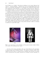
Here I will present a model of [14]. As shown in Fig.1, the musculo-skeletal
system consists of eight segments in the sagittal plane. The triangular foot
interacts with the ground at its heel and/or toe. According to the output
of the neural system, each of twenty ”muscles” generates torque at specific
joints. It is important to note that a number of studies have produced exam-
ples of walking robots, such as the passive dynamic walkers [20, 21] and the
Nonlinear Dynamics of Human Locomotion 191
dynamic running machines [22], which exploit the natural dynamics of the
body. The oscillatory property of the musculo-skeletal system is an important
determinant to establishing a walking pattern.
Our simulated neural system was designed based on the following assump-
tions:
(1) The neural rhythm generator (RG) is composed of neural oscillators,
each of which controls the movement of a corresponding joint. As a model
neural oscillator, we adopt the half-center model, which is composed of two
reciprocally inhibiting neurons and which generates alternative activities be-
tween the two neurons [23].
(2) All of the relevant information about the body and the environment
is taken into account. The angles of the body segments in an earth-fixed
frame of reference and ground reaction forces are available to the sensory
system. Global information on the position of the center of gravity (COG)
with respect to the position of the center of pressure (COP) is also available.
We assume that a gait is represented as a cyclic sequence of what we call
global states: the double-support phase, the first half of the single-support
phase, and the second half of the single-support phase. The global states are
defined by the sensory information on the alternation of the foot contacting
the ground and the orientation of the vector from the COP to the COG.
(3) Reciprocal inhibitions are incorporated between the neural oscillators
on the contralateral side, which generates the anti-phase rhythm of muscles
between the two limbs. Connections between the neural oscillators on the
ipsilateral side change in a phase-dependent manner, using the global state
to generate complex phase relationships of activity among the muscles within
a limb.
(4) Both the local information on the angles of the body segments and the
global information on the entire body are sent to the neural oscillators in a
manner similar to the functional stretch reflex, so that neural oscillation and
body movement are synchronized. Sensory information is sent only during
the relevant phase of the gait cycle by modulating the gains of the sensory
pathways in a phase-dependent manner, which is determined by the global
state.
(5) All of the neural oscillators share tonic input from the higher center,
which is represented by a single parameter. By changing the value of this
parameter, the excitability of each oscillator can be controlled so that different
speeds of locomotion are generated.
(6) While the neural rhythm generator induces the rhythmic movement
of a limb, a posture controller (PC) is responsible for maintaining the static
posture of the stance limb by producing phase-dependent changes in the
impedance of specific joints. The final motor command is a summation of the
signals from the neural rhythm generator and the posture controller.
The computer simulation demonstrated that, given a set of initial condi-
tions and values of various parameters, a stable pattern of walking emerged
192 Gentaro Taga
as an attractor formed in the state space of both the neural and musculo-
skeletal system. Figure 2 shows neural activities, muscle torques and a stick
picture of walking within one gait cycle. The attractor was generated by the
global entrainment between the oscillatory activity of the neural system and
rhythmic movements of the musculo-skeletal system.
When we first proposed the model of bipedal locomotion [5], there were
few studies to suggest the existence of a spinal CPG in humans. More recently,
several studies have shown evidence for a spinal CPG in human subjects
with spinal cord injury [24-26]. Our model is likely to capture the essential
mechanism for the generation of human bipedal locomotion.
Fig. 1. A model of the neuro-musculo-skeletal system for human locomotion [14].
Nonlinear Dynamics of Human Locomotion 193
Fig. 2. The results of computer simulation of emergence of neural activity, muscle
torque and walking movements generated in a self-organized manner.
194 Gentaro Taga
2.2 Real-time flexibility of bipedal locomotion in an
unpredictable environment
When the solution of the differential equations representing the neural and
musculo-skeletal systems converged to a limit cycle that was structurally sta-
ble, walking movement was maintained even with small changes in the initial
conditions and parameter values [15]. For example, when part of the body
was disturbed by a mechanical force, walking was maintained and the steady
state was recovered due to the orbital stability of the limit cycle attractor.
When part of the body was loaded by a mass, which can be applied by chang-
ing the inertial parameters of the musculo-skeletal system, the gait pattern
did not change qualitatively but converged to a new steady state, where the
speed of walking clearly decreased. When the walking path suddenly changed
from level to uneven terrain, the stability of walking was maintained but the
speed and the step length spontaneously changed as shown in Fig. 3. Natu-
rally, the stability of walking was broken for a heavy load and over a steep
and irregular terrain.
This real-time adaptability is attributed not only to the afferent control
based on the proprioceptive information generated by the interaction between
the body and the mechanical environment, but also to the efferent control
of movements based on intention and planning. In this model, a wide range
of walking speeds was available using the nonspecific input from the higher
center to the neural oscillators, which was represented by a single parame-
ter. Changes in the parameter can produce bifurcations of attractors, which
correspond to different motor patterns [5,15].
It is open to question whether a 3D model of the body with a similar
model of the neural system will perform dynamic walking with stability and
flexibility. Designing such a model is a crucial step toward constructing a
humanoid robot that walks in a real environment [27].
Fig. 3. Walking over uneven terrain.
Nonlinear Dynamics of Human Locomotion 195
3 Anticipatory adjustment of locomotion through
visuo-motor coordination
3.1 Anticipatory adjustment of locomotion during obstacle
avoidance
As long as the stability of the attractor is maintained, the locomotor sys-
tem can produce adaptive movements even in an unpredictable environment.
However, this way of generating motor patterns is not sufficient when the at-
tractor loses stability due to drastic changes in the environment. For example,
when we step over an obstacle during walking, the path of limb motion must
be quickly and precisely controlled using visual information that is avail-
able in advance. Given the emergent properties of the neuro-musculo-skeletal
system for producing the basic pattern of walking, how is this anticipatory
adaptation to the environment realized? Neurophysiological studies in cats
have shown that the motor cortex is involved in visuo-motor coordination
during anticipatory modification of the gait pattern [28].
It was examined whether modifications of the basic gait pattern could
produce rapid enough changes so as to clear an obstacle placed in its path.
As shown in Fig. 4, the neural rhythm generator was combined with a system
referred to as a discrete movement generator, which receives both the output
of the neural oscillators and visual information regarding the obstacle and
generates discrete signals for modification of the basic gait pattern [16].
By computer simulation, avoidance of obstacles of varying heights and
proximity was demonstrated, as shown in Fig. 5. An obstacle placed at an
arbitrary position can be cleared by sequential modifications of gait, namely
by modulating the step length when approaching the obstacle and modifying
the trajectory of the swinging limbs while stepping over it. An essential point
is that a dynamic interplay between advance information about the obstacle
and the on-going dynamics of the neural system produces anticipatory move-
ments. This implies that the planning of limb trajectory is not free from the
on-going dynamics of the lower levels of the neural system, body dynamics,
and environmental dynamics.
3.2 A model of the neuro-musculo-skeletal system for human
locomotion
The maintenance of gait when changes in the environment occur quickly rela-
tive to the walking rhythm was possible with the addition of a neural process-
ing component. A further question is what mechanism would be responsible
for adaptation through the action perception cycle of the visuo-motor coor-
dination. For example, how can the precise positioning of a foot on a visible
target on the floor during walking be achieved? The ecological approach of
perception and action argues that adaptation to the complex environment
is achieved not by the construction and the use of internal representations
196 Gentaro Taga
Fig. 4. A model of obstacle avoidance via anticipatory adaptation during walking
[16].
of the world but rather by the use of real time information available in the
optical flow [29]. Time to contact an obstacle or a target, which information
can be obtained directly from a set of invariants present in the optical flow,
has been studied as a key element in the visual control of locomotion [30].
We assumed that the step length modulation command, which was mod-
elled in [a] previous study, was continuously related to optical information
about the time remaining before one reached the target with the current eye-
foot axis [17]. This optical variable in relation to the subject’s own movement
was labelled as time-to-foot (TTF) as shown in Fig. 6. We further assumed
that the current step period was available and that it could be used with
TTF to determine whether the step length must be shortened or lengthened
to position the foot on the target.
Nonlinear Dynamics of Human Locomotion 197
Fig. 5. Result of computer simulation of [16].
Results of computer simulation gave rise to successful pointing behavior
as shown in Fig. 7. The generated behaviors for regulating step length were
similar to those observed in human subjects performing a locomotor pointing
task: namely, the time course of the inter-trial variability of the toe-target
distance and the relationship between the step number at which the regula-
tion is initiated and the total amount of adjustments involved. An important
point of this model was that the adaptation of locomotion emerged from
a perception-action coupling type of control based on temporal information
rather than on the representation of the target. This is the first attempt to
bridge the gap between the ecological approach to perception and the self-
organized control of locomotion based on global entrainment.
4 Computational “lesion” experiments in gait
pathology
It is well known that specific damage to the brain or the peripheral nervous
system leads to locomotor disorders. Although the musculo-skeletal dynam-
ics during walking have been intensively studied in clinical applications of
orthopaedic issues [31], very few studies have taken a modelling approach to
understanding pathological gaits due to brain dysfunctions. A question to be
asked here is whether the generic model for the emergence of a basic gait
can be used to reproduce pathological patterns of gait by changing model
parameters.
198 Gentaro Taga
Fig. 6. Time-to-foot information [17].
Fig. 7. Result of computer simulation of locomotor pointing tasks [17]. a. steady
state walking. b. adjustment of step by shortening of step length. c. adjustment of
step by lengthening of step length.
Nonlinear Dynamics of Human Locomotion 199
As shown in Fig. 8, asymmetric gaits, irregular gaits with changing step
lengths and shuffling gaits with very small step lengths were generated by
changing the values of the parameters of the motor or sensory pathways
asymmetrically, decreasing the strength of the connections between the neu-
ral oscillators and decreasing the tonic input to the rhythm generator, respec-
tively. These patterns of gaits were similar to those of patients with hemi-
plegia, cerebellar disease, and Parkinson’s disease. The results demonstrated
that qualitative changes in gait patterns were produced by the computa-
tional ”legion” study. This inferred that the generation of pathological gaits
can be viewed as a self-organizing process, where dynamic interactions be-
tween remaining parts of the system spontaneously produce specific patterns
of activity.
Fig. 8. Generation of pathological gaits. (a), (b) and (c) in the model show the area
affected by the changes, and the gaits generated by these changes are presented in
the three columns on the right-hand side.
5 Freezing and freeing degrees of freedom in the
development of locomotion
Once we had chosen a structure of the neural system and a set of parameter
values that produced a walking movement as a stable attractor, the model
exhibited flexibility against various changes in the environmental conditions.
However, it was difficult to determine the structure of the model and to
tune the parameters, since the entire system was highly nonlinear. A number
of studies have used a genetic algorithm [32,33] and reinforcement learning
[34] to obtain good locomotor performance in animals and in humans. An-
other approach to overcoming the difficulty of parameter tuning of locomotor
systems is to explore the motor development of infants and to unravel a de-
velopmental principle of the neuro-musculo-skeletal system. Here I show that