Recent Advances in Mechatronics - Ryszard Jabonski et al (Eds) Episode 2 Part 8 pot
Bạn đang xem bản rút gọn của tài liệu. Xem và tải ngay bản đầy đủ của tài liệu tại đây (1.57 MB, 29 trang )
estimated by least square sum analysis involving spectral data and values
obtained from camera CCD sensor for each color reference.
Introduction
For accurate color reproduction there is a need of developing new
tools for calibrating a digital camera system capable of reproducing high
fidelity colors for telemedicine, internet shopping, industrial design and
other high accuracy color reproduction demanding applications. The cam-
era is equipped with three filters S1, S2 and S3, which correspond to red,
green and blue band. These filters have relative spectral sensitivity similar
to primary color sensitive cones in human eye (Fig. 1.).
Macbeth ColorChecker® Chart is used to determine numerical
color description for reference. As an illuminant there is a CIE Daylight 65
(D65) standard used. Under this certain conditions, automatic method of
calibration with use of an integrated spectrometer inside the camera based
on a fiber optic measuring probe has been developed.
Fig.1. Filter spectral characteristics.
Measured color is represented by the tristimulous XYZ values [1].
Color captured by the camera is represented by S1, S2, S3 values, corre-
sponding to pixel value under each filter acquisition. To provide high fi-
delity color reproduction, a conversion matrix between XYZ and S1, S2,
S3 values must be obtained by means of least square sum analysis [2].
XYZ camera principle
For total description of color there can be used so called tristimu-
lous values XYZ [1]. A digital still camera with output containing these
Wavelength [nm]
Relative transmittance
664 M. Kretkowski, H. Suzuki, Y. Shimodaira, R. Jabłoński
values, supported by proper filters and equipped with internal spectrometer
is able to reproduce high fidelity colors in the same way as human eye
would perceive it. Figure 2 shows principle of the camera.
Light passing through the lens is split by a semi-transparent mirror.
Part of the light reflected by the mirror is used for spectral measurement,
while light passing through is projected onto CCD imaging device with S1,
S2, S3 filters put on its way.
Fig. 2. XYZ camera principle.
Basic principle is to take three pictures of photographed scene un-
der each filter acquisition. This gives three layer image called: “S1, S2 and
S3 image”.
The spectrometer is used for calibration of the camera. With an
algorithm described further, by mathematical conversion S1, S2, S3 image
is converted to XYZ image which describes colors contained in the scene
in XYZ color space [1] which is a base for any other color representation.
665Automatic color calibration method for high delity color reproduction digital
Proposed calibration algorithm
Spectral data is collected automatically from Macbeth Chart’s 24
colors by fiber optic probe positioned over picture area along the x and y
axes (Fig. 3.). The positioning mechanism is built inside the camera and its
coordinate system has been calibrated with picture area. This gives a pos-
sibility of acquiring spectral data from any point of the photographed
scene. Described algorithm uses image processing for recognition of each
color on Macbeth Chart and to find coordinates for spectrometer’s probe to
be positioned and perform measurement of each color automatically.
Fig. 3. Block scheme of calibration algorithm.
Calibration starts with acquiring illuminant data (Fig. 3.) by per-
fect reflecting plate. The Macbeth Chart then is placed into the scene and
color pads are recognized and measured. XYZ and S1, S2, S3 data is tabu-
lated and stored into memory. Basic relation between the values is:
White balanci
ng
Color calibr
a
tion
Conversion
m
a
trix
Taking the illuminant
by reflecting plate
Threshold
procedure
Macbeth’s Chart pi
c-
ture taken on a co
n-
trasting Bac
k
ground
Color fields
position estim
a
tion
Fiber optic‘s
positioning sy
s
tem
A
xis
calibr
a
tion
Spe
c
trum
Measur
ement
Dark cu
r
rent
estimation
XYZ Ou
t
put
Lens shading
characterist
i
cs
)1(
3
2
1
333231
232221
312111
=
S
S
S
aaa
aaa
aaa
Z
Y
X
666 M. Kretkowski, H. Suzuki, Y. Shimodaira, R. Jabłoński
The color difference between reference and values obtained from
calibrated camera, is well described by CIE Lab color space derived from
XYZ [1]. The color difference �E principle which describes accuracy of
color reproduction can be expressed with equation (2) (CIE 1932). Where
Li is luminance of compared colors and ai, bi are chroma coefficients.
However at present CIE 2000 is used, to describe �E in more perceptually
uniform color difference representation.
Results and discussion
The calibration of the spectrometer is most important in order to
develop a conversion matrix giving the smallest color difference between
measured XYZ color and X’Y’Z’ obtained from S1, S2 and S3 [2]. Other
major influencing factor is shading of the camera lens (winietting).
Present results show strong dependency between F number and
color difference (Tab. 1.). As we observed color difference decreases as
the F number increases.
Table 1. Color difference and F number.
This is caused by shading characteristics of whole optics including
camera lens, mirror, fiber optics and spectrometer spectral response. To
improve the accuracy of reproduction and reduce �E, amount of light re-
flected from the semi-transparent mirror must be increased to provide bet-
ter signal to noise ratio of the spectrometer.
Above facts lead to conclusion that accurate color reproduction for
still digital camera requires careful calibration. Proposed algorithm uses
color reference and includes various factors for maximum performance.
However in further development it will be investigated for possibility of
calibration using colors of the photographed scene as a reference.
References
[1] R.W.G Hunt “The Reproduction of Colour” ISBN (1995)
[2] T. Ejaz, T. Horiuchi, G. Ohashi, Y. Shimodaira IEICE Trans. Elec-
tron., E89-C (2006)
F of the camera lens
2.8 5.6 8
Color difference (average)
CIE 2000
2.04 1.35 1.13
)2()()()(
2
21
2
21
2
21
bbaaLLE −+−+−=∆
667Automatic color calibration method for high delity color reproduction digital
Coherent noise reduction in optical diffraction
tomography
A. Pakuła*, T. Kozacki
Warsaw University of Technology, Institute of Micromechanics and
Photonics, 8 Sw. A.Boboli St., 02-525 Warsaw, Poland
Abstract
Optical Diffraction Tomography (ODT) is the method for characterization
of 3D distribution of refractive index in micro optical elements. The 3D
distribution is obtained from several measurements taken for different ob-
ject angular orientation taken by means of laser interferometry. Unfortu-
nately the interferometry as a coherent technique suffers from undesirable
coherent noise in measurements results. Additionally the rotation of a
measured object increases the noise influence on the quality of refractive-
index reconstruction due to its amplification in the object area. In this pa-
per we propose the coherent noise suppression technique. The technique
involves modification of ODT setup configuration by introducing off cen-
ter object rotation and applying a modified numerical algorithm of tomo-
graphic reconstruction. The algorithm modification involves introduction
of numerical imaging and detection of element position from its diffraction
spectrum. The received results of noise reduction are shown through nu-
merical simulations and experiments involving optical single mode fiber
measurements.
1. Introduction
The recent rapid growth of microelements with three-dimensional phase
distribution, photonics structures and materials (e.g. photonics crystal fi-
bers, GRIN lenses etc.) which are vastly used in photonics devices results
in need for fast, nondestructive method of 3D refractive index distribution.
The ODT is the method suitable for such measurement tasks [1-6].
Using classical interferometry 2D refractive index distribution, integrated
along optical axis, is obtained for variable angular object orientation. For
every single object angular orientation (range within 1°÷180°) interfero-
gram is recorded and analyzed. Then the 3D refractive index distribution is
obtained using tomographic reconstruction algorithm.
The results obtained by ODT have been recently improved by introduction
of numerical procedure of refocusing to the best focus plane and sample
radial run-out correction algorithm [6], although there is still unsolved
problem of coherent noise, especially if measured sample introduces slight
changes (order of 10
-3
or lower) in refractive index.
2. Coherent noise reduction technique
In principle the ODT involves sample rotation along the axis in the object
centre. This causes the single speckle magnification during the tomo-
graphic algorithm what results in semicircular shape in reconstructed re-
fractive index map (Fig. 1).
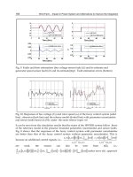
Fig. 1: Coherent noise influence on refractive index reconstruction by tomo-
graphic algorithm – multimode optical fiber [5].
Introduction of substantial radial run-out into sample rotation process
along with sinogram correction correlation technique allows the coherent
noise influence to be reduced. According to simulation results (simulated
object: refractive index distribution: step ∆n=0.01, diameter φ=100λ) the
optimal radial run-out range is 0.75φ - 1.25φ, Fig. 2. In this range the RMS
factor decreases and the S/N ratio rises – Fig. 3.
Although introducing run-out into measurement, according to simulations,
is a significant benefit, it gives some experimental difficulties. Such a
669Coherent noise reduction in optical diraction tomography
modified setup requires larger measurement field of view, which results in
lower magnification. Additionally the defocusing of the sample is inserted
by the sample rotation. For decreasing the influence of factors mentioned
above the numerical sinogram correction and refocusing to the best focus
plane algorithms need to be applied.
a) b)
Fig. 2: Simulations results of tomographic numerical reconstruction:
(a) without radial run-out (b) radial run-out - 2φ.
a) b)
Fig. 3: Simulations’ results due to the sample’s radial run-out:
(a) RMS factor, (b) S/N ratio.
3. Experimental technique
Experimental tomographic setup is based on classical Mach – Zehnder in-
terferometer setup (Fig. 4.). He – Ne laser beam formed by microscope
objective (OB1) is spitted into reference and object beams by a coupler
(FC). The measured object (O) submerged in immersion liquid
(n
633
=1.4584) is illuminated by a plane wave and rotated during the meas-
urement. The imagining system is focused in the sample’s centre area. The
measurement is performed in the following steps. For every sample angu-
lar orientation interferogram are grabbed and analyzed forming phase pro-
670 A. Pakuła, T. Kozacki
jection images data set. Finally from this data set 3D refractive index map
is reconstructed by means of thomographic reconstruction algorithms.
As a proof of principle of our method the experiment involving characteri-
zation of refractive index distribution of single mode telecommunication
fiber (SMF 28) was performed (core diameter 8.2 m, cladding diameter
125 m, ∆n=0.0053) [7]. The experimental run-out was 129.2m.
Fig. 4: Experimental tomographic setup: S – He-Ne laser, λ/2 – halfwave plate,
OB1, OB2, OB3 – fiber coupling objectives, FC – fiber coupler, OB4, OB5 – mi-
croscopic imagining objectives, BS – beam splitter, L – camera objective,
O – measured object, RS – rotation stage, D – detector, PC – central unit.
a) b)
Fig. 5: Results of measurement of single mode fiber (1 pix = 0.2315 m):
(a) reconstruction and cross section without radial run-out, (b) reconstruction and
cross section with 129 m radial run-out.
OB1
O
RS
PC
D
S
λ
/2
FC
OB2
OB3
L
BS
OB4,
OB5
λ
/2
671Coherent noise reduction in optical diraction tomography
The core diameter and difference of average refractive index between the
core and the cladding of the reconstructed fiber from measurements with-
out and with radial run-out are equal adequately: φ = 9.03 m (39 pix),
∆n=0.006 and φ=9.26 m (40 pix) (elliptical deformation occurs),
∆n=0.005. However, as it is shown in Fig. 5b the semicircular shape in
tomographic reconstruction which has its origins in the coherence noise is
removed by introduction of radial run-out the proposed technique intro-
duces deformation of the measured object. The full uncertainty analyses
for the case of the modified procedure have to be performed.
5. Conclusions
The novel method of reduction of the coherence noise influence on tomo-
graphic reconstruction reduction was proposed and verified by the numeri-
cal simulations and the experiment.
References
[1] M. Kujawinska, P. Kniazewski, T. Kozacki “Enhanced interferometric
and photoelastic tomography for 3D studies of phase photonics elements”,
Proc. of the Symposium on Photonics Technologies for 7th Framework
Program, 467- 471, Wroclaw, 2006
[2] W. Gorski “Tomographic microinterferometry of optical fibers”, Opt.
Eng. 45 (12), 2006
[3] B.L. Bachim, T.K.Gaylord „Microinterferometric optical phase tomo-
graphy for measuring small, asymmetric refractive-index differences in the
profiles of optical fibres and fiber devices”, App. Opt., Vol. 44, 2005
[4] P.Guo, A.J.Devaney „Comparison of reconstruction algorithms for op-
tical diffraction tomography”, J. Opt. Soc. Am. A., Vol. 22, 2005
[5] P.Kniazewski, W. Gorski, M. Kujawinska „Microinterferometric to-
mography of photonics phase elements” Proc. SPIE Vol. 5145, 2003
[6] T.Kozacki, M.Kujawińska, P.KniaŜewski „Investigation of limitations
of optical diffraction tomography”, Opto-Electron. Rev., 15, 2007
[7] Cornig Inc. “Cornig SMF 28 Optical Fiber Product Information”, 2002
672 A. Pakuła, T. Kozacki
On micro hole geometry measurement applying
polar co-ordinate laser scanning method
R. Jabłoński, P. Orzechowski
Warsaw University of Technology, Institute of Metrology and Measure-
ment Systems, Sw. A. Boboli Street 8, Warsaw, 02-525, Poland
Abstract
The measurement of long micro hole is often problem in contemporary
technology. Particularly difficult is the measurement of micro holes of the
length to diameter (l/d) ratio higher than 10. The paper presents the new
measurement method based on polar co-ordinate scanning with photon
counter as a detector. When cylindrical micro hole is scanned with ellipti-
cal laser beam, and the hole axis and measurement device axis is not coax-
ial, the measurement results can be ambiguous and dependent on a dis-
tance between axes. This problem can be solved by expansion of results
series into a Fourier series. The ratio of zeroth and first order coefficient of
Fourier expansion, strongly depends on the distance between axes.
1. Introduction
Contemporary technologies make possible the production of small and
long holes. The commonly used parameter in micro hole technology is
length to diameter ratio (l/d). Holes made by photolithographic technolo-
gies (LIGA, DRIE), could have smallest dimensions of single microme-
ters, but its length is limited to 0,5 mm (DRIE), or 3 mm (LIGA). Another
technologies like laser drilling, or EDM, ECM make possible the produc-
tion of micro holes, but l/d ratio is limited for diameter smaller than
approx. 30 m [1]. Measurement techniques, applied for such small ob-
jects are mainly microscope methods [2], but also special contact methods
[3], volumetric, and diffraction methods. Measurement becomes very dif-
ficult when the hole is small and long (l/d>10). The new method presented
in this paper enables measurement of holes of 30 – 100m (diameter), and
l/d>10, with accuracy 5% of measured value
2. Theoretical Considerations
The proposed method consists on illumination of hole by specially shaped
laser beam and measurement the radiation energy passing the hole. It is
obvious that hole light transmission efficiency, understood as ratio of light
radiation energy illuminating the hole, and passing through it, depends
mainly on the hole geometry. The energy passing through the plane (2D)
hole is described by following expression:
Where:
E – energy passing measured hole (expressed in pcs. of photons)
I(ξ,η,p
1
,…., p
n
) – distribution of illuminating light radiation energy (Fig. 1)
S
h
(x,y,r
1
,…, r
n
) – function expressing the hole edge (Fig. 1)
ξ,η - co-ordinates in illuminating light beam system (input beam system)
x,y - co-ordinates in measured hole system (output system)
p
1
,…., p
n
– input parameters (diameter, position, orientation of input beam)
r
1
,…, r
n
– parameters of function expressing measured hole edge
However the above expression is valid for the plane objects it was proved
[4] that, under some conditions, this dependency can be applied for three-
dimensional objects.
Fig.1 Measurement idea scheme.
light beam path
measured hole edge
S
h
(x,y, r
1
r
n
)
light beam area
I(ξ,η, p
1
p
n
)
E
η
y
x
axes shift
(1)
674 R. Jabłoński, P. Orzechowski
The most essential condition is small divergence (less than 0,1 mrad) of
laser beam.
S
h
(x,y,r
1
,…, r
n
) is an unknown function and it can be represented by, for
example as a polynomial. It results from (1) that in order to get more than
one of hole edge function parameters, at least one of input parameters
(p
1
,…., p
n
) has to be variable. The simplest way to change one of the pa-
rameters p
i
is to move the beam. The principle of new method is to rotate
the beam around particular axis so that, the beam can be considered as
scanning in polar co-ordinates. It fits very well to the objects of radial
symmetry ( the beam path has also the radial symmetry). As the result the
function radiation efficiency vs. angle of rotation (Q
(
α
)
) is obtained, after
that function S
h
and its parameters (r
i
) can be calculated using numerical
methods.
The problem discussed in this paper concerns ambiguity caused by uncer-
tainty of the position of hole and beam rotation axis. It can be solved by
expansion the Q
(
α
)
function to Fourier series and than examination the first
to zeroth harmonic ratio. It is obvious that displacement between axes
causes the increase of first harmonic, unfortunately it results in the de-
crease of zeroth harmonic, so that only the ratio between them determines
the axes distance.
3. Measurement stand
Measurement stand used in this experiment is shown on Fig. 2. The stand
consists of laser 1 with beam expander 2, cylindrical lens motor 3, polariz-
ers used as density filter 4, beam splitter 5, measured hole 6, photon
1
2
3
4
5 6
7
8
9
10
Fig. 2 Measurement stand for polar co
-
ordinate scanning
method
675On micro hole geometry measurement applying polar co-ordinate laser
counter 7, additional density filter 8, reference photon counter 9, PC com-
puter 10. The laser beam is shaped by beam expander. The cylindrical lens
is to transform circular beam to elliptical and its rotation makes the laser
beam rotation. Two crossed polarizers are used to adjust the beam inten-
sity. Beam splitter splits beam into two parts, one of them passes the hole,
and its energy is measured by photon counter 7. The energy of the second
beam is measured by photon counter 9; the density filter 8 is used to de-
crease the level of light energy to the required level of photon counter. The
measurement data are collected by PC computer. The measurement results
(light efficiency) are given as ratio of data from two photon counters.
As it was mentioned above, theoretically the incident beam should rotate
around its optical axis and around the hole axis. However unavoidable un-
certainties of rotary movement and cylindrical lens shape, makes the beam
path more complex. The path has been determined by CCD camera, and
applied to calculation of hole real dimensions (S
h
). Beam axis moves with
very small apex angle, (less than 1 mrad) and so this error component was
not taken under consideration. The beam dimensions was determined by
the moving edge method as 100 x 160 �m.
4. Results and Discussion
Fig. 3a shows the measurement results of hole shown on Fig. 3b. The hole
nominal diameter is 80 µm and the length (material thickness) is 3,5 mm.
The hole position is located at the minimum first to zeroth Fourier ratio
(±1µm point “0,0”on the graph in Fig. 5). Fig. 4 shows the light efficiency
(Q
(
α
)
) measurement results after shifting the hole of 20 and 40 µm, from
initial position (“0,0” point), comparing to the results for initial position.
One can see that the line representing measurement results after 20 and 40
µm shift differs comparing to line obtained in the point “0,0” (with mini-
mum first to zeroth harmonic ratio). This difference will create the meas-
urement errors, if the measurement device and measured hole are not coax-
ial.
Fig. 5 shows the 3D graph first to zeroth harmonic ratio vs. hole position.
There is local minimum in the point mentioned above as “0,0” and the line
representing measurement results in this point (Fig. 3a) the best fits the
hole shape shown on microscopic image (Fig. 3b). However small (-5 µm)
systematic error has been observed.
676 R. Jabłoński, P. Orzechowski
Fig. 3 Results of hole measurement after recalculations taking real path of beam
move: a) dimensions [µm] vs. beam position angle [degrees] b) microscopic
image of measured hole in transmitting light
Fig. 4 Results of hole measurement, for increasing shifts between axes of hole
and measurement stand. (light transmission efficiency Q(
α
)) [-] vs. beam position
angle [degrees])
5. Conclusions
The above investigations indicated that, when applying the method with
scanning in polar co-ordinate, the measurement results can be ambiguous
and depends on the hole and the measurement device mutual position.
However the Fourier analysis lets to determine the distance between axes
0
5
10
15
20
25
30
35
40
45
0
13
27
40
53
67
80
93
107
120
133
147
160
173187
200
213
227
240
253
267
280
293
307
320
333
347
0
0,05
0,1
0,15
0,2
0,25
0,3
0
13
27
40
53
67
80
93
107
120
133
147
160
173187
200
213
227
240
253
267
280
293
307
320
333
347
0 um shift 20 um shift 40 um shift
b)
a)
677On micro hole geometry measurement applying polar co-ordinate laser
of hole and measuring device. The results of Fourier analysis can be used
either to set the correct position of hole during the measurement, or to de-
termine corrections used for further calculation of measurement results.
Fig. 5 The first to zeroth harmonic ratio for various distances between nominal
hole axis and nominal measurement stand axis.
The above mentioned systematic error is not explained yet, and it will be
the subject of further investigations.
References:
[1] B. Odom, Manufacturing Engineering 126/2 (2001) 88-102
[2] P. Waurzyniak, Manufacturing Engineering 133/1 (2004) 107-114
[3] M. Yamamoto, I. Kanno, S. Aoki, Proceedings of 30-th Conference on
MEMS (2000) 217-222.
[4] R. Jabłoński, P. Orzechowski, Precision Engineering 30 (2006) 180-
184.
0,736
0,741
0,746
0,751
0,756
0,761
0,766
0,771
0,776
first to zeroth
harmonic ratio [-]
-40 -20 0 20 -40
-20
20
axes shift in x-direction [um]
axes
shift in
y-
directio
n [um]
0
678 R. Jabłoński, P. Orzechowski
Silicon quantum detectors with large
photosensitive surface
A. Baranouski (a), A. Zenevich (b) , E. Novikov (b)
(a) Institute of Applied Physical Problems, Kurchatov str., 7,
Minsk, 220064, Republic of Belarus
(b) Higher state college of communication, Skorina str. 8/2,
Minsk, 220114, Republic of Belarus
Abstract
Semiconductor light detectors are the part of vision and image processing
systems. Pulse amplitude distribution of silicon avalanche photodiodes
with photosensitive surface 7 mm
2
is investigated using measurement
computer system. Amplitude characteristics in the photon-counting mode
are studied depending on the supplied overvoltage, laser intensity and pho-
tosensitive surface area. It is shown that changing the supplied overvoltage
and photosensitive surface area causes increase / decrease of the peaks
number on pulse-amplitude distribution curve due to several microplasmas
in the region of space charge.
1. Introduction
Computer vision and pattern recognition systems require application of
various combinations of optical sensors, laser rangers, microwave sensors.
In this case very simple and inexpensive solution is utilization of charge-
coupled devices, manufactured as multiple-unit matrices, and photodetec-
tors with large area of the photosensitive surface. Progress of microelec-
tronics in the area of such image registration devices development ensures
combination of the high resolution and high rate of imaging with the pos-
sibility for registration of separate photons. A single-quantum registration
or the photon counting method is the most frequently used for registration
of the optical radiation with the extremely week intensity by application of
the solid-state photodetectors with the internal amplification [1].
An ordinary silicon photodetectors with large photosensitive surface areas
possess sufficiently high thermoelectric noise, thereby preventing applica-
tion of the photon counting mode at room temperatures. Hence, the pur-
pose of this work is to show the possibility of realizing the photon count-
ing mode using photodetectors with photosensitive surface area up to sev-
eral square millimeters with a view to register very low intensity light.
2. Experiment and discussion
The avalanche photodetectors with a 7 mm
2
photosensitive area were used.
They featured a metal–resistive layer–semiconductor structure [2] based
on single-crystal silicon substrate with a 1 Ω⋅cm resistivity. Thin undoped
zinc–oxide film of n-type conductivity (d=30 nm, and
ρ
1
= 10
7
Ω cm) was
locally formed, ensuring the formation of an iZnO–Si heterojunction and
acting as a resistive layer, and ZnO : Al film (d� 0.5 µm, and
ρ
2
= 10
-3
Ω
cm) as a transparent conducting electrode.
The photon–counting mode was realized with a passive avalanche quench-
ing circuit [2]. The photodetector acts similarly to a Geiger-Muller quan-
tum counter. Avalanche breakdown voltage U
av
of photodetectors equals
76.8 V. So called overvoltage ∆U = U
s
− U
av
(U
s
– supply voltage) was
used to analyze amplitude characteristics under variation of experiment
conditions. Semiconductor laser with
λ
= 0.68 µm and focusing system
were utilized to light the photosensitive area completely and in part.
Hardware and software package comprising 100 MHz analog-digital con-
verter was used for registration of pulse amplitude characteristics in the
real-time mode.
Pulses with duration 1.0-1.1 µs and rise time less than 100 ns were ob-
served. Their amplitude A depended on supplied overvoltage. The pulse is
called dark when avalanche breakdown initiated by electron as a result of
thermal excitation. And the pulse is called signal when electron generated
via photon absorption.
Amplitude distribution of dark pulses was measured as a function of sup-
ply overvoltage (Fig. 1). The number of peaks on amplitude distribution
increased as supply voltage rose. The similar picture was observed for the
total process of dark and signal pulses.
The number of peaks depends on homogeneity of photosensitive area and
charge carriers multiplication region. Heterogeneities have different gain
and account for avalanche pulses with different amplitude. Such heteroge-
neities in space charge region are called microplasmas.
680 A. Baranouski, A. Zenevich, E. Novikov
Mean M and variance D were calculated versus supplied overvoltage for
dark and signal pulses to characterize statistics of pulse amplitude (Fig. 2).
A, V
0.005 0.010 0.015 0.020 0.025
p(A), V
-1
0
50
100
150
200
250
1
2
3
Fig. 1. Amplitude distribution of dark pulses for three supply overvoltages
(1 - ∆U = –0.3 V; 2 – ∆U = –0.1 V; 3 – ∆U = 0.1 V)
Fig. 2. Mean and variance of avalanche pulse amplitude versus supply overvoltage
(1, 3 – dark pulses, 2, 4 – signal and dark pulses)
681Silicon quantum detectors with large photosensitive surface
The behavior of statistical parameters is similar in presence and absence of
laser irradiation. The variations of mean and variance do not exceed more
then 2-3 times. In this case operating mode is chosen according to the
number of peaks and their magnitude. Demonstrate this fact.
Amplitude distributions of avalanche pulses demonstrated shape changing
as we varied laser stimulation area on photosensitive surface (Fig. 3).
A, V
0.005 0.010 0.015 0.020 0.025
p(A), V
-1
0
50
100
150
200
250
2
1
3
Fig. 3. Amplitude distribution of avalanche pulses for ∆U = 0 V (1 – dark pulses,
2, 3 – laser stimulation in different regions of photosensitive surface area)
Each peak magnitude of the amplitude distribution depended on the laser
beam position on the photosensitive area. By measuring number, position
and magnitude of peaks it is possible to evaluate light intensity on the pho-
tosensitive surface.
3. Conclusion
Amplitude characteristics of avalanche pulses in silicon quantum detector
with large photosensitive surface have been investigated for the purpose of
using the devices in automatic vision systems operating in the photon-
counting mode. It was shown that imperfections in light detection and am-
plification regions resulted in variation of avalanche pulse amplitude.
Therefore, such photodetectors containing several multiplication regions
682 A. Baranouski, A. Zenevich, E. Novikov
may be used in the pattern recognition system under very low light inten-
sity.
References
[1] J. Fraden “Handbook of modern sensors: physics, designs, and applica-
tions” Springer-Verlag, New York, 2004.
[2] I. R. Gulakov, V. B. Zalesskii, A. O. Zenevich, T. R. Leonova, Instru-
ments and experimental techniques. 50, 2 (2007) 249.
683Silicon quantum detectors with large photosensitive surface
Fizeau interferometry with automated fringe
pattern analysis using temporal and spatial
phase shifting
Adam Styk, Krzysztof Patorski
Institute of Micromechanics and Photonics, 8 Sw. A. Boboli St.
Warsaw 02-525, Poland
Abstract
The paper presents a novel approach to measure the parameters of quasi-
parallel plates in a Fizeau interferometer. The beams reflected from the
front and rear surfaces lead to a complicated interferogram intensity distri-
bution. The phase shifting techniques (temporal and spatial) are proposed
to process the interferograms and obtain a two-beam-like fringe pattern
encoding the plate thickness variations. Further pattern processing is con-
ducted using the Vortex transform.
1. Introduction
The surface flatness of transparent plates is frequently tested in a conven-
tional Fizeau interferometer. In case of quasi-parallel plates, however, a
common problem is the interference of more than two beams. They are
reflected from the plate front and rear surfaces and the reference flat. Para-
sitic intensity distribution modulates the two-beam interferogram of the
plate front surface [1,2] and makes the application of phase methods for
automatic fringe pattern analysis [3-6] inefficient. On the other hand para-
sitic fringes contain the information on the light double passage through
the plate. Several methods to suppress unwanted fringe modulations are
available, for example: index matching treatment on the rear surface of the
plate, short-coherence interferometry, grating interferometry, grazing inci-
dence interferometry and wavelength-scanning interferometry [1, 2].
In this paper we present preliminary investigations of a novel proposal of
processing the interferograms of quasi-parallel optical plates. It is based on
the observation that the two-beam interference pattern formed by the
beams reflected from the front and rear plate surfaces only can be readily
derived from the three-beam interference using temporal phase stepping
(TPS) or spatial carrier phase stepping (SCPS) methods. The resulting sin-
gle frame pattern can be processed using the Vortex transform approach
[7,8]. The phase distribution obtained maps the plate thickness variations.
2. Principle and theory of the method
The intensity distribution formed by the three interfering beams in the
Fizeau cavity can be rewritten as:
).cos(2)cos(2
)cos(2
222
brbrrfrf
bfbfbfr
AAAA
AAAAAI
θθθθ
θθ
−+−
+−+++=
(1)
A
r
, A
f
, A
b
,
θ
r
, θ
f
and
θ
b
are the amplitudes and phases of the three beams,
respectively. For notation brevity the (x,y) dependence of all terms has
been omitted.
The goal is to determine the parameters of a quasi-parallel plate such us
the front surface phase
θ
f
and back surface phase
θ
r
using typical fringe
pattern analysis methods. Using the phase shifting method with a mechani-
cal (PZT) phase shift one obtains the interferograms in the form:
)cos(2)cos(2
δθθδθθ
nAAnAADI
brbrrfrf
−−+−−+=
, (2)
where n = 0,1,2,3, and
δ
is the phase shift between frames. When the am-
plitudes of the beams reflected from the front and back surfaces are nearly
equal (A
f
≅ A
b
) the terms before cosine terms in Eq. 2 are equal as well.
Using trigonometric identities the intensity distribution becomes:
−−
+
−
+=
δθ
θθθθ
nAADI
r
bfbf
rf
2
cos
2
cos2
. (3)
A new fringe pattern based on two fringe families multiplied by each other
is obtained. It can be treated as a two beam interferogram with the bias
described by the term D (constant) and the modulation distribution de-
scribed by the first cosine term. The modulation distribution carries the
information on the plate optical thickness variations and the main cosine
term gives the information on the sum of two surfaces. Using conventional
techniques for fringe pattern analysis (TPS, SCPS), it is possible to evalu-
ate the information on the optical thickness variations separately.
685Fizeau interferometry with automated fringe pattern analysis using temporal
Five mutually phase shifted interferograms, Eq. 3, acquired with the phase
shift δ = π/2 and put into the standard TPS five frame algorithm [9,10] for
modulation calculation give the modulation distribution Md in the form:
−
=
2
cos2
bf
rf
AAMd
θθ
. (4)
The calculated distribution may be treated as a fringe pattern without bias.
However, in the form presented by Eq. 4 it cannot be analyzed due to its
highly nonsinusoidal profile as it is a modulus function. To overcome this
difficulty one can square the Md distribution and obtain:
(
)
( )
[ ]
bf
rf
AA
Md
θθ
−+= cos1
2
2
2
2
. (5)
The optical thickness variations of the quasi - parallel plate can be evalu-
ated from Eq. 5. As the presented fringe pattern cannot be intentionally
modified, only the single frame analysis methods can be applied [7,11].
3. Fringe pattern analysis method
In this Section the processing path of the three – beam interference pattern
described by Eq. 3 is introduced, see Fig. 1.
Fig. 1. Three beam interferogram processing path.
In the first step the set of five to seven mutually phase shifted interfero-
grams (with δ = π/2) is recorded. If the TPS technique cannot be imple-
mented, the SCPS technique might be used. In this case only one inter-
ferogram, with intentionally introduced spatial carrier fringes, is sufficient
to evaluate the desired parameters. Unfortunately the interferogram proc-
essing using the SCPS method provides lower accuracy than the TPS
method.
The next step in the three beam interferogram processing path is to calcu-
late the squared interferogram modulation distribution Md
2
. This can be
performed with a specially derived TPS algorithm with high resistance to
Set of phase shifted
interferograms
Md
2
- modu
lation
determination
Fringe pattern bias
removal
Vortex Transform
(VT)
Information on opti-
cal thickness varia-
tions of tested plate
686 A. Styk, K. Patorski
the phase step error [12]. Detailed studies of systematic errors of the most
common TPS algorithms applied to modulation calculations can be found
in [13]. The modulation fringe pattern needs to be processed using single
frame analysis methods. The method presented by Larkin et al [7,8] was
chosen for calculations. This method, called the Vortex Transform (VT), is
based on the two-dimensional Hilbert transform.
4. Experimental results
Experimental work has been conducted using the Fizeau interferometer
with the reference element axially displaced by three PZTs placed along
the optical element circumference (diameter of 50 mm). Figure 2 presents
a three beam interferogram (one from the set of five phase shifted inter-
ferograms) of a microscope cover glass and the calculated squared modu-
lation distribution Md
2
. Figure 3 shows the quadrature signal of the fringe
pattern (Fig. 2b) calculated using VT and the wrapped phase distribution
with information about plate optical thickness variations.
a) b)
Fig. 2. Experimental three - beam interferogram (a) and the calculated squared
modulation distribution Md
2
(b).
a) b)
Fig. 3. Quadrature signal of the fringe pattern presented in Fig 2b (a) and the cal-
culated wrapped phase distribution (b).
687Fizeau interferometry with automated fringe pattern analysis using temporal
5. Conclusions
Preliminary investigations of a novel processing path of interferograms of
quasi-parallel optical plates tested in a Fizeau interferometer were pre-
sented. The processing path is based on the observation that the two-beam
interference pattern formed by the beams reflected from the front and rear
plate surfaces only can be derived from the three-beam interference using
either temporal (TPS) or spatial carrier phase stepping (SCPS) methods.
The evaluated single frame pattern can be subsequently processed using
the vortex transform (VT) approach. The phase distribution obtained cor-
responds to plate optical thickness variations. Experimental investigations
corroborate the theoretical and numerical findings.
Acknowledgments
The authors want to thank Dr. Piotr Szwaykowski for performing the part
of measurements and fruitful discussions.
This work was supported by the grant of the Dean of Faculty of Mecha-
tronics and the statutory founds.
References
[1] P. de Groot, “Measurement of transparent plates with wavelength-
tuned phase-shifting interferometry”, Appl. Opt. 39(16), 2658-2663
(2000).
[2] K. Hibino, B.F. Oreb, P.S. Fairman, and J. Burke, “Simultaneous
measurement of surface shape and variation in optical thickness of a trans-
parent parallel plate in wavelength-scanning Fizeau interferometer”, Appl.
Opt. 43(6), 1241-1249 (2004).
[3] J. Schwider, “Advanced evaluation techniques in interferometry,”
Chap. 4 in Progress in Optics, E. Wolf ed., 28, 271-359, North Holland,
Amsterdam, Oxford, New York, Tokyo, 1990.
[4] J.E. Greivenkamp, and J.H. Brunning, “Phase shifting interferome-
try,” Chap 14 in Optical Shop Testing, D. Malacara ed., 501-598, John
Wiley & Sons, Inc., New York, Chichester, Brisbane, Toronto, Singapore,
1992.
[5] K. Creath, “Temporal phase measurement methods,” Chap. 4 in Inter-
ferogram Analysis: Digital Fringe Pattern Measurement, D.W. Robinson
and G. Reid, eds., 94-140, Institute of Physics Publishing, Bristol, Phila-
delphia, 1993.
688 A. Styk, K. Patorski