Neuronal Control of Eye Movements - part 9 pptx
Bạn đang xem bản rút gọn của tài liệu. Xem và tải ngay bản đầy đủ của tài liệu tại đây (186.76 KB, 21 trang )
Straube A, Büttner U (eds): Neuro-Ophthalmology.
Dev Ophthalmol. Basel, Karger, 2007, vol 40, pp 158–174
Current Models of the Ocular
Motor System
Stefan Glasauer
Center for Sensorimotor Research, Department of Neurology,
Ludwig-Maximilian University Munich, Munich, Germany
Abstract
This chapter gives a brief overview of current models of the ocular motor system.
Beginning with models of the final ocular pathway consisting of eye plant and the neural
velocity-to-position integrator for gaze holding, models of the motor part of the saccadic sys-
tem, models of the vestibulo-ocular reflexes (VORs), and of the smooth pursuit system are
reviewed. As an example, a simple model of the 3-D VOR is developed which shows why the
eyes rotate around head-fixed axes during rapid VOR responses such as head impulses, but
follow a compromise between head-fixed axes and Listing’s law for slow VOR responses.
Copyright © 2007 S. Karger AG, Basel
The ocular motor system is one of the best examined motor systems. Not
only are there numerous studies on behavioral data, but also the neurophysi-
ology and anatomy of the ocular motor system is well documented. This knowl-
edge makes the ocular motor system a perfect candidate for modeling. Models
of the ocular motor system span the range from models at the systems level to
detailed neural networks using firing rate neurons. Spiking neuron models are,
at present, rare. The main reason is that the ocular motor system is composed of
a wealth of neuronal structures which makes a detailed implementation using
spiking neuron models computationally difficult. Moreover, the impressive
explanatory power of models at the systems level has not yet raised the need for
more detailed modeling at the level of single neurons except for restricted sub-
sets of the ocular motor circuitry.
The present chapter attempts to give an overview of the most recent mod-
els related to the ocular motor system, without trying to compile a complete
bibliography or referring to the whole seminal work by D.A. Robinson, starting
Ocular Motor Models 159
in the 1960s, which still is the basis for models of the ocular motor system. The
focus is on the motor system, therefore, models of visual cortical mechanisms
such as computation of motion from retinal sensory inputs will only briefly be
touched upon. However, one should not forget that the question of how retinal
input represented on retinotopic maps is neurally transformed by the brain to
finally result in a motor command for an eye movement is an important aspect
which should not be neglected. In the following, the various models will be pre-
sented in the reverse order, that is, the chapter begins with models focused on
the biomechanics of the eye. Subsequently, models of the neural velocity-to-
position integrator, which is common to all types of eye movements, are con-
sidered. Finally, models of the various types of eye movements and their neural
control are presented.
Eye Plant
The term ‘eye plant’ covers the kinematic and dynamic behavior of the eye.
Thus, models of the eye plant (for review, see also [1]) focus on the relationship
between a motor command generated in the ocular motor nuclei of the brain-
stem and the resulting eye movement. Evidently, this transformation from
motor command to eye movement is determined by the biomechanics of the eye
globe, the extraocular eye muscles, the muscle pulleys (connective tissue pul-
leys that serve as the functional mechanical origin of the muscles), and the
orbital tissues [see Demer, this vol, pp 132–157]. Most models focusing on the
eye plant explicitly deal with the 3-D geometry and kinematics of the eye, and
with specific properties of the plant such as the force-length relationship of the
muscles or the placement of the pulleys. In contrast, models dealing with the
neural control implemented in brainstem structures and above very often treat
the eye plant as a lumped element. Two types of eye plant models can be distin-
guished: static models, concerned with the anatomy of the eye plant, and
dynamic models, also considering the temporal properties involved (e.g. time
constants of the eye plant).
Static models, derived from Robinson’s work [2, 3] have resulted in soft-
ware packages, i.e. Orbit [4], SEEϩϩ [5], designed to help the ophthalmolo-
gist, for example, in strabismus surgery. Other authors have designed static
models to evaluate the role of the eye plant in Listing’s law [6–9]. For a review
on Listing’s law, see Wong [10]. This question is closely related to the problem
of noncommutativity of 3-D rotations. From these theoretical studies, espe-
cially after the existence of muscle pulleys was established [see Demer, this vol,
pp 132–157], it was concluded that, given specific pulley configurations,
Listing’s law (i.e. if eye orientation is expressed as rotation vectors or quaternions,
Glasauer 160
torsion depends linearly on gaze direction) may be implemented by the eye
plant. In other words, a 2-D innervation of the six extraocular eye muscles
would be sufficient to achieve the torsional eye orientations required by
Listing’s law (see also below) in tertiary positions (off the horizontal and verti-
cal meridians). This view has recently been supported by recordings from the
motoneurons during smooth pursuit [11]. This does not mean that the eye plant
constricts eye movements to obey Listing’s law, but it simplifies its implemen-
tation to a great extent.
Dynamics have been implemented mostly in simplified, lumped eye plant
models [12–16], since detailed experimental studies of the 3-D dynamics have
been missing. Recently, the dynamics of the eye plant have been re-evaluated
[17], suggesting that in contrast to previous assumptions of a dominant time
constant of 200 ms, the dynamics have to be described by a wide range of time
constants ranging from about 10 ms to 10 s. A possibly more severe shortcom-
ing of the lumped eye plant models is that they do not account for the fact that
muscle force is a function of innervation and length. According to a more real-
istic model of 3-D dynamics [1], this leads to passive eye position-dependent
torque that has to be compensated for by additional innervation. Thus, while
models using simplified eye plant approximations are useful and valid in many
cases, a more adequate implementation of the eye plant will be necessary to
fully understand the neural mechanisms controlling eye movements.
The Neural Velocity-to-Position Integrator
Together with the ocular motor nuclei in the brainstem, the neural velocity-
to-position integrator [for review, see 18] forms the final neural structure com-
mon to all types of eye movements. The neural commands for eye movements,
which are also sent to the ocular motor nuclei, consist of phasic signals coding
eye velocity (e.g. the saccadic burst command). However, if this were the only
signal sent to the muscles, the eye would not remain in an eccentric position, but
drift back to the equilibrium position determined by the eye plant. Therefore, an
additional signal is necessary to generate the tonic muscle force to hold the eye.
This signal comes from the neural velocity-to-position integrators located in the
brainstem (nucleus prepositus hypoglossi and medial vestibular nucleus) for
horizontal eye movements and the midbrain (interstitial nucleus of Cajal) for
vertical eye movements. Additionally, the cerebellar flocculus plays an impor-
tant role in neural integration in mammals, as shown by lesion experiments in
different species such as rats, cats, and nonhuman primates [19]. As for the eye
plant, many models consider the neural integrator as lumped element, which is
described by a so-called leaky integrator with a time constant of more than 2 s
Ocular Motor Models 161
for primates, which determines the residual centripetal drift. This lumped
description is useful and valid for models interested in other aspects of the ocu-
lar motor system. However, it does not allude as to how the integrator is imple-
mented neurally, or which additional properties it may need.
Specifically when considering 3-D eye movements, it has been shown that
simply using three leaky integrators (as an extension to 1-D models) may not
suffice depending on the coding of velocity information to be integrated,
because 3-D rotations do not commute. This poses a problem especially for the
vestibulo-ocular reflex (VOR): the semicircular canal afferent signal codes
angular velocity, but the integral of angular velocity does not yield orientation
[15]. This problem can, however, be circumvented if the signal to be integrated
is first converted to the derivative of eye orientation (which is not angular
velocity). Thus, in such case, a commutative integrator composed of three par-
allel 1-D integrators can be used [13, 15, 16], and will produce a correct tonic
signal to hold the eye eccentrically, given that the eye plant has the property of
converting this neural command to actual eye orientation. Such a configuration
will also maintain the eye orientation in Listing’s plane if the command is 2-D.
Notably, as mentioned above, eye movements violating Listing’s law (e.g. during
the VOR, or during active eye-head gaze shifts) are still possible, but necessar-
ily require a full 3-D neural command. Additionally, Listing’s law is modified
by vergence and head tilt. Such a modification requires changes in the central
nervous commands, either by altering the pulley configuration or the com-
mands sent to the extraocular muscles. Therefore, an extension to the neural
integrator scheme has been proposed which incorporates additional input from
the otoliths to achieve accurate fixations during head tilt [20, 21].
The neural implementation of the integration is the topic of a considerable
number of studies. It has been suggested that a network of reciprocal inhibition
forms a positive feedback loop which effectively prolongs the short time con-
stants of single neurons to the desired long time constant of the integrating net-
work [18, 22, 23]. Other related models proposed that the positive feedback
loop forming the integrator is excitatory and contains an internal model of the
eye plant dynamics [24, 25]. One of the problems of the original reciprocal
feedback hypothesis was that fine tuning of the synaptic strength is implausible
given that membrane time constants of about 5 ms have to be extended to the
20 s of the network [26]. A possible solution [27] is that the intrinsic time con-
stant of processing is determined by synaptic time constants with values around
100 ms (corresponding to NMDA receptors). Alternative models suggest that
single cell properties determine integration [28, 29].
While the models above mostly assume that the known integrator brain-
stem regions exclusively perform the integration, it has been shown by several
studies that, in mammals, lesions of the cerebellar floccular lobe or the parts of
Glasauer 162
the inferior olive projecting to it decrease the integrator time constant to less
than 2 s. This means that the brainstem integrator alone only needs to achieve
weak integration, the remainder is done by the cerebellum. Models of how the
cerebellum may contribute to the integrator function are relatively sparse, but
suggest that recurrent feedback loops are responsible for this function [19,
30–33].
Saccadic Eye Movements
Saccades rapidly redirect gaze, for example in response to a visual stimulus
[see Thier, this vol, pp 52–75]. The function of the saccadic burst generator in
the brainstem (horizontal: paramedian pontine reticular formation; vertical: ros-
tral interstitial nucleus of the medial longitudinal fasciculus) and its input struc-
tures are the focus of numerous modeling studies. While the first models by
Robinson focused on how the burst generator and the neural integrator cooperate
to achieve an inverse dynamic model of the eye plant to produce rapid and accu-
rate saccades without postsaccadic drift, later studies concentrated, for example,
on how saccadic accuracy is achieved by local feedback loops (e.g. [34]). Such
feedback loops have been proposed since, during an ongoing saccade, visual
feedback for fine endpoint corrections is not available due to the long latency of
visual processing. Subsequently, these 1-D models have been extended to three
dimensions [35, 16, 20] to explain how the 2-D visual input, the retinal error, is
converted to an accurate 3-D motor command, which obeys Listing’s law [10].
Neural network models of the saccadic burst generator, inspired by
Robinson’s work, have shown how the various cell types in the brainstem, such
as omnipause neurons and burst neurons, may interact to generate the saccadic
burst command [36–39].
Another problem tackled by modelers is how the transformation necessary
to generate a temporal, vector-coded command (the saccadic burst) from a spa-
tial representation of retinal error coded in a retinotopic map (e.g. the superior
colliculus) is achieved [40]. Since the exact mechanism of this spatiotemporal
transformation is unknown to date, these models provide important testable
hypotheses [37]. Detailed modeling of map-like structures such as the superior
colliculus necessitates the use of neural network models to represent the spatial
distribution of neural activity. For the superior colliculus, this has been done in
various ways, e.g. as 1-D simplification [41], to complex networks which repre-
sent the collicular map, propose feedback mechanism [42], and also implement
the above-mentioned visuomotor transformation [43]. Even more complex
models of the superior colliculus and saccade generation, such as the ones by
Grossberg et al. [44], incorporate aspects such as multimodality, model cortical
Ocular Motor Models 163
regions such as the frontal eye fields (FEFs), and have been proposed to formu-
lated hypotheses about how the brain may allow for reactive vs. planned sac-
cades, how target selection may work, and how the behavioral differences in
common saccade paradigms, such as gap, overlap, or delayed saccades may be
explained [45].
Another important region implicated in saccade generation, the oculomo-
tor vermis and the fastigial nucleus of the cerebellum, are the focus of only a
few models so far. Their focus is either mainly on the functional role of the
cerebellum [46, 47], or on explaining the possible interaction of superior col-
liculus and cerebellum for saccade generation [32, 48–50]. One of the tests for
the realism of these models is simulation of the profound effects of cerebellar
lesions on saccade execution, thereby providing and testing hypotheses on cere-
bellar function for on-line motor control of rapid movements. The most recent
of these models [50] proposes that the role of the cerebellum goes beyond con-
trolling eye movement in that the cerebellum is considered to control gaze, that
is, the combined action of eye and head in achieving accurate gaze shifts. While
lesion studies have demonstrated the importance of these cerebellar structures
for adaptive modification of saccadic amplitude, even less modeling studies
have touched upon this issue [51–53]. However, since recent experimental stud-
ies [54] on saccade adaptation challenge the prevailing theories of the adaptive
function of the cerebellum and inferior olive [55, 56], an increasing interest in
modeling of these structures can be expected.
Perceptual aspects of the saccadic system, which are further upstream from
motor processing, are also a topic of current models. To name one example,
Niemeier et al. [57] explained the saccadic suppression of displacement by
Bayesian integration of sensory and motor information, thus suggesting that an
apparent flaw in trans-saccadic processing of visual information is, in fact, an
optimal solution.
For readers with deeper interest in computational modeling of the saccadic
system from cortical structures to brainstem, a recent review article [58] pro-
viding a comprehensive overview is recommended.
Vestibulo-Ocular Reflexes
The VOR is the phylogenetically oldest eye movement system and serves
to stabilize the eye in space, and thus the visual image on the retina [see Fetter,
this vol, pp 35–51]. There are two distinct VOR systems, the angular VOR dri-
ven by the semicircular canals stabilizing the retinal image during head rota-
tion, and the translational VOR which gets input from the otolith systems and
compensates for translations. Additionally, the so-called static VOR, which is
Glasauer 164
also driven by the otoliths, compensates for head tilt with respect to gravity and
results in static ocular counterroll and a compensatory tilt of Listing’s plane.
The static VOR plays a minor role in primates due to its weak gain (only about
5Њ of counterroll for a 90Њ head tilt in roll), but is of interest for the clinicians,
since peripheral and central vestibular imbalance causes ocular counterroll. It
has thus been of interest not only to model the static VOR, but also to formalize
hypotheses about possible lesion sites causing pathological counterroll [59, 60].
Another study of interest for clinicians is concerned with the angular VOR after
unilateral or bilateral vestibular lesions [61].
Practically all ocular motor models are based on a firing rate description of
the underlying neural structure. However, there is one exception, a model of the
horizontal angular VOR in the guinea pig which uses realistic spiking neurons
[62]. The model consists of separate brainstem circuits for generation of slow
and quick phases, and thus allows simulation of nystagmus. Due to the bilateral
layout of the network, a simulation of unilateral peripheral vestibular lesions
was also possible.
While the three-neuron arc of the angular VOR and its indirect pathway via
the neural integrator, first modeled by Robinson, has been an excellent example
of an inverse internal model, modeling it regained interest only after consider-
ing the 3-D properties of the VOR [12, 15] (see also the modeling example
below). In parallel to these attempts, models of canal-otolith interaction consid-
ered how the VOR response is influenced by gravity [63, 64], e.g. why there are
differences in pitch VOR if performed in upright vs. supine positions. This
question is closely related to the more general question of how the brain
resolves the ambiguity of otolith signals which do not differentiate between lin-
ear acceleration and gravity, a problem for which various solutions based on
canal-otolith senory fusion have been offered so far [63–67]. While these mod-
els focused on the necessary underlying computations of the proposed interac-
tion of semicircular canal and otolith information for VOR responses, others
investigated how these signals could interact at the brainstem level [68–70].
Some of these models also included visual-vestibular interaction [64, 67],
which played a major role in early models of the angular VOR [71–73], since
the dynamics of the semicircular canals are insufficient to generate the ongoing
nystagmus observed in light in response to continuous whole-body rotation.
This response, called optokinetic nystagmus [see Büttner, this vol, pp 76–89],
and its intimate link to the VOR via the so-called velocity-storage mechanism
have been treated by various models [64, 74, 75].
Of ongoing interest is another feature of the VOR, its adaptability [76]. The
gain of the VOR in darkness can be adapted by changing the visual input during
training, for example, rotating a visual scene with the subject will decrease the
VOR gain. Since adaptability depends on the cerebellum [77], several models
Ocular Motor Models 165
have been proposed which explain gain adaptation by assuming synaptic plas-
ticity at the level of the cerebellar flocculus [78]. Other models suggest on the
basis of experimental evidence that plasticity also occurs at the level of the
vestibular nuclei [75, 79, 80]. Recent papers suggest that VOR adaptation may,
in fact, be ‘plant adaptation’, since the experimental modification is applied to
the visual rather than the vestibular input [30, 33]. Consequently, in those mod-
els the adaptation takes place in a floccular feedback loop carrying an efference
copy of the motor command rather than changing the weights of the vestibular
input.
A Modeling Example: A 3-D Model of the Angular VOR
As an example of how a model is formulated in mathematical terms, I shall
now develop a model of the 3-D rotational VOR. 3-D eye position can be
expressed by rotation vectors [6]. The rotation vector expresses the rotation of
the eye with respect to a reference direction, e.g. straight ahead. The direction
of the rotation vector corresponds to the rotation axis, and its length is approxi-
mately proportional to half the angle of the rotation. Since the VOR is driven by
the afferent signal from the semicircular canals, which is proportional to angu-
lar head velocity, we need a relation between rotational position and angular
velocity. This relation is given by a differential equation which expresses the
temporal derivative of a rotation vector r
·
ϭ dr
/dt by angular velocity and the
rotation vector r [81]:
r
и
ϭ (
ϩ ( ᭺ r) и r ϩϫ r)/2 (1)
From this differential equation, angular position is obtained by integration.
The model developed below is based on work by Tweed [15], who origi-
nally used quaternions to describe rotations. Note that, for our purpose, both
methods are equivalent. According to the linear plant hypothesis (see above),
the extraocular motor neurons code a weighted combination of eye position, the
output of the neural integrator, and its temporal derivative (rather than angular
velocity). The brain has thus to convert the angular velocity vector supplied by
the semicircular canal system to the temporal derivative of eye position. This
conversion can be performed by equation 1. Tweed [15] suggested a simplified
version of quaternion multiplication, which, expressed in rotation vectors, leads
to the following formulation:
r
и
ഠ (
ϩϫ r)/2 ഠ R и :ϭ ½ [1 r
x
Ϫr
y
; Ϫr
z
1 0; r
y
0 1] и (2)
with r being an eye position in Listing’s plane, i.e. r
x
Ϸ 0. The latter pre-
requisite is fulfilled for real VOR eye movements, since frequent vestibular
Glasauer 166
quick phases keep the eye close to Listing’s plane [82, 83]. Equation 2 also
shows that using this relation there is no longer a difference between Tweed’s
3-component quaternions [15] and the rotation vector computation. Even
though this formula is sufficient for most purposes, it does not capture a main
feature of the VOR, the quarter-angle rule [84]. Therefore, instead of equation
2, the following relationship is proposed
r
j
и
ϭ [½ 0 0; 0 1 0; 0 0 1] и R и
(3)
which sets the gain of the torsional component of the derivative of eye
position to 0.5. R is the eye position-dependent matrix defined in equation 2.
Note that this is not equivalent to setting the gain of the torsional
angular velocity to 0.5. This equation already reproduces both the low gain of
the torsional VOR and the quarter-angle rule (on close inspection, this is
exactly what is proposed by Tweed [15] in the simulation source code in his
appendix A).
However, it was shown that the rapid VOR, for example in response to
head impulses, does not follow the quarter-angle rule but remains head fixed
[85]. This finding, which is not explained by Tweed’s model, can easily be
accounted for by the combination of a direct pathway carrying an accurate
derivative of eye position (equation 2) and the integrator pathway following
equation 3. This results in the following motor command m
:
m ϭиR и r
и
ϩ r
j
(4)
with r being computed according to equation 3, and being the dominant
time constant of the eye plant (200 ms). The so-called linear plant can be
expressed by
e
·
ϭ (m – e)/ (5)
with e being true eye position (in contrast to r, which signifies an internal
estimate of eye position). Equations 2–5 thus constitute a simple dynamical
model, which captures the main features of the 3-D VOR: low torsional VOR
gain, quarter-angle rule for low frequencies, and head-fixed rotation axes for
high frequencies.
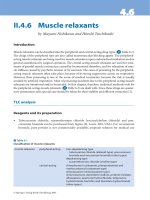
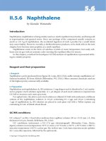
The model necessarily requires feedback connections from the neural inte-
grators to vestibular nuclei to achieve the conversion from angular velocity to
the derivative of eye position (fig. 1). Indeed, feedback connections to the
vestibular nuclei have been shown anatomically from both the nucleus preposi-
tus hypoglossi and the interstitial nucleus of Cajal. For a numerical simulation
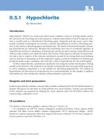
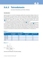
of the model comparing responses to slow and fast head movements, see figure 2.
This modeling example not only demonstrates the importance of taking into
account that eye movements are 3-D, but also that models based on eye velocity
as model output are often not sufficient.
Ocular Motor Models 167
Semicircular
canals
Head rotation
Neural
integrator
Vestibular
nuclei
Eye plant
Eye rotation
+
+
Direct pathway
Ocular motor
nuclei
e
I
r
· R ·
m
I
r
I
r
.
r
.
Fig. 1. Block diagram of the model of the VOR described in the main text. The sym-
bols correspond to the variables used in the mathematical description (equations 1–5); the
boxes contain the differential equations or other mathematical relations translating input to
output.
Ϫ50 0 50
0
50
100
150
200
250
Torsional (degrees/s)
Horizontal (degrees/s)
Ϫ1 0 1
0
1
2
3
4
5
6
Torsional (degrees/s)
Horizontal (degrees/s)
Straight ahead
30˚ down
30˚ up
ab
Fig. 2. Simulation of VOR responses to purely horizontal head rotations (amplitude 5Њ)
with a model of the 3-D VOR (see text). a Rapid VOR, duration 50 ms. b Slow VOR, duration
2 s. Solid lines: horizontal angular eye velocity plotted over torsional angular eye velocity.
Note the difference in velocity scales. Black: gaze straight ahead; dark grey: gaze 30° down;
light grey: gaze 30° up. Dashed lines: quarter-angle rule prediction for relation between tor-
sional and horizontal eye velocity at the respective gaze elevation. The model thus simulates
how rapid VOR responses can be purely head fixed, while slow VOR follows the quarter-
angle rule, as demonstrated experimentally [85].
Glasauer 168
Smooth Pursuit Eye Movements
The smooth pursuit system [for overview, see Büttner, this vol, pp 76–89]
has received considerable interest by modelers. In contrast to saccadic eye
movements, it has to be modeled as a closed-loop system, since the pursuit eye
movement changes the visual input by attempting to stabilize the target on the
retina. Even though it shares some pathways and properties with the saccadic
system (for review, see [86]), most of its structure can be regarded as imple-
menting a separate stream of processing [review: 87]. Most importantly,
smooth pursuit relies on an intact cerebellum (flocculus, paraflocculus, and
dorsal vermis), while saccades are possible even without it. One group of mod-
els assumes that the eye movement response is based on a combination of eye
acceleration, eye velocity, and sometimes eye position signals, which are com-
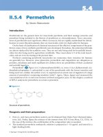
bined to drive the pursuit controller (e.g. [88]). Alternatively, a positive feed-
back loop within the visual cortex is proposed (fig. 3) which has a similar
effect as using combined retinal velocity and acceleration signals [90].
Another group building on the earliest modeling attempts [91, 92] assumes an
internal reconstruction of target velocity from retinal slip and an efference
Afferent
pathways
Efferent
pathways
Target motion
Retinal
slip
Motor
pathways
Pursuit
command
ϩ
Ϫ
Retina
Afferent
pathways
Efferent
pathways
Target motion
Retinal
slip
Motor
pathways
Pursuit
command
ϩ
Ϫ
Retina
Internal
model
ϩ
ϩ
Reconstructed
target motion
Efference
copy
a
b
Fig. 3. Two basic hypotheses for the processing of retinal slip information for smooth
pursuit [after 89]. a The pursuit command is generated in a simple feedback loop. b An inter-
nal model of motor pathways and afferent pathways driven by an efference copy of the pur-
suit command generates a signal suited to reconstruct target motion. This signal is used to
generate the pursuit command.
Ocular Motor Models 169
copy of eye velocity, which then drives pursuit (e.g. [93]). The latter approach
has some advantages, especially regarding the problem of the long latency of
visual processing, which makes the use of a simple high-gain feedback loop
problematic. It is also supported by recent experimental evidence: it has been
shown that the cortical middle superior temporal area (MST) contains neurons
which code target motion in space (for review, see [87]), and that thalamic neu-
rons carry smooth pursuit signals which are suited to convey an efference copy
to the cortex [94].
While figure 2 shows the basic information processing of the two hypothe-
ses, the various boxes in this processing scheme may contain mathematical
descriptions of the underlying processing from simple gain elements, delays or
linear differential equations, as in [91], to more complex systems of nonlinear
differential equations which are used to model neural networks. It is worth to
note that all dynamic computational models, whether on a systems level or
describing in detail the dynamics of ion channels of single neurons, rely on the
same basic building blocks, coupled differential equations.
All pathways for pursuit pass through the cerebellum; therefore, most
models have concentrated on the role of the cerebellar pathways, especially
those passing through the floccular lobe. The role of the cortical structures (pur-
suit region of the FEFs and MST), their downstream pathways (dorsolateral
pontine nuclei and nucleus reticularis tegmenti pontis), and their specific con-
tribution has received less attention so far. Neurophysiology suggests that the
FEF pathway is more related to signals on eye acceleration, i.e. changes in pur-
suit velocity, while MST is thought to convey signals related to ongoing pursuit
[95]. FEF has also been implicated in pursuit gain control [96]: rapid variations
in target velocity have a greater effect if pursuit velocity is high. Similarly, pur-
suit onset is slower than pursuit offset. While earlier models assumed a switch
in pursuit pathways [97], one pathway for pursuit onset, and one for offset, a
continuous gain control is now discussed [98, 99].
Gain control may also be related to another relevant feature of smooth pur-
suit, its predictive nature. Despite the visual latency, pursuit tracking of simple
motions, such as ramp-like or sinusoidal target movement, can reach unity gain
with zero latency. Thus, some form of predictive control must take place.
Current models propose memory-based mechanisms [100] or adaptive control
implementing a predictive model of target dynamics [101] to explain the exper-
imental findings. It is also not clear whether the predictive aspects of pursuit
control are implemented in cortical areas [101], or in the cerebellum [102], or
in both.
Since pursuit movements are almost always accompanied by saccadic eye
movements, a recent model proposes how switching between both modes can
be achieved together with predictive aspects of saccades and pursuit [103].
Glasauer 170
Combined Eye-Head Movements
Combined eye-head movements occur when the head is passively per-
turbed and the eyes compensate by the VOR. However, under natural circum-
stances saccadic gaze shifts and smooth pursuit consist of a combination of eye
and head movements, especially when the target eccentricity is too large to be
reached with the eye alone. Active combined eye-head movements raise several
questions [104], for example whether the VOR is active during the gaze shift, or
whether the local feedback loops in the saccadic system operate on gaze (eye
plus head) or eye-in-head signals. While it is usually accepted that the VOR is
shut off during the gaze shift, models on combined eye-head gaze shifts reached
different conclusions concerning the feedback loops: while most models
assume that gaze is the controlled variable [105–107], others propose that eye
and head movements are controlled separately with the head controller influ-
encing the saccadic burst generator for the eye [108]. The 3-D behavior of eye
and head during gaze shifts has successfully been explained by an elegant
model [109] which shows how the eye may anticipate the final head position.
Finally, a recent neural network model showed how superior colliculus and
cerebellum may interact for combined eye-head gaze shifts [50].
Conclusions
Basically for all aspects of eye movement control, computational models do
exist. The vast majority of these models are based on a systems level approach or
use neural networks with firing rate neurons. While most models concentrate on
specific aspects of eye movements, there are some attempts to provide models
putting together several of the ocular motor subsystems. To be useful, future mod-
els need to pursue such a holistic approach to eye movements, and at the same
time try to link the systems level approach to the underlying neural mechanisms.
References
1 Quaia C, Optican LM: Dynamic eye plant models and the control of eye movements. Strabismus
2003;11:17–31.
2 Robinson DA: A quantitative analysis of extraocular muscle cooperation and squint. Invest
Ophthalmol Vis Sci 1975;14:801–825.
3 Miller JM, Robinson DA: A model of the mechanics of binocular alignment. Comput Biomed Res
1984;17:436–470.
4 Miller JM, Pavlovski DS, Shamaeva I: Orbit
TM
1.8 Gaze Mechanics Simulation. Eidactics, Suite
404, 1450 Greenwich St., San Francisco, CA 94109, USA, 1999.
5 Haslwanter T, Buchberger M, Kaltofen T, Hoerantner R, Priglinger S: SEEϩϩ: a biomechanical
model of the oculomotor plant. Ann N Y Acad Sci 2005;1039:9–14.
Ocular Motor Models 171
6 Haustein W: Considerations on listing’s law and the primary position by means of amatrix descrip-
tion of eye position control. Biol Cybern 1989;60:411–420.
7 Porrill J, Warren PA, Dean P: A simple control law generates Listing’s positions in a detailed
model of the extraocular muscle system. Vision Res 2000;40:3743–3758.
8 Koene AR, Erkelens CJ: Properties of 3D rotations and their relation to eye movement control.
Biol Cybern 2004;90:410–417.
9 Warren PA, Porrill J, Dean P: Consistency of Listing’s law and reciprocal innervation with pseudo-
inverse control of eye position in 3-D. Biol Cybern 2004;91:1–9.
10 Wong AM: Listing’s law: clinical significance and implications for neural control. Surv
Ophthalmol 2004;49:563–575.
11 Ghasia FF, Angelaki DE: Do motoneurons encode the noncommutativity of ocular rotations?
Neuron 2005;47:281–293.
12 Schnabolk C, Raphan T: Modeling three-dimensional velocity-to position transformation in ocu-
lomotor control. J Neurophysiol 1994;71:623–638.
13 Tweed D, Misslisch H, Fetter M: Testing models of the oculomotor velocity-to-position transfor-
mation. J Neurophysiol 1994;72:1425–1429.
14 Raphan T: Modeling control of eye orientation in three dimensions; in Fetter M, Haslwanter T,
Misslisch H, Tweed D (eds): Three-Dimensional Kinematics of Eye, Head, and Limb Movements.
The Netherlands, Harwood Academic Publishing, 1997, pp 359–374.
15 Tweed D: Velocity-to-position transformation in the VOR and the saccadic system; in Fetter M,
Haslwanter T, Misslisch H, Tweed D (eds): Three-Dimensional Kinematics of Eye, Head, and
Limb Movements. The Netherlands, Harwood Academic Publishing, 1997, pp 375–386.
16 Quaia C, Optican LM: Commutative saccadic generator is sufficient to control a 3-D ocular plant
with pulleys. J Neurophysiol 1998;79:3197–3215.
17 Sklavos S, Porrill J, Kaneko CR, Dean P: Evidence for wide range of time scales in oculomotor
plant dynamics: implications for models of eye-movement control. Vision Res 2005;45:1525–1542.
18 Robinson DA: Integrating with neurons. Annu Rev Neurosci 1989;12:33–45.
19 Zee D, Yamazaki A, Butler PH, Gücer G: Effects of ablation of flocculus and paraflocculus on eye
movements in primate. J Neurophysiol 1981;46:878–899.
20 Glasauer S, Dieterich M, Brandt T: Central positional nystagmus simulated by a mathematical
ocular motor model of otolith-dependent modification of Listing’s plane. J Neurophysiol 2001;86:
1546–1554.
21 Crawford JD, Tweed DB, Vilis T: Static ocular counterroll is implemented through the 3-D neural
integrator. J Neurophysiol 2003;90:2777–2784.
22 Anastasio TJ: Nonuniformity in the linear network model of the oculomotor integrator produces
approximately fractional-order dynamics and more realistic neuron behavior. Biol Cybern 1998;
79:377–391.
23 Sklavos SG, Moschovakis AK: Neural network simulations of the primate oculomotor system IV.
A distributed bilateral stochastic model of the neural integrator of the vertical saccadic system.
Biol Cybern 2002;86:97–109.
24 Galiana HL, Outerbridge JS: A bilateral model for central neural pathways in vestibuloocular
reflex. J Neurophysiol 1984;51:210–241.
25 Green AM, Galiana HL: Hypothesis for shared central processing of canal and otolith signals.
J Neurophysiol 1998;80:2222–2228.
26 Seung HS: How the brain keeps the eyes still. Proc Natl Acad Sci USA 1996;93:13339–13344.
27 Seung HS, Lee DD, Reis BY, Tank DW: Stability of the memory of eye position in a recurrent net-
work of conductance-based model neurons. Neuron 2000;26:259–271.
28 Koulakov AA, Raghavachari S, Kepecs A, Lisman JE: Model for a robust neural integrator. Nat
Neurosci 2002;5:775–782.
29 Loewenstein Y, Sompolinsky H: Temporal integration by calcium dynamics in a model neuron.
Nat Neurosci 2003;6:961–967.
30 Dean P, Porrill J, Stone JV: Decorrelation control by the cerebellum achieves oculomotor plant
compensation in simulated vestibulo-ocular reflex. Proc Biol Sci 2002;269:1895–1904.
31 Ebadzadeh M, Darlot C: Cerebellar learning of bio-mechanical functions of extra-ocular muscles:
modeling by artificial neural networks. Neuroscience 2003;122:941–966.
Glasauer 172
32 Glasauer S: Cerebellar contribution to saccades and gaze holding: a modeling approach. Ann NY
Acad Sci 2003;1004:206–219.
33 Porrill J, Dean P, Stone JV: Recurrent cerebellar architecture solves the motor-error problem. Proc
Biol Sci 2004;271:789–796.
34 Jürgens R, Becker W, Kornhuber HH: Natural and drug-induced variations of velocity and dura-
tion of human saccadic eye movements: evidence for a control of the neural pulse generator by
local feedback. Biol Cybern 1981;39:87–96.
35 Crawford JD, Guitton D: Visual-motor transformations required for accurate and kinematically
correct saccades. J Neurophysiol 1997;78:1447–1467.
36 Scudder CA: A new local feedback model of the saccadic burst generator. J Neurophysiol
1988;59:1455–1475.
37 Moschovakis AK: Neural network simulations of the primate oculomotor system. II. Frames of
reference. Brain Res Bull 1996;40:337–345.
38 Gancarz G, Grossberg S: A neural model of the saccade generator in the reticular formation.
Neural Netw 1998;11:1159–1174.
39 Jackson ME, Litvak O, Gnadt JW: Analysis of the frequency response of the saccadic circuit:
numerical simulations. Neural Netw 2001;14:1357–1376.
40 Moschovakis AK, Highstein SM: The anatomy and physiology of primate neurons that control
rapid eye movements. Annu Rev Neurosci 1994;17:465–488.
41 Bozis A, Moschovakis AK: Neural network simulations of the primate oculomotor system. III. An
one-dimensional, one-directional model of the superior colliculus. Biol Cybern 1998;79:215–230.
42 Arai K, Das S, Keller EL, Aiyoshi E: A distributed model of the saccade system: simulations of tem-
porally perturbed saccades using position and velocity feedback. Neural Netw 1999;12:1359–1375.
43 Smith MA, Crawford JD: Distributed population mechanism for the 3-D oculomotor reference
frame transformation. J Neurophysiol 2005;93:1742–1761.
44 Grossberg S, Roberts K, Aguilar M, Bullock D: A neural model of multimodal adaptive saccadic
eye movement control by superior colliculus. J Neurosci 1997;17:9706–9725.
45 Brown JW, Bullock D, Grossberg S: How laminar frontal cortex and basal ganglia circuits interact
to control planned and reactive saccades. Neural Netw 2004;17:471–510.
46 Dean P: Modelling the role of the cerebellar fastigial nuclei in producing accurate saccades: the
importance of burst timing. Neuroscience 1995;68:1059–1077.
47 Enderle JD, Engelken EJ: Effects of cerebellar lesions on saccade simulations. Biomed Sci
Instrum 1996;32:13–21.
48 Lefevre P, Quaia C, Optican LM: Distributed model of control of saccades by superior colliculus
and cerebellum. Neural Netw 1998;11:1175–1190.
49 Quaia C, Lefèvre P, Optican LM: Model of the control of saccades by superior colliculus and
cerebellum. J Neurophysiol 1999;82:999–1018.
50 Wang X, Jin J, Jabri M: Neural network models for the gaze shift system in the superior colliculus
and cerebellum. Neural Netw 2002;15:811–832.
51 Schweighofer N, Arbib MA, Dominey PF: A model of the cerebellum in adaptive control of
saccadic gain. I. The model and its biological substrate. Biol Cybern 1996;75:19–28.
52 Schweighofer N, Arbib MA, Dominey PF: A model of the cerebellum in adaptive control of
saccadic gain. II. Simulation results. Biol Cybern 1996;75:29–36.
53 Ebadzadeh M, Darlot C: Cerebellar learning of bio-mechanical functions of extra-ocular muscles:
modeling by artificial neural networks. Neuroscience 2003;122:941–966.
54 Catz N, Dicke PW, Thier P: Cerebellar complex spike firing is suitable to induce as well as to
stabilize motor learning. Curr Biol 2005;15:2179–2189.
55 Marr D: A theory of cerebellar cortex. J Physiol 1969;202:437–470.
56 Albus JS: A theory of cerebellar function. Math Biosci 1971;10:5–61.
57 Niemeier M, Crawford JD, Tweed DB: Optimal transsaccadic integration explains distorted spatial
perception. Nature 2003;422:76–80.
58 Girard B, Berthoz A: From brainstem to cortex: computational models of saccade generation
circuitry. Prog Neurobiol 2005;77:215–251.
59 Glasauer S, Dieterich M, Brandt T: Three-dimensional modeling of static vestibulo-ocular
brainstem syndromes. Neuroreport 1998;9:3841–3845.
Ocular Motor Models 173
60 Glasauer S, Dieterich M, Brandt T: Simulation of pathological ocular counterroll and skew-torsion
by a 3-D mathematical model. Neuroreport 1999;10:1843–1848.
61 Galiana HL, Smith HL, Katsarkas A: Modelling non-linearities in the vestibulo-ocular reflex (VOR)
after unilateral or bilateral loss of peripheral vestibular function. Exp Brain Res 2001;137:369–386.
62 Cartwright AD, Gilchrist DP, Burgess AM, Curthoys IS: A realistic neural-network simulation of
both slow and quick phase components of the guinea pig VOR. Exp Brain Res 2003;149:299–311.
63 Merfeld DM, Young LR: The vestibulo-ocular reflex of the squirrel monkey during eccentric rota-
tion and roll tilt. Exp Brain Res 1995;106:111–122.
64 Zupan LH, Merfeld DM, Darlot C: Using sensory weighting to model the influence of canal,
otolith and visual cues on spatial orientation and eye movements. Biol Cybern 2002;86:209–230.
65 Glasauer S: Interaction of semicircular canals and otoliths in the processing structure of the sub-
jective zenith. Ann NY Acad Sci 1992;656:847–849.
66 Mergner T, Glasauer S: A simple model of vestibular canal-otolith signal fusion. Ann NY Acad Sci
1999;871:430–434.
67 Reymond G, Droulez J, Kemeny A: Visuovestibular perception of self-motion modeled as a
dynamic optimization process. Biol Cybern 2002;87:301–314.
68 Green AM, Galiana HL: Hypothesis for shared central processing of canal and otolith signals.
J Neurophysiol 1998;80:2222–2228.
69 Green AM, Angelaki DE: Resolution of sensory ambiguities for gaze stabilization requires a sec-
ond neural integrator. J Neurosci 2003;23:9265–9275.
70 Green AM, Angelaki DE: An integrative neural network for detecting inertial motion and head ori-
entation. J Neurophysiol 2004;92:905–925.
71 Robinson DA: Vestibular and optokinetic symbiosis: an example of explaining by modeling; in Baker
R, Berthoz A (eds): Control of Gaze by Brain Stem Neurons. Elsevier, Amsterdam, 1977, pp 49–58.
72 Furman JM, Hain TC, Paige GD: Central adaptation models of the vestibulo-ocular and optoki-
netic systems. Biol Cybern 1989;61:255–264.
73 Raphan T, Sturm D: Modeling the spatiotemporal organization of velocity storage in the vestibu-
loocular reflex by optokinetic studies. J Neurophysiol 1991;66:1410–1421.
74 Schweigart G, Mergner T, Barnes G: Eye movements during combined pursuit, optokinetic and
vestibular stimulation in macaque monkey. Exp Brain Res 1999;127:54–66.
75 Hirata Y, Takeuchi I, Highstein SM: A dynamical model for the vertical vestibuloocular reflex and
optokinetic response in primate. Neurocomputing 2003;52–54:531–540.
76 Miles FA, Lisberger SG: Plasticity in the vestibulo-ocular reflex: a new hypothesis. Annu Rev
Neurosci 1981;4:273–299.
77 Blazquez PM, Hirata Y, Highstein SM: The vestibulo-ocular reflex as a model system for motor
learning: what is the role of the cerebellum? Cerebellum 2004;3:188–192.
78 Gomi H, Kawato M: Adaptive feedback control models of the vestibulocerebellum and spinocere-
bellum. Biol Cybern 1992;68:105–114.
79 Lisberger SG: Neural basis for motor learning in the vestibuloocular reflex of primates. III.
Computational and behavioral analysis of the sites of learning. J Neurophysiol 1994;72:974–998.
80 Galiana HL, Green AM: Vestibular adaptation: how models can affect data interpretations.
Otolaryngol Head Neck Surg 1998;119:231–243.
81 Hepp K: Oculomotor control: Listing’s law and all that. Curr Opin Neurobiol 1994;4:862–868.
82 Lee C, Zee DS, Straumann D: Saccades from torsional offset positions back to listing’s plane.
J Neurophysiol 2000;83:3241–3253.
83 Schneider E, Glasauer S, Dieterich M, Kalla R, Brandt T: Diagnosis of vestibular imbalance in the
blink of an eye. Neurology 2004;63:1209–1216.
84 Misslisch H, Tweed D, Fetter M, Sievering D, Koenig E: Rotational kinematics of the human
vestibulo-ocular reflex III. Listing’s law. J Neurophysiol 1994;72:2490–2502.
85 Palla A, Straumann D, Obzina H: Eye-position dependence of three-dimensional ocular rotation-
axis orientation during head impulses in humans. Exp Brain Res 1999;129:127–133.
86 Krauzlis RJ: The control of voluntary eye movements: new perspectives. Neuroscientist 2005;11:
124–137.
87 Thier P, Ilg U: The neural basis of smooth-pursuit eye movements. Curr Opin Neurobiol 2005;15:
645–652.
Glasauer 174
88 Krauzlis RJ, Lisberger SG: A model of visually-guided smooth pursuit eye movements based on
behavioral observations. J Comput Neurosci 1994;1:265–283.
89 Lisberger SG, Morris EJ, Tychsen L: Visual motion processing and sensory-motor integration for
smooth pursuit eye movements. Annu Rev Neurosci 1987;10:97–129.
90 Tabata H, Yamamoto K, Kawato M: Computational study on monkey VOR adaptation and smooth
pursuit based on the parallel control-pathway theory. J Neurophysiol 2002;87:2176–2189.
91 Robinson DA, Gordon JL, Gordon SE: A model of the smooth pursuit eye movement system. Biol
Cybern 1986;55:43–57.
92 Yasui S, Young LR: Perceived visual motion as effective stimulus to pursuit eye movement system.
Science 1975;190:906–908.
93 Marti S, Straumann D, Glasauer S: The origin of downbeat nystagmus: an asymmetry in the dis-
tribution of on-directions of vertical gaze-velocity Purkinje cells. Ann N Y Acad Sci 2005;1039:
548–553.
94 Tanaka M: Involvement of the central thalamus in the control of smooth pursuit eye movements.
J Neurosci 2005;25:5866–5876.
95 Ono S, Das VE, Economides JR, Mustari MJ: Modeling of smooth pursuit-related neuronal
responses in the DLPN and NRTP of the rhesus macaque. J Neurophysiol 2005;93:108–116.
96 Chou IH, Lisberger SG: The role of the frontal pursuit area in learning in smooth pursuit eye
movements. J Neurosci 2004;24:4124–4133.
97 Huebner WP, Saidel GM, Leigh RJ: Nonlinear parameter estimation applied to a model of smooth
pursuit eye movements. Biol Cybern 1990;62:265–273.
98 Keating EG, Pierre A: Architecture of a gain controller in the pursuit system. Behav Brain Res
1996;81:173–181.
99 Nuding U, Glasauer S, Büttner U: Nonlinear Systems Analysis of the Smooth-Pursuit Gain-Control
Mechanism. Abstract for the Japan-Germany Symposium on Computational Neuroscience, Tokio,
2006
100 Barnes G, Grealy M: Modelling prediction in ocular pursuit; in Becker W, Deubel H, Mergner T
(eds): Current Oculomotor Research. New York, Plenum Press, 1999, pp 97–107.
101 Shibata T, Tabata H, Schaal S, Kawato M: A model of smooth pursuit in primates based on learn-
ing the target dynamics. Neural Netw 2005;18:213–224.
102 Kettner RE, Suh M, Davis D, Leung HC: Modeling cerebellar flocculus and paraflocculus
involvement in complex predictive smooth eye pursuit in monkeys. Ann N Y Acad Sci 2002;978:
455–467.
103 Lee WJ, Galiana HL: An internally switched model of ocular tracking with prediction. IEEE Trans
Neural Syst Rehabil Eng 2005;13:186–193.
104 Crawford JD, Martinez-Trujillo JC, Klier EM: Neural control of three-dimensional eye and head
movements. Curr Opin Neurobiol 2003;13:655–662.
105 Galiana HL, Guitton D: Central organization and modeling of eye-head coordination during ori-
enting gaze shifts. Ann N Y Acad Sci 1992;656:452–471.
106 Goossens HH, Van Opstal AJ: Human eye-head coordination in two dimensions under different
sensorimotor conditions. Exp Brain Res 1997;114:542–560.
107 Wagner R, Galiana HL: Hybrid gaze control: ffusing visual/vestibular senses, slow/fast processes,
and eye/head coordination. Proc 31st Int Symp Robotics ISR 2000;220–225.
108 Freedman EG: Interactions between eye and head control signals can account for movement kine-
matics. Biol Cybern 2001;84:453–462.
109 Tweed D: Three-dimensional model of the human eye-head saccadic system. J Neurophysiol
1997;77:654–666.
Stefan Glasauer
Department of Neurology
Klinikum Grosshadern
Marchioninistrasse 15
DE–81377 Munich (Germany)
Tel. ϩ49 89 7095 4835, Fax ϩ49 89 7095 4801, E-Mail
Straube A, Büttner U (eds): Neuro-Ophthalmology.
Dev Ophthalmol. Basel, Karger, 2007, vol 40, pp 175–192
Therapeutic Considerations for Eye
Movement Disorders
A. Straube
Department of Neurology, University of Munich, Munich, Germany
Abstract
Advances made in understanding the pathophysiology of eye movement disorders have
only recently with the publication of the first well-planned studies been translated into better
treatment strategies. The following chapter summarizes the pharmacological treatment options
for a variety of oculomotor syndromes. Cortisone is useful, for example, for acute vestibular
neuritis to improve the restitution of the labyrinthine function. For the widespread benign
paroxysmal positioning nystagmus, a series of liberatory movements that free the semicircular
canal from the causative otoconia is now a well-established therapy. Treatment for the central
vestibular syndrome of up- and downbeat nystagmus consists of drugs like the potassium canal
blocker 4-aminopyridine, which influence the cerebellar circuits involved in the disorder’s
pathophysiology. Acquired pendular nystagmus, one of the oculomotor syndromes often cau-
sed by multiple sclerosis, results in the severe impairment of reduced visual acuity. Memantine,
a weak NMDA antagonist, has now been proven effective here. Finally, anticonvulsants like
carbamazepine are the drugs of choice for disorders involving a nerve-blood vessel contact that
induces symptoms of vestibular paroxysmia or superior oblique myokymia.
Copyright © 2007 S. Karger AG, Basel
The common goal of voluntary as well as most reflexive eye movements is
to stabilize images on the retina (especially the central fovea, the area of the
highest resolution) in order to prevent retinal slip. Abnormal involuntary eye
movements may cause excessive motion of images on the retina, leading to
blurred vision and to the illusion that the perceived world is moving (oscillop-
sia). Clinical examination of such pathological eye movements often allows the
topological diagnosis of the lesion causing the abnormalities. Despite our exten-
sive knowledge of the anatomy and physiology of eye movements, very little is
known about pharmacological aspects of the ocular motor system. Thus, our
treatment options for abnormal eye movements remain fairly limited. Most drug
treatments are based on case reports. Only recently have a few controlled trials
Straube 176
been published (overview in [1–5]). Several drugs can themselves cause nystag-
mus, for example, anticonvulsants, sedatives, and antihistaminergic drugs
induce gaze-evoked nystagmus; nicotinergic substances induce a nystagmus that
can disclose an underlying vestibular tone imbalance; and intoxication due to
lithium or phenytoin can lead to downbeat nystagmus as well as opsoclonus.
This chapter summarizes the most recent publications on pharmacological
treatment options for the different eye movement syndromes and also gives a
short overview of the clinical aspects and pathophysiology of these syndromes.
Eye movement syndromes are generally differentiated into those character-
ized by a pathological jerk nystagmus, pendular nystagmus, atypical nystagmus,
or saccadic oscillations. All interfere with the normal foveal fixation of a target.
Table 1. Practical treatment of oculomotor signs/syndromes
Ocular sign/disorder Substance Dosage Contraindications
Vestibular neuritis Acute: dimenhydrinate 50–100 mg General
Prednisolone 1 mg/kg body weight contraindications for
per day for 5 days, cortisone and
or starting with 100 mg dimenhydrinate
Menière’s disease Acute: dimenhydrinate 50–100 mg General
Prophylaxis: betahistine 8/16–32 mg/day contraindications for
Gentamicin Locally in the middle ear dimenhydrinate and
betahistine
Ototoxic (hearing loss)
Vestibular paroxysmia Carbamazepine 2 ϫ 200–600 mg slow- Drowsiness, ataxia
release formulation Vertigo, dry mouth
Gabapentin 3–4 ϫ 300–600 mg Enzyme induction
Superior oblique Carbamazepine 2 ϫ 200–600mg slow- Drowsiness, ataxia
myokymia release formulation Vertigo, dry mouth
Gabapentin 3–4 ϫ 300–600 mg Enzyme induction
Downbeat nystagmus Clonazepam 2 ϫ 0.5–1 mg daily Sedation
Baclofen 3 ϫ 5–10 mg daily Ataxia, weakness
4-aminopyridine 3 ϫ 10 mg Seizures
Upbeat nystagmus Baclofen 3 ϫ 5–10 mg Sedation; weakness
4-aminopyridine Seizures
Periodic alternating Baclofen 3 ϫ 5–10 mg Sedation
nystagmus Ataxia, weakness
Acquired pendular Memantine 3–4 ϫ 10mg Somnolence, confusion,
nystagmus Gabapentin 3–4 ϫ 300–600mg dry mouth, edema
Treatment of Oculomotor Disorders 177
Peripheral and Central Vestibular Disorders
Pathophysiology
The vestibulo-ocular reflex (VOR) is one of the most basic reflexes. It can
even be observed in fish. After a short latency, the VOR generates eye rotations
in the same plane as the head rotation that elicits them [6]. To do this, the ocu-
lomotor system uses information provided by the three pairs of orthogonally
oriented semicircular canals. The right and left sides work together in a tradeoff
manner (i.e. when one labyrinth increases the neuronal activity, the other
decreases it) [6]. Disorders of the vestibular periphery cause nystagmus in a
direction that is determined by the pattern of labyrinthine semicircular canals
involved [6]. The complete, unilateral loss of one labyrinth causes a mixed hor-
izontal-torsional nystagmus that is suppressed by visual fixation. Another con-
sequence of peripheral vestibular lesions is a change in the size (gain) of the
overall dynamic VOR response, i.e. the gain of the VOR for head movements
toward the affected ear becomes smaller, and the subject has to refixate the
object after the head movement by a saccade. The head-impulse test uses this
feature clinically. As a result, patients may complain of oscillopsia during rapid
head movements.
Central vestibular disorders are caused by lesions of pathways or areas
involved in the adjustment of the VOR (e.g. the cerebellar connections to the
vestibular nuclei) [2, 6]. These lesions result in upbeat, downbeat, torsional nys-
tagmus or central positional vertigo.
Vestibular Neuritis
Clinical Aspects
The presenting sign of vestibular neuritis is an acute onset of severe rota-
tory vertigo that lasts for hours to days [7]. Hearing loss is normally not a sign
of vestibular neuritis [7]. The horizontal contraversive beating spontaneous nys-
tagmus has a torsional component and causes postural instability with a ten-
dency to fall to the ipsiversive side.
Etiology
Recent findings support the view that an inflammation of parts of the
vestibular nerve is the cause of vestibular neuritis and acute labyrinthitis. Most
studies have shown the presence of latent herpes simplex virus type 1 in human
vestibular ganglia [8, 9]. The imaging of 2 patients with vestibular neuritis using
3-tesla MRI and high-dose contrast medium revealed isolated enhancement of
Straube 178
the vestibular nerve only on the affected side [10], a sign of a disturbed blood-
brain barrier due to the inflammation.
Treatment
Treatment options consist primarily of vestibular sedatives (e.g. dimenhy-
drinate, 50–100 mg) [11] in the first 3 days administered in combination with
steroids. Kitahara et al. [12] examined 36 patients who were treated for up to 2
years after onset either with or without steroids. Although the treatment onset
was rather late, the group on steroids showed a tendency for more improve-
ment. A more detailed study published in 2004 [13] reported on a total of 141
patients who were randomized within 3 days after onset of symptoms to one of
four treatment options – placebo, methylprednisolone (starting with 100 mg
daily), valacyclovir , or a combination of valacyclovir and methylprednisolone.
The main finding of this study was that the groups receiving methyl-
prednisolone had a better final outcome (caloric testing showed about 60%
recovery of peripheral vestibular function) after 12 months than the placebo/
valacyclovir groups (36–39%). The combination of valacyclovir and
methylprednisolone provided no additional benefit. It has also been reported
that patients should be mobilized early to accelerate the recovery of vestibu-
lospinal function [14].
Menière’s Disease
Clinical Aspects
Menière’s disease is characterized by spontaneous attacks of vertigo, fluc-
tuating sensorineural hearing loss, aural fullness, and tinnitus that lasts for
hours to a few days [11, 15]. Key symptoms of such an attack are a horizontal
rotatory nystagmus, postural instability, and nausea/vomiting. The symptoms
only rarely include the opposite ear. Only 5 of 101 patients in a 2-year follow-
up developed symptoms in the contralateral ear [16]. In addition to a typical
history, the finding of a unilateral hearing deficit on the audiogram and a
reduced reaction to caloric vestibular testing also support the diagnosis [15].
Etiology
The cause of Menière’s disease is still not known. It has been shown
histopathologically that endolymphatic hydrops and concomitant distortion of
the membranous labyrinth can cause Menière’s disease [15]. Other candidates
include immunological causes and inflammation. An increased prevalence of
migraine has also been described in patients with Menière’s disease [17]. The
pathophysiological link between both diseases may be allergic mechanisms [17].