World of Microbiology and Immunology vol 1 - part 5 doc
Bạn đang xem bản rút gọn của tài liệu. Xem và tải ngay bản đầy đủ của tài liệu tại đây (636.32 KB, 37 trang )
Cowpox
WORLD OF MICROBIOLOGY AND IMMUNOLOGY
138
•
•
A bacterial suspension is best analyzed in the Coulter
counter when the suspension has been thoroughly shaken
beforehand. This step disperses the bacteria. Most bacteria
tend to aggregate together in a suspension. If not dispersed, a
clump of bacteria passing through the orifice of the counter
could be counted as a single bacterium. This would produce an
underestimate of the number of bacteria in the suspension.
The Coulter counter has been used for many applica-
tions, both biological and nonbiological. In the 1970s, the
device was reconfigured to incorporate a laser beam. This
allowed the use of fluorescent labeled monoclonal antibodies
to detect specific types of cells (e.g., cancer cells) or to detect
a specific species of bacteria. This refinement of the Coulter
counter is now known as flow cytometry.
See also Bacterial growth and division; Laboratory techniques
in microbiology
COWPOX
Cowpox
Cowpox refers to a disease that is caused by the cowpox or
catpox virus. The virus is a member of the orthopoxvirus fam-
ily. Other
viruses in this family include the smallpox and vac-
cinia viruses. Cowpox is a rare disease, and is mostly
noteworthy as the basis of the formulation, over 200 years ago,
of an injection by
Edward Jenner that proved successful in
curing smallpox.
The use of cowpox virus as a means of combating
smallpox, which is a much more threatening disease to
humans, has remained popular since the time of Jenner.
Once a relatively common malady in humans, cowpox
is now confined mostly to small mammals in Europe and the
United Kingdom. The last recorded case of a cow with cow-
pox was in the United Kingdom in 1978. Occasionally the dis-
ease is transmitted from these sources to human. But this is
very rare. Indeed, only some 60 cases of human cowpox have
been reported in the medical literature.
The natural reservoir for the cowpox virus is believed to
be small woodland animals, such as voles and wood mice.
Cats and cows, which can harbor the virus, are thought to be
an accidental host, perhaps because of their contact with the
voles or mice.
The cowpox virus, similar to the other orthopoxvirus, is
best seen using the
electron microscopic technique of negative
staining. This technique reveals surface details. The cowpox
virus is slightly oval in shape and has a very ridged-appearing
surface.
Human infection with the cowpox virus is thought to
require direct contact with an infected animal. The virus gains
entry to the bloodstream through an open cut. In centuries past,
farmers regularly exposed to dairy cattle could acquire the dis-
ease from hand milking the cows, for example. Cowpox is typ-
ically evident as pus-filled sores on the hands and face that
subsequently turn black before fading away. While present, the
lesions are extremely painful. There can be scars left at the site
of the infection. In rare instances, the virus can become more
widely disseminated through the body, resulting in death.
Both males and females are equally as likely to acquire
cowpox. Similarly, there no racial group is any more suscepti-
ble to infection. There is a predilection towards acquiring the
infection in youth less than 18 years of age. This may be
because of a closer contact with animals such as cats by this age
group, or because of lack of administration of smallpox
vaccine.
Treatment for cowpox tends to be ensuring that the
patient is as comfortable as possible while waiting for the
infection to run its course. Sometimes, a physician may wish
to drain the pus from the skin sores to prevent the spread of the
infection further over the surface of the skin. In cases where
symptoms are more severe, an immune globulin known as
antivaccinia gamaglobulin may be used. This immunoglobulin
is reactive against all viruses of the orthopoxvirus family. The
use of this treatment needs to be evaluated carefully, as there
can be side effects such as kidney damage. Antibodies to the
vaccinia virus may also be injected into a patient, as these
antibodies also confer protection against cowpox.
See also Vaccination; Virology; Zoonoses
COXIELLA BURNETII
• see Q FEVER
C
RANBERRY JUICE AS AN ANTI
-ADHE-
SION METHOD
• see A
NTI-
ADHESION METHODS
CREUTZFELDT-JAKOB DISEASE (CJD)
•
see BSE
AND CJD DISEASE
CRICK, FRANCIS (1916- )
Crick, Francis
English molecular biologist
Francis Crick is one half of the famous pair of molecular biol-
ogists who unraveled the mystery of the structure of
DNA
(deoxyribonucleic acid), the carrier of genetic information,
thus ushering in the modern era of
molecular biology. Since
this fundamental discovery, Crick has made significant contri-
butions to the understanding of the
genetic code and gene
action, as well as the understanding of molecular neurobiol-
ogy. In Horace Judson’s book The Eighth Day of Creation,
Nobel laureate
Jacques Lucien Monod is quoted as saying,
“No one man created molecular biology. But Francis Crick
dominates intellectually the whole field. He knows the most
and understands the most.” Crick shared the Nobel Prize in
medicine in 1962 with
James Watson and Maurice Wilkins for
the elucidation of the structure of DNA.
The eldest of two sons, Francis Harry Compton Crick
was born to Harry Crick and Anne Elizabeth Wilkins in
Northampton, England. His father and uncle ran a shoe and
boot factory. Crick attended grammar school in Northampton,
and was an enthusiastic experimental scientist at an early age,
producing the customary number of youthful chemical explo-
womi_C 5/6/03 2:05 PM Page 138
Crick, Francis
WORLD OF MICROBIOLOGY AND IMMUNOLOGY
139
•
•
sions. As a schoolboy, he won a prize for collecting wildflow-
ers. In his autobiography, What Mad Pursuit, Crick describes
how, along with his brother, he “was mad about tennis,” but
not much interested in other sports and games. At the age of
fourteen, he obtained a scholarship to Mill Hill School in
North London. Four years later, at eighteen, he entered
University College, London. At the time of his matriculation,
his parents had moved from Northampton to Mill Hill, and this
allowed Crick to live at home while attending university.
Crick obtained a second-class honors degree in physics, with
additional work in mathematics, in three years. In his autobi-
ography, Crick writes of his education in a rather light-hearted
way. Crick states that his background in physics and mathe-
matics was sound, but quite classical, while he says that he
learned and understood very little in the field of chemistry.
Like many of the physicists who became the first molecular
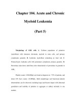
Francis Crick (right) and James Watson (left), who deduced the structure of the DNA double helix (shown between them).
womi_C 5/6/03 2:05 PM Page 139
Crick, Francis
WORLD OF MICROBIOLOGY AND IMMUNOLOGY
140
•
•
biologists and who began their careers around the end of
World War II, Crick read and was impressed by Erwin
Schrödinger’s book What Is Life?, but later recognized its lim-
itations in its neglect of chemistry.
Following his undergraduate studies, Crick conducted
research on the viscosity of water under pressure at high tem-
peratures, under the direction of Edward Neville da Costa
Andrade, at University College. It was during this period that
he was helped financially by his uncle, Arthur Crick. In 1940,
Crick was given a civilian job at the Admiralty, eventually
working on the design of mines used to destroy shipping.
Early in the year, Crick married Ruth Doreen Dodd. Their son
Michael was born during an air raid on London on November
25, 1940. By the end of the war, Crick was assigned to scien-
tific intelligence at the British Admiralty Headquarters in
Whitehall to design weapons.
Realizing that he would need additional education to
satisfy his desire to do fundamental research, Crick decided to
work toward an advanced degree. Crick became fascinated
with two areas of biology, particularly, as he describes it in his
autobiography, “the borderline between the living and the non-
living, and the workings of the brain.” He chose the former
area as his field of study, despite the fact that he knew little
about either subject. After preliminary inquiries at University
College, Crick settled on a program at the Strangeways
Laboratory in Cambridge under the direction of Arthur
Hughes in 1947, to work on the physical properties of
cyto-
plasm
in cultured chick fibroblast cells. Two years later, he
joined the Medical Research Council Unit at the Cavendish
Laboratory, ostensibly to work on protein structure with
British chemists Max Perutz and John Kendrew (both future
Nobel Prize laureates), but eventually to work on the structure
of DNA with Watson.
In 1947, Crick was divorced, and in 1949, married
Odile Speed, an art student whom he had met during the war.
Their marriage coincided with the start of Crick’s Ph.D. thesis
work on the x-ray diffraction of proteins. X-ray diffraction is
a technique for studying the crystalline structure of molecules,
permitting investigators to determine elements of three-
dimensional structure. In this technique, x rays are directed at
a compound, and the subsequent scattering of the x-ray beam
reflects the molecule’s configuration on a photographic plate.
In 1941 the Cavendish Laboratory where Crick worked
was under the direction of physicist Sir William Lawrence
Bragg, who had originated the x-ray diffraction technique
forty years before. Perutz had come to the Cavendish to apply
Bragg’s methods to large molecules, particularly proteins. In
1951, Crick was joined at the Cavendish by James Watson, a
visiting American who had been trained by Italian physician
Salvador Edward Luria and was a member of the Phage
Group, a group of physicists who studied bacterial
viruses
(known as bacteriophages, or simply phages). Like his phage
colleagues, Watson was interested in discovering the funda-
mental substance of genes and thought that unraveling the
structure of DNA was the most promising solution. The infor-
mal partnership between Crick and Watson developed, accord-
ing to Crick, because of their similar “youthful arrogance” and
similar thought processes. It was also clear that their experi-
ences complemented one another. By the time of their first
meeting, Crick had taught himself a great deal about x-ray dif-
fraction and protein structure, while Watson had become well
informed about phage and bacterial genetics.
Both Crick and Watson were aware of the work of bio-
chemists Maurice Wilkins and Rosalind Franklin at King’s
College, London, who were using x-ray diffraction to study
the structure of DNA. Crick, in particular, urged the London
group to build models, much as American chemist Linus
Pauling had done to solve the problem of the alpha helix of
proteins. Pauling, the father of the concept of the chemical
bond, had demonstrated that proteins had a three-dimensional
structure and were not simply linear strings of amino acids.
Wilkins and Franklin, working independently, preferred a
more deliberate experimental approach over the theoretical,
model-building scheme used by Pauling and advocated by
Crick. Thus, finding the King’s College group unresponsive to
their suggestions, Crick and Watson devoted portions of a two-
year period discussing and arguing about the problem. In early
1953, they began to build models of DNA.
Using Franklin’s x-ray diffraction data and a great deal
of trial and error, they produced a model of the DNA molecule
that conformed both to the London group’s findings and to the
data of Austrian-born American biochemist Erwin Chargaff.
In 1950, Chargaff had demonstrated that the relative amounts
of the four nucleotides, or bases, that make up DNA con-
formed to certain rules, one of which was that the amount of
adenine (A) was always equal to the amount of thymine (T),
and the amount of guanine (G) was always equal to the
amount of cytosine (C). Such a relationship suggests pairings
of A and T, and G and C, and refutes the idea that DNA is noth-
ing more than a tetranucleotide, that is, a simple molecule con-
sisting of all four bases.
During the spring and summer of 1953, Crick and
Watson wrote four papers about the structure and the supposed
function of DNA, the first of which appeared in the journal
Nature on April 25. This paper was accompanied by papers by
Wilkins, Franklin, and their colleagues, presenting experimen-
tal evidence that supported the Watson-Crick model. Watson
won the coin toss that placed his name first in the authorship,
thus forever institutionalizing this fundamental scientific
accomplishment as “Watson-Crick.”
The first paper contains one of the most remarkable
sentences in scientific writing: “It has not escaped our notice
that the specific pairing we have postulated immediately sug-
gests a possible copying mechanism for the genetic material.”
This conservative statement (it has been described as “coy”
by some observers) was followed by a more speculative paper
in Nature about a month later that more clearly argued for the
fundamental biological importance of DNA. Both papers
were discussed at the 1953 Cold Spring Harbor Symposium,
and the reaction of the developing community of molecular
biologists was enthusiastic. Within a year, the Watson-Crick
model began to generate a broad spectrum of important
research in genetics.
Over the next several years, Crick began to examine
the relationship between DNA and the genetic code. One of
his first efforts was a collaboration with Vernon Ingram,
womi_C 5/6/03 2:05 PM Page 140
Cryoprotection
WORLD OF MICROBIOLOGY AND IMMUNOLOGY
141
•
•
which led to Ingram’s 1956 demonstration that sickle cell
hemoglobin differed from normal hemoglobin by a single
amino acid. Ingram’s research presented evidence that a
molecular genetic disease, caused by a Mendelian mutation,
could be connected to a DNA-protein relationship. The
importance of this work to Crick’s thinking about the func-
tion of DNA cannot be underestimated. It established the
first function of “the genetic substance” in determining the
specificity of proteins.
About this time, South African-born English geneticist
and molecular biologist
Sydney Brenner joined Crick at the
Cavendish Laboratory. They began to work on the coding
problem, that is, how the sequence of DNA bases would spec-
ify the amino acid sequence in a protein. This work was first
presented in 1957, in a paper given by Crick to the
Symposium of the Society for Experimental Biology and
entitled “On Protein Synthesis.” Judson states in The Eighth
Day of Creation that “the paper permanently altered the logic
of biology.” While the events of the
transcription of DNA and
the synthesis of protein were not clearly understood, this
paper succinctly states “The Sequence Hypothesis assumes
that the specificity of a piece of nucleic acid is expressed
solely by the sequence of its bases, and that this sequence is
a (simple) code for the amino acid sequence of a particular
protein.” Further, Crick articulated what he termed “The
Central Dogma” of molecular biology, “that once ‘informa-
tion’ has passed into protein, it cannot get out again. In more
detail, the transfer of information from nucleic acid to nucleic
acid, or from nucleic acid to protein may be possible, but
transfer from protein to protein, or from protein to nucleic
acid is impossible.” In this important theoretical paper, Crick
establishes not only the basis of the genetic code but predicts
the mechanism for
protein synthesis. The first step, tran-
scription, would be the transfer of information in DNA to
ribonucleic acid (RNA), and the second step, translation,
would be the transfer of information from RNA to protein.
Hence, the genetic message is transcribed to a messenger, and
that message is eventually translated into action in the syn-
thesis of a protein. Crick is credited with developing the term
“codon” as it applies to the set of three bases that code for one
specific amino acid. These codons are used as “signs” to
guide protein synthesis within the cell.
A few years later, American geneticist Marshall Warren
Nirenberg and others discovered that the nucleic acid
sequence U-U-U (polyuracil) encodes for the amino acid
phenylalanine, and thus began the construction of the
DNA/RNA dictionary. By 1966, the DNA triplet code for
twenty amino acids had been worked out by Nirenberg and
others, along with details of protein synthesis and an elegant
example of the control of protein synthesis by French geneti-
cist
François Jacob, Arthur Pardée, and French biochemist
Jacques Lucien Monod. Brenner and Crick themselves turned
to problems in developmental biology in the 1960s, eventually
studying the structure and possible function of histones, the
class of proteins associated with
chromosomes.
In 1976, while on sabbatical from the Cavendish, Crick
was offered a permanent position at the Salk Institute for
Biological Studies in La Jolla, California. He accepted an
endowed chair as Kieckhefer Professor and has been at the
Salk Institute ever since. At the Salk Institute, Crick began to
study the workings of the brain, a subject that he had been
interested in from the beginning of his scientific career. While
his primary interest was consciousness, he attempted to
approach this subject through the study of vision. He pub-
lished several speculative papers on the mechanisms of
dreams and of attention, but, as he stated in his autobiogra-
phy, “I have yet to produce any theory that is both novel and
also explains many disconnected experimental facts in a con-
vincing way.”
During his career as an energetic theorist of modern
biology, Francis Crick has accumulated, refined, and synthe-
sized the experimental work of others, and has brought his
unusual insights to fundamental problems in science.
See also Cell cycle (eukaryotic), genetic regulation of; Cell
cycle (prokaryotic), genetic regulation of; Genetic identifica-
tion of microorganisms; Genetic mapping; Genetic regulation
of eukaryotic cells; Genetic regulation of prokaryotic cells;
Genotype and phenotype; Immunogenetics
CRYOPROTECTION
Cryoprotection
Cryopreservation refers to the use of a very low temperature
(below approximately –130° C [–202° F]) to store a living
organism. Organisms (including many types of
bacteria,
yeast, fungi, and algae) can be frozen for long periods of time
and then recovered for subsequent use.
This form of long-term storage minimizes the chances
of change to the microorganism during storage. Even at refrig-
eration temperature, many
microorganisms can grow slowly
and so might become altered during storage. This behavior has
been described for strains of Pseudomonas aeruginosa that
produce an external slime layer. When grown on a solid
agar
surface, the colonies of such strains appear like mucous drops.
However, when recovered from refrigeration storage, the
mucoid appearance can be lost. Cryopreservation of mucoid
strains maintains the mucoid characteristic.
Cryostorage of bacteria must be done at or below the
temperature of –130° C [–202° F], as it is at this temperature
that frozen water can form crystals. Because much of the inte-
rior of a bacterium and much of the surrounding membrane(s)
are made of water, crystal formation would be disastrous to the
cell. The formation of crystals would destroy structure, which
would in turn destroy function.
Ultralow temperature freezers have been developed that
achieve a temperature of –130° C . Another popular option for
cryopreservation is to immerse the sample in a compound
called liquid nitrogen. Using liquid nitrogen, a temperature of
–196° C [–320.8° F] can be achieved.
Another feature of bacteria that must be taken into
account during cryopreservation is called osmotic pressure.
This refers to the balance of ions on the outside versus the
inside of the cell. An imbalance in osmotic pressure can cause
water to flow out of or into a bacterium. The resulting shrink-
age or ballooning of the bacterium can be lethal.
womi_C 5/6/03 2:05 PM Page 141
Cryptococci and cryptococcosis
WORLD OF MICROBIOLOGY AND IMMUNOLOGY
142
•
•
To protect against crystal formation and osmotic pres-
sure shock to the bacteria, bacterial suspensions are typically
prepared in a so-called cryoprotectant solution. Glycerol is an
effective cryoprotective agent for many bacteria. For other
bacteria, such as cyanobacteria, methanol and dimethyl sul-
foxide are more suitable.
The microorganisms used in the cryoprotection process
should be in robust health. Bacteria, for example, should be
obtained from the point in their growth cycle where they are
actively growing and divided. In conventional liquid growth
media, this is described as the mid-logarithmic phase of
growth. In older cultures, where nutrients are becoming
depleted and waste products are accumulating, the cells can
deteriorate and change their characteristics.
For bacteria, the cryoprotectant solution is added
directly to an agar
culture of the bacteria of interest and bac-
teria are gently dislodged into the solution. Alternately, bacte-
ria in a liquid culture can be centrifuged and the “pellet” of
bacteria resuspended in the cryoprotectant solution. The
resulting bacterial suspension is then added to several spe-
cially designed cryovials. These are made of plastic that can
withstand the ultralow temperature.
The freezing process is done as quickly as possible to
minimize crystal formation. This is also referred to as “snap
freezing.” Bacterial suspensions in t cryoprotectant are ini-
tially at room temperature. Each suspension is deep-frozen in
a step-wise manner. First, the suspensions are chilled to refrig-
erator temperature. Next, they are stored for a few hours at
–70° C [–94° F]. Finally, racks of cryovials are either put into
the ultralow temperature freezer or plunged into liquid nitro-
gen. The liquid nitrogen almost instantaneously brings the
samples to –196° C [–320.8° F]. Once at this point, the sam-
ples can be stored indefinitely.
Recovery from cryostorage must also be rapid to avoid
crystal formation. Each suspension is warmed rapidly to room
temperature. The bacteria are immediately recovered by cen-
trifugation and the pellet of bacteria is resuspended in fresh
growth medium. The suspension is allowed to adapt to the
new temperature for a few days before being used.
Cryoprotection can be used for other purposes than the
long-term storage of samples. For example, cryoelectron
microscopy involves the rapid freezing of a sample and
examination of portions of the sample in an electro
micro-
scope
under conditions where the ultralow temperature is
maintained. If done correctly, cryoelectron microscopy will
revel features of microorganisms that are not otherwise evi-
dent in conventional electron microscopy. For example, the
watery
glycocalyx, which is made of chains of sugar, col-
lapses onto the surface of a bacterium as the sample is dried
out during preparation for conventional electron microscopy.
But glycocalyx structure can be cryopreserved. In another
example, cryoelectron microscopy has also maintained
external structural order on virus particles, allowing
researchers to deduce how these structures function in the
viral infection of tissue.
See also Bacterial ultrastructure; Donnan equilibrium; Quality
control in microbiology
C
RYPTOCOCCI AND CRYPTOCOCCOSIS
Cryptococci and cryptococcosis
Cryptococcus is a yeast that has a capsule surrounding the
cell. In the yeast classification system, Cryptococcus is a
member of the Phylum Basidimycota, Subphylum Basidi-
mycotina, Order Sporidiales, and Family Sporidiobolaceae.
There are 37 species in the genus Cryptococcus. One of
these, only one species is disease-causing, Cryptococcus neo-
formans. There are three so-called varieties of this species,
based on antigenic differences in the capsule, some differ-
ences in biochemical reactions such as the use of various sug-
ars as nutrients, and in the shape of the spores produced by the
yeast cells. The varieties are Cryptococcus neoformans var.
gatti, grubii, and neoformans. The latter variety causes the
most cryptococcal infections in humans.
Cryptococcus neoformans has a worldwide distribution.
It is normally found on plants, fruits and in birds, such as
pigeons and chicken. Transmission via bird waste is a typical
route of human infection.
Cryptococcus neoformans causes an infection known as
cryptococcosis. Inhalation of the microorganism leads to the
persistent growth in the lungs. For those whose
immune sys-
tem
is compromised, such as those having Acquired
Immunodeficiency Syndrome (AIDS), the pulmonary infection
can be life-threatening. In addition, yeast cells may become
distributed elsewhere in the body, leading to
inflammation of
nerve lining in the brain (
meningitis). A variety of other infec-
tions and symptoms can be present, including infections of the
eye (conjunctivitis), ear (otitis), heart (myocarditis), liver
(
hepatitis), and bone (arthritis).
The most common illness caused by the cryptococcal
fungus is cryptococcal meningitis. Those at most risk of devel-
oping cryptococcosis are AIDS patients. Those who have
received an organ, are receiving
chemotherapy for cancer or
have Hodgkin’s disease are also at risk, since frequently their
immune systems are suppressed. As the incidence of AIDS
and the use of immunosupressant drugs have grown over the
past decade, the number of cases of cryptococcosis has risen.
Until then, cases of cryptococcus occurred only rarely. Even
today, those with a well-functioning immune system are sel-
dom at risk for cryptococcosis. For these individuals a slight
skin infection may be the only adverse effect of exposure to
Cryptococcus.
Cryptococcus begins with the inhalation of Crypto-
coccus neoformans. Likely, the inhaled yeast is weakly encap-
sulated and is relatively small. This allows the cells to pene-
trate into the alveoli of the lungs. There the production of
capsule occurs. The capsule surrounding each yeast cell aids
the cell in avoiding the immune response of the host, particu-
larly the engulfing of the yeast by macrophage cells (which is
called
phagocytosis). The capsule is comprised of chains of
sugars, similar to the capsule around
bacteria. The capsule of
Cryptococcus neoformans is very negatively charged. Because
cells such as macrophages are also negatively charged, repul-
sive forces will further discourage interaction of macrophages
with the capsular material.
Another important virulence factor of the yeast is an
enzyme called phenol oxidase. The enzyme operates in the
womi_C 5/6/03 2:05 PM Page 142
Cryptosporidium and cryptosporidiosis
WORLD OF MICROBIOLOGY AND IMMUNOLOGY
143
•
•
production of melanin. Current thought is that the phenol oxi-
dase prevents the formation of charged hydroxy groups, which
can be very damaging to the yeast cell. The yeast may actually
recruit the body’s melanin producing machinery to make the
compound.
Cryptococcus neoformans also has other
enzymes that
act to degrade certain proteins and the
phospholipids that
make up cell membranes. These enzymes may help disrupt the
host cell membrane, allowing the yeast cells penetrate into
host tissue more easily.
Cryptococcus neoformans is able to grow at body tem-
perature. The other Cryptococcus species cannot tolerate this
elevated temperature.
Yet another virulence factor may operate. Evidence
from laboratory studies has indicated that antigens from the
yeast can induce a form of
T cells that down regulates the
immune response of the host. This is consistent with the
knowledge that survivors of cryptococcal meningitis display a
poorly operating immune system for a long time after the
infection has ended. Thus, Cryptococcus neoformans may not
only be capable of evading an immune response by the host,
but may actually dampen down that response.
If the infection is treated while still confined to the
lungs, especially in patients with a normally operative immune
system, the prospects for full recovery are good. However,
spread to the central nervous system is ominous, especially in
immunocompromised patients.
The standard treatment for cryptococcal meningitis is
the intravenous administration of a compound called ampho-
tericin B. Unfortunately the compound has a raft of side
effects, including fever, chills, headache, nausea with vomit-
ing, diarrhea, kidney damage, and suppression of bone mar-
row. The latter can lead to a marked decrease in red blood
cells. Studies are underway in which amphotericin B is
enclosed in bags made of lipid material (called liposomes).
The use of liposomes can allow the drug to be more specifi-
cally targeted to the site where treatment is most needed,
rather than flooding the entire body with the drug. Hopefully,
the use of liposome-delivered amphotericin B will lessen the
side effects of therapy.
See also Fungi; Immunomodulation; Yeast, infectious
CRYPTOSPORIDIUM AND
CRYPTOSPORIDIOSIS
Cryptosporidium and cryptosporidiosis
Cryptosporidum is a protozoan, a single-celled parasite that
lives in the intestines of humans and other animals. The organ-
ism causes an intestinal malady called cryptosporidiosis
(which is commonly called “crypto”).
The members of the genus Cryptosporidium infects
epithelial cells, especially those that line the walls of the intes-
tinal tract. One species, Cryptosporidium muris, infects labo-
ratory tests species, such as rodents, but does not infect
humans. Another species, Cryptosporidium parvum, infects a
wide variety of mammals, including humans. Calculations
have indicated that cattle alone release some five tons of the
parasite each year in the United States alone.
Non-human mammals are the reservoir of the organism
for humans. Typically, the organism is ingested when in water
that has been contaminated with Cryptosporidium-containing
feces. Often in an environment such as water, Crypto-
sporidium exists in a form that is analogous to a bacterial
spore. In the case of Cryptosporidium, this dormant and envi-
ronmentally resilient form is called an oocyst.
An oocyst is smaller than the growing form of
Cryptosporidium. The small size can allow the oocyst to pass
through some types of filters used to treat water. In addition,
an oocyst is also resistant to the concentrations of chlorine that
are widely used to disinfect drinking water. Thus, even drink-
ing water from a properly operating municipal treatment plant
has the potential to contain Cryptosporidium.
The organism can also be spread very easily by contact
with feces, such as caring with someone with diarrhea or
changing a diaper. Spread of cryptosporidiosis in nursing
homes and day care facilities is not uncommon.
Only a few oocytes need to be ingested to cause cryp-
tosporidiosis. Studies using volunteers indicate that an infec-
tious dose is anywhere from nine to 30 oocysts. When an
oocyte is ingested, it associates with intestinal epithelial cells.
Then, four bodies called sporozoites, which are contained
inside the oocyst, are released. These burrow inside the neigh-
bouring epithelial cells and divide to form cells that are called
merozoites. Eventually, the host cell bursts, releasing the
merozoites. The freed cells go on to attack neighbouring
epithelial cells and reproduce. The new progeny are released
and the cycle continues over and over. The damage to the
intestinal cells affects the functioning of the intestinal tract.
Cryptosporidium and its oocyte form have been known
since about 1910. Cryptosporidium parvum was first
described in 1911. Cryptosporidiosis has been a veterinary
problem for a long time. The disease was recognized as a
human disease in the 1970s. In the 1980s, the number of
human cases rose sharply along with the cases of
AIDS.
There have been many outbreaks of cryptosporidiosis
since the 1980s. In 1987, 13,000 in Carrollton, Georgia con-
tracted cryptosporidiosis via their municipal drinking water.
This incident was the first case of the spread of the disease
through water that had met all state and federal standards for
microbiological quality. In 1993, an outbreak of cryp-
tosporidiosis, again via contaminated municipal drinking water
that met the current standards, sickened 400,000 people and
resulted in several deaths. Outbreaks such as these prompted a
change in
water quality standards in the United States.
Symptoms of cryptosporidiosis are diarrhea, weight
loss, and abdominal cramping. Oocysts are released in the
feces all during the illness. Even when the symptoms are gone,
oocysts continue to be released in the feces for several weeks.
Even though known for a long time, detection of the
organism and treatment of the malady it causes are still chal-
lenging. No
vaccine for cryptosporidiosis exists. A well-func-
tioning
immune system is the best defense against the disease.
Indeed, estimates are that about 30% of the population has
antibodies to Cryptosporidium parvum, even though no symp-
womi_C 5/6/03 2:05 PM Page 143
Culture
WORLD OF MICROBIOLOGY AND IMMUNOLOGY
144
•
•
toms of cryptosporidiosis developed. The malady is most
severe in immunocompromised people, such as those infected
with
HIV (the virus that causes AIDS), or those receiving
chemotherapy for cancer or after a transplant. For those who
are diabetic, alcoholic, or pregnant, the prolonged diarrhea can
be dangerous.
In another avenue of infection, some of the merozoites
grow bigger inside the host epithelial cell and form two other
types of cells, termed the macrogametocyte and microgameto-
cyte. The macrogametocytes contain macrogametes. When
these combine with the microgametes released from the
microgametocytes, a zygote is formed. An oocyst wall forms
around the zygote and the genetic process of meiosis results in
the creation of four sporozoites inside the oocyst. The oocyst
is released to the environment in the feces and the infectious
cycle is started again.
The cycle from ingestion to the release of new infectious
oocytes in the feces can take about four days. Thereafter, the
production of a new generation of
parasites takes as little as
twelve to fourteen hours. Internally, this rapid division can cre-
ate huge numbers of organisms, which crowd the intestinal
tract. Cryptosporidiosis can spread to secondary sites, like the
duodenum and the large intestine. In people whose immune sys-
tems are not functioning properly, the spread of the organism
can be even more extensive, with parasites being found in the
stomach, biliary tract, pancreatic ducts, and respiratory tract.
Detection of Cryptosporidium in water is complicated
by the lack of a
culture method and because large volumes of
water (hundreds of gallons) need to be collected and concen-
trated to collect the few oocytes that may be present. Presently,
oocysts are detected using a microscopic method involving the
binding of a specific fluorescent probe to the oocyte wall.
There are many other noninfectious species of Crypto-
sporidium in the environment that react with the probe used in
the test. Furthermore, the test does not distinguish a living
organism from one that is dead. So a positive test result is not
always indicative of the presence of an infectious organism.
Skilled analysts are required to perform the test and so the
accuracy of detection varies widely from lab to lab.
See also Giardia and giardiasis; Water quality; Water purifi-
cation
CULTURE
Culture
A culture is a single species of microorganism that is isolated
and grown under controlled conditions. The German bacteri-
ologist
Robert Koch first developed culturing techniques in the
late 1870s. Following Koch’s initial discovery, medical scien-
tists quickly sought to identify other pathogens. Today
bacte-
ria cultures are used as basic tools in microbiology and
medicine.
The ability to separate bacteria is important because
microorganisms exist as mixed populations. In order to study
individual species, it is necessary to first isolate them. This
isolation can be accomplished by introducing individual bac-
terial cells onto a culture medium containing the necessary
elements microbial growth. The medium also provides condi-
tions favorable for growth of the desired species. These con-
ditions may involve
pH, osmotic pressure, atmospheric
oxygen, and moisture content. Culture media may be liquids
(known broths) or solids. Before the culture can be grown, the
media must be sterilized to prevent growth of unwanted
species. This
sterilization process is typically done through
exposure to high temperatures. Some tools like the metal loop
used to introduce bacteria to the media, may be sterilized by
exposure to a flame. The media itself may be sterilized by
treatment with steam-generated heat through a process known
as autoclaving.
To grow the culture, a number of the cells of the
microorganism must be introduced to the sterilized media.
This process is known as inoculation and is typically done by
exposing an inoculating loop to the desired strain and then
placing the loop in contact with the sterilized surface. A few of
the cells will be transferred to the growth media and under the
proper conditions, that species will begin to grow and form a
pure
colony. Cells in the colony can reproduce as often as
every 20 minutes and under the ideal conditions, this rate of
cell division could result in the production of 500,000 new
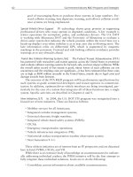
Liquid cultures of luminescent bacteria.
womi_C 5/6/03 2:05 PM Page 144
Cytoplasm, eukaryotic
WORLD OF MICROBIOLOGY AND IMMUNOLOGY
145
•
•
cells after six hours. Such rapid growth rates help to explain
the rapid development of disease, food spoilage, decay, and
the speed at which certain chemical processes used in industry
take place. Once the culture has been grown, a variety of
observation methods can be used to record the strain’s charac-
teristics and chart its growth.
See also Agar and agarose; Agar diffusion; American type cul-
ture collection; Antibiotic resistance, tests for; Bacterial
growth and division; Bacterial kingdoms; Epidemiology,
tracking diseases with technology; Laboratory techniques in
microbiology
CYCLOSPORIN
• see ANTIBIOTICS
C
YTOGENETICS
• see M
OLECULAR BIOLOGY AND
MOLECULAR GENETICS
CYTOKINES
Cytokines
Cytokines are a family of small proteins that mediate an
organism’s response to injury or infection. Cytokines operate
by transmitting signals between cells in an organism. Minute
quantities of cytokines are secreted, each by a single cell type,
and regulate functions in other cells by binding with specific
receptors. Their interactions with the receptors produce sec-
ondary signals that inhibit or enhance the action of certain
genes within the cell. Unlike endocrine hormones, which can
act throughout the body, most cytokines act locally, near the
cells that produced them.
Cytokines are crucial to an organism’s self-defense.
Cells under attack release a class of cytokines known as
chemokines. Chemokines participate in a process called
chemotaxis, signaling white blood cells to migrate toward the
threatened region. Other cytokines induce the white blood
cells to produce
inflammation, emitting toxins to kill
pathogens and enzymes to digest both the invaders and the
injured tissue. If the inflammatory response is not enough to
deal with the problem, additional
immune system cells are
also summoned by cytokines to continue the fight.
In a serious injury or infection, cytokines may call the
hematopoietic, or blood-forming system into play. New white
blood cells are created to augment the immune response, while
additional red blood cells replace any that have been lost.
Ruptured blood vessels emit chemokines to attract platelets,
the element of the blood that fosters clotting. Cytokines are
also responsible for signaling the nervous system to increase
the organism’s metabolic level, bringing on a fever that
inhibits the proliferation of pathogens while boosting the
action of the immune system.
Because of the central role of cytokines in fighting infec-
tion, they are being studied in an effort to find better treatments
for diseases such as
AIDS. Some have shown promise as thera-
peutic agents, but their usefulness is limited by the tendency of
cytokines to act locally. This means that their short amino acid
chains are likely either to be destroyed by enzymes in the
bloodstream or tissues before reaching their destination, or to
act on other cells with unintended consequences.
Other approaches to developing therapies based on
research into cytokines involve studying their receptor sites on
target cells. If a molecule could be developed that would bind
to the receptor site of a specific cytokine, it could elicit the
desired action from the cell, and might be more durable in the
bloodstream or have other advantages over the native
cytokine. Alternatively, a drug that blocked receptor sites
could potentially prevent the uncontrolled inflammatory
responses seen in certain autoimmune diseases.
See also Autoimmunity and autoimmune diseases;
Immunochemistry; Immunodeficiency disease syndromes;
Immunodeficiency diseases
C
YTOPLASM, EUKARYOTIC
Cytoplasm, eukaryotic
The cytoplasm, or cytosol of eukaryotic cells is the gel-like,
water-based fluid that occupies the majority of the volume of
the cell. Cytoplasm functions as the site of energy production,
storage, and the manufacture of cellular components. The vari-
ous organelles that are responsible for some of these functions
in the eukaryotic cell are dispersed throughout the cytoplasm, as
are the compounds that provide structural support for the cell.
The cytoplasm is the site of almost all of the chemical
activity occurring in a eukaryotic cell. Indeed, the word cyto-
plasm means “cell substance.”
Despite being comprised mainly of water (about 65%
by volume), the cytoplasm has the consistency of gelatin.
Unlike gelatin, however, the cytoplasm will flow. This enables
eukaryotes such as the amoeba to adopt different shapes, and
makes possible the formation of pseudopods that are used to
engulf food particles. The consistency of the cytoplasm is the
result of the other constituents of the cell that are floating in
fluid. These constituents include salts, and organic molecules
such as the many
enzymes that catalyze the myriad of chemi-
cal reactions that occur in the cell.
When viewed using the transmission electron
micro-
scope
, the cytoplasm appears as a three-dimensional lattice-
work of strands. In the early days of
electron microscopy there
was doubt as to whether this appearance reflected the true
nature of the cytoplasm, or was an artifact of the removal of
water from the cytoplasm during the preparation steps prior to
electron microscopic examination. However, development of
techniques that do not perturb the natural structure biological
specimens has confirmed that this latticework is real.
The lattice is made of various cytoplasmic proteins.
They are scaffolding structures that assist in the process of cell
division and in the shape of the cell. The shape-determinant is
referred to as the cytoskeleton. It is a network of fibers com-
posed of three types of proteins. The proteins form three fila-
mentous structures known as microtubules, intermediate
filaments, and microfilaments. The filaments are connected to
most of organelles located in the cytoplasm and serve to hold
together the organelles.
womi_C 5/6/03 2:05 PM Page 145
Cytoplasm, prokaryotic
WORLD OF MICROBIOLOGY AND IMMUNOLOGY
146
•
•
The microtubules are tubes that are formed by a spiral
arrangement of the constituent protein. They function in the
movement of the
chromosomes to either pole of the cell dur-
ing the cell division process. The microtubules are also known
as the spindle apparatus. Microfilaments are a composed of
two strands of protein that are twisted around one another.
They function in the contraction of muscle in higher eukary-
otic cells and in the change in cell shape that occurs in organ-
isms such as the amoeba. Finally, the intermediate filaments
act as more rigid scaffolding to maintain the cell shape.
The organelles of the cell are dispersed throughout the
cytoplasm. The
nucleus is bound by its own membrane to pro-
tect the genetic material from potentially damaging reactions
that occur in the cytoplasm. Thus, the cytoplasm is not a part
of the interior of the organelles.
The cytoplasm also contains
ribosomes, which float
around and allow protein to be synthesized all through the cell.
Ribosomes are also associated with a structure called the
endoplasmic reticulum. The golgi apparatus is also present, in
association with the endoplasmic reticulum. Enzymes that
degrade compounds are in the cytoplasm, in organelles called
lysosomes. Also present throughout the cytoplasm are the
mitochondria, which are the principal energy generating struc-
tures of the cell. If the eukaryotic cell is capable of photosyn-
thetic activity, then
chlorophyll containing organelles known
as chloroplasts are also present.
The cytoplasm of eukaryotic cells also functions to
transport dissolved nutrients around the cell and move waste
material out of the cell. These functions are possible because
of a process dubbed cytoplasmic streaming.
See also Eukaryotes
CYTOPLASM, PROKARYOTIC
Cytoplasm, prokaryotic
The cytoplasm of a prokaryotic cell is everything that is pres-
ent inside the bacterium. In contrast to a eukaryotic cell, there
is not a functional segregation inside
bacteria. The cytoplasm
houses all the chemicals and components that are used to sus-
Scanning electron micrograph of an eukaryotic cell, showing the nucleus in the center surrounded by the cytoplasm.The oval objects to the lower
left are ribosomes.
womi_C 5/6/03 2:05 PM Page 146
Cytoplasm, prokaryotic
WORLD OF MICROBIOLOGY AND IMMUNOLOGY
147
•
•
tain the life of a bacterium, with the exception of those com-
ponents that reside in the membrane(s), and in the
periplasm
of Gram-negative bacteria.
The cytoplasm is bounded by the cytoplasmic mem-
brane. Gram-negative bacteria contain another outer mem-
brane. In between the two membranes lies the periplasm.
When viewed in the light
microscope, the cytoplasm of
bacteria is transparent. Only with the higher magnification
available using the transmission
electron microscope does the
granular nature of the cytoplasm become apparent. The exact
structure of the cytoplasm may well be different than this
view, since the cytoplasm is comprised mainly of water. The
dehydration necessary for conventional electron microscopy
likely affect the structure of the cytoplasm.
The cytoplasm of prokaryotes and
eukaryotes is similar
in texture. Rather than being a free-flowing liquid the cyto-
plasm is more of a gel. The consistency has been likened to
that of dessert gel, except that the bacterial gel is capable of
flow. The ability of flow is vital, since the molecules that
reside in the cytoplasm must be capable of movement within
the bacterium as well as into and out of the cytoplasm.
The genetic material of the bacteria is dispersed
throughout the cytoplasm. Sometimes, the
deoxyribonucleic
acid
genome can aggregate during preparation for microscopy.
Then, the genome is apparent as a more diffuse area within the
granular cytoplasm. This artificial structure has been called
the nucleoid. Smaller, circular arrangements of genetic mate-
rial called
plasmids can also be present. The dispersion of the
bacterial genome throughout the cytoplasm is one of the fun-
damental distinguishing features between prokaryotic and
eukaryotic cells.
Also present throughout the cytoplasm is the
ribonu-
cleic acid
, various enzymes, amino acids, carbohydrates,
lipids, ions, and other compounds that function in the bac-
terium. The constituents of the membrane(s) are manufac-
tured in the cytoplasm and then are transported to their final
destination.
Some bacteria contain specialized regions known as
cytoplasmic inclusions that perform specialized functions.
These inclusions can be stored products that are used for the
nutrition of the bacteria. Examples of such inclusions are
glycogen, poly-B-hydroxybutyrate, and sulfur granules. As
well, certain bacteria contain gas-filled vesicles that act to
buoy the bacterium up to a certain depth in the water, or mem-
branous structures that contain
chlorophyll. The latter function
to harvest light for energy in photosynthetic bacteria.
The cytoplasm of prokaryotic cells also houses the
ribo-
somes required for the manufacture of protein. There can be
many ribosomes in the cytoplasm. For example, a rapidly
growing bacterium can contain upwards of 15,000 ribosomes.
The processes of
transcription, translation, protein
import and export, and at least some degradation of com-
pounds occurs in the cytoplasm. In Gram-negative bacteria,
some of these functions also occur in the periplasmic fluid.
The mechanisms that underlie the proper sequential orchestra-
tion of these functions are still yet to be fully determined.
See also Bacterial ultrastructure
womi_C 5/6/03 2:05 PM Page 147
D
149
•
•
D’HÉRELLE, FÉLIX (1873-1949)
d’Hérelle, Félix
Canadian bacteriologist
Félix d’Hérelle’s major contribution to science was the dis-
covery of the
bacteriophage, a microscopic agent that appears
in conjunction with and destroys disease-producing
bacteria in
a living organism. Like many researchers, d’Hérelle spent
much of his life exploring the effects of his major discovery.
He was also well-traveled; in the course of his life he lived for
long or short periods of time in Canada, France, the
Netherlands, Guatemala, Mexico, Indochina, Egypt, India, the
United States, and the former Soviet Union.
D’Hérelle was born in Montreal, Quebec, Canada. His
father, Félix d’Hérelle—a member of a well-established
French Canadian family, died when the young Félix was six
years old. After his father’s death, he moved with his mother,
Augustine Meert d’Hérelle, a Dutch woman, to Paris, France.
In Paris, d’Hérelle received his secondary education at the
Lycée Louis-le-Grand and began his medical studies. He com-
pleted his medical program at the University of Leiden in the
Netherlands. He married Mary Kerr, of France, in 1893, and
the couple eventually had two daughters. In 1901, d’Hérelle
moved to Guatemala City, Guatemala, to become the director
of the bacteriology laboratory at the general hospital and to
teach microbiology at the local medical school. In 1907, he
moved to Merida, Yucatan, Mexico, to study the
fermentation
of sisal hemp, and in 1908, the Mexican government sent him
back to Paris to further his microbiological studies. D’Hérelle
became an assistant at Paris’s Pasteur Institute in 1909,
became chief of its laboratory in 1914, and remained at the
Institute until 1921.
During his time at the Pasteur Institute, d’Hérelle stud-
ied a bacterium called Coccobacillus acridiorum, which
caused enteritis (
inflammation of the intestines) in locusts and
grasshoppers of the acrididae family of insects, with a view
toward using the microbe to destroy locusts. In growing the
bacteria on
culture plates, d’Hérelle observed empty spots on
the plates and theorized that these spots resulted from a virus
that grew along with and killed the bacteria. He surmised that
this phenomenon might have great medical significance as an
example of an organism fighting diseases of the digestive
tract. In 1916, he extended his investigation to cultures of the
bacillus that caused
dysentery and again observed spots free
of the microbe on the surface of the cultures. He was able to
filter out a substance from the feces of dysentery victims that
consumed in a few hours a culture broth of the bacillus. On
September 10, 1917, he presented to the French Academy of
Sciences a paper announcing his discovery entitled “Sur un
microbe invisible, antagoniste du bacille dysentérique.” He
named the bacteria–destroying substance bacteriophage (liter-
ally, “eater of bacteria”). He devoted most of his research and
writing for the rest of his life to the various types of bacterio-
phage which appeared in conjunction with specific types of
bacteria. He published several books dealing with his findings.
From 1920 to the late 1930s, d’Hérelle traveled and
lived in many parts of the world. In 1920, he went to French
Indochina under the auspices of the Pasteur Institute to study
human dysentery and septic pleuropneumonia in buffaloes. It
was during the course of this expedition that he perfected his
techniques for isolating bacteriophage. From 1922 to 1923, he
served as an assistant professor at the University of Leiden. In
1924, he moved to Alexandria, Egypt, to direct the
Bacteriological Service of the Egyptian Council on Health and
Quarantine. In 1927, he went to India at the invitation of the
Indian Medical Service to attempt to cure cholera through the
use of the bacteriophage associated with that disease.
D’Hérelle served as professor of bacteriology at Yale
University from 1928 to 1933, and in 1935 the government of
the Soviet Socialist Republic of Georgia requested that
d’Hérelle establish institutes dedicated to the study of bacte-
riophage in Tiflis, Kiev, and Kharkov. However, unstable civil
conditions forced d’Hérelle’s departure from the Soviet Union
in 1937, and he returned to Paris, where he lived, continuing
his study of bacteriophage, for the remainder of his life.
D’Hérelle attempted to make use of bacteriophage in
the treatment of many human and animal diseases, including
womi_D 5/6/03 2:08 PM Page 149
Darwin, Charles Robert
WORLD OF MICROBIOLOGY AND IMMUNOLOGY
150
•
•
dysentery, cholera, plague, and staphylococcus and strepto-
coccus infections. Such treatment was widespread for a time,
especially in the Soviet Union. However, use of bacteriophage
for this purpose was superseded by the use of chemical drugs
and
antibiotics even within d’Hérelle’s lifetime. Today bacte-
riophage is employed primarily as a diagnostic ultravirus. Of
the many honors d’Hérelle received, his perhaps most notable
is the Leeuwenhoek Medal given to him by the Amsterdam
Academy of Science in 1925; before d’Hérelle,
Louis Pasteur
had been the only other French scientist to receive the award.
D’Hérelle was presented with honorary degrees from the
University of Leiden and from Yale, Montreal, and Laval
Universities. He died after surgery in Paris at the age of 75.
See also Bacteriophage and bacteriophage typing
DARWIN, CHARLES ROBERT (1809-1882)
Darwin, Charles Robert
English naturalist
Charles Robert Darwin is credited with popularizing the con-
cept of organic
evolution by means of natural selection.
Though Darwin was not the first naturalist to propose a model
of biological evolution, his introduction of the mechanism of
the “survival of the fittest,” and discussion of the evolution of
humans, marked a revolution in both science and natural phi-
losophy.
Darwin was born in Shrewsbury, England and showed
an early interest in the natural sciences, especially geology.
His father, Robert Darwin, a wealthy physician, encouraged
Charles to pursue studies in medicine at the University of
Edinburg. Darwin soon tired of the subject, and his father sent
him to Cambridge to prepare for a career in the clergy. At
Cambridge, Darwin rekindled his passion for the natural sci-
ences, often devoting more time to socializing with
Cambridge scientists than to his clerical studies. With guid-
ance from his cousin, entomologist William Darwin Fox
(1805–1880), Darwin became increasingly involved in the
growing circle of natural scientists at Cambridge. ox intro-
duced Darwin to clergyman and biologist John Stevens
Henslow (1796–1861). Henslow became Darwin’s tutor in
mathematics and theology, as well as his mentor in his per-
sonal studies of botany, geology, and zoology. Henslow pro-
foundly influenced Darwin, and it was he who encouraged
Darwin to delay seeking an appointment in the Church of
England in favor of joining an expedition team and venturing
overseas. After graduation, Darwin agreed to an unpaid posi-
tion as naturalist aboard the H.M.S. Beagle. The expedition
team was initially chartered for a three year voyage and sur-
vey of South America’s Pacific coastline, but the ship pursued
other ventures after their work was complete and Darwin
remained part of H.M.S. Beagle’s crew for five years.
Darwin used his years aboard the Beagle to further his
study of the natural sciences. In South America, Darwin
became fascinated with geology. He paid close attention to
changes in the land brought about by earthquakes and volca-
noes. His observations led him to reject catastrophism (a the-
ory that land forms are the result of single, catastrophic
events), and instead espoused the geological theories of grad-
ual development proposed by English geologist Charles Lyell
(1797–1875) in his 1830 work, Principles of Geology. Yet,
some of his observations in South America did not fit with
Lyell’s theories. Darwin disagreed with Lyell’s assertion that
coral reefs grew atop oceanic volcanoes and rises, and con-
cluded that coral reefs built upon themselves. When Darwin
returned to England in 1836, he and Lyell became good
friends. Lyell welcomed Darwin’s new research on coral reefs,
and encouraged him to publish other studies from his voyages.
Darwin was elected a fellow of the Geological Society
in 1836, and became a member of the Royal Society in 1839.
That same year, he published his Journal of Researches into
the Geology and Natural History of the Various Countries
Visited by H.M.S. Beagle. Though his achievements in geol-
ogy largely prompted his welcoming into Britain’s scientific
community, his research interests began to diverge from the
discipline in the early 1840s. Discussions with other natural-
ists prompted Darwin’s increasing interest in population diver-
sity of fauna, extinct animals, and the presumed fixity of
species. Again, he turned to notes of his observations and var-
ious specimens he gathered while on his prior expedition. The
focus of his new studies was the Galápagos Islands off the
Pacific coast of Ecuador. While there, Darwin was struck by
the uniqueness of the island’s tortoises and birds. Some neigh-
boring islands had animal populations, which were largely
similar to that of the continent, while others had seemingly
different variety of species. After analyzing finch specimen
from the Galápagos, Darwin concluded that species must have
some means of transmutation, or ability of a species to alter
over time. Darwin thus proposed that as species modified, and
as old species disappeared, new varieties could be introduced.
Thus, Darwin proposed an evolutionary model of animal pop-
ulations.
The idea of organic evolution was not novel. French
naturalist, Georges Buffon (1707–1788) had theorized that
species were prone to development and change. Darwin’s own
grandfather, Erasmus Darwin, also published research regard-
ing the evolution of species. Although the theoretical concept
of evolution was not new, it remained undeveloped prior to
Charles Darwin. Just as he had done with Lyell’s geological
theory, Darwin set about the further the understanding of evo-
lution not merely as a philosophical concept, but as a practical
scientific model for explaining the diversity of species and
populations. His major contribution to the field was the intro-
duction of a mechanism by which evolution was accom-
plished. Darwin believed that evolution was the product of an
ongoing struggle of species to better adapt to their environ-
ment, with those that were best adapted surviving to reproduce
and replace less-suited individuals. He called this phenome-
non “survival of the fittest,” or natural selection. In this way,
Darwin believed that traits of maximum adaptiveness were
transferred to future generations of the animal population,
eventually resulting in new species.
Darwin finished an extensive draft of his theories in
1844, but lacked confidence in his abilities to convince others
of the merits of his discoveries. Years later, prompted by
rumors that a colleague was about to publish a theory similar
womi_D 5/6/03 2:09 PM Page 150
Davies, Julian E.
WORLD OF MICROBIOLOGY AND IMMUNOLOGY
151
•
•
to his own, Darwin decided to release his research. On the
Origin of Species by Means of Natural Selection, or The
Preservation of Favoured Races in the Struggle for Life, was
published November 1859, and became an instant bestseller.
A common misconception is that On the Origin of
Species was the introduction of the concept of human evolu-
tion. In fact, a discussion of human antiquity is relatively
absent from the book. Darwin did not directly address the rela-
tionship between animal and human evolution until he pub-
lished The Descent of Man, and Selection in Relation to Sex in
1871. Darwin introduced not only a model for the biological
evolution of man, but also attempted to chart the process of
man’s psychological evolution. He further tried to break down
the barriers between man and animals in 1872 with his work
The Expression of the Emotions in Man and Animals. By
observing facial features and voice sounds, Darwin asserted
that man and non-human animals exhibited signs of emotion
in similar ways. In the last years of his career, Darwin took the
concept of organic evolution to its logical end by applying nat-
ural selection and specialization to the plant kingdom.
Darwin’s works on evolution met with both debate from
the scientific societies, and criticism from some members of
the clergy. On the Origin of Species and The Descent of Man
were both published at a time of heightened religious evangel-
icalism in England. Though willing to discuss his theories with
colleagues in the sciences, Darwin refrained from participating
in public debates concerning his research. In the last decade of
his life, Darwin was disturbed about the application of his evo-
lutionary models to social theory. By most accounts, he con-
sidered the emerging concept of the social and cultural
evolution of men and civilizations, which later became known
as Social Darwinism, to be a grievous misinterpretation of his
works. Regardless of his opposition, he remained publicly tac-
iturn about the impact his scientific theories on theology, sci-
entific methodology, and social theory. Closely guarding his
privacy, Darwin retired to his estate in Down. He died at Down
House in 1882. Though his wishes were to receive an informal
burial, Parliament immediately ordered a state burial for the
famous naturalist at Westminster Abby. By the time of his
death, the scientific community had largely accepted the argu-
ments favoring his theories of evolution. Although the later dis-
coveries in genetics and
molecular biology radically
reinterpreted Darwin’s evolutionary mechanisms, evolutionary
theory is the key and unifying theory in all biological science.
See also Evolution and evolutionary mechanisms; Evolu-
tionary origin of bacteria and viruses
DAVIES, JULIAN E. (1932- )
Davies, Julian E.
Welsh bacteriologist
Julian Davies is a bacteriologist renowned for his research
concerning the mechanisms of bacterial resistance to
antibi-
otics, and on the use of antibiotics as research tools.
Davies was born in Casrell Nedd, Morgannwg, Cymru,
Wales. He received his education in Britain. His university
education was at the University of Nottingham, where he
received a B.Sc. (Chemistry, Physics, Math) in 1953 and a
Ph.D. (Organic Chemistry) in 1956. From 1959 to 1962, he
was Lecturer at the University of Manchester. Davies then
moved to the United States where he was an Associate at the
Harvard Medical School from 1962 until 1967. From 1965 to
1967, he was also a Visiting Professor at the Institute Pasteur
in Paris. In 1967, Davies became an Associate Professor in the
Department of
Biochemistry at the University of Wisconsin.
He attained the rank of Professor in 1970 and remained at
Wisconsin until 1980. In that year, Davies took up the post of
Research Director at Biogen in Geneva. In 1983, he became
President of Biogen. Two years later, Davies assumed the
position of Chief of Genetic Microbiology at the Institute
Pasteur, where he remained until 1992. In that year, he
returned to North America to become Professor and Head of
the Department of Microbiology and
Immunology at UBC. He
retained this position until his retirement in 1997. Presently he
remains affiliated with UBC as Emeritus Professor in the same
department.
While in British Columbia, Davies returned to commer-
cial
biotechnology. In 1996, he founded and became President
and CEO of TerraGen Diversity Inc. Davies assumed the post
of Chief Scientific Officer from 1998 to 2000. From 2000 to
the present, he is Executive Vice President, technology devel-
opment of Cubist Pharmaceuticals, Inc.
Davies has made fundamental discoveries in the area of
bacterial
antibiotic resistance, including the origin and evolu-
tion
of antibiotic resistance genes. He has identified bacterial
plasmids that carry genes that carry the information that deter-
mines the resistance of
bacteria to certain antibiotics.
Furthermore, he demonstrated that this information could be
transferred from one bacterium to another. These discoveries
have crucial to the efforts to develop drugs that can overcome
such antibiotic resistance.
Another facet of research has demonstrated how genetic
information can be transferred between bacteria that are dis-
tantly related. This work has had a fundamental influence on
the understanding of how bacteria can acquire genetic traits,
especially those that lead to antimicrobial resistance.
Davies has also developed a technique whereby genes
can be “tagged” and their path from one bacterium to another
followed. This technique is now widely used to follow
gene
transfer between prokaryotic and eukaryotic cells. In another
research area, Davies has explored the use of antibiotics as
experimental tools to probe the mechanisms of cellular
biochemistry, and the interaction between various molecules
in cells.
This prodigious research output has resulted in over 200
publications in peer-reviewed journals, authorship of six
books and numerous guest lectures.
Davies has also been active as an undergraduate and
graduate teacher and a mentor to a number of graduate stu-
dents. These research, commercial and teaching accomplish-
ments have been recognized around the world. He is a Fellow
of the Royal Society (London) and the Royal Society of
Canada, and is a past President of the American Society for
Microbiology. In 2000, he received a lifetime achievement
womi_D 5/6/03 2:09 PM Page 151
Broglie, Louis Victor de
WORLD OF MICROBIOLOGY AND IMMUNOLOGY
152
•
•
award in recognition of his development of the biotechnology
sector in British Columbia.
See also Microbial genetics
BROGLIE, LOUIS VICTOR DE (1892-1987)
Broglie, Louis Victor de
French physicist
Louis Victor de Broglie, a theoretical physicist and member of
the French nobility, is best known as the father of wave
mechanics, a far-reaching achievement that significantly
changed modern physics. Wave mechanics describes the
behavior of matter, including subatomic particles such as elec-
trons, with respect to their wave characteristics. For this
groundbreaking work, de Broglie was awarded the 1929
Nobel Prize for physics. De Broglie’s work contributed to the
fledgling science of microbiology in the mid-1920s, when he
suggested that electrons, as well as other particles, should
exhibit wave-like properties similar to light. Experiments on
electron beams a few years later confirmed de Broglie’s
hypothesis. Of importance to
microscope design was the fact
that the wavelength of electrons is typically much smaller than
the wavelength of light. Therefore, the limitation imposed on
the light microscope of 0.4 micrometers could be significantly
reduced by using a beam of electrons to illuminate the speci-
men. This fact was exploited in the 1930s in the development
of the
electron microscope.
Louis Victor Pierre Raymond de Broglie was born on
August 15, 1892, in Dieppe, France, to Duc Victor and Pauline
d’Armaille Broglie. His father’s family was of noble
Piedmontese origin and had served French monarchs for cen-
turies, for which it was awarded the hereditary title Duc from
King Louis XIV in 1740, a title that could be held only by the
head of the family.
The youngest of five children, de Broglie inherited a
familial distinction for formidable scholarship. His early edu-
cation was obtained at home, as befitted a great French family
of the time. After the death of his father when de Broglie was
fourteen, his eldest brother Maurice arranged for him to obtain
his secondary education at the Lycée Janson de Sailly in Paris.
After graduating from the Sorbonne in 1909 with bac-
calaureates in philosophy and mathematics, de Broglie entered
the University of Paris. He studied ancient history, paleogra-
phy, and law before finding his niche in science, influenced by
the writings of French theoretical physicist Jules Henri
Poincaré. The work of his brother Maurice, who was then
engaged in important, independent experimental research in x
rays and radioactivity, also helped to spark de Broglie’s inter-
est in theoretical physics, particularly in basic atomic theory.
In 1913, he obtained his Licencié ès Sciences from the
University of Paris’s Faculté des Sciences.
De Broglie’s studies were interrupted by the outbreak of
World War I, during which he served in the French army. Yet,
even the war did not take the young scientist away from the
country where he would spend his entire life; for its duration,
de Broglie served with the French Engineers at the wireless
station under the Eiffel Tower. In 1919, de Broglie returned to
his scientific studies at his brother’s laboratory. Here he began
his investigations into the nature of matter, inspired by a
conundrum that had long been troubling the scientific com-
munity: the apparent physical irreconcilability of the experi-
mentally proven dual nature of light. Radiant energy or light
had been demonstrated to exhibit properties associated with
particles as well as their well-documented wave-like charac-
teristics. De Broglie was inspired to consider whether matter
might not also exhibit dual properties. In his brother’s labora-
tory, where the study of very high frequency radiation using
spectroscopes was underway, de Broglie was able to bring the
problem into sharper focus. In 1924, de Broglie, with over two
dozen research papers on electrons, atomic structure, and x
rays already to his credit, presented his conclusions in his doc-
toral thesis at the Sorbonne. Entitled “Investigations into the
Quantum Theory,” it consolidated three shorter papers he had
published the previous year.
In his thesis, de Broglie postulated that all matter—
including electrons, the negatively charged particles that orbit
an atom’s
nucleus—behaves as both a particle and a wave.
Wave characteristics, however, are detectable only at the
atomic level, whereas the classical, ballistic properties of mat-
ter are apparent at larger scales. Therefore, rather than the
wave and particle characteristics of light and matter being at
odds with one another, de Broglie postulated that they were
essentially the same behavior observed from different per-
spectives. Wave mechanics could then explain the behavior of
all matter, even at the atomic scale, whereas classical
Newtonian mechanics, which continued to accurately account
for the behavior of observable matter, merely described a spe-
cial, general case. Although, according to de Broglie, all
objects have “matter waves,” these waves are so small in rela-
tion to large objects that their effects are not observable and no
departure from classical physics is detected. At the atomic
level, however, matter waves are relatively larger and their
effects become more obvious. De Broglie devised a mathe-
matical formula, the matter wave relation, to summarize his
findings.
American physicist Albert Einstein appreciated the sig-
nificant of de Broglie’s theory; de Broglie sent Einstein a
copy of his thesis on the advice of his professors at the
Sorbonne, who believed themselves not fully qualified to
judge it. Einstein immediately pronounced that de Broglie
had illuminated one of the secrets of the Universe. Austrian
physicist Erwin Schrödinger also grasped the implications of
de Broglie’s work and used it to develop his own theory of
wave mechanics, which has since become the foundation of
modern physics.
De Broglie’s wave matter theory remained unproven
until two separate experiments conclusively demonstrated the
wave properties of electrons—their ability to diffract or bend,
for example. American physicists Clinton Davisson and Lester
Germer and English physicist George Paget Thomson all
proved that de Broglie had been correct. Later experiments
would demonstrate that de Broglie’s theory also explained the
behavior of protons, atoms, and even molecules. These prop-
erties later found practical applications in the development of
magnetic lenses, the basis for the electron microscope.
womi_D 5/6/03 2:09 PM Page 152
Dengue fever
WORLD OF MICROBIOLOGY AND IMMUNOLOGY
153
•
•
In 1928, de Broglie was appointed professor of theoret-
ical physics at the University of Paris’s Faculty of Science. De
Broglie was a thorough lecturer who addressed all aspects of
wave mechanics. Perhaps because he was not inclined to
encourage an interactive atmosphere in his lectures, he had no
noted record of guiding young research students.
During his long career, de Broglie published over
twenty books and numerous research papers. His preoccupa-
tion with the practical side of physics is demonstrated in his
works dealing with cybernetics, atomic energy, particle accel-
erators, and wave-guides. His writings also include works on
x rays, gamma rays, atomic particles, optics, and a history of
the development of contemporary physics. He served as hon-
orary president of the French Association of Science Writers
and, in 1952, was awarded first prize for excellence in science
writing by the Kalinga Foundation. In 1953, Broglie was
elected to London’s Royal Society as a foreign member and,
in 1958, to the French Academy of Arts and Sciences in
recognition of his formidable output. With the death of his
older brother Maurice two years later, de Broglie inherited the
joint titles of French duke and German prince. De Broglie died
of natural causes on March 19, 1987, at the age of ninety-four.
See also Electron microscope, transmission and scanning;
Electron microscopic examination of microorganisms;
Microscope and microscopy
D
EFECTS OF CELLULAR IMMUNITY
• see
I
MMUNODEFICIENCY DISEASE SYNDROMES
DEFECTS OF T CELL MEDIATED IMMU-
NITY
• see IMMUNODEFICIENCY DISEASE SYNDROMES
DENGUE FEVER
Dengue fever
Dengue fever is a debilitating and sometimes hemorrhagic
fever (one that is associated with extensive internal bleeding).
The disease is caused by four slightly different types of a virus
from the genus Flavivirus that is designated as DEN. The four
antigenic types are DEN-1, DEN-2, DEN-3, and DEN-4.
The dengue virus is transmitted to humans via the bite
of a mosquito. The principle mosquito species is known as
Aedes aegypti. This mosquito is found all over the world, and,
throughout time, became adapted to urban environments. For
example, the mosquito evolved so as to be capable of living
year round in moist storage containers, rather than relying on
the seasonal patterns of rainfall. Another species, Aedes
albopictus (the “Tiger mosquito”), is widespread throughout
Asia. Both mosquitoes are now well established in urban cen-
ters. Accordingly, dengue fever is now a disease of urbanized,
developed areas, rather than rural, unpopulated regions.
The dengue virus is passed to humans exclusively by
the bite of mosquito in search of a blood meal. This mode of
transmission makes the dengue virus an arbovirus (that is, one
that is transmitted by an arthropod). Studies have demon-
strated that some species of monkey can harbor the virus.
Thus, monkeys may serve as a reservoir of the virus.
Mosquitoes who bite the monkey may acquire the virus and
subsequently transfer the virus to humans.
The disease has been known for centuries. The first
reported cases were in 1779–1780, occurring almost simulta-
neously in Asia, Africa, and North America. Since then, peri-
odic outbreaks of the disease have occurred in all areas of the
world where the mosquito resides. In particular, an outbreak
that began in Asia after World War II, spread around the world,
and has continued to plague southeast Asia even into 2002. As
of 2001, dengue fever was the leading cause of hospitalization
and death among children in southeast Asia.
Beginning in the 1980s, dengue fever began to increase
in the Far East and Africa. Outbreaks were not related to eco-
nomic conditions. For example, Singapore had an outbreak of
dengue fever from 1990 to 1994, even after a mosquito control
program that had kept the disease at minimal levels for over
two decades. The example of Singapore illustrates the impor-
tance of an ongoing program of mosquito population control.
The disease is a serious problem in more than 100 coun-
tries in Africa, North and South America, the Eastern
Mediterranean, South-East Asia, and the Western Pacific.
Unlike other bacterial or viral diseases, which can be
controlled by
vaccination, the four antigenic types of the
dengue virus do not confer cross-protection. Thus, it is possi-
ble for an individual to be sickened with four separate bouts of
dengue fever.
Following the transfer of the virus from mosquito to
humans, the symptoms can be varied, ranging from nonspe-
cific and relatively inconsequential ailments to severe and fatal
hemorrhaging. The incubation period of the virus is typically 5
to 8 days, but symptoms may develop after as few as three
days or as many as 15 days. The onset of symptoms is sudden
and dramatic. Initially, chills tend to develop, followed by a
headache. Pain with the movement of the eyes leads to more
generalized and extreme pain in the legs and joints. A high
fever can be produced, with temperatures reaching 104° F
[40° C]. Also, a pale rash may appear transiently on the face.
These symptoms can persist for up to 96 hours. Often,
the fever then rapidly eases. After a short period when symp-
toms disappear, the fever reappears. The temperature elevates
rapidly but the fever is usually not as high as in previous
episodes. The palms of the hands and soles of the feet may
turn bright red and become very swollen.
In about 80% of those who are infected, recovery is
complete after a convalescent period of several weeks with
general weakness and lack of energy. However, in some 20%
of those who are infected a severe form of dengue fever devel-
ops. This malady is characterized by the increased leakage of
fluid from cells and by the abnormal clotting of the blood.
These factors produce the hemorrhaging that can be a hallmark
of the disease, which is called dengue hemorrhagic fever. Even
then, recovery can be complete within a week. Finally, in some
of those who are infected, a condition called dengue shock syn-
drome can result in convulsions. In addition, a failure of the cir-
culatory system can occur, resulting in death.
womi_D 5/6/03 2:09 PM Page 153
Desiccation
WORLD OF MICROBIOLOGY AND IMMUNOLOGY
154
•
•
The reasons for the varied degrees of severity and symp-
toms that the viral infection can elicit are still unclear. Not sur-
prisingly, there is currently no cure for dengue, nor is there a
vaccine. Treatment for those who are afflicted is palliative, that
is, intended to ease the symptoms of the disease. Upon recov-
ery,
immunity to the particular antigenic type of the virus is in
place for life. However, an infection with one antigenic type of
dengue virus is not protective against the other three antigenic
types. Currently, the only preventive measure that can be taken
is to eradicate the mosquito vector of the virus.
See also Epidemics, viral; Zoonoses
DEOXYRIBONUCLEIC ACID
• see DNA
(DEOXYRIBONUCLEIC ACID)
DESICCATION
Desiccation
Desiccation is the removal of water from a biological system.
Usually this is accomplished by exposure to dry heat. Most
biological systems are adversely affected by the loss of water.
Microorganisms are no exception to this, except for those that
have evolved defensive measures to escape the loss of viabil-
ity typically associated with water loss.
Desiccation also results from the freezing of water, such
as in the polar regions on Earth. Water is present at these
regions, but is unavailable.
Microorganisms depend on water for their structure and
function. Cell membranes are organized with the water-loving
portions of the membrane lipids positioned towards the exte-
rior and the water-hating portions pointing inward. The loss of
water can throw this structure into disarray. Furthermore, the
interior of microorganisms such as
bacteria is almost entirely
comprised of water. Extremely rapid freezing of the water can
be a useful means of preserving bacteria and other microor-
ganisms. However, the gradual loss of water will produce
lethal changes in the chemistry of the interior
cytoplasm of
cells, collapse of the interior structure, and an alteration in the
three-dimensional structure of
enzymes. These drastic
changes caused by desiccation are irreversible.
In the laboratory, desiccation techniques are used to
help ensure that glassware is free of viable microbes.
Typically, the glassware is placed in a large dry-heat oven and
heated at 160° to 170° C [320° to 338° F] for up to two hours.
The effectiveness of
sterilization depends on the penetration of
heat into a biological sample.
Some microorganisms have evolved means of coping
with desiccation. The formation of a spore by bacteria such as
Bacillus and Clostridium allows the genetic material to sur-
vive the removal of water. Cysts produced by some protozoans
can also resist the destruction of desiccation for long periods
of time. Bacterial biofilms might not be totally dehydrated if
they are thick enough. Bacteria buried deep within the biofilm
might still be capable of growth.
The fact that some microbes on Earth can resist desic-
cation and then resuscitate when moisture becomes available
holds out the possibility of life on other bodies in our solar
system, particularly Mars. The snow at the poles of Mars is
proof that water is present. If liquid water becomes transiently
available, then similar resuscitation of dormant Martian
microorganisms could likewise occur.
See also Cryoprotection
DETECTION OF MUTANTS
• see LABORATORY
TECHNIQUES IN MICROBIOLOGY
D
IATOMS
Diatoms
Algae are a diverse group of simple, nucleated, plant-like
aquatic organisms that are primary producers. Primary pro-
ducers are able to utilize
photosynthesis to create organic
molecules from sunlight, water, and carbon dioxide.
Ecologically vital, algae account for roughly half of photosyn-
thetic production of organic material on Earth in both fresh-
water and marine environments. Algae exist either as single
cells or as multicellular organizations. Diatoms are micro-
scopic, single-celled algae that have intricate glass-like outer
cell walls partially composed of silicon. Different species of
diatom can be identified based upon the structure of these
walls. Many diatom species are planktonic, suspended in the
water column moving at the mercy of water currents. Others
remain attached to submerged surfaces. One bucketful of
water may contain millions of diatoms. Their abundance
makes them important food sources in aquatic ecosystems.
When diatoms die, their cell walls are left behind and sink to
the bottom of bodies of water. Massive accumulations of
diatom-rich sediments compact and solidify over long periods
of time to form rock rich in fossilized diatoms that is mined for
use in abrasives and filters.
Diatoms belong to the taxonomic phylum Bacillario-
phyta. There are approximately 10,000 known diatom species.
Of all algae phyla, diatom species are the most numerous. The
diatoms are single-celled, eukaryotic organisms, having
genetic information sequestered into subcellular compart-
ments called nuclei. This characteristic distinguishes the group
from other single-celled photosynthetic aquatic organisms,
like the
blue-green algae that do not possess nuclei and are
more closely related to
bacteria. Diatoms also are distinct
because they secrete complex outer cell walls, sometimes
called skeletons. The skeleton of a diatom is properly referred
to as a frustule.
Diatom frustules are composed of pure hydrated silica
within a layer of organic, carbon containing material.
Frustules are really comprised of two parts: an upper and
lower frustule. The larger upper portion of the frustule is
called the epitheca. The smaller lower piece is the hypotheca.
The epitheca fits over the hypotheca as the lid fits over a shoe-
womi_D 5/6/03 2:09 PM Page 154
Dictyostelium
WORLD OF MICROBIOLOGY AND IMMUNOLOGY
155
•
•
box. The singular algal diatom cell lives protected inside the
frustule halves like a pair of shoes snuggled within a shoebox.
Frustules are ornate, having intricate designs delineated
by patterns of holes or pores. The pores that perforate the frus-
tules allow gases, nutrients, and metabolic waste products to
be exchanged between the watery environment and the algal
cell. The frustules themselves may exhibit bilateral symmetry
or radial symmetry. Bilaterally symmetric diatoms are like
human beings, having a single plane through which halves are
mirror images of one another. Bilaterally symmetric diatoms
are elongated. Radially symmetric diatom frustules have many
mirror image planes. No matter which diameter is used to
divide the cell into two halves, each half is a mirror image of
the other. The combination of symmetry and perforation pat-
terns of diatom frustules make them beautiful biological struc-
tures that also are useful in identifying different species.
Because they are composed of silica, an inert material, diatom
frustules remain well preserved over vast periods of time
within geologic sediments.
Diatom frustules found in sedimentary rock are micro-
fossils. Because they are so easily preserved, diatoms have an
extensive fossil record. Specimens of diatom algae extend
back to the Cretaceous Period, over 135 million years ago.
Some kinds of rock are formed nearly entirely of fossilized
diatom frustules. Considering the fact that they are micro-
scopic organisms, the sheer numbers of diatoms required to
produce rock of any thickness is staggering. Rock that has rich
concentrations of diatom fossils is known as diatomaceous
earth, or diatomite. Diatomaceous earth, existing today as
large deposits of chalky white material, is mined for commer-
cial use in abrasives and in filters. The fine abrasive quality of
diatomite is useful in cleansers, like bathtub scrubbing pow-
der. Also, many toothpaste products contain fossil diatoms.
The fine porosity of frustules also makes refined diatoma-
ceous earth useful in fine water filters, acting like microscopic
sieves that catch very tiny particles suspended in solution.
Fossilized diatom collections also tell scientists a lot
about the environmental conditions of past eras. It is known
that diatom deposits can occur in layers that correspond to
environmental cycles. Certain conditions favor mass deaths of
diatoms. Over many years, changes in diatom deposition rates
in sediments, then, are preserved as diatomite, providing clues
about prehistoric climates.
Diatom cells within frustules contain chloroplasts, the
organelles in which photosynthesis occurs. Chloroplasts con-
tain
chlorophyll, the pigment molecule that allows plants and
other photosynthetic organisms to capture solar energy and
convert it into usable chemical energy in the form of simple
sugars. Because of this, and because they are extremely abun-
dant occupants of freshwater and saltwater habitats, diatoms
are among the most important
microorganisms on Earth.
Some estimates calculate diatoms as contributing 20–25% of
all carbon fixation on Earth. Carbon fixation is a term describ-
ing the photosynthetic process of removing atmospheric car-
bon in the form of carbon dioxide and converting it to organic
carbon in the form of sugar. Due to this, diatoms are essential
components of aquatic food chains. They are a major food
source for many microorganisms, aquatic animal larvae, and
grazing animals like mollusks (snails). Diatoms are even
found living on land. Some species can be found in moist soil
or on mosses. Contributing to the abundance of diatoms is
their primary mode of reproduction, simple asexual cell divi-
sion. Diatoms divide asexually by mitosis. During division,
diatoms construct new frustule cell walls. After a cell divides,
the epitheca and hypotheca separate, one remaining with each
new daughter cell. The two cells then produce a new
hypotheca. Diatoms do reproduce sexually, but not with the
same frequency.
See also Autotrophic bacteria; Fossilization of bacteria;
Photosynthesis; Photosynthetic microorganisms; Plankton and
planktonic bacteria
DICTYOSTELIUM
Dictyostelium
Dictyostelium discoideum, also know as slime mold, are sin-
gle-celled soil amoeba which naturally occur amongst decay-
ing leaves on the forest floor. Their natural food sources are
bacteria that are engulfed by phagocytosis. Amoeba are
eukaryotic organisms, that is, they organize their genes onto
chromosomes. Dictyostelium may be either haploid (the vast
majority) or diploid (approximately 1 in 10,000 cells).
There is no true sexual phase of development, although
two haploid cells occasionally coalesce into a diploid organ-
ism. Diploid cells may lose chromosomes one by one to tran-
sition back to a haploid state. When food sources are plentiful,
D. discoideum reproduces by duplicating its genome and
dividing into two identical diploid daughter cells. Under star-
vation conditions, Dictyostelium enter an extraordinary alter-
nate life cycle in which large populations of cells
spontaneously aggregate and begin to behave much like a
multicellular organism. Aggregation is initiated when a small
proportion of cells emit pulses of cyclic AMP drawing in cells
in the immediate vicinity. In this phase of the life cycle, groups
of 100,000 cells coalesce and develop a surface sheath to form
well-defined slugs (pseudoplasmodia), which can migrate
together as a unit. As the pseudoplasmodium phase nears its
end, cells near the tip of the slug begin to produce large quan-
tities of cellulose that aids the slug in standing erect. This new
phase is called culmination. At this stage, cells from the under-
lying mound move upward toward the vertical tip where they
are encapsulated into spores forming the fruiting body. Spores
then are dispersed into the environment where they can remain
dormant until favorable conditions arise to resume the primary
life mode as independent organisms. Spores are resistant to
heat, dehydration, and lack of food sources. When a source of
amino acids is detected in the environment, spores open lon-
gitudinally, releasing a small but normal functioning amoeba.
Dictyostelium are valuable biological model organisms
for studying the principals of morphological development and
signaling pathways.
See also Microbial genetics
womi_D 5/6/03 2:09 PM Page 155
Dilution theory and techniques
WORLD OF MICROBIOLOGY AND IMMUNOLOGY
156
•
•
D
IFFUSION
• see C
ELL MEMBRANE TRANSPORT
DIGEORGE SYNDROME
• see IMMUNODEFICIENCY
DISEASE SYNDROMES
DILUTION THEORY AND TECHNIQUES
Dilution theory and techniques
Dilution allows the number of living bacteria to be determined
in suspensions that contain even very large numbers of bacteria.
The number of bacteria obtained by dilution of a
culture
can involve growth of the living bacteria on a solid growth
source, the so-called dilution plating technique. The objective
of dilution plating is to have growth of the bacteria on the sur-
face of the medium in a form known as a
colony. Theoretically
each colony arises from a single bacterium. So, a value called
the colony-forming unit can be obtained. The acceptable range
of colonies that needs to be present is between 30 and 300. If
there is less than 30 colonies, the sample has been diluted too
much and there is too a great variation in the number of
colonies in each milliliter (ml) of the dilution examined.
Confidence cannot be placed in the result. Conversely, if there
are more than 300 colonies, the over-crowded colonies cannot
be distinguished from one another.
To use an example, if a sample contained 100 living bac-
teria per ml, and if a single milliliter was added to the growth
medium, then upon incubation to allow the bacteria to grow
into colonies, there should be 100 colonies present. If, how-
ever, the sample contained 1,000 living bacteria per ml, then
plating a single ml onto the growth medium would produce far
too many colonies to count. What is needed in the second case
is an intervening step. Here, a volume is withdrawn from the
sample and added to a known volume of fluid. Typically either
one ml or 10 ml is withdrawn. These would then be added to
nine or 90 ml of fluid, respectively. The fluid used is usually
something known as a
buffer, which is fluid that does not pro-
vide nutrients to the bacteria but does provide the ions needed
to maintain the bacteria in a healthy state. The original culture
would thus have been diluted by 10 times. Now, if a milliliter
of the diluted suspension was added to the growth medium, the
number of colonies should be one-tenth of 1,000 (= 100). The
number of colonies observed is then multiplied by the dilution
factor to yield the number of living bacteria in the original cul-
ture. In this example, 100 colonies multiplied by the dilution
factor of 10 yields 1,000 bacteria per ml of the original culture.
In practice, more than a single ten-fold dilution is
required to obtain a countable number of bacterial colonies.
Cultures routinely contain millions of living bacteria per mil-
liliter. So, a culture may need to be diluted millions of times.
This can be achieved in two ways. The first way is known as
serial dilution. An initial 10-times dilution would be prepared
as above. After making sure the bacteria are evenly dispersed
throughout, for example, 10 ml of buffer, one milliliter of the
dilution would be withdrawn and added to nine milliliters of
buffer. This would produce a 10-times dilution of the first dilu-
tion, or a 100-times dilution of the original culture. A milliliter
of the second dilution could be withdrawn and added to
another nine milliliters of buffer (1,000 dilution of the original
culture) and so on. Then, one milliliter of each dilution can be
added to separate plates of growth medium and the number of
colonies determined after incubation. Those plates that contain
between 30 and 300 colonies could be used to determine the
number of living bacteria in the original culture.
The other means of dilution involves diluting the sam-
ple by 100 times each time, instead of 10 times. Taking one
milliliter of culture or dilution and adding it to 99 ml of buffer
accomplish this. The advantage of this dilution scheme is that
dilution is obtained using fewer materials. However, the dilu-
tion steps can be so great that the countable range of 30-300 is
missed, necessitating a repeat of the entire procedure.
Another dilution method is termed the “most probable
number” method. Here, 10-fold dilutions of the sample are
made. Then, each of these dilutions is used to inoculate tubes
of growth medium. Each dilution is used to inoculate either a
set of three or five tubes. After incubation the number of tubes
that show growth are determined. Then, a chart is consulted
and the number of positive tubes in each set of each sample
dilution is used to determine the most probable number
(MPN) of bacteria per milliliter of the original culture.
See also Agar and agarose; Laboratory techniques in microbi-
ology; Qualitative and quantitative techniques in microbiology
D
INOFLAGELLATES
Dinoflagellates
Dinoflagellates are microorganisms that are regarded as algae.
Their wide array of exotic shapes and, sometimes, armored
appearance is distinct from other algae. The closest microor-
ganism in appearance are the
diatoms.
Dinoflagellates are single-celled organisms. There are
nearly 2000 known living species. Some are bacterial in size,
while the largest, Noctiluca, can be up to two millimeters in
size. This is large enough to be seen by the unaided eye.
Ninety per cent of all known dinoflagellates live in the
ocean, although freshwater species also exist. In fact, dinofla-
gellates have even been isolated from snow. In these environ-
ments, the organisms can exist as free-living and independent
forms, or can take up residence in another organism. A num-
ber of photosynthetic dinoflagellates inhabit sponges, corals,
jellyfish, and flatworms. The association is symbiotic. The
host provides a protective environment and the growth of the
dinoflagellates impart nutritive carbohydrates to the host.
As their name implies, flagella are present. Indeed, the
term dinoflagellate means whirling flagella. Typically, there
are two flagella. One of these circles around the body of the
cell, often lying in a groove called the cingulum. The other fla-
gellum sticks outward from the surface of the cell. Both fla-
gella are inserted into the dinoflagellate at the same point. The
arrangement of the flagella can cause the organism to move in
a spiral trajectory.
The complex appearance, relative to other algae and
bacteria, is carried onward to other aspects of dinoflagellate
behavior and growth. Some dinoflagellates feed on other
microorganisms, while others produce energy using photosyn-
womi_D 5/6/03 2:09 PM Page 156
Diphtheria
WORLD OF MICROBIOLOGY AND IMMUNOLOGY
157
•
•
thesis. Still other dinoflagellates can do both. The life cycle of
the organisms is also complex, involving forms that are
immobile and capable of movement and forms that are capa-
ble of sexual or asexual reproduction (bacteria, for example,
reproduce asexually, by the self-replication of their genetic
material and other constituents). Dinoflagellates are primarily
asexual in reproduction.
Some dinoflagellates contain plates of cellulose that lie
between the two surface membranes that cover the organism.
These plates function as protective armor.
Dinoflagellates are noteworthy for several reasons.
They are one of the bedrocks of the food chain, particularly in
the oceans and lakes of the world. Their numbers can be so
great that they are evident as a mass of color on the surface of
the water. Sometimes satellite cameras can even visualize
these blooms. This abundant growth can consume so much
oxygen that survival of other species in the area is threatened.
As well, some dinoflagellates can produce toxins that can find
their way into higher species, particularly those such as shell-
fish that feed by filtering water through them. Paralytic shell-
fish poisoning, which harms the neurological system of
humans, is an example of a malady associated with the con-
sumption of clams, mussels, and oysters that are contaminated
with dinoflagellate toxins known as saxitoxin and brevitoxin.
Saxitoxin is extremely potent, exerting its effect on the neuro-
logical system at concentrations 10,000 times lower than that
required by cocaine. Another example of a dinoflagellate-
related malady is a disease called ciguatera, which results
from eating toxin-contaminated fish.
A third distinctive feature of dinoflagellates concerns
their
nucleus. The deoxyribonucleic acid shares some features
with the
DNA of eukaryotes, such as the presence of repeated
stretches of DNA. But, other eukaryotic features, such as the
supportive structures known as histones, have as yet not been
detected. Also, the amount of DNA in dinoflagellates is far
greater than in eukaryotes. The nucleus can occupy half the
volume of the cell.
As with other microorganisms, dinoflagellates have been
present on the Earth for a long time. Fossils of Arpylorus
antiquus have been found in rock that dates back 400 million
years. And, fossils that may be dinoflagellate cysts have been
found in rock that is almost two billion years old. Current
thought is that dinoflagellates arose when a bacterium was swal-
lowed but not digested by another microorganism. The bacteria
became symbiotic with the organism that swallowed them. This
explanation is also how mitochondria are thought to have arisen.
Dinoflagellates cysts are analogous to the cysts formed
by other microorganisms. They function to protect the genetic
material during periods when conditions are too harsh for
growth. When conditions become more favorable, resuscita-
tion of the cyst and growth of the dinoflagellate resumes.
Dinoflagellates are sometimes referred to as
Pyrrhophyta, which means fire plants. This is because of their
ability to produce biological luminescence, akin to that of the
firefly. Often, these luminescent dinoflagellates can be seen in
the wake of ocean-going ships at night.
See also Bioluminescence; Red tide; Snow blooms
D
IPHTHERIA
Diphtheria
Diphtheria is a potentially fatal, contagious bacterial disease
that usually involves the nose, throat, and air passages, but
may also infect the skin. Its most striking feature is the forma-
tion of a grayish membrane covering the tonsils and upper part
of the throat.
Like many other upper respiratory diseases, diphtheria
is most likely to break out during the winter months. At one
time it was a major childhood killer, but it is now rare in devel-
oped countries because of widespread
immunization. Since
1988, all confirmed cases in the United States have involved
visitors or immigrants. In countries that do not have routine
immunization against this infection, the mortality rate varies
from 1.5% to 25%.
Persons who have not been immunized may get diph-
theria at any age. The disease is spread most often by droplets
from the coughing or sneezing of an infected person or carrier.
The incubation period is two to seven days, with an average of
three days. It is vital to seek medical help at once when diph-
theria is suspected, because treatment requires emergency
measures for adults as well as children.
The symptoms of diphtheria are caused by toxins pro-
duced by the diphtheria bacillus, Corynebacterium diphthe-
riae (from the Greek for “rubber membrane”). In fact, toxin
production is related to infections of the bacillus itself with a
particular
bacteria virus called a phage (from bacteriophage;
a virus that infects bacteria). The intoxication destroys healthy
tissue in the upper area of the throat around the tonsils, or in
open wounds in the skin. Fluid from the dying cells then coag-
ulates to form the telltale gray or grayish green membrane.
Inside the membrane, the bacteria produce an exotoxin, which
is a poisonous secretion that causes the life-threatening symp-
toms of diphtheria. The exotoxin is carried throughout the
body in the bloodstream, destroying healthy tissue in other
parts of the body.
The most serious complications caused by the exotoxin
are inflammations of the heart muscle (myocarditis) and dam-
age to the nervous system. The risk of serious complications is
increased as the time between onset of symptoms and the
administration of antitoxin increases, and as the size of the
membrane formed increases. The myocarditis may cause dis-
turbances in the heart rhythm and may culminate in heart fail-
ure. The symptoms of nervous system involvement can
include seeing double (diplopia), painful or difficult swallow-
ing, and slurred speech or loss of voice, which are all indica-
tions of the exotoxin’s effect on nerve functions. The exotoxin
may also cause severe swelling in the neck (“bull neck”).
The signs and symptoms of diphtheria vary according to
the location of the infection. Nasal diphtheria produces few
symptoms other than a watery or bloody discharge. On exam-
ination, there may be a small visible membrane in the nasal
passages. Nasal infection rarely causes complications by
itself, but it is a
public health problem because it spreads the
disease more rapidly than other forms of diphtheria.
Pharyngeal diphtheria gets its name from the pharynx,
which is the part of the upper throat that connects the mouth
and nasal passages with the larynx. This is the most common
womi_D 5/6/03 2:09 PM Page 157
Disinfection and disinfectants
WORLD OF MICROBIOLOGY AND IMMUNOLOGY
158
•
•
form of diphtheria, causing the characteristic throat mem-
brane. The membrane often bleeds if it is scraped or cut. It is
important not to try to remove the membrane because the
trauma may increase the body’s absorption of the exotoxin.
Other signs and symptoms of pharyngeal diphtheria include
mild sore throat, fever of 101–102°F (38.3–38.9°C), a rapid
pulse, and general body weakness.
Laryngeal diphtheria, which involves the voice box or
larynx, is the form most likely to produce serious complica-
tions. The fever is usually higher in this form of diphtheria
(103–104°F or 39.4–40°C) and the patient is very weak.
Patients may have a severe cough, have difficulty breathing, or
lose their voice completely. The development of a “bull neck”
indicates a high level of exotoxin in the bloodstream.
Obstruction of the airway may result in respiratory compro-
mise and death.
The skin form of diphtheria, which is sometimes called
cutaneous diphtheria, accounts for about 33% of diphtheria
cases. It is found chiefly among people with poor
hygiene.
Any break in the skin can become infected with diphtheria.
The infected tissue develops an ulcerated area and a diphthe-
ria membrane may form over the wound but is not always
present. The wound or ulcer is slow to heal and may be numb
or insensitive when touched.
The diagnosis of diphtheria can be confirmed by the
results of a
culture obtained from the infected area. Material
from the swab is put on a
microscope slide and stained using
a procedure called
Gram’s stain. The diphtheria bacillus is
called Gram-positive because it holds the dye after the slide is
rinsed with alcohol. Under the microscope, diphtheria bacilli
look like beaded rod-shaped cells, grouped in patterns that
resemble Chinese characters. Another laboratory test involves
growing the diphtheria bacillus on Loeffler’s medium.
The most important treatment is prompt administration
of diphtheria antitoxin. The antitoxin is made from horse
serum and works by neutralizing any circulating exotoxin. The
physician must first test the patient for sensitivity to animal
serum. Patients who are sensitive (about 10%) must be desen-
sitized with diluted antitoxin, since the antitoxin is the only
specific substance that will counteract diphtheria exotoxin. No
human antitoxin is available for the treatment of diphtheria.
Antibiotics are given to wipe out the bacteria, to prevent
the spread of the disease, and to protect the patient from devel-
oping
pneumonia. They are not a substitute for treatment with
antitoxin. Both adults and children may be given
penicillin,
ampicillin, or erythromycin. Erythromycin appears to be more
effective than penicillin in treating people who are carriers
because of better penetration into the infected area. Cutaneous
diphtheria is usually treated by cleansing the wound thor-
oughly with soap and water, and giving the patient antibiotics
for 10 days.
Universal immunization is the most effective means of
preventing diphtheria. The standard course of immunization
for healthy children is three doses of DPT (diphtheria-tetanus-
pertussis) preparation given between two months and six
months of age, with booster doses given at 18 months and at
entry into school. Adults should be immunized at 10-year
intervals with Td (tetanus-diphtheria) toxoid. A toxoid is a
bacterial toxin that is treated to make it harmless but still can
induce
immunity to the disease.
Diphtheria patients must be isolated for one to seven days
or until two successive cultures show that they are no longer
contagious. Because diphtheria is highly contagious and has a
short incubation period, family members and other contacts of
diphtheria patients must be watched for symptoms and tested to
see if they are carriers. They are usually given antibiotics for
seven days and a booster shot of diphtheria/tetanus toxoid.
Reporting is necessary to track potential
epidemics, to
help doctors identify the specific strain of diphtheria, and to
see if resistance to penicillin or erythromycin has developed.
In 1990, an outbreak of diphtheria began in Russia and spread
within four years to all of the newly independent states of the
former Soviet Union. By the time that the epidemic was con-
tained, over 150,000 cases and 5000 deaths were reported. A
vast public health immunization campaign largely confined
the epidemic by 1999.
See also Bacteria and bacterial infection; Epidemics, bacterial;
Public health, current issues
D
IRECT MICROSCOPIC COUNT
• see
L
ABORATORY TECHNIQUES IN MICROBIOLOGY
DISEASE OUTBREAKS
• see EPIDEMICS AND PAN
-
DEMICS
DISINFECTION AND DISINFECTANTS
Disinfection and disinfectants
Disinfection and the use of chemical disinfectants is one key
strategy of infection control. Disinfection refers to the reduc-
tion in the number of living microorganisms to a level that is
considered to be safe for the particular environment.
Typically, this entails the destruction of those microbes that
are capable of causing disease.
Disinfection is different from
sterilization, which is the
complete destruction of all microbial life on the surface or in
the liquid. The steam-heat technique of autoclaving is an
example of sterilization.
There are three levels of disinfection, with respect to
power of the disinfection. High-level disinfection will kill all
organisms, except for large concentrations of bacterial spores,
using a chemical agent that has been approved as a so-called
sterilant by the United States Food and Drug Administration.
Intermediate level disinfection is that which kills mycobacte-
ria, most
viruses, and all types of bacteria. This type of disin-
fection uses a chemical agent that is approved as a
tuberculocide by the United States Environmental Protection
Agency (EPA). The last type of disinfection is called low-level
disinfection. In this type, some viruses and bacteria are killed
using a chemical compound designated by the EPA as a hos-
pital disinfectant.
There are a variety of disinfectants that can be used to
reduce the microbial load on a surface or in a solution. The
womi_D 5/6/03 2:09 PM Page 158
Disinfection and disinfectants
WORLD OF MICROBIOLOGY AND IMMUNOLOGY
159
•
•
disinfectant that is selected and the use of the particular disin-
fectant depend on a number of factors. The nature of the sur-
face is important. A smoother surface is easier to disinfect, as
there are not as many crevasses for organisms to hide.
Generally, a smoother surface requires less time to disinfect
than a rough surface. The surface material is also important.
For example, a wooden surface can soak up liquids that can
act as nutrients for the microorganisms, while a plastic surface
that is more
hydrophobic (water-hating) will tend to repel liq-
uids and so present a more hostile environment for microbes.
Another factor in the
selection of a disinfectant is the
number of living microorganisms present. Generally, more
organisms require a longer treatment time and sometimes a
more potent disinfectant. The nature of the microbial growth
is also a factor. Bacteria growing a slime-encased
biofilm are
hardier than bacteria that are not growing in biofilms. Other
resistance mechanisms can operate. A general order of resist-
ance, from the most to the least resistant, is: bacterial spores,
mycobacteria (because of their unusual cell wall composition),
viruses that repel water,
fungi, actively growing bacteria, and
viruses whose outer surface is mostly lipid.
Alcohol is a disinfectant that tends to be used on the skin
to achieve a short-term disinfection. It can be used on surfaces
as a spray. However, because alcohol evaporates quickly, it may
not be present on a surface long enough to adequately disinfect
the surface. A type of disinfectant known as tamed iodines, or
iodophors, are also useful as skin disinfectants. In hospital set-
tings, iodophors are used as a replacement for hand soap.
A better choice of disinfectant for surfaces is sodium
hypochlorite. It can also be added to drinking water, where
dissociation to produce free chlorine provides disinfection
power. Bacteria such as Escherichia coli are susceptible to
chlorine.
Chlorination of drinking water is the most popular
choice of water treatment in the world. If left for five minutes,
sodium hypochlorite performs as an intermediate level disin-
fectant on surfaces. However, chlorine bleach can be corrosive
to metal surfaces and irritating to mucous membranes of the
eye and nose.
Another surface disinfectant is compounds that contain a
phenol group. A popular commercial brand known as Lysol is a
phenolic disinfectant. Phenolics are intermediate level disinfec-
tants, derived from coal tar, that are effective on contaminated
surfaces. However, certain types of viruses and some bacteria
are resistant to the killing action of phenolic compounds.
Another disinfectant is chlorhexidine. It is effective
against fungus and
yeast, but is not as effective against Gram-
negative bacteria. Nor will it inactivate viruses whose surfaces
are water loving. In situations where the contaminant is
expected to be fungi or yeast, chlorhexidine is a suitable
choice of disinfectant.
Aldehyde compounds, such as formaldehyde and glu-
taraldehyde, are very effective disinfectants. Glutaraldehyde
has other uses as well, such as preserving specimens prior to
their examination by the technique of electron microscopy.
Glutaraldehyde kills many microorganisms, and all known
disease-causing microorganisms, after only a few minutes
exposure. Another effective general disinfectant is those that
contain quaternary ammonium.
Many disinfectants are non-specific in their action.
They will act against any biological material that is present.
These are referred to as broad-spectrum disinfectants.
Examples of broad-spectrum disinfectants are glutaraldehyde,
sodium hypochlorite (the active ingredient in common house-
hold bleach), and hydrogen peroxide. Disinfectants such as
phenolics and quaternary ammonium compounds are very
specific. Other disinfectants lie in between the highly specific
and broadly based categories. For example, alcohol is effec-
tive against actively growing bacteria and viruses with a lipid-
based outer surface, but is not effective against bacterial
spores or viruses that prefer watery environments.
The potency of a disinfectant can also be affected by the
concentration that is used. For example, pure alcohol is less
effective than alcohol diluted with water, because the more
dilute form can penetrate farther into biological specimens
than the pure form can.
Another factor that can decrease the effectiveness of
disinfectants can be the presence of organic (carbon-contain-
ing) material. This can be a great problem in the chlorine dis-
infection of surface water. The vegetation in the water can
bind the chlorine, leaving less of the disinfectant available to
act on the microorganisms in the water. Proteins can also bind
disinfectants. So, the presence of blood or blood products,
Disinfection of hands.
womi_D 5/6/03 2:09 PM Page 159
Disposal of infectious microorganisms
WORLD OF MICROBIOLOGY AND IMMUNOLOGY
160
•
•
other body fluids, and fecal waste material can compromise
disinfectant performance.
Microorganisms can develop resistance to disinfectants,
or can even have built-in, or intrinsic, resistance. For example,
application of some disinfectants to contaminated surfaces for
too short a time can promote the development of resistance in
those bacteria that survive the treatment.
See also Bacteriocidal and bacteriostatic; Fungicide
DISPOSAL OF INFECTIOUS
MICROORGANISMS
Disposal of infectious microorganisms
In research and clinical settings, the safe disposal of microor-
ganisms
is of paramount importance. Microbes encountered
in the hospital laboratory have often been isolated from
patients. These organisms can be the cause of the malady that
has hospitalized the patient. Once examination of the microor-
ganisms has ended, they must be disposed of in a way that
does not harm anyone in the hospital or in the world outside of
the hospital. For example, if solutions of the living microor-
ganisms were simply dumped down the sink, the infectious
organisms could find their way to the water table, or could
become aerosolized and infect those who happened to inhale
the infectious droplets.
A similar scenario operates in the research laboratory.
Research can involve the use of hazardous microorganisms.
Facilities can be constructed to minimize the risk to
researchers who work with the organisms, such as fume
hoods, glove boxes and, in special circumstances, whole
rooms designed to contain the microbes. However, steps need
to be taken to ensure that the organisms that are disposed of no
longer present a risk of infection.
In addition to the cultures of microorganisms, anything
that the organisms contacted must be disposed of carefully.
Such items include tissues, syringes, the bedding in animal
cages,
microscope slides, razors, and pipettes. Often glass-
ware and syringe are disposed of in sturdy plastic containers,
which can be sterilized. The so-called “sharps” container pre-
vents the sharp glass or syringe tip from poking out and cut-
ting those handling the waste.
Depending on the material, there are several means by
which items can be treated. The most common methods of
treatment and disposal are
disinfection using chemicals, steril-
ization
using steam (such as in an autoclave), and burning at
high temperature (which is also called incineration).
Disinfection can be done using chemicals. For example,
a common practice in a microbiology laboratory is to wipe off
the lab bench with alcohol both before and after a work ses-
sion. Other liquid chemicals that are used as disinfectants
include formaldehyde and chlorine-containing compounds
(that are commonly referred to as bleach). Chemical disinfec-
tion can be achieved using a gas. The most common example
is the use of ethylene oxide. Gas disinfection is advantageous
when the sample is such that scrubbing of inner surfaces can-
not be done, such as in tubing.
A second means of waste treatment is sterilization. This
is the complete elimination of living organisms. A very common
means of sterilization is the use of steam. The most common
form of steam sterilization in laboratory settings is the auto-
clave. For example, in disinfection procedures and other labo-
ratory procedures, items such as the adsorbent material used to
wipe the bench and plastic gloves are usually put into a special
biohazard bag. The bag is sealed when it is full and is sterilized,
typically in an autoclave. The seal is typically an indicator tape
that displays marking if the sterilization conditions have been
achieved. The inclusion in the load being autoclaved of a solu-
tion containing spores of Bacillus sterothermophilus is typically
done at regular intervals. Attempts to grow the contents of the
solution after autoclaving should be unsuccessful if the sterili-
zation procedure worked. After successful sterilization, the bag
can be treated as normal waste.
An autoclave is essentially a large pressure cooker.
Samples to be treated are placed in a chamber and a door can
be tightly sealed. The seal is so tight that air cannot escape.
Steam is introduced into the chamber at high pressure. At
higher pressure a higher temperature can be achieved than the
100° C [212° F] possible at atmospheric pressure.
Biohazard technician handles suspected infectious microorganisms.
womi_D 5/6/03 2:09 PM Page 160
DNA (deoxyribonucleic acid)
WORLD OF MICROBIOLOGY AND IMMUNOLOGY
161
•
•
The relationship between time and temperature deter-
mines the speed of sterilization. The higher the temperature
the more quickly a sample can be sterilized. Typical combina-
tions of temperature and pressure are 115° C [239° F]–10
pounds per square inch (psi), 121° C [249.8° F]–15 psi, and
132° C [269.6° F]–27psi. Which combination is used depends
on the material being sterilized. For example, a large and
bulky load, or a large volume of
culture should be kept in
longer. Shorter sterilizations times are sufficient for contami-
nated objects such as surgical dressing, instruments, and
empty glassware.
The third method of treatment of microorganisms and
material contaminated with microorganisms is incineration.
On a small scale incineration is practiced routinely in a
microbiology laboratory to sterilize the metal loops used to
transfer microorganisms from one place to another. Exposing
the metal loop to a gas flame will burn up and vaporize any
living microbes that are on the loop, ensuring that infectious
organisms are not inadvertently transferred elsewhere. The
method of incineration is also well suited to the treatment of
large volumes of contaminated fluids or solids. Incineration is
carried out in specially designed furnaces that achieve high
temperatures and are constructed to be airtight. The use of a
flame source such as a fireplace is unsuitable. The incinera-
tion needs to occur very quickly and should not leave any
residual material. The process needs to be smoke-free, other-
wise microbes that are still living could be wafted away in the
rising smoke and hot air to cause infection elsewhere.
Another factor in proper incineration is the rate at which sam-
ple is added to the flame. Too much sample can result in an
incomplete burn.
Disposal of microorganisms also requires scrupulous
record keeping. The ability to back track and trace the disposal
of a sample is very important. Often institutions will have
rules in place that dictate how samples should be treated, the
packaging used for disposal, the labeling of the waste, and the
records that must be maintained.
See also Laboratory techniques in microbiology; Steam pres-
sure sterilizer
DNA (DEOXYRIBONUCLEIC ACID)
DNA (deoxyribonucleic acid)
DNA, or deoxyribonucleic acid, is the genetic material that
codes for the components that make life possible. Both
prokaryotic and eukaryotic organisms contain DNA. An
exception is a few
viruses that contain ribonucleic acid,
although even these viruses have the means for producing
DNA.
The DNA of
bacteria is much different from the DNA
of eukaryotic cells such as human cells. Bacterial DNA is dis-
persed throughout the cell, while in eukaryotic cells the DNA
is segregated in the
nucleus, a membrane-bound region. In
eukaryotics, structures called mitochondria also contain DNA.
The dispersed bacterial DNA is much shorter than eukaryotic
DNA. Hence the information is packaged more tightly in bac-
terial DNA. Indeed, in DNA of
microorganisms such as
viruses, several genes can overlap with each other, providing
information for several proteins in the same stretch of nucleic
acid. Eukaryotic DNA contains large intervening regions
between genes.
The DNA of both prokaryotes and
eukaryotes is the
basis for the transfer of genetic traits from one generation to
the next. Also, alterations in the genetic material (
mutations)
can produce changes in structure,
biochemistry, or behavior
that might also be passed on to subsequent generations.
Genetics is the science of heredity that involves the
study of the structure and function of genes and the methods
by which genetic information contained in genes is passed
from one generation to the next. The modern science of genet-
ics can be traced to the research of Gregor Mendel
(1823–1884), who was able to develop a series of laws that
described mathematically the way hereditary characteristics
pass from parents to offspring. These laws assume that hered-
itary characteristics are contained in discrete units of genetic
material now known as genes.
The story of genetics during the twentieth century is, in
one sense, an effort to discover the
gene itself. An important
breakthrough came in the early 1900s with the work of the
American geneticist, Thomas Hunt Morgan (1866–1945).
Working with fruit flies, Morgan was able to show that genes
are somehow associated with the
chromosomes that occur in
the nuclei of cells. By 1912, Hunt’s colleague, American
geneticist A. H. Sturtevant (1891–1970) was able to construct
the first chromosome map showing the relative positions of
different genes on a chromosome. The gene then had a con-
crete, physical referent; it was a portion of a chromosome.
During the 1920s and 1930s, a small group of scientists
looked for a more specific description of the gene by focusing
their research on the gene’s molecular composition. Most
researchers of the day assumed that genes were some kind of
protein molecule. Protein molecules are large and complex.
They can occur in an almost infinite variety of structures. This
quality is expected for a class of molecules that must be able
to carry the enormous variety of genetic traits.
A smaller group of researchers looked to a second fam-
ily of compounds as potential candidates as the molecules of
heredity. These were the nucleic acids. The nucleic acids were
first discovered in 1869 by the Swiss physician Johann
Miescher (1844–1895). Miescher originally called these com-
pounds “nuclein” because they were first obtained from the
nuclei of cells. One of Miescher’s students, Richard Altmann,
later suggested a new name for the compounds, a name that
better reflected their chemical nature: nucleic acids.
Nucleic acids seemed unlikely candidates as molecules
of heredity in the 1930s. What was then known about their
structure suggested that they were too simple to carry the vast
array of complex information needed in a molecule of hered-
ity. Each nucleic acid molecule consists of a long chain of
alternating sugar and phosphate fragments to which are
attached some sequence of four of five different nitrogen
bases: adenine, cytosine, guanine, uracil and thymine (the
exact bases found in a molecule depend slightly on the type of
nucleic acid).
womi_D 5/6/03 2:09 PM Page 161
DNA (deoxyribonucleic acid)
WORLD OF MICROBIOLOGY AND IMMUNOLOGY
162
•
•
It was not clear how this relatively simple structure
could assume enough different conformations to “code” for
hundreds of thousands of genetic traits. In comparison, a sin-
gle protein molecule contains various arrangements of twenty
fundamental units (amino acids) making it a much better can-
didate as a carrier of genetic information.
Yet, experimental evidence began to point to a possible
role for nucleic acids in the transmission of hereditary charac-
teristics. That evidence implicated a specific sub-family of the
nucleic acids known as the deoxyribose nucleic acids, or
DNA. DNA is characterized by the presence of the sugar
deoxyribose in the sugar-phosphate backbone of the molecule
and by the presence of adenine, cytosine, guanine, and
thymine, but not uracil.
As far back as the 1890s, the German geneticist
Albrecht Kossel (1853–1927) obtained results that pointed to
the role of DNA in heredity. In fact, historian John Gribbin has
suggested that the evidence was so clear that it “ought to have
been enough alone to show that the hereditary informa-
tion must be carried by the DNA.” Yet, somehow, Kossel
himself did not see this point, nor did most of his colleagues
for half a century.
As more and more experiments showed the connection
between DNA and genetics, a small group of researchers in the
1940s and 1950s began to ask how a DNA molecule could
code for genetic information. The two who finally resolved
this question were
James Watson, a 24-year-old American
trained in genetics, and
Francis Crick, a 36-year-old
Englishman, trained in physics and self-taught in chemistry.
The two met at the Cavendish Laboratories of Cambridge
University in 1951. They shared the view that the structure of
DNA held the key to understanding how genetic information
is stored in a cell and how it is transmitted from one cell to its
daughter cells.
The key to lay in a technique known as x-ray crystal-
lography. When x rays are directed at a crystal of some mate-
rial, such as DNA, they are reflected and refracted by atoms
that make up the crystal. The refraction pattern thus produced
consists of a collection of spots and arcs. A skilled observer
can determine from the refraction pattern the arrangement of
atoms in the crystal.
Watson and Crick were fortunate in having access to
some of the best x-ray diffraction patterns that then existed.
These “photographs” were the result of work being done by
Maurice Wilkins and Rosalind Elsie Franklin at King’s College
in London. Although Wilkins and Franklin were also working
on the structure of DNA, they did not recognize the informa-
tion their photographs contained. Indeed, it was only when
Watson accidentally saw one of Franklin’s photographs that he
suddenly saw the solution to the DNA puzzle.
Watson and Crick experimented with tinker-toy-like
models of the DNA molecule, shifting atoms around into var-
ious positions. They were looking for an arrangement that
would give the kind of x-ray photograph that Watson had seen
in Franklin’s laboratory. On March 7, 1953, the two scientists
found the answer. They built a model consisting of two helices
(corkscrew-like spirals), wrapped around each other. Each
helix consisted of a backbone of alternating sugar and phos-
phate groups. To each sugar was attached one of the four nitro-
gen bases, adenine, cytosine, guanine, or thymine. The
sugar-phosphate backbone formed the outside of the DNA
molecule, with the nitrogen bases tucked inside. Each nitrogen
base on one strand of the molecule faced another nitrogen base
on the opposite strand of the molecule. The base pairs were not
arranged at random, however, but in such a way that each ade-
nine was paired with a thymine, and each cytosine with a gua-
nine.
The Watson-Crick model was a remarkable achieve-
ment, for which the two scientists won the 1954 Nobel Prize
in Chemistry. The molecule had exactly the shape and dimen-
sions needed to produce an x-ray photograph like that of
Franklin’s. Furthermore, Watson and Crick immediately saw
how the molecule could “carry” genetic information. The
sequence of nitrogen bases along the molecule, they said,
could act as a
genetic code. A sequence, such as A-T-T-C-G-
C-T etc., might tell a cell to make one kind of protein (such
Computer-generated image of the DNA double helix, showing the
deoxyribose backbone (vertical ribbons) and the linking nucloetides
(horizontal bars).
womi_D 5/6/03 2:09 PM Page 162
DNA (deoxyribonucleic acid)
WORLD OF MICROBIOLOGY AND IMMUNOLOGY
163
•
•
Diagram depicting semiconservative DNA replication.
as that for red hair), while another sequence, such as G-C-T-
C-T-C-G etc., might code for a different kind of protein
(such as that for blonde hair). Watson and Crick themselves
contributed to the deciphering of this genetic code, although
that process was long and difficult and involved the efforts of
dozens of researchers over the next decade.
Watson and Crick had also considered, even before
their March 7th discovery, what the role of DNA might be in
the manufacture of proteins in a cell. The sequence
that they outlined was that DNA in the nucleus of
a cell might act as a template for the formation of
a second type of nucleic acid,
RNA (ribonucleic
acid). RNA would then leave the nucleus, emigrate
to the
cytoplasm and then itself act as a template
for the production of protein. That theory, now
known as the Central Dogma, has since been
womi_D 5/6/03 2:09 PM Page 163