World of Microbiology and Immunology vol 1 - part 9 docx
Bạn đang xem bản rút gọn của tài liệu. Xem và tải ngay bản đầy đủ của tài liệu tại đây (547.1 KB, 37 trang )
Immunity: active, passive, and delayed
WORLD OF MICROBIOLOGY AND IMMUNOLOGY
288
•
•
ent antigens and antibodies in serum. Immunobiology also
advanced.
Frank Macfarlane Burnet suggested that animals did
not produce antibodies to substances they had encountered
very early in life; Peter Medawar proved this idea in 1953
through experiments on mouse embryos.
In 1957, Burnet put forth his clonal
selection theory to
explain the biology of immune responses. On meeting an anti-
gen, an immunologically responsive cell (shown by C. S.
Gowans (1923– ) in the 1960s to be a lymphocyte) responds
by multiplying and producing an identical set of plasma cells,
which in turn manufacture the specific antibody for that anti-
gen. Further cellular research has shown that there are two
types of lymphocytes (nondescript lymph cells): B-lympho-
cytes, which secrete antibody, and
T-lymphocytes, which reg-
ulate the B-lymphocytes and also either kill foreign substances
directly (killer
T cells) or stimulate macrophages to do so
(helper T cells). Lymphocytes recognize antigens by charac-
teristics on the surface of the antigen-carrying molecules.
Researchers in the 1980s uncovered many more intricate bio-
logical and chemical details of the immune system compo-
nents and the ways in which they interact.
Knowledge about the immune system’s role in rejection
of transplanted tissue became extremely important as organ
transplantation became surgically feasible. Peter Medawar’s
work in the 1940s showed that such rejection was an immune
reaction to antigens on the foreign tissue. Donald Calne
(1936– ) showed in 1960 that immunosuppressive drugs,
drugs that suppress immune responses, reduced transplant
rejection, and these drugs were first used on human patients in
1962. In the 1940s, George Snell (1903–1996) discovered in
mice a group of tissue-compatibility genes, the
MHC, that
played an important role in controlling acceptance or resist-
ance to tissue grafts. Jean Dausset found human MHC, a set of
antigens to human leucocytes (white blood cells), called
HLA.
Matching of HLA in donor and recipient tissue is an important
technique to predict compatibility in transplants. Baruj
Benacerraf in 1969 showed that an animal’s ability to respond
to an antigen was controlled by genes in the MHC complex.
Exciting new discoveries in the study of the immune
system are on the horizon. Researchers are investigating the
relation of HLA to disease; certain types of HLA molecules
may predispose people to particular diseases. This promises to
lead to more effective treatments and, in the long run, possible
prevention. Autoimmune reaction, in which the body has an
immune response to its own substances, may also be a cause
of a number of diseases, like multiple sclerosis, and research
proceeds on that front. Approaches to cancer treatment also
involve the immune system. Some researchers, including
Burnet, speculate that a failure of the immune system may be
implicated in cancer. In the late 1960s, Ion Gresser (1928– )
discovered that the protein interferon acts against cancerous
tumors. After the development of genetically engineered inter-
feron in the mid-1980s finally made the substance available in
practical amounts, research into its use against cancer acceler-
ated. The invention of monoclonal antibodies in the mid-
1970s was a major breakthrough. Increasingly sophisticated
knowledge about the workings of the immune system holds
out the hope of finding an effective method to combat one of
the most serious immune system disorders,
AIDS.
Avenues of research to treat AIDS includes a focus on
supporting and strengthening the immune system. (However,
much research has to be done in this area to determine whether
strengthening the immune system is beneficial or whether it
may cause an increase in the number of infected cells.) One
area of interest is
cytokines, proteins produced by the body
that help the immune system cells communicate with each
other and activate them to fight infection. Some individuals
infected with the AIDS virus
HIV (human immunodeficiency
virus
) have higher levels of certain cytokines and lower levels
of others. A possible approach to controlling infection would
be to boost deficient levels of cytokines while depressing lev-
els of cytokines that may be too abundant. Other research has
found that HIV may also turn the immune system against itself
by producing antibodies against its own cells.
Advances in immunological research indicate that the
immune system may be made of more than 100 million highly
specialized cells designed to combat specific antigens. While
the task of identifying these cells and their functions may be
daunting, headway is being made. By identifying these spe-
cific cells, researchers may be able to further advance another
promising area of immunologic research, the use of recombi-
nant
DNA technology, in which specific proteins can be mass-
produced. This approach has led to new cancer treatments that
can stimulate the immune system by using synthetic versions
of proteins released by
interferons.
See also Antibody and antigen; Antibody formation and kinet-
ics; Antibody, monoclonal; Antibody-antigen, biochemical
and molecular reactions; B cells or B lymphocytes; Bacteria
and bacterial infection; Germ theory of disease; Immunity,
active, passive and delayed; Immunity, cell mediated;
Immunity, humoral regulation; Immunochemistry;
Immunodeficiency; Immunogenetics; Immunologic therapies;
Immunological analysis techniques; Immunology, nutritional
aspects; Immunology; Immunosuppressant drugs; Infection
and resistance; Invasiveness and intracellular infection; Major
histocompatibility complex (MHC); T cells or T-lymphocytes;
Transmission of pathogens; Transplantation genetics and
immunology; Viruses and responses to viral infection
IMMUNITY: ACTIVE, PASSIVE, AND
DELAYED
Immunity: active, passive, and delayed
Active, passive, and delayed immunity are all variations on
the operation of the
immune system, whereby antibodies are
produced in response to the presence of an
antigen considered
to be foreign.
Active immunity occurs due to the production of an
antibody as a result of the presence of the target antigen either
as part of an intact infecting organism, or because of the intro-
duction of the specific antigen in the form of a
vaccine. The
immunity is provided by an individual’s own immune system.
womi_I 5/6/03 3:23 PM Page 288
Immunity: active, passive, and delayed
WORLD OF MICROBIOLOGY AND IMMUNOLOGY
289
•
•
The type of immunity invoked by the active response
tends to be permanent. Once the antibody has been produced, an
individual will be protected against the presence of the target
antigen for a lifetime. The immune system has a capacity for
memory of the antigen. If presented with the antigen challenge
again, the immune machinery responsible for the formation of
the corresponding antibody is rapidly triggered into action.
An example of active immunity is the injection into
healthy individuals of the disabled toxins of
bacteria such as
Corynebacterium diphtheriae, the agent causing
diphtheria,
and Clostridium tetani, the agent that causes
tetanus. This
rational was first proposed by
Paul Ehrlich. In 1927, Gaston
Ramon attempted his suggestion. He separately injected inac-
tivated version of the bacterial toxins and was able to demon-
strate an immune response to both toxins. This rationale has
carried forward to the present day. A combination vaccine con-
taining both inactivated toxins is a routine inoculation in
childhood.
Another historical development associated with active
immunity involved
Louis Pasteur. In 1884, Pasteur used
weakened cultures of Bacillus anthracis, the causative agent
of
anthrax, and inactivated sample from the spinal cords of
rabbits infected with the rabies virus to produce immunity to
anthrax and rabies. Pasteur’s method spurred the development
of other active immune protective vaccines. Just one example
is the oral
poliomyelitis vaccine developed by Albert Sabin in
the 1950s.
Passive immunity also results in the presence of anti-
body. However, the particular individual does not produce the
antibody. Rather, the antibody, which has been produced in
someone else, is introduced to the recipient. An example is
the transfer of antibodies from a mother to her unborn child
in the womb. Such antibodies confer some immune protection
to the child in the first six months following birth. Indeed, the
transient nature of the protection is a hallmark of passive
immunity. Protection fades over the course of weeks or a few
months following the introduction of the particular antibody.
For example, a newborn carries protective maternal antibod-
ies to several diseases, including
measles, mumps and
rubella. But by the end of the individual’s first year of life,
vaccination with the MMR vaccine is necessary to maintain
the protection.
Another example of passive
immunization is the admin-
istration to humans of tetanus antitoxin that is produced in a
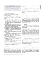
Vaccination against hepatitis.
womi_I 5/6/03 3:23 PM Page 289
Immunity, cell mediated
WORLD OF MICROBIOLOGY AND IMMUNOLOGY
290
•
•
horse in response to the inactivated tetanus toxin. This proce-
dure is typically done if someone has been exposed to a situa-
tion where the possibility of contracting tetanus exists. Rather
than rely on the individual’s immune system to respond to the
presence of the toxin, neutralizing antibodies are administered
right away.
Active and passive immunity are versions of what is
known as antibody-mediated immunity. That is, antibodies
bind to the antigen and this binding further stimulates the
immune system to respond to the antigen threat. Antibody-
mediated immunity is also called humoral immunity.
A third type of immunity, which is known as delayed
immunity or delayed-type hypersensitivity, is represents a dif-
ferent sort of immunity. Delayed immunity is a so-called cell-
mediated immunity. Here, immune components called T-cells
bind to the surface of other cells that contain the antigen on
their surface. This binding triggers a further response by the
immune system to the foreign antigen. The response can
involve components such as white blood cells.
An example of delayed immunity is the tuberculin test
(or the Mantoux test), which tests for the presence of
Mycobacterium tuberculosis, the bacterium that causes
tuber-
culosis
. A small amount of bacterial protein is injected into the
skin. If the individual is infected with the bacteria, or has ever
been infected, the injection site becomes inflamed within 24
hours. The response is delayed in time, relative to the imme-
diate response of antibody-based immunity. Hence, the name
of the immunity.
See also Antibody formation and kinetics; Immunization
IMMUNITY, CELL MEDIATED
Immunity, cell mediated
The immune system is a network of cells and organs that work
together to protect the body from infectious organisms. Many
different types of organisms such as
bacteria, viruses, fungi,
and
parasites are capable of entering the human body and
causing disease. It is the immune system’s job to recognize
these agents as foreign and destroy them.
The immune system can respond to the presence of a
foreign agent in one of two ways. It can either produce solu-
ble proteins called antibodies, which can bind to the foreign
agent and mark them for destruction by other cells. This type
of response is called a humoral response or an
antibody
response. Alternately, the immune system can mount a cell-
mediated immune response. This involves the production of
special cells that can react with the foreign agent. The reacting
cell can either destroy the foreign agents, or it can secrete
chemical signals that will activate other cells to destroy the
foreign agent.
During the 1960s, it was discovered that different types
of cells mediate the two major classes of immune responses.
The T lymphocytes, which are the main effectors of the cell-
mediated response, mature in the thymus, thus the name T cell.
The
B cells, which develop in the adult bone marrow, are
responsible for producing antibodies. There are several differ-
ent types of
T cells performing different functions. These
diverse responses of the different T cells are collectively
called the “cell-mediated immune responses.”
There are several steps involved in the cell-mediated
response. The pathogen (bacteria, virus, fungi, or a parasite),
or foreign agent, enters the body through the blood stream, dif-
ferent tissues, or the respiratory tract. Once inside the body,
the foreign agents are carried to the spleen, lymph nodes, or
the mucus-associated lymphoid tissue (MALT) where they
will come in contact with specialized cells known as antigen-
presenting cells (APC). When the foreign agent encounters the
antigen-presenting cells, an immune response is triggered.
These
antigen presenting cells digest the engulfed material,
and display it on their surface complexed with certain other
proteins known as the Major
Histocompatibility Class (MHC)
of proteins.
Next, the T cells must recognize the antigen.
Specialized receptors found on some T cells are capable of
recognizing the MHC-antigen complexes as foreign and bind-
ing to them. Each T cell has a different receptor in the cell
membrane that is capable of binding a specific antigen. Once
the T cell receptor binds to the antigen, it is stimulated to
divide and produce large amounts of identical cells that are
specific for that particular foreign antigen. The T lymphocytes
also secrete various chemicals (
cytokines) that can stimulate
this proliferation. The cytokines are also capable of amplify-
ing the immune defense functions that can eventually destroy
and remove the antigen.
In cell-mediated immunity, a subclass of the T cells
mature into cytotoxic T cells that can kill cells having the for-
eign antigen on their surface, such as virus-infected cells, bac-
terial-infected cells, and tumor cells. Another subclass of T cells
called helper T cells activates the B cells to produce antibodies
that can react with the original antigen. A third group of T cells
called the suppressor T cells is responsible for regulating the
immune response by turning it on only in response to an antigen
and turning it off once the antigen has been removed.
Some of the B and T lymphocytes become “memory
cells,” that are capable of remembering the original antigen. If
that same antigen enters the body again while the memory
cells are present, the response against it will be rapid and
heightened. This is the reason the body develops permanent
immunity to an infectious disease after being exposed to it.
This is also the principle behind
immunization.
See also Antibody and antigen; Antibody-antigen, biochemi-
cal and molecular reactions; Antibody formation and kinetics;
Antibody, monoclonal; Antigenic mimicry; Immune stimula-
tion, as a vaccine; Immune synapse; Immune system;
Immunity, active, passive and delayed; Immunity, humoral
regulation; Immunization; Immunochemistry
IMMUNITY, HUMORAL REGULATION
Immunity, humoral regulation
One way in which the immune system responds to pathogens
is by producing soluble proteins called antibodies. This is
known as the humoral response and involves the activation of
a special set of cells known as the
B lymphocytes, because
womi_I 5/6/03 3:23 PM Page 290
Immunization
WORLD OF MICROBIOLOGY AND IMMUNOLOGY
291
•
•
they originate in the bone marrow. The humoral immune
response helps in the control and removal of pathogens such
as
bacteria, viruses, fungi, and parasites before they enter host
cells. The antibodies produced by the
B cells are the mediators
of this response.
The antibodies form a family of plasma proteins
referred to as
immunoglobulins. They perform two major
functions. One function of an
antibody is to bind specifically
to the molecules of the foreign agent that triggered the
immune response. A second antibody function is to attract
other cells and molecules to destroy the pathogen after the
antibody molecule is bound to it.
When a foreign agent enters the body, it is engulfed by
the antigen-presenting cells, or the B cells. The B cell that has
a receptor (surface immunoglobulin) on its membrane that
corresponds to the shape of the
antigen binds to it and engulfs
it. Within the B cell, the antigen-antibody pair is partially
digested, bound to a special class of proteins called MHC-II,
and then displayed on the surface of the B cell. The helper
T
cells
recognize the pathogen bound to the MHC-II protein as
foreign and becomes activated.
These stimulated T cells then release certain chemicals
known as
cytokines (or lymphokines) that act upon the primed
B cells (B cells that have already seen the antigen). The B cells
are induced to proliferate and produce several identical cells
capable of producing the same antibody. The cytokines also
signal the B cells to mature into antibody producing cells. The
activated B cells first develop into lymphoblasts and then
become plasma cells, which are essentially antibody produc-
ing factories. A subclass of B cells does not differentiate into
plasma cells. Instead, they become memory cells that are
capable of producing antibodies at a low rate. These cells
remain in the immune system for a long time, so that the body
can respond quickly if it encounters the same antigen again.
The antibody destroys the pathogen in three different
ways. In neutralization, the antibodies bind to the bacteria or
toxin and prevent it from binding and gaining entry to a host
cell. Neutralization leads to a second process called
opsoniza-
tion
. Once the antibody is bound to the pathogen, certain other
cells called macrophages engulf these cells and destroy them.
This process is called
phagocytosis. Alternately, the
immunoglobulin IgM or IgG can bind to the surface of the
pathogen and activate a class of serum proteins called the
complement, which can cause lysis of the cells bearing that
particular antigen.
In the humoral immune response, each B cell produces
a distinct antibody molecule. There are over a million differ-
ent B lymphocytes in each individual, which are capable of
recognizing a corresponding million different antigens. Since
each antibody molecule is composed of two different proteins
(the light chain and the heavy chain), it can bind two different
antigens at the same time.
See also Antibody and antigen; Antibody-antigen, biochemi-
cal and molecular reactions; Antibody formation and kinetics;
Immune system; Immunity, active, passive and delayed;
Immunity, cell mediated
I
MMUNIZATION
Immunization
When a foreign disease-causing agent (pathogen) enters the
body, a protective system known as the
immune system comes
into play. This system consists of a complex network of organs
and cells that can recognize the pathogen and mount an
immune response against it.
Any substance capable of generating an immune
response is called an
antigen or an immunogen. Antigens are
not the foreign
bacteria or viruses themselves; they are sub-
stances such as toxins or
enzymes that are produced by the
microorganism. In a typical immune response, certain cells
known as the antigen-presenting cells trap the antigen and
present it to the immune cells (lymphocytes). The lympho-
cytes that have receptors specific for that antigen binds to it.
The process of binding to the antigen activates the lympho-
cytes and they secrete a variety of cytokines that promotes the
growth and maturation of other immune cells such as cyto-
toxic T lymphocytes. The cytokines also act on B cells stimu-
lating them to divide and transform into
antibody secreting
cells. The foreign agent is then either killed by the cytotoxic
T
cells
or neutralized by the antibodies.
The process of inducing an immune response is called
immunization. It may be either natural, i.e., acquired after
infection by a pathogen, or, the
immunity may be artificially
acquired with serum or vaccines.
In order to make vaccines for immunization, the organ-
ism, or the poisonous toxins of the microorganism that can
cause diseases, are weakened or killed. These vaccines are
injected into the body or are taken orally. The body reacts to
the presence of the vaccine (foreign agent) by making anti-
bodies. This is known as active immunity. The antibodies
accumulate and stay in the system for a very long time, some-
times for a lifetime. When antibodies from an actively immu-
nized individual are transferred to a second non-immune
subject, it is referred to as passive immunity. Active immunity
is longer lasting than passive immunity because the memory
cells remain in the body for an extended time period.
Immunizations are the most powerful and cost-effective
way to prevent infectious disease in children. Because they
have received antibodies from their mother’s blood, babies are
immune to many diseases when they are born. However, this
immunity wanes during the first year of life. Immunization
programs, therefore, are begun during the first year of life.
Each year in the United States, thousands of adults die
needlessly from vaccine-preventable diseases or their compli-
cations. Eight childhood diseases (
measles, mumps, rubella,
diphtheria, tetanus, pertussis, Hemophilus influenzae type b,
and polio) are preventable by immunization. With the excep-
tion of tetanus, all the other diseases are contagious and could
spread rapidly, resulting in
epidemics in an unvaccinated pop-
ulation. Hence, vaccinations are among the safest and most
cost-efficient public health measures. Vaccinations against flu
(
influenza), hepatitis A, and pneumococcal disease are also
recommended for some adolescents and adults. The vaccines
indicated for adults will vary depending on lifestyle factors,
occupation, chronic medical conditions and travel plans.
womi_I 5/6/03 3:23 PM Page 291
Immunochemistry
WORLD OF MICROBIOLOGY AND IMMUNOLOGY
292
•
•
See also Antibody and antigen; Antibody formation and kinet-
ics; Immunity, active, passive and delayed; Immunity, cell
mediated; Immunity, humoral regulation
IMMUNOCHEMISTRY
Immunochemistry
Immunochemistry is the study of the chemistry of immune
responses.
An immune response is a reaction caused by the inva-
sion of the body by an
antigen. An antigen is a foreign sub-
stance that enters the body and stimulates various defensive
responses. The cells mainly involved in this response are
macrophages and T and
B lymphocytes. A macrophage is a
large, modified white blood cell. Before an antigen can stimu-
late an immune response, it must first interact with a
macrophage. The macrophage engulfs the antigen and trans-
ports it to the surface of the lymphocytes. The macrophage (or
neutrophil) is attracted to the antigen by chemicals that the
antigen releases. The macrophage recognizes these chemicals
as alien to the host body. The local cells around the infection
will also release chemicals to attract the macrophages; this is
a process known as chemotaxis. These chemicals are a
response to the infection. This process of engulfing the foreign
body is called
phagocytosis, and it leads directly to painful
swelling and
inflammation of the infected area.
Lymphocytes are also cells that have been derived from
white blood cells (leucocytes). Lymphocytes are found in
lymph nodes, the spleen, the thymus, bone marrow, and circu-
lating in the blood plasma. Those lymphocytes that mature
inside mammalian bone marrow are called
B cells. Once B
cells have come into contact with an antigen, they proliferate
and differentiate into antibody secreting cells. An antibody is
any protein that is released in the body in direct response to
infection by an antigen. Those lymphocytes that are formed
inside the thymus are called T lymphocytes or
T cells. After
contact with an antigen, T cells secrete lymphokines—a group
of proteins that do not interact with the antigens themselves,
instead they stimulate the activity of other cells. Lymphokines
are able to gather uncommitted T cells to the site of infection.
They are also responsible for keeping T cells and macrophages
at the site of infection. Lymphokines also amplify the number
of activated T cells, stimulate the production of more lym-
phokines, and kill infected cells. There are several types of T
cells. These other types include T helper cells that help B cells
mature into antibody-secreting cells, T suppresser cells that
halt the action of B and T cells, T cytotoxic cells that attack
infected or abnormal cells, and T delayed hypersensitivity
cells that react to any problems caused by the initial infection
once it has disappeared. This latter group of cells are long
lived and will rapidly attack any remaining antigens that have
not been destroyed in the major first stages of infection.
Once the antibodies are released by the B and T cells,
they interact with the antigen to attempt to neutralize it. Some
antibodies act by causing the antigens to stick together; this is a
process known as agglutination. Antibodies may also cause the
antigens to fall apart, a process known as cell lysis. Lysis is
caused by
enzymes known as lytic enzymes that are secreted by
the antibodies. Once an antigen has been lysed, the remains of
the antigen are removed by phagocytosis. Some antigens are
still able to elicit a response even if only a small part of the anti-
gen remains intact. Sometimes the same antibody will cause
agglutination and then lysis. Some antibodies are antitoxins,
which directly neutralize any toxins secreted by the antigens.
There are several different forms of antibody that carry out this
process depending upon the type of toxin that is produced.
Once antibodies have been produced for a particular
antigen they tend to remain in the body. This provides
immu-
nity
. Sometimes immunity is long term and once exposed to a
disease we will never catch the disease again. At other times,
immunity may only be short lived. The process of active
immunity is when the body produces its own antibodies to
confer immunity. Active immunity occurs after an initial expo-
sure to the antigen. Passive immunity is where antibodies are
passed form mother to child through the placenta. This form of
immunity is short lived. Artificial immunity can be conferred
by the action of
immunization. With immunization, a vaccine
is injected into the body. The vaccine may be a small quantity
of antigen, it may be a related antigen that causes a less seri-
ous form of the disease, it may be a fragment of the antigen,
or it may be the whole antigen after it has been inactivated. If
a fragment of antigen is used as a vaccine, it must be sufficient
to elicit an appropriate response from the body. Quite often
viral coat proteins are used for this. The first vaccine was
developed by
Edward Jenner (1749–1823) in 1796 to inocu-
late against
smallpox. Jenner used the mild disease cowpox to
confer immunity for the potentially fatal but biochemically
similar smallpox.
Within the blood there are a group of blood serum pro-
teins called
complement. These proteins become activated by
antigen antibody reactions. Immunoglobulin is an antibody
secreted by lymphoid cells called plasma cells.
Immuno-
globulins
are made of two long polypeptide chains and two
short polypeptide chains. These chains are bound together in a
Y-shaped arrangement, with the short chains forming the inner
parts of the Y. Each arm of the Y has specific antigen binding
properties. There are five different classes of immunoglobulin
that are based on their antigen-binding properties. Different
classes of immunoglobulins come into play at different stages
of infection. Immunoglobulins have specific binding sites
with antigens.
One class of compounds in animals has antigens that
can be problematical. This is the group called the
histocom-
patibility
complex. This is the group of usually surface pro-
teins that are responsible for rejections and incompatibilities
in organ transplants. These antigens are genetically encoded
and they are present on the surface of cells. If the cells or tis-
sues are transferred from one organism to another or the body
does not recognize the antigens, it will elicit a response to try
to rid the body of the foreign tissue. A body is not interested
where foreign proteins come from. It is interested in the fact
that they are there when they should not be. Even if an organ
is human in origin, it must be genetically similar to the host
body or it will be rejected. Because an organ is much larger
than a small infection of an antigen when it elicits an immune
response, it can be a greater problem. With an organ trans-
womi_I 5/6/03 3:23 PM Page 292
Immunodeficiency
WORLD OF MICROBIOLOGY AND IMMUNOLOGY
293
•
•
plant, there can be a massive cascade reaction of antibody pro-
duction. This will include all of the immune responses of
which the body is capable. Such a massive response can over-
load the system and it can cause death. Thus, tissue matching
in organ transplants is vitally important. Often, a large range
of immunosuppressor drugs are employed until the body inte-
grates a particular organ. In some cases, this may necessitate a
course of drugs for the rest of the individuals life.
Histocompatibility problems also exist with blood.
Fortunately, the proteins in blood are less specific and blood
transfusions are a lot easier to perform than organ transplants.
The blood-typing systems that are in use are indications of the
proteins that are present. If blood is mixed from the wrong
types, it can cause lethal clotting. The main blood types are A,
B, O, and AB. Group O individuals are universal donors, they
can give blood to anyone. Group AB are universal recipients
because they can accept blood from anyone. Type A blood has
A antigens on the blood cells and B antibodies in the plasma.
The combination of B antibodies and B antigens will cause
agglutination. There are also subsidiary blood proteins such as
the rhesus factor (rh) that can be positive (present) or negative
(absent). If only small amounts of blood are transfused, it is
not a problem due to the dilution factor.
Immunochemistry is the chemistry of the
immune sys-
tem
. Most of the chemicals involved in immune responses are
proteins. Some chemicals inactivate invading proteins, others
facilitate this response. The histocompatibility complex is a
series of surface proteins on organs and tissues that elicit an
immune response when placed in a genetically different indi-
vidual.
See also Biochemistry; History of immunology; Immune
stimulation, as a vaccine; Immunity, active, passive and
delayed; Immunity, cell mediated; Immunity, humoral regula-
tion; Immunization; Immunological analysis techniques;
Laboratory techniques in immunology; Major histocompati-
bility complex (MHC)
IMMUNODEFICIENCY
Immunodeficiency
The immune system is the body’s main system to fight infec-
tions. Any defect in the immune system decreases a person’s
ability to fight infections. A person with an immunodeficiency
disorder may get more frequent infections, heal more slowly,
and have a higher incidence of some cancers.
The normal immune system involves a complex inter-
action of certain types of cells that can recognize and attack
“foreign” invaders, such as
bacteria, viruses, and fungi. It also
plays a role in fighting cancer. The immune system has both
innate and adaptive components. Innate
immunity is made up
of immune protections present at birth. Adaptive immunity
develops the immune system to fight off specific invading
organisms throughout life. Adaptive immunity is divided into
two components: humoral immunity and cellular immunity.
The innate immune system is made up of the skin
(which acts as a barrier to prevent organisms from entering the
body), white blood cells called phagocytes, a system of pro-
teins called the complement system, and chemicals called
interferons. When phagocytes encounter an invading organ-
ism, they surround and engulf it to destroy it. The complement
system also attacks bacteria. The elements in the complement
system create a hole in the outer layer of the target cell, which
leads to the death of the cell.
The adaptive component of the immune system is
extremely complex, and is still not entirely understood.
Basically, it has the ability to recognize an organism or tumor
cell as not being a normal part of the body, and to develop a
response to attempt to eliminate it.
The humoral response of adaptive immunity involves a
type of cell called
B lymphocytes. B lymphocytes manufacture
proteins called antibodies (which are also called
immunoglob-
ulins
). Antibodies attach themselves to the invading foreign
substance. This allows the phagocytes to begin engulfing and
destroying the organism. The action of antibodies also acti-
vates the complement system. The humoral response is partic-
ularly useful for attacking bacteria.
The cellular response of adaptive immunity is useful for
attacking viruses, some
parasites, and possibly cancer cells.
The main type of cell in the cellular response is T lympho-
cytes. There are helper T lymphocytes and killer T lympho-
cytes. The helper T lymphocytes play a role in recognizing
invading organisms, and they also help killer T lymphocytes to
multiply. As the name suggests, killer T lymphocytes act to
destroy the target organism.
Defects can occur in any component of the immune sys-
tem or in more than one component (combined immunodefi-
ciency). Different
immunodeficiency diseases involve
different components of the immune system. The defects can
be inherited and/or present at birth (congenital), or acquired.
Congenital immunodeficiency is present at the time of
birth, and is the result of genetic defects. Even though more
than 70 different types of congenital immunodeficiency disor-
ders have been identified, they rarely occur. Congenital
immunodeficiencies may occur as a result of defects in B lym-
phocytes, T lymphocytes, or both. They can also occur in the
innate immune system.
If there is an abnormality in either the development or
function of B lymphocytes, the ability to make antibodies will
be impaired. This allows the body to be susceptible to recur-
rent infections. Bruton’s agammaglobulinemia, also known as
X-linked agammaglobulinemia, is one of the most common
congenital immunodeficiency disorders. The defect results in
a decrease or absence of B lymphocytes, and therefore a
decreased ability to make antibodies. People with this disorder
are particularly susceptible to infections of the throat, skin,
middle ear, and lungs. It is seen only in males because it is
caused by a genetic defect on the X chromosome. Since males
have only one X chromosome, they always have the defect if
the
gene is present. Females can have the defective gene, but
since they have two X
chromosomes, there will be a normal
gene on the other X chromosome to counter it. Women may
pass the defective gene on to their male children.
Another type of B lymphocyte deficiency involves a
group of disorders called selective immunoglobulin deficiency
syndromes. Immunoglobulin is another name for
antibody,
womi_I 5/6/03 3:23 PM Page 293
Immunodeficiency
WORLD OF MICROBIOLOGY AND IMMUNOLOGY
294
•
•
and there are five different types of immunoglobulins (called
IgA, IgG, IgM, IgD, and IgE). The most common type of
immunoglobulin deficiency is selective IgA deficiency. The
amounts of the other antibody types are normal. Some patients
with selective IgA deficiency experience no symptoms, while
others have occasional lung infections and diarrhea. In another
immunoglobulin disorder, IgG and IgA antibodies are defi-
cient and there is increased IgM. People with this disorder
tend to get severe bacterial infections.
Common variable immunodeficiency is another type of
B lymphocyte deficiency. In this disorder, the production of
one or more of the immunoglobulin types is decreased and the
antibody response to infections is impaired. It generally devel-
ops around the age of 10-20. The symptoms vary among
affected people. Most people with this disorder have frequent
infections, and some will also experience anemia and rheuma-
toid arthritis. Many people with common variable immunode-
ficiency develop cancer.
Severe defects in the ability of T lymphocytes to mature
results in impaired immune responses to infections with
viruses, fungi, and certain types of bacteria. These infections
are usually severe and can be fatal.
DiGeorge syndrome is a T lymphocyte deficiency that
starts during fetal development, but it isn’t inherited. Children
with DiGeorge syndrome either do not have a thymus or have
an underdeveloped thymus. Since the thymus is a major organ
that directs the production of
T-lymphocytes, these patients have
very low numbers of T-lymphocytes. They are susceptible to
recurrent infections, and usually have physical abnormalities as
well. For example, they may have low-set ears, a small reced-
ing jawbone, and wide-spaced eyes. In some cases, no treatment
is required for DiGeorge syndrome because T lymphocyte pro-
duction improves. Either an underdeveloped thymus begins to
produce more T lymphocytes or organ sites other than the thy-
mus compensate by producing more T lymphocytes.
Some types of immunodeficiency disorders affect both
B lymphocytes and T lymphocytes. For example,
severe com-
bined immunodeficiency
disease (SCID) is caused by the
defective development or function of these two types of lym-
phocytes. It results in impaired humoral and cellular immune
responses. SCID is usually recognized during the first year of
life. It tends to cause a fungal infection of the mouth (
thrush),
diarrhea, failure to thrive, and serious infections. If not treated
with a bone marrow transplant, a person with SCID will gen-
erally die from infections before age two.
Disorders of innate immunity affect phagocytes or the
complement system. These disorders also result in recurrent
infections.
Acquired immunodeficiency is more common than
congenital immunodeficiency. It is the result of an infectious
process or other disease. For example, the
Human Immu-
nodeficiency Virus
(HIV) is the virus that causes acquired
immunodeficiency syndrome (
AIDS). However, this is not the
most common cause of acquired immunodeficiency. Acquired
immunodeficiency often occurs as a complication of other
conditions and diseases. For example, the most common
causes of acquired immunodeficiency are malnutrition, some
types of cancer, and infections. People who weigh less than
70% of the average weight of persons of the same age and
gender are considered to be malnourished. Examples of types
of infections that can lead to immunodeficiency are chicken-
pox, cytomegalovirus, German
measles, measles, tuberculo-
sis
, infectious mononucleosis (Epstein-Barr virus), chronic
hepatitis, lupus, and bacterial and fungal infections.
Sometimes, acquired immunodeficiency is brought on
by drugs used to treat another condition. For example, patients
who have an organ transplant are given drugs to suppress the
immune system so the body will not reject the organ. Also,
some
chemotherapy drugs, which are given to treat cancer,
have the side effect of killing cells of the immune system.
During the period of time that these drugs are being taken, the
risk of infection increases. It usually returns to normal after
the person stops taking the drugs.
Congenital immunodeficiency is caused by genetic
defects, and they generally occur while the fetus is developing
in the womb. These defects affect the development and/or
function of one or more of the components of the immune sys-
tem. Acquired immunodeficiency is the result of a disease
process, and it occurs later in life. The causes, as described
above, can be diseases, infections, or the side effects of drugs
given to treat other conditions.
People with an immunodeficiency disorder tend to
become infected by organisms that don’t usually cause disease
in healthy persons. The major symptoms of most immunode-
ficiency disorders are repeated infections that heal slowly.
These chronic infections cause symptoms that persist for long
periods of time.
Laboratory tests are used to determine the exact nature
of the immunodeficiency. Most tests are performed on blood
samples. Blood contains antibodies, lymphocytes, phagocytes,
and complement components—all of the major immune com-
ponents that might cause immunodeficiency. A blood cell
count will determine if the number of phagocytic cells or lym-
phocytes is below normal. Lower than normal counts of either
of these two cell types correlates with immunodeficiencies.
The blood cells are also checked for their appearance.
Sometimes a person may have normal cell counts, but the cells
are structurally defective. If the lymphocyte cell count is low,
further testing is usually done to determine whether any par-
ticular type of lymphocyte is lower than normal. A lymphocyte
proliferation test is done to determine if the lymphocytes can
respond to stimuli. The failure to respond to stimulants corre-
lates with immunodeficiency. Antibody levels can be meas-
ured by a process called
electrophoresis. Complement levels
can be determined by immunodiagnostic tests.
There is no cure for immunodeficiency disorders.
Therapy is aimed at controlling infections and, for some dis-
orders, replacing defective or absent components.
In most cases, immunodeficiency caused by malnutri-
tion is reversible. The health of the immune system is directly
linked to the nutritional health of the patient. Among the
essential nutrients required by the immune system are pro-
teins, vitamins, iron, and zinc. For people being treated for
cancer, periodic relief from chemotherapy drugs can restore
the function of the immune system.
womi_I 5/6/03 3:23 PM Page 294
Immunodeficiency disease syndromes
WORLD OF MICROBIOLOGY AND IMMUNOLOGY
295
•
•
In general, people with immunodeficiency disorders
should maintain a healthy diet. This is because malnutrition
can aggravate immunodeficiencies. They should also avoid
being near people who have colds or are sick because they can
easily acquire new infections. For the same reason, they
should practice good personal
hygiene, especially dental care.
People with immunodeficiency disorders should also avoid
eating undercooked food because it might contain bacteria that
could cause infection. This food would not cause infection in
normal persons, but in someone with an immunodeficiency,
food is a potential source of infectious organisms. People with
immunodeficiency should be given
antibiotics at the first indi-
cation of an infection.
There is no way to prevent a congenital immunodefi-
ciency disorder. However, someone with a congenital immun-
odeficiency disorder might want to consider getting genetic
counseling before having children to find out if there is a
chance they will pass the defect on to their children.
Some of the infections associated with acquired immun-
odeficiency can be prevented or treated before they cause
problems. For example, there are effective treatments for
tuberculosis and most bacterial and fungal infections. HIV
infection can be prevented by practicing “safe sex” and not
using illegal intravenous drugs. These are the primary routes
of transmitting the virus. For people who don’t know the HIV
status of the person with whom they are having sex, safe sex
involves using a condom.
See also AIDS, recent advances in research and treatment;
Immunity, active, passive and delayed; Immunity, cell medi-
ated; Immunity, humoral regulation; Immunodeficiency dis-
ease syndromes; Immunodeficiency diseases; Infection and
resistance
IMMUNODEFICIENCY DISEASE
SYNDROMES
Immunodeficiency disease syndromes
An effective immune system requires that any antigens that are
not native to the body be quickly recognized and destroyed,
and that none of the antigens native to the body be identified as
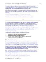
Scanning electron microscope image of the Human Immunodeficiency Virus (HIV) on a hemocyte.
womi_I 5/6/03 3:23 PM Page 295
Immunodeficiency diseases, genetic causes
WORLD OF MICROBIOLOGY AND IMMUNOLOGY
296
•
•
foreign. Excesses in the latter constitute the autoimmune dis-
eases. Deficiencies in the body’s ability to recognize antigens
as foreign or a diminished capacity to respond to recognized
antigens constitute the
immunodeficiency syndromes.
There are many causes associated with immunodefi-
ciencies. Primary immunodeficiencies are inherited conditions
in which specific genes or
gene families are corrupted by
mutations or chromosome deletions. These syndromes are dis-
cussed elsewhere in this volume. Secondary immunodeficien-
cies are acquired conditions that may result from infections,
cancers, aging, exposure to drugs, chemicals or radiation, or a
variety of other disease processes.
Bacteria, viral, fungi, protozoa, and even parasitic infec-
tions can result in specific deficiencies of
B cells, T cells,
macrophages, and granulocytes. The best characterized of the
infectious diseases is the acquired immunodeficiency syn-
drome (
AIDS).
Infection by two
viruses, HIV-1 and HIV-2, is associ-
ated with a wide range of responses in different people from
essentially asymptomatic to a full-blown AIDS in which cell-
mediated
immunity is seriously compromised. HIV-1 and HIV-
2 are
retroviruses that attack humans and compromise cellular
function. In contrast, the human T cell lymphotrophic viruses
(
HTLV) tend to provoke lymphoid neoplasms and neurologic
disease. AIDS is most often associated with HIV-1 infection.
The chance of developing AIDS following infection with HIV-
1 is approximately one to two percent per year initially, and
increases to around five percent per year after the fifth year of
infection. Roughly, half of those infected with the virus will
develop AIDS within ten years. In between those who are
asymptomatic, and those with AIDS who are symptomatic
with conditions associated with AIDS.
In AIDS, cellular immunity mechanisms are disrupted.
Some immunologic cells are reduced in number and others,
such as natural killer cells, have reduced activity despite their
normal numbers.
HIV infects primarily T helper lymphocyte
cells and a variety of cells outside of the lymphoid system
such as macrophages, endothelial, and epithelial cells.
Because the T helper cells normally express a surface glyco-
protein called CD4, counts of CD4 cells are helpful in pre-
dicting immunologic depression in HIV-infected individuals.
The amount of viral
RNA in circulation is also a helpful pre-
dictor of immunologic compromise. In addition to cell-medi-
ated immunity,
antibody responses (humoral immunity) are
also muted in individuals with AIDS.
Initially, there is a period of several weeks to months
where the host remains HIV antibody negative and viral repli-
cation occurs rapidly. Some subjects develop an acute response
that appears like the flu or
mononucleosis. Symptoms typically
include fever, malaise, joint pain, and swollen lymph nodes. As
the initial symptoms dissipate, patients enter an antibody posi-
tive phase without symptoms associated with AIDS. A variety
of relatively mild symptoms like
thrush, diarrhea, fever, or
other viral infections may manifest along with a wide array of
partial anemias. Nerve function can become compromised
resulting in weakness, pain, or sensory loss. Eventually, life
threatening opportunistic infections resulting from decreased
immunologic function occur and may be accompanied by
wasting, dementia, meningitis, and encephalitis. Drug therapy
in the form of antiretroviral agents is directed toward inhibition
of proteases and reverse transcriptase
enzymes which are crit-
ical for replication of the viruses.
Although not nearly as well known as AIDS, there are a
variety of other acquired immunodeficiencies. Infections other
than HIV can significantly alter the numbers and functions of
other cells within the immune system. While individually
these various infections may appear to be relatively uncom-
mon, depression in the numbers of platelets, T cells, B cells,
natural killer (NK) cells, and granulocytes can lead to
immunologic dysfunction. The manifestations of these various
conditions will depend on the specific cell population that is
involved and its normal function within the immune system. B
cell deficiencies tend to result in an increased susceptibility to
bacterial infections. Decreased natural killer cell activity can
result in the survival of tumor cells which would otherwise be
destroyed by the immune system.
Chemical and physical agents (such as radiation) also
can potentially depress various fractions of cells within the
immune system, and like the immunodeficiencies caused by
infectious agents, the manifestations of these agents will differ
depending on the cells which are influenced. Cancer
chemotherapeutic agents are often immunosuppressive.
Likewise, immune function often declines with age. T cell
populations (including the T helper cells) decline as the thy-
mus gland activity decreases. Frequently, B cell populations
proliferate at an accelerated rate in older people. Over produc-
tion of cells within the immune system such as leukemias,
lymphomas, and related disorders also may disturb immune
function by radically altering the distribution of white cells. A
number of other diverse disease processes can alter or com-
promise immune function. These include diabetes, liver dis-
ease, kidney disease, sickle cell anemia, Down syndrome, and
many of the autoimmune diseases.
See also AIDS, recent advances in research and treatment;
Autoimmunity and autoimmune diseases; Immunodeficiency
diseases, genetic causes
IMMUNODEFICIENCY DISEASES, GENETIC
CAUSES
Immunodeficiency diseases, genetic causes
The complex workings of the immune system requires the
cooperation of various organs, tissues, cells and proteins and
thus, it can be compromised in a number of different ways.
People who have normal immune function at birth who later
acquire some form of
immunodeficiency are said to have sec-
ondary or acquired immunodeficiency diseases. Examples
would include
AIDS, age-related immune depression, and
other immune deficiencies caused by infections, drug reac-
tions, radiation sickness, or cancer. Individuals who are born
with an intrinsically reduced capacity for immunologic
activity usually have some genetic alteration present at birth.
There are varieties of different genes involved, and they ren-
der people susceptible to infection by an assortment of dif-
womi_I 5/6/03 3:23 PM Page 296
Immunodeficiency diseases, genetic causes
WORLD OF MICROBIOLOGY AND IMMUNOLOGY
297
•
•
ferent germs. Some of these diseases are relatively mild with
onset in adolescence or adulthood. Others are severely debil-
itating and severely compromise daily activity. Clinically
significant primary immunodeficiencies are relatively rare
with 1 in 5,000 to 1 in 10,000 people in developed countries
afflicted.
The most common form of primary immunodeficiency,
selective IgA deficiency, is a very mild deficiency and may
affect as many as 1 in every 300 persons, most of whom will
never realize they have an immunodeficiency at all. B-cells
are lymphocytes that produce antibodies and this component
of the immune system is often called humoral
immunity.
Defects in humoral immunity predispose the body to viral
infections. T-cells are lymphocytes that are processed in the
thymus gland. Granulocytes are cells which consume an
destroy
bacteria.
There are now thought to be around 70 different primary
immunodeficiency diseases. Of the more common forms, the
vast majority of these conditions are recessive. This means that
a single working copy of the
gene is generally sufficient to per-
mit normal immune functioning. Some of the genes are found
on the X chromosome. Since males receive only a single X
chromosome, recessive
mutations of these genes will result in
disease. Females have two copies of the X chromosome, and so
rarely will express X-linked recessive diseases.
The most widely known of the primary immunodefi-
ciencies is severe combined immune deficiency (
SCID) and it
conjures pictures of a child who must live his life encased in
a plastic bubble to keep out germs. SCID is manifest in early
childhood as a severe combined T cell and B cell deficiency,
and can be caused by a number of different gene mutations.
The most common form is X-linked, and so primarily affects
boys. It can also be caused by an enzyme called adenosine
deaminase. When ADA is deficient, toxic chemicals kill off
the lymphocytes. Until recently, SCID was uniformly lethal.
In recent years, the elucidation of the genes responsible has
made possible interventions based on gene therapy. SCID
often presents in early childhood as persistent diaper rash or
thrush. Pneumonia, meningitis, blood poisoning, and many
common viral infections are serious threats to children born
with SCID. Diagnosis demands immediate medical attention
and bone marrow transplants are a common form of treatment
for SCID. Children with ADA deficiency may be treated with
ADA infusions to correct the enzyme deficiency. Partial com-
bined immune deficiencies are milder conditions in which
cellular and humoral immunity are both compromised but not
completely shut down. These are generally accompanied by
other physical symptoms and so constitute syndromes.
Wiskott-Aldrich syndrome, for example, is an X-linked par-
tial combined syndrome in which the repeated infections are
combined with eczema and a tendency toward bleeding.
Another combined B and T cell deficiency is ataxia telang-
iectasia (AT). In AT, the combined B and T cell deficiency
causes repeated respiratory infections, and is accompanied by
a jerky movement disorder and dilated blood vessels in the
eyes and skin. The thymus gland where T-cells are processed
is underdeveloped.
Deficiency of the B cell population results in decreased
antibody production and thus, an increased risk of viral or bac-
terial infection
. X-linked agammaglobulinemia (XLA) is a
condition in which boys (because it is X-linked) produce little
to no antibodies due to an absence of
B cells and plasma cells
in circulation. As these children grow, they deplete the anti-
bodies transmitted through the mother, and they become
susceptible to repeated infections. Common variable immun-
odeficiency (CVID) is a group of disorders in which the num-
ber of B cells is normal, but the levels of antibody production
are reduced.
DiGeorge anomaly is an example of a T cell deficiency
produced by an underdeveloped thymus gland. Children with
DiGeorge anomaly often have characteristic facial features,
developmental delays, and certain kinds of heart defects usu-
ally stemming from small deletions on chromosome 22 (or
more rarely, chromosome 10). In rare cases, there is an auto-
somal dominant gene mutation rather than a chromosome
deletion.
Phagocytosis, the ability of the granulocytes to ingest
and destroy bacteria, can also be the chief problem. One
example of this is chronic granulomatous disease (CGD).
There are four known genes that cause CGD; all are reces-
sive. One is on the X chromosome, and the other three are on
autosomes. These children do well until around age three
when they begin to have problems with staphylococcal infec-
tions and infections with
fungi which are generally benign in
other people. Their granulosa cells may aggregate in tissues
forming tumor like masses. Similarly, leukocyte adhesion
defect (LAD) is a condition in which granulocytes fail to
work because they are unable to migrate to the site of infec-
tions. In Chediak-Higashi syndrome (CHS), not only granu-
locytes, but also melanocytes and platelets are diminished.
CHS is generally fatal in adolescence unless treated by bone
marrow transplantation.
One other class of primary immunodeficiencies, the
complement system defects, result from the body’s inability to
recognize and/or destroy germs that have been bound by anti-
bodies. Complement fixation is a complex multi step process,
and thus a number of different gene mutations can potentially
corrupt the normal pathway. Complement system defects are
rare and often not expressed until later in life.
The prospect of the development of effective and safe
gene therapies holds hope for the primary immunodeficiency
diseases. As these genes and their genetic pathways are more
fully understood, interventions which replace the missing gene
product will likely provide effective treatments.
See also Immunity, cell mediated; Immunity, humoral regula-
tion; Microbial genetics; Microbiology, clinical
IMMUNOELECTRON MICROSCOPY,
THEORY, TECHNIQUES AND USES
• see
E
LECTRON MICROSCOPIC EXAMINATION OF MICROORGANISMS
womi_I 5/6/03 3:23 PM Page 297
Immunoelectrophoresis
WORLD OF MICROBIOLOGY AND IMMUNOLOGY
298
•
•
I
MMUNOELECTROPHORESIS
Immunoelectrophoresis
Immunoelectrophoresis is a technique that separates proteins
on the basis of both their net charge (and so their movement in
an electric field) and on the response of the
immune system to
the proteins. The technique is widely used in both clinical and
research laboratories as a diagnostic tool to probe the protein
composition of serum.
Petr Nikolaevich Grabar, a French immunologist,
devised the technique in the 1950s. In essence, immunoelec-
trophoresis separates the various proteins in a sample in an
electric field and then probes the separated proteins using the
desired
antiserum.
The most widely used version of the technique employs
an apparatus, which consists basically of a
microscope slide-
sized plate. The plate is the support for a gel that is poured
over top and allowed to congeal. The construction of the gel
can vary, depending on the separation to be performed.
Agar,
such as that used in microbiological growth media, and
another material called agarose can be used. Another popular
choice is a linked network of a chemical known as acrylamide.
The linked up acrylamide chains form what is designated as
polyacrylamide.
The different types of gel networks can be most pro-
ductively envisioned as a three-dimensional overlay of the
crossed linked chains. The effect is to produce snaking tunnels
through the matrix of various diameters. These diameters,
which are also referred to as pore sizes, can be changed to a
certain extent by varying the concentrations of some of the
ingredients of the gel suspension. Depending on the size and
the shape of the protein, movement through this matrix will be
relatively slow or fast. As well, depending on the net charge a
protein molecule has, the protein will migrate towards the pos-
itively charged electrode or the negatively charged electrode
when the electric current is passed through the gel matrix.
Thus, the various species of protein will separate from each
other along the length of the gel.
In some configurations of the immunoelectrophoretic
set-up, the samples that contain the proteins to be analyzed are
added to holes on either side of the gel plate. For example, one
sample could contain serum from a health individual and
another sample could contain serum from someone with an
infection. The middle portion of the plate contains a trough,
into which a single purified species of
antibody or known mix-
ture of antibodies is added. The antibody molecules diffuse
outward from the trough solution into the gel. Where an anti-
body encounters a corresponding
antigen, a reaction causes
the formation of a visual precipitate. Typically, the precipita-
tion occurs in arc around the antigen-containing sample. In the
example, the pattern of precipitation can reveal antigenic dif-
ferences between the normal serum and the serum from a
infected person.
This type of immunoelectrophoresis provides a qualita-
tive (“yes or no”) answer with respect to the presence or
absence of proteins, and can be semi-quantitative. The shape
of the arc of precipitation is also important. An irregularly
shaped arc can be indicative of an abnormal protein or the
presence of more than one antigenically similar protein.
Immunoelectrophoresis can also be used to detect a par-
ticular antigenic site following the transfer of the proteins
from a gel to a special support, such as nitrocellulose. Addition
of the antibody followed by a chemical to which bound anti-
body reacts produces a darkening on the support wherever
antibody has bound to antigen. One version of this technique
is termed Western Blotting. An advantage of this technique is
that, by running two gels and using just one gel for the trans-
fer of proteins to the nitrocellulose, the immune detection of a
protein can be performed without affecting the protein resid-
ing in the other gel.
Another application of immunoelectrophoresis is
known as capillary immunoelectrophoresis. In this applica-
tion, a sample can be simultaneously drawn up into many cap-
illary tubes. The very small diameter of the tubes means that
little sample is required to fill a tube. Thus, a sample can be
subdivided into very many sub volumes. Each volume can be
tested against a different antibody preparation. Often, the reac-
tion between antigen and antibody can be followed by the use
of compounds that fluoresces when exposed to laser light of a
specific wavelength. Capillary immunoelectrophoresis is
proving to be useful in the study of Bovine Spongiform
Encephalopathy in cattle, where sample sizes can be very
small.
In the clinical laboratory setting, immunoelectophoresis
is used to examine alterations in the content of serum, espe-
cially changes concerned with
immunoglobulins. Change in
the immunoglobulin profile can be the result of immunodefi-
ciencies, chronic bacterial or viral infections, and infections of
a fetus. The immunoglobulin most commonly assayed for are
IgM, IgG, and IgA. Some of the fluids that can be examined
using immunoelectrophoresis include urine, cerebrospinal
fluid and serum. When concerned with immunoglobulins, the
technique can also be called gamma globulin
electrophoresis
or immunoglobulin electrophoresis.
See also Antibody-antigen, biochemical and molecular reac-
tions; Immunological analysis techniques
IMMUNOFLUORESCENCE
Immunofluorescence
Immunofluorescence refers to the combination of an antibody
and a compound that will fluoresce when illuminated by light
of a specific wavelength. The duo is also referred to as a fluo-
rescently labeled antibody. Such an antibody can be used to
visually determine the location of a target
antigen in biologi-
cal samples, typically by microscopic observation.
The fluorescent compound that is attached to an anti-
body is able to absorb light of a certain wavelength, the par-
ticular wavelength being dependent on the molecular
construction of the compound. The absorption of the light con-
fers additional energy to the compound. The energy must be
relieved. This is accomplished by the emission of light, at a
higher wavelength (and so a different color) than the absorbed
radiation. It is this release of radiant energy that is the under-
pinning for immunofluorescence.
womi_I 5/6/03 3:23 PM Page 298
Immumogenetics
WORLD OF MICROBIOLOGY AND IMMUNOLOGY
299
•
•
Immunofluorescence microscopy can revel much detail
about the processes inside cells. In a light microscopic applica-
tion of the technique, sections of sample are exposed to the flu-
orescently labeled antibody. The large wavelength of visible
light, relative to other forms of illumination such as laser light,
does not allow details to be revealed at the molecular level.
Still, details of the trafficking of a protein from the site of its
manufacture to the surface of a cell, for example, is possible,
by the application of different antibodies. The antibodies can
be labeled with the same fluorescent compound but are applied
at different times. An example of the power of this type of
approach is the information that has been obtained concerning
the pathway that the
yeast known as Saccharomyces cervisiae
uses to shuttle proteins out of the cell.
Resolution of details to the molecular level has been
made possible during the 1990s with the advent of the tech-
nique of confocal laser microscopy. This technique employs a
laser to sequentially scan samples at selected depths through
the sample. These so-called optical sections can be obtained
using laser illumination at several different wavelengths
simultaneously. Thus, the presence of different antibodies that
are labeled with fluorescent compounds that fluoresce at the
different wavelengths can produce an image of the location of
two antigens in the same sample at the same time.
The use of immunofluorescent compounds in combina-
tion with confocal microscopy has allowed the fluorescent
probing of samples which do not need to be chemically pre-
served (or “fixed”) prior to examination. The thin sections of
sample that are examined in light microscopy often require
such chemical fixation. While the fixation regimens have been
designed to avoid change of the sample’s internal structure,
especially the chemistry and three-dimensional structure of
the site of the antigen to which the antibody will bind, the
avoidance of any form of chemical modification is preferred.
There are a multitude of fluorescent compounds avail-
able. Collectively these compounds are referred to as fluo-
rochromes. A well-known example in biological and
microbiological studies is the green fluorescent protein. This
molecule is ring-like in structure. It fluoresces green when
exposed to light in the ultraviolet or blue wavelengths. Other
compounds such as fluorescein, rhodamine, phycoerythrin,
and Texas Red, fluoresce at different wavelength and can pro-
duce different colors.
Immunofluorescence can be accomplished in a one-step
or two-step reaction. In the first option, the fluorescently
labeled antibody directly binds to the target antigen molecule.
In the second option the target antigen molecule binds a so-
called secondary antibody. Then, other antigenic sites in the
sample that might also bind the fluorescent antibody are
“blocked” by the addition of a molecule that more globally
binds to antigenic sites. The secondary antibody then can itself
be the target to which the fluorescently labeled antibody binds.
The use of antibodies to antigen that are critical to dis-
ease processes in
microorganisms allow immunofluorescence
to act as a detection and screening tool in the monitoring of a
variety of materials. Foe example, research to adapt immuno-
fluorescence to food monitoring is an active field. In the pres-
ent, immunofluorescence provides the means by which
organisms can be sorted using the technique of flow cytome-
try. As individual
bacteria, for example, pass by a detector, the
presence of fluorescence will register and cause the bacterium
to be shuttled to a special collection reservoir. Thus, bacteria
with a certain surface factor can be separated from the other
bacteria in the population that do not possess the factor
See also Fluorescent dyes; Microscopy
IMMUNOGENETICS
Immumogenetics
Immunogenetics is the study of the mechanisms of autoim-
mune diseases, tolerance in organ transplantation, and
immu-
nity to infectious diseases—with a special emphasis on the
role of the genetic make-up of an organism in these processes.
The
immune system evolved essentially to protect vertebrates
from a myriad species of potentially harmful infectious agents
such as
bacteria, virus, fungi and various eukaryotic parasites.
However, the growing understanding of the immune system
has influenced a variety of different biomedical disciplines,
and is playing an increasingly important role in the study and
treatment of many human diseases such as cancer and autoim-
mune conditions.
There are two broad types of immune systems. The
innate immune system of defense depends on invariant recep-
tors that recognize common features of pathogens, but are not
varied enough to recognize all types of pathogens, or specific
enough to act effectively against re-infection by the same
pathogen. Although effective, this system lacks both speci-
ficity and the ability to acquire better receptors to deal with the
same infectious challenge in the future, a phenomenon called
immunological memory. These two properties, specificity and
memory, are the main characteristics of the second type of
immune system, known as the specific or adaptive immune
system, which is based on
antigen specific receptors. Besides
these two families of different receptors that help in immune
recognition of foreign infectious agents, both the innate and
the adaptive immune systems rely on soluble mediators like
the different
cytokines and kemokines that allow the different
cells involved in an immune response to communicate with
each other. The major focus of immunogeneticists is the iden-
tification, characterization, and sequencing of genes coding
for the multiple receptors and mediators of immune responses.
Historically, the launch of immunogenetics could be
traced back to the demonstration of Mendelian inheritance of
the human ABO blood groups in 1910. The importance of this
group of molecules is still highlighted by their important in
blood transfusion and organ transplantation protocols. Major
developments that contributed to the emergence of immuno-
genetics as an independent discipline in
immunology were the
rediscovery of allograft reactions during the Second World
War and the formulation of an immunological theory of allo-
graft reaction as well as the formulation of the clonal
selection
hypothesis by Burnett in 1959. This theory proposed that
clones of immunocompetent cells with unique receptors exist
prior to exposure to antigens, and only cells with specific
receptors are selected by antigen for subsequent activation.
womi_I 5/6/03 3:23 PM Page 299
Immunoglobulins and immunoglobulin deficiency syndromes
WORLD OF MICROBIOLOGY AND IMMUNOLOGY
300
•
•
The molecular understanding of how the diverse repertoire of
these receptors is generated came with the discovery of
somatic
recombination of receptor genes, which is the para-
digm for studying
gene rearrangement during cell maturation.
The most important influence on the development of
immunogenetics is, however, the studies of a gene family
known as the MCH, or
major histocompatibility complex.
These highly polymorphic genes, first studied as white-cell
antigens of the blood and therefore named human leukocyte
antigens (
HLA), influence both donor choice in organ trans-
plantation and the susceptibility of an organism to chronic dis-
eases. The
MHC is also linked with most of all the important
autoimmune diseases such as rheumatoid arthritis and diabetes.
The discovery in 1972 that these MHC molecules are
intimately associated with the specific immune response to
viruses led to an explosion in immunogenetic studies of these
molecules. This has led to the construction of very detailed
genetic and physical maps of this complex and ultimately to its
complete sequence in an early stage of the human genome-
sequencing project.
Other clusters of immune recognition molecules that are
well established at the center of the immunogenetics discipline
are the large arrays of rearranging gene segments that deter-
mine B-cell
immunoglobulins and T-cell receptors.
Immunoglobulins, which mediate the humoral immune
response of the adaptive immune system, are the antibodies
that circulate in the bloodstream and diffuse in other body flu-
ids, where they bind specifically to the foreign antigen that
induced them. This interaction with the antigen most often
leads to its clearance. T cell receptors, which are involved in
the cell-mediated immune response of the adaptive immune
system, are the principle partners of the MHC molecules in
mounting a specific immune response. An antigen that is taken
up by specialized cells called antigen presenting cells is usu-
ally presented on the surface of this cell in complex with either
MHC class I or class II to
T cells that use specific receptors to
recognize and react to the infectious agent. The reacting T
cells can kill the host cells that bear the foreign antigen or
secrete mediators (cytokines and lynphokines) that activate
professional phagocytic cells of the immune system that elim-
inate the antigen. It is believed that during disease
epidemics,
some forms of class I and class II MHC molecules stimulate
T-cell responses that better favor survival. Which MHC mole-
cule is more favorable depends on the infectious agents
encountered. Consequently, human populations that were geo-
graphically separated and have different disease histories dif-
fer in the sequences and frequencies of the HLA class I and
class II alleles.
Other immune recognition molecules that were studied
in great details in immunogenetics are two families of genes
that encode receptors on the surface for natural killer (NK)
cells. These large lymphocytes participate in the innate
immune system and provide early defense from a pathogens
attack, a response that distinguish them from B and T cells
which become useful after days of infection. Some NK-cell
receptors bind polymorphic determinants of MHC class I mol-
ecules and appear to be modulated by the effects that infectious
agents have upon the conformation of these determinants.
One of the most important applications of immuno-
genetics in clinical medicine is HLA-typing in order to help
match organ donors and recipients during transplantation sur-
gery. Transplantation is a procedure in which an organ or tis-
sue that is damaged and is no longer functioning is replaced
with one obtained from another person. Because HLA anti-
gens can be recognized as foreign by another person’s immune
system, surgeons and physicians try to match as many of the
HLA antigens as possible, between the donated organ and the
recipient. In order to do this, the HLA type of every potential
organ recipient is determined. When a potential organ donor
becomes available, the donor’s HLA type is determined as
well to make absolutely sure that the donor organ is suitable
for the recipient.
See also Autoimmunity and autoimmune diseases; Immunity,
active, passive and delayed; Immunity, cell mediated;
Immunity, humoral regulation; Immunologic therapies;
Immunosuppressant drugs; In vitro and in vivo research;
Laboratory techniques in immunology; Major histocompati-
bility complex (MHC); Medical training and careers in
immunology; Molecular biology and molecular genetics;
Mutations and mutagenesis; Oncogenetic research;
Transplantation genetics and immunology; Viral genetics
IMMUNOGLOBULIN DEFICIENCY
• see
I
MMUNODEFICIENCY DISEASE SYNDROMES
IMMUNOGLOBULINS AND IMMUNOGLOBU-
LIN DEFICIENCY SYNDROMES
Immunoglobulins and immunoglobulin deficiency syndromes
Immunoglobulins are proteins that are also called antibodies.
The five different classes of immunoglobulins are formed in
response to the presence of antigens. The specificity of an
immunoglobulin for a particular
antibody is exquisitely precise
The five classes of immunoglobulins are designated
IgA, IgD, IgG, IgE, and IgM. These share a common structure.
Two so-called heavy chains form a letter “Y” shape, with two
light chains linked to each of the upper arms of the Y. The
heavy chains are also known as alpha, delta, gamma, epsilon,
or mu. The light chains are termed lambda or kappa.
The IgG class of immunoglobulin is the most common.
IgG antibody is routinely produced in response to bacterial and
viral infections and to the presence of toxins. IgG is found in
many tissues and in the plasma that circulates throughout the
body. IgM is the first antibody that is produced in an immune
response. IgA is also produced early in a body’s immune
response, and is commonly found in saliva, tears, and other
such secretions. The activity of IgD is still not clear. Finally, the
IgE immunoglobulin is found in respiratory secretions.
The different classes of immunoglobulins additionally
display differences in the sequence of amino acids comprising
certain regions within the immunoglobulin molecule. For
example, differences in the antigen-binding region, the variable
region, accounts for the different
antigen binding specificities
womi_I 5/6/03 3:23 PM Page 300
Immunologic therapies
WORLD OF MICROBIOLOGY AND IMMUNOLOGY
301
•
•
of the various immunoglobulins. Differences in their structure
outside of the antigen binding region, in an area known as the
constant region, accounts for differences in the immunoglobu-
lin in other functions. These other functions are termed effec-
tor functions, and include features such as the recognition and
binding to regions on other cells, and the stimulation of activ-
ity of an immune molecule known as
complement.
The vast diversity of immunoglobulin specificity is due
to the tremendous number of variations that are possible in the
variable region of an immunoglobulin. A certain immune cell
known as a B cell produces each particular immunoglobulin.
Thus, at any particular moment in time, there are a myriad of
B cells actively producing a myriad of different immunoglob-
ulins, in response to antigenic exposure.
Immunoglobulins can exist in two forms. They can be
fixed to the surface of the B cells that have produced them. Or
they can float freely in body fluids, essentially patrolling until a
recognizable antigen is encountered. The protection of the body
from invading antigens depends on the production of
immunoglobulins of the required type and in sufficient quantity.
Conditions where an individual has a reduced number
of immunoglobulins or none at all of a certain type is known
as an immunoglobulin deficiency syndrome. Such syndromes
are typically the result of damage to B cells.
People with immunoglobulin deficiencies are prone to
more frequent illness than those people whose immune sys-
tems are fully functional. Often the illnesses are caused by
bacteria, in particular bacteria that are able to form a capsule
surrounding them. A capsule is not easily recognized by even
an optimally performing
immune system. As well,
immunoglobulin deficiency can render a person more suscep-
tible to some viral infections, in particular those caused by
echovirus, enterovirus, and
hepatitis B.
Immunoglobulin deficiencies can take the form of a pri-
mary disorder or a secondary disorder. A secondary deficiency
results from some other ongoing malady or treatment. For
example,
chemotherapy for a cancerous illness can compro-
mise the immune system, leading to an
immunodeficiency.
Once the treatment is stopped the immunodeficiency can be
reversed. A primary immunodeficiency is not the result of an
illness or medical treatment. Rather, it is the direct result of a
genetic disorder or a defect to B cells or other immune cells.
X-linked agammaglobulinemia results in an inability of
B cells to mature. This results in the production of fewer B
cells and in a lack of “memory” of an infection. Normally, the
immune system is able to rapidly respond to antigen that has
been encountered before, because of the “memory” of the B
cells. Without this ability, repeated infections caused by the
same agent can result.
Another genetically based immunoglobulin deficiency
is known as selective IgA deficiency. Here, B cells fail to
switch from producing IgM to produce IgA. The limited
amount of IgA makes someone more prone to infections of
mucosal cells. Examples of such infections include those in
the nose, throat, lungs, and intestine.
Genetic abnormalities cause several other immunodefi-
ciency syndromes. A missing stretch of information in the
gene that codes for the heavy chain of IgG results in the pro-
duction of an IgG that is structurally incomplete. The result is
a loss of function of the IgG class of antibodies, as well as the
IgA and IgE classes. On a subtler level, another genetic mal-
function affects the four subclasses of antibodies within the
IgG class. The function of some of the subclasses are affected
more so than other subclasses. Finally, another genetic muta-
tion destroys the ability of B cells to switch from making IgM
to manufacture IgG. The lack of flexibility in the antibody
capability of the immune system adversely affects the ability
of the body to successfully fight infections.
Transient hypogammaglobulinemia is an immunodefi-
ciency syndrome that is not based on a genetic aberration.
Rather, the syndrome occurs in infants and is of short-term in
duration. The
T cells of the immune system do not function
properly. Fewer than normal antibodies are produced, and
those that are made are poor in their recognition of the anti-
genic target. However, as the immune system matures with
age the proper function of the T cells is established. The cause
of the hypogammaglobulinemia is not known.
Immunoglobulin deficiency syndromes are curable only
by a bone marrow transplant, an option exercised in life-
threatening situations. Normally, treatment rather than cure is
the option. Prevention of infection, through the regular use of
antimicrobial drugs and scrupulous oral health are important
to maintain health in individuals with immunoglobulin defi-
ciency syndromes.
See also Immunochemistry; Immunodeficiency disease syn-
dromes; Immunodeficiency diseases; Immunodeficiency;
Immunogenetics; Immunologic therapies; Immunological
analysis techniques; Immunology; Immunosuppressant drugs
IMMUNOLOGIC THERAPIES
Immunologic therapies
Immunologic therapy is defined as the use of medicines that
act to enhance the body’s immune response as a means of
treating disease. The drugs can also aid in the recovery of the
body from the harmful effects of immune-compromising treat-
ments like
chemotherapy and radiation.
Both microorganism-related infections and other mal-
adies that are due to immune deficiency or cell growth defects
are targets of immunologic therapy.
The emphasis in immunologic therapy is the application
of synthetic compounds that mimic immune substances that
are naturally produced in the body. For example, a compound
called aldesleukin is an artificial form of interkeukin-2, a nat-
ural compound that assists white blood cells in recognizing
and dealing with foreign material. Other examples are filgras-
tim and sargramostim, which are synthetic version of
colony
stimulating factors, which stimulate bone marrow to make the
white blood cells, and epoetin, an artificial version of erythro-
poietin, which stimulates the marrow to produce red blood
cells. Thrombopoietin encourages the manufacture of
platelets, which are plate-shaped components of the blood that
are vital in the clotting of blood. As a final example, synthetic
forms of interferon are available and can be administered to
womi_I 5/6/03 3:23 PM Page 301
Immunological analysis techniques
WORLD OF MICROBIOLOGY AND IMMUNOLOGY
302
•
•
aid the natural forms of interferon in battling infections and
even cancer.
Research has provided evidence that the infusion of spe-
cific
enzymes can produce positive results with respect to
some neurological disorders. While not strictly an immuno-
logic therapy, the supplementation of the body’s natural com-
ponents is consistent with the aim of the immune approach.
The use of immunologic therapy is not without risk.
Paradoxically, given their longer-term enhancement of the
immune defenses, some of the administered drugs reduce the
body’s ability to fight off infection because of a short-term
damping-down of some aspects of the
immune system. As
well, certain therapies carry a risk of reduced clotting of the
blood and of seizures.
As with other therapies, the use of immunologic therapies
is assessed in terms of the risks of the therapy versus the health
outcome if therapy is not used. Typically, the immediate health
threat to a patient outweighs the possible side effects from ther-
apy. Immunologic therapies are always administered under a
physician’s care, almost always in a hospital setting. As well,
frequent monitoring of the patients is done, both for the abate-
ment of the malady and the development of adverse effects.
Immunologic therapy can provide continued treatment
following chemotherapy or the use of radiation. The latter two
treatments cannot be carried on indefinitely, due to toxic reac-
tions in the body. Immunologic therapy provides another
avenue of treatment. For example, some tumors that are resist-
ant to chemical therapy are susceptible to immune attack. By
enhancing the immune response, such tumors may be produc-
tively treated. Moreover, despite their side effects, immuno-
logic therapies usually are less toxic than either chemotherapy
or the use of radiation.
See also Immune system; Laboratory techniques in
immunology
IMMUNOLOGICAL ANALYSIS TECHNIQUES
Immunological analysis techniques
Immunological techniques are the wide varieties of methods
and specialized experimental protocols devised by immunolo-
gists for inducing, measuring, and characterizing immune
responses. They allow the immunologists to alter the
immune
system
through cellular, molecular and genetic manipulation.
These techniques are not restricted to the field of
immunology,
but are widely applied by basic scientists in many other bio-
logical disciplines and by clinicians in human and veterinary
medicine.
Most immunological techniques available are focused
on the study of the adaptive immune system. They classically
involve the experimental induction of an immune response
using methods based on
vaccination protocols. During a typi-
cal experiment called
immunization, immunologists inject a
test
antigen to an animal or human subject and monitor for the
appearance of immune responses in the form of specific anti-
bodies and effector
T cells. Monitoring the antibody response
usually involves the analysis of crude preparations of serum
from the immunized subject. The analysis of the immune
responses mediated by T cells are usually performed only on
experimental animals and involves the preparation of these
cells from blood or from the lymphoid organs, such as the
spleen and the lymph nodes. Typically, any substance that has
a distinctive structure or conformation that may be recognized
by the immune system can serve as an antigen. A wide range
of substances from simple chemicals like sugars, and small
peptides to complex macromolecules and
viruses can induce
the immune system. Although the antigenic determinant of a
test substance is usually a minor part of that substance called
the epitope, a small antigen referred to as a hapten can rarely
elicit an immune response on its own. It is not an immunogen
and would therefore need to be covalently linked to a carrier
in order to elicit an immune response. The induction of such a
response to even large immunogenic antigen is not easy to
achieve and the dose, the form and route of administration of
that antigen can profoundly affect whether a response can
occur. Especially the use of certain substances called adju-
vants is necessary to alert the immune system and produce a
strong immune response.
According to the clonal
selection theory, antibodies pro-
duced in a typical immunization experiment are products of
different clones of B-lymphocytes that are already committed
to making antibodies to the corresponding antigen. These
polyclonal antibodies are multi-subunit proteins that belong to
the
immunoglobulins family. They have a basic Y-shaped
structure with two identical Fab domains, which form the arms
and interact with the antigen, and one Fc domain that forms
the stem and determines the isotype subclass of each antibody.
There are five different isotype subclasses, IgM, Ig G, IgA,
IgE, and IgD, which show different tissue distribution and
half-life in vivo. They determine the biological function of the
antibodies and appear during different stages of the immu-
nization process. Knowledge about the biosynthesis and struc-
ture of these antibodies is important for their detection and use
both as diagnostic and therapeutic tools.
Antibodies are highly specific for their corresponding
antigen, and are able to detect one molecule of a protein anti-
gen out of around a billion similar molecules. The amount and
specificity of an antibody in a test serum can be measured by
its direct binding to the antigen in assays usually referred to as
primary interaction immunoassays. Commonly used direct
assays are radioimmunoassay (RIA), enzyme-linked
immunosorbent assay (
ELISA), and immunoblotting tech-
niques. In both ELISA and RIA, an enzyme or a radioisotope
is covalently linked to the pure antigen or antibody. The unla-
beled component, which most often is the antigen, is attached
to the surface of a plastic well. The labeled antibody is allowed
to bind to the unlabeled antigen. The plastic well is subse-
quently washed with plenty of
buffer that will remove any
excess non-bound antibody and prevent non-specific binding.
Antibody binding is measured as the amount of radioactivity
retained by the coated wells in radioimmunoassay or as fluo-
rescence emitted by the product of an enzymatic reaction in
the case of ELISA. Modifications of these assays known as
competitive inhibition assays can be used that will allow quan-
tifying the antigen (or antibody) in a mixture and determining
the affinity of the antibody-antigen interaction by using math-
womi_I 5/6/03 3:23 PM Page 302
Immunology
WORLD OF MICROBIOLOGY AND IMMUNOLOGY
303
•
•
ematical models. Immunoblotting is usually performed in the
form of Western blotting, which is reserved to the detection of
proteins and involves an
electrophoresis separation step fol-
lowed by electroblotting of the separated proteins from the gel
to a membrane and then probing with an antibody. Detection
of the antigen protein antibody interaction is made in a similar
way as in RIA or ELISA depending on whether a radiolabeled
or enzyme-coupled antibody is used.
Antibodies can also be monitored through immunoas-
says that are based on the ability of antibodies to alter the
physical state of their corresponding antigens and typically
involve the creation of a precipitate in a solid or liquid
medium. The hemmaglutination assay used to determine the
ABO type of blood groups and match compatible donors and
recipients for blood transfusion is based on this assay.
Currently, the most common application of this immunoassay
is in a procedure known as immunoprecipitation. This method
allows antibodies to form complexes with their antigen in a
complex mixture like the cytosol, the
nucleus or membrane
complexes of the cell. The antigen-antibody complex is pre-
cipitated either by inducing the formation of even larger com-
plexes through the addition of excess amounts of
anti-immunoglobulin antibodies or by the addition of agarose
beads coupled to a special class of bacterial proteins that bind
the Fc region of the antibody. The complex can also be pre-
cipitated by covalently linking the antibody to agarose beads
forming a special affinity matrix. This procedure will also
allow the purification of the antigen by immunoaffinity, a spe-
cial form of affinity chromatography. Immunoprecipitation is
a valuable technique that led to major discoveries in immunol-
ogy an all disciplines of molecular and cellular biology. It
allows the precipitation of the antigen in complex with other
interacting proteins and reagents and therefore gives an idea
on the function of the antigen.
The T cell immune response is detected by using mon-
oclonal antibodies, a specific family of antibodies that recog-
nize surface markers that are expressed by lymphocytes upon
their activation. These monoclonal antibodies are highly spe-
cific, and are produced by special techniques from single
clones of
B cells and are therefore, homogenous groups of
immunoglobulins with the same isotype and antigen binding
affinity. These antibodies are used to identify characterize
cells by flow cytometry (FACS), immunocytochemistry,
immunofluorescence techniques. The difficulty to isolate anti-
gen specific T cells is due to the fact that these T cells recog-
nize the antigen in the context of a tri-molecular complex
involving the T cell receptor and the
MHC molecules on the
surface of specialized cells called antigen-presenting cells.
These interactions are subtle, have low affinity and are
extremely complex to study. Novel and powerful techniques
using tetramers of MHC molecules were developed in 1997
that are now used to identify and isolate antigen specific T cell
clones. These tetramer-based assays are proving useful in sep-
arating very rare cells, and could be used in clinical medicine.
In fact, virus and tumor specific T cells usually give a stronger
response and are usually more effective in killing virus
infected and tumor cells. Testing for the function of activated,
antigen specific T cells known as effector T cells is routinely
done in vitro by testing for cytokine production, cytotoxicity
to other cells and proliferation in response to antigen stimula-
tion. Local reactions in the skin of animals and humans pro-
vide information about T cell responses to an antigen, a
procedure that is very used in testing for allergic reactions and
the efficacy of vaccination procedures. Experimental manipu-
lations of the immune system in vivo are performed to reveal
the functions of each component of the immune system in
vivo.
Mutations through irradiation, or mutations produced by
gene targeting (e.g., knock-out and knock-in techniques), as
well as animal models produced by transgenic breeding, are
proving helpful to researchers in evaluating this highly com-
plex system.
See also Immune complex test; Immune stimulation, as a vac-
cine; Immune synapse; Immunity, active, passive and delayed;
Immunity, cell mediated; Immunity, humoral regulation;
Immunization; Immunochemistry; Immunodeficiency;
Immunoelectrophoresis; Immunofluorescence; Immunogene-
tics; Immunologic therapies; Immunology; Immunomodu-
lation; Immunosuppressant drugs; In vitro and in vivo
research; Laboratory techniques in immunology
IMMUNOLOGICAL ASPECTS OF REPRO-
DUCTION
• see R
EPRODUCTIVE IMMUNOLOGY
IMMUNOLOGY
Immunology
Immunology is the study of how the body responds to foreign
substances and fights off infection and other disease.
Immunologists study the molecules, cells, and organs of the
human body that participate in this response.
The beginnings of our understanding of
immunity date
to 1798, when the English physician
Edward Jenner
(1749–1823) published a report that people could be protected
from deadly
smallpox by sticking them with a needle dipped
in the material from a
cowpox boil. The French biologist and
chemist
Louis Pasteur (1822–1895) theorized that such immu-
nization
protects people against disease by exposing them to a
version of a microbe that is harmless but is enough like the
disease-causing organism, or pathogen, that the
immune sys-
tem
learns to fight it. Modern vaccines against diseases such
as
measles, polio, and chicken pox are based on this principle.
In the late nineteenth century, a scientific debate was
waged between the German physician
Paul Ehrlich
(1854–1915) and the Russian zoologist Élie Metchnikoff
(1845–1916). Ehrlich and his followers believed that proteins
in the blood, called antibodies, eliminated pathogens by stick-
ing to them; this phenomenon became known as humoral
immunity. Metchnikoff and his students, on the other hand,
noted that certain white blood cells could engulf and digest
foreign materials: this cellular immunity, they claimed, was
the real way the body fought infection.
Modern immunologists have shown that both the
humoral and cellular responses play a role in fighting disease.
womi_I 5/6/03 3:23 PM Page 303
Immunology
WORLD OF MICROBIOLOGY AND IMMUNOLOGY
304
•
•
They have also identified many of the actors and processes
that form the immune response.
The immune response recognizes and responds to
pathogens via a network of cells that communicate with each
other about what they have “seen” and whether it “belongs.”
These cells patrol throughout the body for infection, carried by
both the blood stream and the lymph ducts, a series of vessels
carrying a clear fluid rich in immune cells.
The
antigen presenting cells are the first line of the
body’s defense, the scouts of the immune army. They engulf
foreign material or
microorganisms and digest them, display-
ing bits and pieces of the invaders—called antigens—for other
immune cells to identify. These other immune cells, called T
lymphocytes, can then begin the immune response that attacks
the pathogen.
The body’s other cells can also present antigens,
although in a slightly different way. Cells always display anti-
gens from their everyday proteins on their surface. When a
cell is infected with a virus, or when it becomes cancerous, it
will often make unusual proteins whose antigens can then be
identified by any of a variety of cytotoxic T lymphocytes.
These “killer cells” then destroy the infected or cancerous cell
to protect the rest of the body. Other T lymphocytes generate
chemical or other signals that encourage multiplication of
other infection-fighting cells. Various types of T lymphocytes
are a central part of the cellular immune response; they are
also involved in the humoral response, encouraging
B lym-
phocytes
to turn into antibody-producing plasma cells.
The body cannot know in advance what a pathogen will
look like and how to fight it, so it creates millions and millions
of different lymphocytes that recognize random antigens.
When, by chance, a B or T lymphocyte recognizes an antigen
being displayed by an antigen presenting cell, the lymphocyte
divides and produces many offspring that can also identify and
attack this antigen. The way the immune system expands cells
that by chance can attack an invading microbe is called clonal
selection.
Some researchers believe that while some B and T lym-
phocytes recognize a pathogen and begin to mature and fight
an infection, others stick around in the bloodstream for months
or even years in a primed condition. Such memory cells may
be the basis for the immunity noted by the ancient Chinese and
by Thucydides. Other immunologists believe instead that trace
amounts of a pathogen persist in the body, and their continued
presence keeps the immune response strong over time.
Substances foreign to the body, such as disease-causing
bacteria, viruses, and other infectious agents (known as anti-
gens), are recognized by the body’s immune system as
invaders. The body’s natural defenses against these infectious
agents are antibodies—proteins that seek out the antigens and
help destroy them. Antibodies have two very useful charac-
teristics. First, they are extremely specific; that is, each
anti-
body
binds to and attacks one particular antigen. Second,
some antibodies, once activated by the occurrence of a dis-
ease, continue to confer resistance against that disease; clas-
sic examples are the antibodies to the childhood diseases
chickenpox and measles.
The second characteristic of antibodies makes it possi-
ble to develop vaccines. A
vaccine is a preparation of killed or
weakened bacteria or viruses that, when introduced into the
body, stimulates the production of antibodies against the anti-
gens it contains.
It is the first trait of antibodies, their specificity, that
makes monoclonal antibody technology so valuable. Not only
can antibodies be used therapeutically, to protect against dis-
ease; they can also help to diagnose a wide variety of illnesses,
and can detect the presence of drugs, viral and bacterial prod-
ucts, and other unusual or abnormal substances in the blood.
Given such a diversity of uses for these disease-fighting
substances, their production in pure quantities has long been
the focus of scientific investigation. The conventional method
was to inject a laboratory animal with an antigen and then,
after antibodies had been formed, collect those antibodies
from the blood serum (antibody-containing blood serum is
called
antiserum). There are two problems with this method:
It yields antiserum that contains undesired substances, and it
provides a very small amount of usable antibody.
Monoclonal antibody technology allows the production
of large amounts of pure antibodies in the following way. Cells
that produce antibodies naturally are obtained along with a
class of cells that can grow continually in cell
culture. The
hybrid resulting from combining cells with the characteristic
of “immortality” and those with the ability to produce the
desired substance, creates, in effect, a factory to produce anti-
bodies that work around the clock.
A myeloma is a tumor of the bone marrow that can be
adapted to grow permanently in cell culture. Fusing myeloma
cells with antibody-producing mammalian spleen cells, results
in hybrid cells, or hybridomas, producing large amounts of
monoclonal antibodies. This product of cell fusion combined
the desired qualities of the two different types of cells, the
ability to grow continually, and the ability to produce large
amounts of pure antibody. Because selected hybrid cells pro-
duce only one specific antibody, they are more pure than the
polyclonal antibodies produced by conventional techniques.
They are potentially more effective than conventional drugs in
fighting disease, because drugs attack not only the foreign
substance but also the body’s own cells as well, sometimes
producing undesirable side effects such as nausea and allergic
reactions. Monoclonal antibodies attack the target molecule
and only the target molecule, with no or greatly diminished
side effects.
While researchers have made great gains in understand-
ing immunity, many big questions remain. Future research
will need to identify how the immune response is coordinated.
Other researchers are studying the immune systems of non-
mammals, trying to learn how our immune response evolved.
Insects, for instance, lack antibodies, and are protected only by
cellular immunity and chemical defenses not known to be
present in higher organisms.
Immunologists do not yet know the details behind
allergy, where antigens like those from pollen, poison ivy, or
certain kinds of food make the body start an uncomfortable,
unnecessary, and occasionally life-threatening immune
response. Likewise, no one knows exactly why the immune
womi_I 5/6/03 3:23 PM Page 304
Immunomodulation
WORLD OF MICROBIOLOGY AND IMMUNOLOGY
305
•
•
system can suddenly attack the body’s tissues—as in autoim-
mune diseases like rheumatoid arthritis, juvenile diabetes, sys-
temic lupus erythematosus, or multiple sclerosis.
The hunt continues for new vaccines, especially against
parasitic organisms like the
malaria microbe that trick the
immune system by changing their antigens. Some researchers
are seeking ways to start an immune response that prevents or
kills cancers. A big goal of immunologists is the search for a
vaccine for
HIV, the virus that causes AIDS. HIV knocks out
the immune system—causing immunodeficiency—by infect-
ing crucial T lymphocytes. Some immunologists have sug-
gested that the chiefly humoral response raised by
conventional vaccines may be unable to stop HIV from getting
to lymphocytes, and that a new kind of vaccine that encour-
ages a cellular response may be more effective.
Researchers have shown that transplant rejection is just
another kind of immune response, with the immune system
attacking antigens in the transplanted organ that are different
from its own. Drugs that suppress the immune system are
now used to prevent rejection, but they also make the patient
vulnerable to infection. Immunologists are using their
increased understanding of the immune system to develop
more subtle ways of deceiving the immune system into
accepting transplants.
See also AIDS, recent advances in research and treatment;
Antibody, monoclonal; Biochemical analysis techniques;
BSE, scrapie and CJD: recent advances in research; History of
immunology; Immunochemistry; Immunodeficiency disease
syndromes; Immunodeficiency diseases; Immunodeficiency;
Immunogenetics; Immunological analysis techniques;
Immunology, nutritional aspects; Immunosuppressant drugs;
Infection and resistance; Laboratory techniques in immunol-
ogy; Reproductive immunology; Transplantation genetics and
immunology
IMMUNOLOGY, HISTORY OF
• see HISTORY OF
IMMUNOLOGY
IMMUNOLOGY, NUTRITIONAL ASPECTS
Immunology, nutritional aspects
The role of nutrition is central to the development and modu-
lation of the
immune system. The importance of nutrition has
been made clear by the burgeoning field of sports medicine. It
appears the immune system is enhanced by moderate to severe
exercise, although many components of the immune response
exhibit adverse change for a period of from 3 to 72 hours after
prolonged intense exertion. This “window of opportunity” for
opportunistic bacterial and viral infections seems to be
increased for “elite” athletes that are more prone to over-train.
The elements of the immune response most affected by the
strenuous activity that leads to the impairment of the immune
system are lymphocyte concentrations, depressed natural
killer activity, and elevated levels of IgA in the saliva.
The possible basis for this prolonged immunosuppres-
sion may include reduced plasma glutamine concentrations,
altered plasma glucose levels, and proliferation of neutrophils
and monocytes that increases prostaglandin concentrations.
Exercise produces oxidative stress and so concomitantly, there
are elevated free radical levels along with an attendant deple-
tion of antioxidant levels. Therefore, antioxidants that help
protect against oxidative stress are considered the most prom-
ising for further study, but those nutrients that heal the gut
show potential also. These nutrients include Vitamin E,
Vitamin C, zinc, and glutamine. Glutamine and nucleotides
show a direct effect on lymphocyte proliferation. Free radicals
and other reactive oxygen species that can damage cells as
well as tissues are an integral part of the immune system, so
the body has developed systems that protect from their dam-
age. These products function by destroying invading organ-
isms and damaged tissues, as well as enhance interleukin-I,
Interleukin-8 and tumor necrosis factor concentrations as part
of the inflammatory response. The purpose of supplementing
the diet is to provide a balance to the immune system’s pro-
oxidant function. Carbohydrate supplementation has addition-
ally shown impressive results. Increased plasma levels, a
depressed cortisol and growth hormone response, fewer fluc-
tuations in blood levels of immune competent cells, decreased
granulocyte and monocyte
phagocytosis, reduced oxidative
stress and a diminished pro-inflammatory and anti-inflamma-
tory cytokine response are all associated with an increase in
complex carbohydrate consumption.
Besides exercise-associated immune suppression, mal-
nutrition plays a pivotal role in modulating the immune
response. Nowhere is this more important then during preg-
nancy and gestation. Besides genetics, no other factor is more
important for the developing immune system then optimal
nutrition. The immune response of low-birth-weight babies is
compromised as well as those of children born to mothers
without adequate nutrition. Especially important is the role of
Vitamin E and selenium in preventing immune impairment.
Animal studies showed that progeny of Vitamin E and sele-
nium-deficient mothers never adequately developed immune
competent cell lines.
Because nutrition plays such a vital role in the immune
response, a special branch of
immunology is developing called
immunonutrition. These scientists are particularly interested in
the interaction of genetics and nutrition. Preliminary work
suggests that individual genotypes vary in their response to
healing, infection, and dietary supplementation.
See also Immunogenetics; Infection and resistance;
Metabolism
IMMUNOMODULATION
Immunomodulation
From a therapeutic point of view, immunomodulation refers to
any process in which an immune response is altered to a
desired level.
Microorganisms are also capable of modulating
the response of the
immune system to their presence, in order
womi_I 5/6/03 3:23 PM Page 305
Immunosuppressant drugs
WORLD OF MICROBIOLOGY AND IMMUNOLOGY
306
•
•
to establish or consolidate an infection. Thus, immunomodu-
lation can be beneficial or detrimental to a host.
Many providers of nutritional supplements claim that a
product enhances certain aspects of the immune system so as
to more vigorously shield the body from infection or the
development of maladies such as cancer. However, rigorous
testing of these claims is typically lacking and so the claims of
nutritional links with immune system improvement are at
present tenuous.
A firmer link exists between exercise and immunomod-
ulation. Moderately active people are known to have
macrophages that are more capable of killing tumors, due to
the increased production of a compound called nitric oxide.
This population also displays lower incidence rates for cancer
and other chronic diseases. Even sporadic exercise increases
the ability of an immune system component called natural
killer cells to eradicate tumors.
Conversely, too much exercise is associated with
increased susceptibility to respiratory tract infections, indicat-
ing that the immune system is impaired in the ability to thwart
infections.
Immunomodulation by microorganisms is directed at
several aspects of the immune system. One target are the small
molecules known as
cytokines, which function as messengers
of the immune system. In other words, cytokines stimulate
various immune responses such as
inflammation of the manu-
facture of antibodies. Other cytokines are involved in down-
regulating the immune responses.
Some microorganisms are able to produce and excrete
proteins that mimic the structure and function of cytokines.
Often the result is a suppression of the host’s inflammatory
response. Examples of microbes that produce cytokine-like
molecules are the
Epstein-Barr virus, poxvirus, vaccinia virus.
Other microbes, such as the protozoan Trypanosoma
cruzi blocks the activation of cytokines by an as yet unknown
mechanism. The result is a severe suppression of the immune
system.
Adenoviruses also block cytokine expression, at the
level of
transcription.
The manipulation of cytokine expression and action
may also be exploited to produce vaccines. For example, vac-
cines designed to nullify or enhance the activity of certain
cytokines could cause greater activity of certain components
of the immune system. While vaccines have yet to achieve this
level of activity, specific experimental targeting of
deoxyri-
bonucleic acid
has suppressed certain cytokines.
Another portion of the immune system capable of
immunomodulation is
complement. Herpes simplex virus types
1 and 2, the
viruses responsible for cold sores and genital her-
pes in humans, resist the action of complement. The presence of
specific viral proteins are required, and may act by disrupting a
key enzyme necessary for complement manufacture.
Vaccinia virus can also evade complement action, via a
protein that structurally resembles a host protein to which
complement binds. Also, another viral protein, called the
inflammation modulatory protein, acts to decrease the inflam-
matory response at the site of infection, thus preserving host
tissue from damage and providing the virus particles with rel-
atively undamaged cells in which to grow.
The protozoan Trypanosoma cruzi can regulate the
activity of complement before infecting human cells. Once
inside the cells of the host, the parasite can evade an immune
response.
A variety of
bacteria, viruses, and parasites are also
able to modulate the immune system by affecting the way anti-
gens are exposed on their surfaces.
Antigen presentation is a
complex series of steps. By controlling or modulating even
one of these steps, the antigen presentation process can be dis-
rupted. The formation of
antibody is thus affected.
Aside from biological agents, physiological factors can
cause immunomodulation. For example, stress is known to be
capable of suppressing various aspects of the cellular immune
response. The release of various hormones may disrupt in the
normal expression of cytokines. Specifically, those cytokines
that suppress inflammation are more evident, either because of
their increased production or the decreased production of
cytokines that activate inflammation.
See also Immunologic therapies
IMMUNOPRECIPITATION
• see ANTIBODY-ANTIGEN,
BIOCHEMICAL AND MOLECULAR REACTIONS
IMMUNOSUPPRESSANT DRUGS
Immunosuppressant drugs
Immunosuppressant drugs are medications that reduce the
ability of the
immune system to recognize and respond to the
presence of foreign material. Such drugs were developed and
still have an important use as a means of ensuring that trans-
planted organs and tissues are not rejected by the recipient.
Rejection of transplanted organs or tissue is a natural
reaction of a person’s immune system. In a very real sense, the
transplanted material is foreign and is treated, as would be an
infectious microorganism. The immune system attacks and
tries to destroy the foreign matter. Suppressing the immune
system allows the transplanted material to be retained.
Drugs to suppress the immune system are available only
with a physician’s authorization. Some commonly prescribed
drugs are azathioprine, cyclosporine, prednisolone, and
tacrolimus. These can be taken orally, both in solid and liquid
forms, or can be injected.
The main target of such immunosuppressant drugs are
the white blood cells (which are also called lymphocytes). The
main function of lymphocytes is to patrol the body and root
out foreign material. Then these cells, in combination with
other immune system components, destroy the foreign mate-
rial.
Transplantation of animal kidneys into humans was
tried in the early 1900s, and human-to-human transplant
attempts were first made in 1933. These attempts were unsuc-
cessful. It was not until the years of World War II that the
immunological basis for these failures was deciphered. Then,
Peter Medawar observed that a skin graft survived about a
week before being rejected, but a subsequent graft was
womi_I 5/6/03 3:23 PM Page 306
In vitro and in vivo research
WORLD OF MICROBIOLOGY AND IMMUNOLOGY
307
•
•
rejected much more quickly. This led him to propose that an
immunological response was at play in the rejection of trans-
planted material. This led to the first successful transplant in
1954, when the kidney of one identical twin was transplanted
to the other twin. In the twins, the absence of genetic differ-
ences in their tissues would eliminate an immunological
response.
As the role of the immune system in transplantation fail-
ure became more clear, the use of compounds to suppress the
immune system began in the 1960s. In the 1960s and 1970s,
the antigenic basis of immune recognition of foreign and non-
foreign tissue became evident. With these discoveries came
the recognition that the suppression of the immune system
could aid in maintaining transplanted tissue. Successful trans-
plantation of the liver was achieved in 1963, of the heart and
small bowel in 1967.
In the 1980s, cyclosporin was discovered and shown to
be effective in maintaining transplanted material. The clinical
use of cyclosporin became standard. By the end of that decade,
the use of immunosuppressant drugs just prior to and forever
after a transplant had boosted the one-year transplant success
rate to more than 80 per cent for all transplants except for the
small intestine. In the present day, the survival rate of a kidney
transplant is 86 percent even after five years.
Immunosuppressant drugs have other uses as well.
Suppressing the immune system can lessen the disfigurement
caused by severe forms of skin disorders such as psoriasis.
Other examples include rheumatoid arthritis, Crohn’s disease
(which is an ongoing
inflammation of the intestinal tract) and
alopecia areata (nonuniform hair loss). In such cases the use of
immunosuppressant therapy needs to be evaluated carefully,
especially when the condition is not life threatening. This is
because the deliberate suppression of the immune system can
leave the individual vulnerable to other infections. Also, the
clotting of blood can be inhibited, which could produce
uncontrolled bleeding.
Another potential risk in the use of immunosuppres-
sant drugs involves the administration of vaccines. The use
of vaccines is not advisable when immunosuppressant drugs
are being used, especially vaccines that utilize living but
weakened
bacteria or a virus as the agent designed to elicit
protection. The deliberately immunocompromised individual
could develop the disease for which the
vaccine is intended
to prevent.
The same risk analysis applies to the possible side
effects of immunosuppressant drugs, which can include a
higher than normal risk of developing some kinds of cancer
later in life. The link between immunosuppressant drugs and
cancer is not yet clear. The link was assumed to be a conse-
quence of the interference with the ability of the body to detect
and respond to cancerous cells. Conversely, cancer develop-
ment has been viewed as being due partially to a failure of the
immune system. Yet people with acquired
immunodeficiency
system, whose immune systems are also compromised, do not
show increased rates of cancer. Instead, immunosuppressant
drugs such as cyclosporine may themselves encourage the
development of cancer by activating a cellular factor that
makes cells more invasive.
It is now well known that the deliberate suppression of
the immune system carries risks. However, the risks of a side
effect or developing another illness, is usually less than the
immediate health risk associated with not suppressing the
immune system.
See also Autoimmunity and autoimmune diseases;
Immunodeficiency
IN VITRO AND IN VIVO RESEARCH
In vitro and in vivo research
In vitro research is generally referred to as the manipulation of
organs, tissues, cells, and biomolecules in a controlled, artifi-
cial environment. The characterization and analysis of bio-
molecules and biological systems in the context of intact
organisms is known as in vivo research.
The basic unit of living organisms is the cell, which in
terms of scale and dimension is at the interface between the
molecular and the microscopic level. The living cell is in turn
divided into functional and structural domains such as the
nucleus, the cytoplasm, and the secretory pathway, which are
composed of a vast array of biomolecules. These molecules of
life carry out the chemical reactions that enable a cell to inter-
act with its environment, use and store energy, reproduce, and
grow. The structure of each biomolecule and its subcellular
localization determines in which chemical reactions it is able
to participate and hence what role it plays in the cell’s life
process. Any manipulation that breaks down this unit of life,
that is, the cell into its non-living components is, considered
an in vitro approach. Thus, in vitro, which literally means “in
glass,” refers to the experimental manipulation conducted
using cell-free extracts and purified or partially purified bio-
molecules in test tubes. Most of the biochemical and molecu-
lar biological approaches and techniques are considered
genetic manipulation research. Molecular
cloning of a gene
with the aim of expressing its protein product includes some
steps that are considered in vitro experiments such as the
PCR
amplification of the gene and the ligation of that gene to the
expression vector. The expression of that gene in a host cell is
considered an in vivo procedure. What characterizes an in
vitro experiment is in principle the fact the conditions are arti-
ficial and are reconstructions of what might happen in vivo.
Many in vitro assays are approximate reconstitutions of bio-
logical processes by mixing the necessary components and
reagents under controlled conditions. Examples of biological
processes that can be reconstituted in vitro are enzymatic reac-
tions, folding and refolding of proteins and
DNA, and the repli-
cation of DNA in the PCR reaction.
The definition of in vitro and in vivo research depends
on the experimental model used. Microbiologists and
yeast
geneticists working with single cells or cell populations are
conducting in vivo research while an immunologist who works
with purified lymphocytes in tissue
culture usually considers
his experiments as an in vitro approach. The in vivo approach
involves experiments performed in the context of the large
system of the body of an experimental animal. In the case of
in vitro fertilization, physicians and reproductive biologists
womi_I 5/6/03 3:23 PM Page 307
Indicator species
WORLD OF MICROBIOLOGY AND IMMUNOLOGY
308
•
•
are manipulating living systems, and many of the biological
processes involved take place inside the living egg and sperm.
This procedure is considered an in vitro process in order to
distinguish it from the natural fertilization of the egg in the
intact body of the female.
In vivo experimental research became widespread with
the use
microorganisms and animal models in genetic manip-
ulation experiments as well as the use of animal models to
study drug toxicity in pharmacology. Geneticists have used
prokaryotic, unicellular
eukaryotes like yeast, and whole
organisms like Drosophila, frogs, and mice to study genetics,
molecular biology and toxicology. The function of genes has
been studied by observing the effects of spontaneous
muta-
tions
in whole organisms or by introducing targeted mutations
in cultured cells. The introduction of gene cloning and in vitro
mutagenesis has made it possible to produce specific muta-
tions in whole animals thus considerably facilitating in vivo
research. Mice with extra copies or altered copies of a gene in
their genome can be generated by transgenesis, which is now
a well-established technique. In many cases, the function of a
particular gene can be fully understood only if a mutant ani-
mal that does not express the gene can be obtained. This is
now achieved by gene knock-out technology, which involves
first isolating a gene of interest and then replacing it in vivo
with a defective copy.
Both in vitro and in vivo approaches are usually com-
bined to obtain detailed information about structure-function
relationships in genes and their protein products, either in cul-
tured cells and test tubes or in the whole organism.
See also Immunogenetics; Immunologic therapies;
Immunological analysis techniques; Laboratory techniques in
immunology; Laboratory techniques in microbiology;
Molecular biology and molecular genetics
INDICATOR SPECIES
Indicator species
Indicator organisms are used to monitor water, food or other
samples for the possibility of microbial
contamination. The
detection of the designated species is an indication that harm-
ful microbes, which are found in the same environment as the
indicator species, may be present in the sample.
Indicator organisms serve as a beacon of fecal contami-
nation. The most common fecal microorganism that is used
have in the past been designated as fecal coliforms. Now, with
more specific growth media available, testing for Escherichia
coli can be done directly. The detection of Escherichia coli
indicates the presence of fecal material from warm-blooded
animals, and so the possible presence of disease producing
bacteria, such as Salmonella, Shigella, and Vibrio.
To be an indicator organism, the bacteria must fulfill
several criteria. The species should always be present in the
sample whenever the bacterial pathogens are present. The
indicator should always be present in greater numbers than the
pathogen. This increases the chances of detecting the indica-
tor. Testing directly for the pathogen, which can be more
expensive and time-consuming, might yield a negative result
if the numbers of the pathogen are low. Thirdly, the indicator
bacterial species should be absent, or present in very low num-
bers, in clean water or other uncontaminated samples. Fourth,
the indicator should not grow more abundantly than the
pathogen in the same environment. Fifth, the indicator should
respond to
disinfection or sterilization treatments in the same
manner as the pathogen does. For example, Escherichia coli
responds to water disinfection treatments, such as
chlorina-
tion
, ozone, and ultra-violet irradiation, with the same sensi-
tivity as does Salmonella. Thus, if the indicator organism is
killed by the water treatment, the likelihood of Salmonella
being killed also is high.
Another indicator bacterial species that is used are of
the fecal Streptococcus group. These have been particularly
useful in salt water monitoring, as they persist longer in the
salt water than does Escherichia coli. In addition, the ratio of
fecal coliform bacteria to fecal
streptococci can provide an
indication of whether the fecal contamination is from a human
or another warm-blooded animal.
The use of indicator bacteria has long been of funda-
mental importance in the monitoring of drinking water.
Similar indicator organisms will be needed to monitor water
against the emerging protozoan threats of
giardia and cryp-
tosporidium.
See also Antibiotic resistance, tests for; Water quality
INDUSTRIAL MICROBIOLOGY
• see ECONOMIC
USES AND BENEFITS OF MICROORGANISMS
INFECTION AND RESISTANCE
Infection and resistance
Infection describes the process whereby harmful microorgan-
isms
enter the body, multiply, and cause disease. Normally the
defense mechanisms of the body’s
immune system keep infec-
tious microorganisms from becoming established. Those
organisms, however, that can evade or diffuse the immune sys-
tem and therapeutic strategies (e.g., the application of
antibi-
otics
) are able to increase their population numbers faster than
they can be killed. The population increase usually results in
host illness.
There are a variety of ways by which harmful microor-
ganisms can be acquired. Blood contaminated with microbes,
such as the viral agents of
hepatitis and acquired immunodefi-
ciency
syndrome, is one source. Infected food or water is
another source that causes illness and death to millions of peo-
ple around the world every year. A prominent example is the
food and water-borne transmission of harmful strains of
Escherichia coli
bacteria. Harmful microbes can enter the
body through close contact with infected creatures.
Transmission of the
rabies virus by an infected raccoon bite
and of encephalitis virus via mosquitoes are but two examples.
Finally, breathing contaminated air can cause illness. Bacterial
spores of the causative bacterial agent of
anthrax are readily
aerosolized and inhaled into the lungs, where, if sufficient in
womi_I 5/6/03 3:23 PM Page 308
Infection and resistance
WORLD OF MICROBIOLOGY AND IMMUNOLOGY
309
•
•
large enough numbers, can germinate and cause severe illness
and even death.
To establish an infection, microbes must defeat two lines
of defense of the body. The first line of defense is at body sur-
faces that act as a barrier guard the boundaries between the body
and the outside world. These barriers include the skin, mucous
membranes in the nose and throat, and tiny hairs in the nose that
act to physically block invading organisms. Organisms can be
washed away from body surfaces by tears, bleeding, and sweat-
ing. These are non-specific mechanisms of resistance.
The body’s second line of defense involves the specific
mechanisms of the immune system, a coordinated response
involving a variety of cells and protein antibodies, whereby an
invading microorganism is recognized and destroyed. The
immune system can be strengthened by
vaccination, which
supplies or stimulates the creation of antibodies to an organ-
ism that the body has not yet encountered.
An increasing cause of
bacterial infection is the ability
of the bacteria to resist the killing action of antibiotics. Within
the past decade, the problem of antibiotic resistant bacteria has
become a significant clinical issue. Part of the reason for the
development of resistance has been the widespread and some-
times inappropriate use of antibiotics (e.g., use of antibiotics
for viral illness because antibiotics are not effective against
viruses).
Resistance can have molecular origins. The mem-
brane(s) of the bacteria may become altered to make entry of
the antibacterial compound more difficult. Also,
enzymes can
be made that will destroy or inactivate the antibacterial agent.
These resistance mechanisms can be passed on to subsequent
generations of bacteria that will then be able to survive in
increasing numbers.
Bacteria can also acquire resistance to antibiotics and
other antibacterial agents, even components of the immune
system, by growing on body surfaces, passages, and tissues. In
this mode of growth, termed a biofilm, the bacteria are
enmeshed in a sticky polymer produced by the cells. The poly-
mer and the slow, almost dormant, growth rate of the bacteria
protect them from antibacterial compounds that would other-
wise kill them, and can encourage the bacteria to become
A group of people with leprosy in the Middle East.
womi_I 5/6/03 3:23 PM Page 309
Infection control
WORLD OF MICROBIOLOGY AND IMMUNOLOGY
310
•
•
resistant to the compounds. Examples of such resistance
includes the chronic Pseudomonas aeruginosa lung infections
experienced by those with cystic fibrosis and infection of arti-
ficially implanted material, such as urinary catheters and heart
pacemakers.
Bacteria and viruses can also evade immune destruction
by entering host cells and tissues. Once inside the host struc-
tures they are shielded from immune recognition.
See also Antibody and antigen; Antibody formation and kinet-
ics; Antibody-antigen, biochemical and molecular reactions;
Bacteria and bacterial infection; Bacterial adaptation; Biofilm
formation and dynamic behavior; Immune system; Immunity,
active, passive and delayed; Immunity, cell mediated;
Immunity, humoral regulation; Immunodeficiency;
Microbiology, clinical; Viruses and responses to viral infection
I
NFECTION CONTROL
Infection control
Microorganisms are easily transmitted from place to place via
vectors such as insects or animals, by humans that can harbor
the infectious organism and shed them to the environment, and
via movement through the air (in the case of some
bacteria,
yeast, and viruses). Microorganisms can adapt to antimicro-
bial treatments (the best example being the acquisition of
inheritable
antibiotic resistance by bacteria). Thus, the poten-
tial for the spread of infection by disease-causing microbes is
substantial unless steps are taken to limit the spread. Such
strategies are collectively termed infection control.
For many microorganisms, particularly bacteria, contact
transmission is a common means of spread of infection. This
can involve the fecal-oral route, where hands soiled by expo-
sure to feces are placed in the mouth. Day care workers and
the infants under their charge are a significant focus of such
Escherichia coli infections. As well, touching a contaminated
inanimate surface is a means of transmitting an infectious
microorganism.
The contact route of transmission is the most common
route in the hospital setting. Various steps can be taken to con-
trol the spread of infection through contact with contaminated
surfaces. Proper handwashing, in fact, is the single most effec-
tive means of preventing the spread of infection. Thorough
handwashing prevents spread of bacteria to others and also
prevents
contamination of work or food preparation surfaces.
The operating theatre is an example of a place where the
importance of infection control measures is apparent. In the
nineteenth century, before the importance of hygienic proce-
dures was recognized, operations were used as a last resort
because of the extremely high mortality rate after surgery.
Pioneering efforts by scientists such as
Joseph Lister made
operating rooms much cleaner, which resulted in a drop in the
death rate attributable to surgically acquired infections. In the
present day, operating rooms are places where personal
hygiene is meticulous, instruments and clothing is sterile, and
where post-operative clean up is scrupulous.
In hospitals and particularly in research settings, the
control of infections involves the use of filters that can be
placed in the ventilation systems. Such filters prevent the
movement of particles even as small as viruses from a con-
tainment area to other parts of a building. Work surfaces are
kept free of clutter and are exposed to disinfectant both
before and after work with microbes, to kill any transient
organisms that may be on the inanimate surface.
Laboratories often contain containment structures called
fume hoods, in which organisms can be worked with isolated
from the airflow of the remainder of the lab. Even the nature
of the work surface is designed to thwart infection. Surfaces
are constructed so as to be very smooth and to be watertight.
The presence of crevasses and cracks at the junction between
surfaces are ideal spots for the collection and breeding of
infectious microorganisms.
Some infectious microorganisms can be transferred by
animal or insect vectors. One example is the viral agent of
Yellow Fever, which is transmitted to humans via the mos-
quito. Control of such an infection can be challenging.
Typically a concerted campaign to kill the breeding popula-
tion of the vector is required, along with measures to protect
people from those vectors that might escape the eradication
campaign. To use the Yellow Fever example, spraying in
mosquito breeding sites could be supplemented with the use
of mosquito netting over the beds of people in particularly
susceptible regions.
Another strategy of infection control is the use of
antimicrobial or antiviral agents in an effort to either defeat an
infection or, in the case of vaccines, to protect against the
spread of an infection.
Antibiotics are an antimicrobial agent.
They have been in common use for less than 75 years, and
already history is showing that antibiotics achieve success but
that this success should not be assumed to be everlasting.
Bacteria are proving to be adept at acquiring resistance to
many antibiotics. Indeed, already strains of enterococci and
Staphylococcus aureus are known to be resistant to virtually
all antibiotics currently in use.
Immunization against infection is a widely practiced
and successful infection control strategy. Depending upon the
target microbe, the
vaccination program may be undertaken
to prevent the seasonal occurrence of a malady such as
influenza, or to eradicate the illness on a worldwide scale. An
example of the latter is the World Health Organization’s effort
to eradicate polio.
One breeding ground for the development of resistant
microbial populations is the hospital. Antibiotics and disinfec-
tants are an important part of the infection control strategy in
place in most hospitals. Bacteria are constantly exposed to
antibacterial agents. The pressure to adapt is constant.
The degree of infection control is tailored to the insti-
tution. For example, in a day care facility, the observance of
proper hygiene and proper food preparation may be adequate
to protect staff and children. However, in a hospital or nurs-
ing home, where people are frequently immunocompro-
mised, additional measures need to be taken to ensure that
microbes do not spread. Such measures can include regular
disinfection of surfaces, one-time use of specific medical
equipment such as disposable needles, and well-functioning
ventilation systems.
womi_I 5/6/03 3:23 PM Page 310
Influenza
WORLD OF MICROBIOLOGY AND IMMUNOLOGY
311
•
•
The focus of infection control strategies has shifted with
the emerging knowledge in the 1970s and 1980s of the exis-
tence and medical relevance of the adherent bacterial popula-
tions known as biofilms. These adherent growths can remain
viable on surfaces after being treated with concentrations of
chemicals that swiftly kill their free-floating counterparts.
Infection control in areas such as physician and dentist offices,
now focus on ensuring that equipment is free from biofilms,
because the bacteria could be easily transferred from the
equipment to a patient.
See also Bacteria and bacterial infection; Disinfection and dis-
infectants; Epidemics and pandemics; Hygiene
INFLAMMATION
Inflammation
Inflammation is a localized, defensive response of the body to
injury, usually characterized by pain, redness, heat, swelling,
and, depending on the extent of trauma, loss of function. The
process of inflammation, called the inflammatory response, is
a series of events, or stages, that the body performs to attain
homeostasis (the body’s effort to maintain stability). The
body’s inflammatory response mechanism serves to confine,
weaken, destroy, and remove
bacteria, toxins, and foreign
material at the site of trauma or injury. As a result, the spread
of invading substances is halted, and the injured area is pre-
pared for regeneration or repair. Inflammation is a nonspecific
defense mechanism; the body’s physiological response to a
superficial cut is much the same as with a burn or a
bacterial
infection
. The inflammatory response protects the body
against a variety of invading pathogens and foreign matter,
and should not be confused with an immune response, which
reacts to specific invading agents. Inflammation is described
as acute or chronic, depending on how long it lasts.
Within minutes after the body’s physical barriers, the skin
and mucous membranes, are injured or traumatized (for exam-
ple, by bacteria and other
microorganisms, extreme heat or
cold, and chemicals), the arterioles and capillaries dilate, allow-
ing more blood to flow to the injured area. When the blood ves-
sels dilate, they become more permeable, allowing plasma and
circulating defensive substances such as antibodies, phagocytes
(cells that ingest microbes and foreign substances), and fibrino-
gen (blood-clotting chemical) to pass through the vessel wall to
the site of the injury. The blood flow to the area decreases and
the circulating phagocytes attach to and digest the invading
pathogens. Unless the body’s defense system is compromised
by a preexisting disease or a weakened condition, healing takes
place. Treatment of inflammation depends on the cause. Anti-
inflammatory drugs such as aspirin, acetaminophen, ibuprofen,
or a group of drugs known as NSAIDs (non-steroidal anti-
inflammatory drugs) are sometimes taken to counteract some of
the symptoms of inflammation.
INFLUENZA
Influenza
Influenza (commonly known as flu) is a highly contagious
illness caused by a group of
viruses called the orthomyx-
Young children lying on beds in a hospital ward.
An example of inflammation, showing the rash associated with hives.
womi_I 5/6/03 3:23 PM Page 311
Influenza
WORLD OF MICROBIOLOGY AND IMMUNOLOGY
312
•
•
oviruses. Infection with these viruses leads to a self-limiting
illness usually characterized by fever, muscle aches, fatigue,
and upper respiratory infection and
inflammation. Children
and young adults usually recover from influenza within 3–7
days with no complications; however, in older adults, espe-
cially those over 65 with underlying conditions such as heart
disease or lung illnesses, influenza can be deadly. Most of
the hospitalizations and deaths from influenza occur in this
age group. Although an influenza
vaccine is available, it
does not confer complete protection against all strains of
influenza viruses.
Like all viruses, orthomyxoviruses cause illness by enter-
ing host cells and replicating within them. The new viruses then
burst from the host cell and infect other cells.
Orthomyxoviruses are sphere-shaped viruses that contain
ribonucleic acid (RNA). The viruses use this RNA as a blue-print
for replication within host cells. The outer envelope of an
orthomyxovirus is studded with protein spikes that help the
virus invade host cells. Two different types of spikes are present
on the virus’s outer envelope. One type, composed of
hemag-
glutinin
protein (HA), fuses with the host cell membrane, allow-
ing the virus particle to enter the cell. The other type of spike,
composed of the protein neuraminidase (NA), helps the newly
formed virus particles to bud out from the host cell membrane.
The only way a virus can be neutralized and stopped is
through the body’s immune response. At the present time, no
cure or treatment is available that completely destroys viruses
within the body. The HA spikes and proteins in the orthomyx-
ovirus envelope stimulate the production of antibodies,
immune proteins that mark infected cells for destruction by
other immune cells. In a healthy person, it takes about three
days for antibodies to be formed against an invading virus.
People with impaired immune function (such as people with
Acquired Immune Deficiency Syndrome, the elderly, or peo-
ple with underlying conditions) may not be able to mount an
effective immune response to the influenza virus. Therefore,
these people may develop serious complications, such as
pneumonia, that may lead to hospitalization or death.
Three types of orthomyxoviruses cause illness in
humans and animals: types A, B, and C. Type A causes epi-
demic influenza, in which large numbers of people become
infected during a short period of time. Flu
epidemics caused
by Type A orthomyxoviruses include the worldwide outbreaks
of 1918, 1957, 1968, and 1977. Type A viruses infect both
humans and animals and usually originate in the Far East,
where a large population of ducks and swine incubate the virus
and pass it to humans. The Far East also has a very large
human population that provides a fertile ground for viral repli-
cation. In 1997, a new strain of influenza A jumped from the
poultry population in Hong Kong to the human population.
H5N1, as the strain was named, was contracted through con-
tact with the feces of chicken. The illness it caused (dubbed
avian flu) was severe, and sometimes fatal. Although it was
strongly believed that humans could not get the disease from
eating properly cooked chicken, the decision was ultimately
made to destroy and bury all of the chickens in Hong Kong.
This massive effort was carried out in December 1997.
Type B influenza viruses are not as common as type
A viruses. Type B viruses cause outbreaks of influenza
about every two to four years. Type C viruses are the least
common type of influenza virus and cause sporadic and
milder infections.
The hallmark of all three kinds of influenza viruses is
that they frequently mutate. Due to the small amount of RNA
genetic material within a virus, mutation of the genetic mate-
rial is very common. The result of this frequent mutation is
that each flu virus is different, and people who have become
immune to one flu virus are not immune to other flu viruses.
The ability to mutate frequently therefore allows these viruses
to cause frequent outbreaks.
Influenza is characterized by a sudden onset of fever,
cough, and malaise. The incubation period of influenza is
short, only 1–3 days. The cells that the influenza virus target
are the cells of the upper respiratory tract, including the
sinuses, bronchi, and alveoli. The targeting of the upper respi-
ratory tract by the viruses accounts for the prominence of res-
piratory symptoms of flu. In fact, flu viruses are rarely found
outside the respiratory tract. Most of the generalized symp-
toms of flu, such as muscle aches, are probably due to toxin-
like substances produced by the virus.
Symptoms last for about 3–6 days; however, lethargy
and cough may persist for several days to weeks after a bout
with the flu. Children may have more severe symptoms due to
a lack of general
immunity to influenza viruses. Children also
have smaller airways, and thus may not be as able to compen-
sate for respiratory impairment as well as adults.
The most common complication of influenza is pneu-
monia. Pneumonia may be viral or bacterial. The viral form of
pneumonia that occurs with influenza can be very severe. This
form of pneumonia has a high mortality rate. Another form of
pneumonia that is seen with influenza is a bacterial pneumo-
nia. If the respiratory system becomes severely obstructed dur-
ing influenza,
bacteria may accumulate in the lungs. This type
of pneumonia occurs 5–10 days after onset of the flu. Because
it is bacterial in origin, it can be treated with
antibiotics.
Microscopic view of Infuenza virus.
womi_I 5/6/03 3:23 PM Page 312