WORLD OF MICROBIOLOGY AND IMMUNOLOGY VOL 2 - PART 1 docx
Bạn đang xem bản rút gọn của tài liệu. Xem và tải ngay bản đầy đủ của tài liệu tại đây (498.63 KB, 34 trang )
WORLD of
MICROBIOLOGY
AND IMMUNOLOGY
WOMI2.tpgs 5/8/03 6:01 PM Page 1
WORLD of
MICROBIOLOGY
AND IMMUNOLOGY
Brigham Narins, Editor
V olume 2
M-Z
General Index
WOMI2.tpgs 5/8/03 6:01 PM Page 3
M
359
•
•
MACLEOD, COLIN MUNRO (1909-1972)
MacLeod, Colin Munro
Canadian-born American microbiologist
Colin Munro MacLeod is recognized as one of the founders of
molecular biology for his research concerning the role of
deoxyribonucleic acid (DNA) in bacteria. Along with his col-
leagues Oswald Avery and
Maclyn McCarty, MacLeod con-
ducted experiments on bacterial
transformation which
indicated that DNA was the active agent in the genetic trans-
formation of bacterial cells. His earlier research focused on the
causes of
pneumonia and the development of serums to treat
it. MacLeod later became chairman of the department of
microbiology at New York University; he also worked with a
number of government agencies and served as White House
science advisor to President John F. Kennedy.
MacLeod, the fourth of eight children, was born in Port
Hastings, in the Canadian province of Nova Scotia. He was the
son of John Charles MacLeod, a Scottish Presbyterian minister,
and Lillian Munro MacLeod, a schoolteacher. During his child-
hood, MacLeod moved with his family first to Saskatchewan
and then to Quebec. A bright youth, he skipped several grades
in elementary school and graduated from St. Francis College, a
secondary school in Richmond, Quebec, at the age of fifteen.
MacLeod was granted a scholarship to McGill University in
Montreal but was required to wait a year for admission because
of his age; during that time he taught elementary school. After
two years of undergraduate work in McGill’s premedical pro-
gram, during which he became managing editor of the student
newspaper and a member of the varsity ice hockey team,
MacLeod entered the McGill University Medical School,
receiving his medical degree in 1932.
Following a two-year internship at the Montreal
General Hospital, MacLeod moved to New York City and
became a research assistant at the Rockefeller Institute for
Medical Research. His research there, under the direction of
Oswald Avery, focused on pneumonia and the Pneumococcal
infections which cause it. He examined the use of animal anti-
serums (liquid substances that contain proteins that guard
against antigens) in the treatment of the disease. MacLeod also
studied the use of
sulfa drugs, synthetic substances that coun-
teract bacteria, in treating pneumonia, as well as how
Pneumococci develop a resistance to sulfa drugs. He also
worked on a mysterious substance then known as “C-reactive
protein,” which appeared in the blood of patients with acute
infections.
MacLeod’s principal research interest at the Rockefeller
Institute was the phenomenon known as bacterial transforma-
tion. First discovered by Frederick Griffith in 1928, this was a
phenomenon in which live bacteria assumed some of the char-
acteristics of dead bacteria. Avery had been fascinated with
transformation for many years and believed that the phenom-
enon had broad implications for the science of biology. Thus,
he and his associates, including MacLeod, conducted studies
to determine how the bacterial transformation worked in
Pneumococcal cells.
The researchers’ primary problem was determining the
exact nature of the substance which would bring about a trans-
formation. Previously, the transformation had been achieved
only sporadically in the laboratory, and scientists were not able
to collect enough of the transforming substance to determine its
exact chemical nature. MacLeod made two essential contribu-
tions to this project: He isolated a strain of Pneumococcus
which could be consistently reproduced, and he developed an
improved nutrient
culture in which adequate quantities of the
transforming substance could be collected for study.
By the time MacLeod left the Rockefeller Institute in
1941, he and Avery suspected that the vital substance in these
transformations was DNA. A third scientist, Maclyn McCarty,
confirmed their hypothesis. In 1944, MacLeod, Avery, and
McCarty published “Studies of the Chemical Nature of the
Substance Inducing Transformation of Pneumococcal Types:
Induction of Transformation by a Deoxyribonucleic Acid
Fraction Isolated from Pneumococcus Type III” in the Journal
of Experimental Medicine. The article proposed that DNA was
the material which brought about genetic transformation.
Though the scientific community was slow to recognize the
womi_M 5/7/03 7:52 AM Page 359
Magnetotactic bacteria
WORLD OF MICROBIOLOGY AND IMMUNOLOGY
360
•
•
article’s significance, it was later hailed as the beginning of a
revolution that led to the formation of molecular biology as a
scientific discipline.
MacLeod married Elizabeth Randol in 1938; they even-
tually had one daughter. In 1941, MacLeod became a citizen
of the United States, and was appointed professor and chair-
man of the department of microbiology at the New York
University School of Medicine, a position he held until 1956.
At New York University he was instrumental in creating a
combined program in which research-oriented students could
acquire both an M.D. and a Ph.D. In 1956, he became profes-
sor of research medicine at the Medical School of the
University of Pennsylvania. MacLeod returned to New York
University in 1960 as professor of medicine and remained in
that position until 1966.
From the time the United States entered World War II
until the end of his life, MacLeod was a scientific advisor to
the federal government. In 1941, he became director of the
Commission on Pneumonia of the United States Army
Epidemiological Board. Following the unification of the mili-
tary services in 1949, he became president of the Armed
Forces Epidemiological Board and served in that post until
1955. In the late 1950s, MacLeod helped establish the Health
Research Council for the City of New York and served as its
chairman from 1960 to 1970. In 1963, President John F.
Kennedy appointed him deputy director of the Office of
Science and Technology in the Executive Office of the
President; from this position he was responsible for many pro-
gram and policy initiatives, most notably the United
States/Japan Cooperative Program in the Medical Sciences.
In 1966, MacLeod became vice-president for Medical
Affairs of the Commonwealth Fund, a philanthropic organiza-
tion. He was honored by election to the National Academy of
Sciences, the American Philosophical Society, and the
American Academy of Arts and Sciences. MacLeod was en
route from the United States to Dacca, Bangladesh, to visit a
cholera laboratory when he died in his sleep in a hotel at the
London airport in 1972. In the Yearbook of the American
Philosophical Society, Maclyn McCarty wrote of MacLeod’s
influence on younger scientists, “His insistence on rigorous
principles in scientific research was not enforced by stern dis-
cipline but was conveyed with such good nature and patience
that it was simply part of the spirit of investigation in his lab-
oratory.”
See also Bacteria and bacterial infection; Microbial genetics;
Pneumonia, bacterial and viral
MAD COW DISEASE
• see BSE AND CJD DISEASE
MAGNETOTACTIC BACTERIA
Magnetotactic bacteria
Magnetotactic bacteria are bacteria that use the magnetic field
of Earth to orient themselves. This phenomenon is known as
magnetotaxis. Magnetotaxis is another means by which bacte-
ria can actively respond to their environment. Response to
light (phototaxis) and chemical concentration (chemotaxis)
exist in other species of bacteria.
The first magnetotactic bacterium, Aquasprilla magne-
totactum was discovered in 1975 by Richard Blakemore. This
organism, which is now called Magnetospirillum magneto-
tacticum, inhabits swampy water, where because of the
decomposition of organic matter, the oxygen content in the
water drops off sharply with increasing depth. The bacteria
were shown to use the magnetic field to align themselves. By
this behavior, they were able to position themselves at the
region in the water where oxygen was almost depleted, the
environment in which they grow best. For example, if the bac-
teria stray too far above or below the preferred zone of habi-
tation, they reverse their direction and swim back down or up
the lines of the magnetic field until they reach the preferred
oxygen concentration. The bacteria have flagella, which
enables them to actively move around in the water. Thus, the
sensory system used to detect oxygen concentration is coordi-
nated with the movement of the flagella.
Magnetic orientation is possible because the magnetic
North Pole points downward in the Northern Hemisphere. So,
magnetotactic bacteria that are aligned to the fields are also
pointing down. In the Northern Hemisphere, the bacteria
would move into oxygen-depleted water by moving north
along the field. In the Southern Hemisphere, the magnetic
North Pole points up and at an angle. So, in the Southern
Hemisphere, magnetotactic bacteria are south-seeking and
also point downward. At the equator, where the magnetic
North Pole is not oriented up or down, magnetotactic bacteria
from both hemispheres can be found.
Since the initial discovery in 1975, magnetotactic bac-
teria have been found in freshwater and salt water, and in oxy-
gen rich as well oxygen poor zones at depths ranging from the
near-surface to 2000 meters beneath the surface.
Magnetotactic bacteria can be spiral-shaped, rods and spheres.
In general, the majority of magnetotactic bacteria discovered
so far gather at the so-called oxic-anoxic transition zone; the
zone above which the oxygen content is high and below which
the oxygen content is essentially zero.
Magnetotaxis is possible because the bacteria contain
magnetically responsive particles inside. These particles are
composed of an iron-rich compound called magnetite, or var-
ious iron and sulfur containing compounds (ferrimagnetite
greigite, pyrrhotite, and pyrite). Typically, these compounds
are present as small spheres arranged in a single chain or sev-
eral chains (the maximum found so far is five) in the
cyto-
plasm
of each bacterium. The spheres are enclosed in a
membrane. This structure is known as a magnetosome. Since
many bacterial membranes selectively allow the movement of
molecules across them, magnetosome membranes may func-
tion to create a unique environment within the bacterial cyto-
plasm in which the magnetosome crystal can form. The
membranes may also be a means of extending the chain of
magnetosome, with a new magnetosome forming at the end of
the chain.
Magnetotactic bacteria may not inhabit just Earth.
Examination of a 4.5 billion-year-old Martian meteorite in
womi_M 5/7/03 7:52 AM Page 360
Major histocompatibility complex (MHC)
WORLD OF MICROBIOLOGY AND IMMUNOLOGY
361
•
•
2000 revealed the presence of magnetite crystals, which on
Earth are produced only in magnetotactic bacteria. The mag-
netite crystals found in the meteorite are identical in shape,
size and composition to those produced in Magnetospirillum
magnetotacticum. Thus, magnetite is a “biomarker,” indicat-
ing that life may have existed on Mars in the form of magne-
totactic bacteria. The rationale for the use of magnetotaxis in
Martian bacteria is still a point of controversy. The Martian
atmosphere is essentially oxygen-free and the magnetic field
is nearly one thousand times weaker than on Earth.
Magnetotactic bacteria are also of scientific and indus-
trial interest because of the quality of their magnets. Bacterial
magnets are much better in performance than magnets of com-
parable size that are produced by humans. Substitution of
man-made micro-magnets with those from magnetotactic bac-
teria could be both feasible and useful.
See also Bacterial movement
MAJOR HISTOCOMPATIBILITY COMPLEX
(MHC)
Major histocompatibility complex (MHC)
In humans, the proteins coded by the genes of the major his-
tocompatibility
complex (MHC) include human leukocyte
antigens (
HLA), as well as other proteins. HLA proteins are
present on the surface of most of the body’s cells and are
important in helping the
immune system distinguish “self”
from “non-self” molecules, cells, and other objects.
The function and importance of MHC is best under-
stood in the context of a basic understanding of the function of
the immune system. The immune system is responsible for
distinguishing foreign proteins and other antigens, primarily
with the goal of eliminating foreign organisms and other
invaders that can result in disease. There are several levels of
defense characterized by the various stages and types of
immune response.
Present on chromosome 6, the major histocompatibility
complex consists of more than 70 genes, classified into class
I, II, and III MHC. There are multiple alleles, or forms, of each
HLA
gene. These alleles are expressed as proteins on the sur-
face of various cells in a co-dominant manner. This diversity
is important in maintaining an effective system of specific
immunity. Altogether, the MHC genes span a region that is
four million base pairs in length. Although this is a large
region, 99% of the time these closely linked genes are trans-
mitted to the next generation as a unit of MHC alleles on each
chromosome 6. This unit is called a haplotype.
Class I MHC genes include HLA-A, HLA-B, and HLA-
C. Class I MHC are expressed on the surface of almost all
cells. They are important for displaying
antigen from viruses
or parasites to killer T-cells in cellular immunity. Class I MHC
is also particularly important in organ and tissue rejection fol-
lowing transplantation. In addition to the portion of class I
MHC coded by the genes on chromosome 6, each class I MHC
protein also contains a small, non-variable protein component
called beta 2-microglobulin coded by a gene on chromosome
15. Class I HLA genes are highly polymorphic, meaning there
are multiple forms, or alleles, of each gene. There are at least
57 HLA-A alleles, 111 HLA-B alleles, and 34 HLA-C alleles.
Class II MHC genes include HLA-DP, HLA-DQ, and
HLA-DR. Class II MHC are particularly important in humoral
immunity. They present foreign antigen to helper T-cells,
which stimulate B-cells to elicit an
antibody response. Class II
MHC is only present on antigen presenting cells, including
phagocytes and B-cells. Like Class I MHC, there are hundreds
of alleles that make up the class II HLA gene pool.
Class III MHC genes include the
complement system
(i.e. C2, C4a, C4b, Bf). Complement proteins help to activate
and maintain the inflammatory process of an immune response.
When a foreign organism enters the body, it is encoun-
tered by the components of the body’s natural immunity.
Natural immunity is the non-specific first-line of defense car-
ried out by phagocytes, natural killer cells, and components of
the complement system. Phagocytes are specialized white
blood cells that are capable of engulfing and killing an organ-
ism. Natural killer cells are also specialized white blood cells
that respond to cancer cells and certain viral infections. The
complement system is a group of proteins called the class III
MHC that attack antigens. Antigens consist of any molecule
capable of triggering an immune response. Although this list is
not exhaustive, antigens can be derived from toxins, protein,
carbohydrates,
DNA, or other molecules from viruses, bacte-
ria
, cellular parasites, or cancer cells.
The natural immune response will hold an infection at
bay as the next line of defense mobilizes through acquired, or
specific, immunity. This specialized type of immunity is usu-
ally what is needed to eliminate an infection and is dependent
on the role of the proteins of the major histocompatibility
complex. There are two types of acquired immunity. Humoral
immunity is important in fighting infections outside the body’s
cells, such as those caused by bacteria and certain viruses.
Other
types of viruses and parasites that invade the cells are
better fought by cellular immunity. The major players in
acquired immunity are the antigen-presenting cells (APCs), B-
cells, their secreted antibodies, and the T-cells. Their functions
are described in detail below.
In humoral immunity, antigen-presenting cells, includ-
ing some B-cells, engulf and break down foreign organisms.
Antigens from these foreign organisms are then brought to the
outside surface of the antigen-presenting cells and presented
in conjunction with class II MHC proteins. The helper T-cells
recognize the antigen presented in this way and release
cytokines, proteins that signal B-cells to take further action. B-
cells are specialized white blood cells that mature in the bone
marrow. Through the process of maturation, each B-cell devel-
ops the ability to recognize and respond to a specific antigen.
Helper T-cells aid in stimulating the few B-cells that can rec-
ognize a particular foreign antigen. B-cells that are stimulated
in this way develop into plasma cells, which secrete antibod-
ies specific to the recognized antigen. Antibodies are proteins
that are present in the circulation, as well as being bound to the
surface of B-cells. They can destroy the foreign organism from
which the antigen came. Destruction occurs either directly, or
by tagging the organism, which will then be more easily rec-
womi_M 5/7/03 7:52 AM Page 361
Major histocompatibility complex (MHC)
WORLD OF MICROBIOLOGY AND IMMUNOLOGY
362
•
•
ognized and targeted by phagocytes and complement proteins.
Some of the stimulated B-cells go on to become memory cells,
which are able to mount an even faster response if the antigen
is encountered a second time.
Another type of acquired immunity involves killer T-
cells and is termed cellular immunity. T-cells go through a
process of maturation in the organ called the thymus, in which
T-cells that recognized self-antigens are eliminated. Each
remaining T-cell has the ability to recognize a single, specific,
non-self antigen that the body may encounter. Although the
names are similar, killer T-cells are unlike the non-specific
natural killer cells in that they are specific in their action.
Some viruses and parasites quickly invade the body’s cells,
where they are hidden from antibodies. Small pieces of pro-
teins from these invading viruses or parasites are presented on
the surface of infected cells in conjunction with class I MHC
proteins, which are present on the surface of most all of the
body’s cells. Killer T-cells can recognize antigen bound to
class I MHC in this way, and they are prompted to release
chemicals that act directly to kill the infected cell. There is
also a role for helper T-cells and antigen-presenting cells in
cellular immunity. Helper T-cells release cytokines, as in the
humoral response, and the cytokines stimulate killer T-cells to
multiply. Antigen-presenting cells carry foreign antigen to
places in the body where additional killer T-cells can be
alerted and recruited.
The major histocompatibility complex clearly performs
an important role in functioning of the immune system.
Related to this role in disease immunity, MHC is also impor-
tant in organ and tissue transplantation, as well as playing a
role in susceptibility to certain diseases. HLA typing can also
provide important information in parentage, forensic, and
anthropologic studies.
There is significant variability of the frequencies of
HLA alleles among ethnic groups. This is reflected in anthro-
pologic studies attempting to use HLA-types to determine pat-
terns of migration and evolutionary relationships of peoples of
various ethnicity. Ethnic variation is also reflected in studies
of HLA-associated diseases. Generally, populations that have
been subject to significant patterns of migration and assimila-
tion with other populations tend to have a more diverse HLA
gene pool. For example, it is unlikely that two unrelated indi-
viduals of African ancestry would have matched HLA types.
Conversely, populations that have been isolated due to geog-
raphy, cultural practices, and other historical influences may
display a less diverse pool of HLA types, making it more
likely for two unrelated individuals to be HLA-matched.
There is a role for HLA typing of individuals in various
settings. Most commonly, HLA typing is used to establish if an
organ or tissue donor is appropriately matched to the recipient
for key HLA types, so as not to elicit a rejection reaction in
which the recipient’s immune system attacks the donor tissue.
In the special case of bone marrow transplantation, the risk is
for graft-versus-host disease (GVHD), as opposed to tissue
rejection. Because the bone marrow contains the cells of the
immune system, the recipient effectively receives the donor’s
immune system. If the donor immune system recognizes the
recipient’s tissues as foreign, it may begin to attack, causing the
inflammatory and other complications of GVHD. As advances
occur in transplantation medicine, HLA typing for transplanta-
tion occurs with increasing frequency and in various settings.
There is an established relationship between the inheri-
tance of certain HLA types and susceptibility to specific dis-
eases. Most commonly, these are diseases that are thought to
be autoimmune in nature. Autoimmune diseases are those
characterized by inflammatory reactions that occur as a result
of the immune system mistakenly attacking self tissues. The
basis of the HLA association is not well understood, although
there are some hypotheses. Most autoimmune diseases are
characterized by the expression of class II MHC on cells of the
body that do not normally express these proteins. This may
confuse the killer T-cells, which respond inappropriately by
attacking these cells. Molecular mimicry is another hypothe-
sis. Certain HLA types may look like antigens from foreign
organisms. If an individual is infected by such a foreign virus
or bacteria, the immune system mounts a response against the
invader. However, there may be a cross-reaction with cells dis-
playing the HLA type that is mistaken for foreign antigen.
Whatever the underlying mechanism, certain HLA-types are
known factors that increase the relative risk for developing
specific autoimmune diseases. For example, individuals who
carry the HLA B-27 allele have a relative risk of 150 for devel-
oping ankylosing spondylitis—meaning such an individual
has a 150-fold chance of developing this form of spinal and
pelvic arthritis, as compared to someone in the general popu-
lation. Selected associations are listed below (disease name is
first, followed by MHC allele and then the approximate corre-
sponding relative risk of disease).
• Type 1 diabetes, DR3, 5
• Type 1 diabetes, DR4, 5
• Type 1 diabetes, DR3 + DR4, 20-40
• Narcolepsy, DR2, 260-360
• Ankylosing spondylitis, B27, 80-150
• Reiter’s disease, B27, 37
• Rheumatoid arthritis, DR4, 3-6
• Myasthenia gravis, B8, 4
• Lupus, DR3, 2
• Graves disease, DR3, 5
• Multiple sclerosis, DR2, 3
• Celiac disease, DR3 and DR7, 5-10
• Psoriasis vulgaris, Cw6, 8
In addition to autoimmune disease, HLA-type less com-
monly plays a role in susceptibility to other diseases, includ-
ing cancer, certain infectious diseases, and metabolic diseases.
Conversely, some HLA-types confer a protective advantage
for certain types of infectious disease. In addition, there are
rare immune deficiency diseases that result from inherited
mutations of the genes of components of the major histocom-
patibility complex.
Among other tests, HLA typing can sometimes be used
to determine parentage, most commonly paternity, of a child.
This type of testing is not generally done for medical reasons,
but rather for social or legal reasons.
womi_M 5/7/03 7:52 AM Page 362
Malaria and the physiology of parasitic infections
WORLD OF MICROBIOLOGY AND IMMUNOLOGY
363
•
•
HLA-typing can provide valuable DNA-based evidence
contributing to the determination of identity in criminal cases.
This technology has been used in domestic criminal trials.
Additionally, it is a technology that has been applied interna-
tionally in the human-rights arena. For example, HLA-typing
had an application in Argentina following a military dictator-
ship that ended in 1983. The period under the dictatorship was
marked by the murder and disappearance of thousands who
were known or suspected of opposing the regime’s practices.
Children of the disappeared were often adopted by military
officials and others. HLA-typing was one tool used to deter-
mine non-parentage and return children of the disappeared to
their biological families.
HLA-typing has proved to be an invaluable tool in the
study of the evolutionary origins of human populations. This
information, in turn, contributes to an understanding of cul-
tural and linguistic relationships and practices among and
within various ethnic groups.
See also Antibody and antigen; Immunity, cell mediated;
Immunity, humoral regulation; Immunodeficiency disease
syndromes; Immunodeficiency diseases; Immunogenetics;
Immunological analysis techniques; Transplantation genetics
and immunology
MALARIA AND THE PHYSIOLOGY OF
PARASITIC INFECTIONS
Malaria and the physiology of parasitic infections
Malaria is a disease caused by a unicellular parasite known as
Plasmodium. Although more than 100 different species of
Plasmodium exist, only four types are known to infect humans
including, Plasmodium falciparum, vivax, malariae, and
ovale. While each type has a distinct appearance under the
microscope, they each can cause a different pattern of symp-
toms. Plasmodium falciparum is the major cause of death in
Africa, while Plasmodium vivax is the most geographically
widespread of the species and the cause of most malaria cases
diagnosed in the United States. Plasmodium malariae infec-
tions produce typical malaria symptoms that persist in the
blood for very long periods, sometimes without ever produc-
ing symptoms. Plasmodium ovale is rare, and is isolated to
West Africa. Obtaining the complete sequence of the
Plasmodium genome is currently under way.
The life cycle of Plasmodium relies on the insect host
(for example, the Anopheles mosquito) and the carrier host
(humans) for its propagation. In the insect host, the
Plasmodium parasite undergoes sexual reproduction by unit-
ing two sex cells producing what are called sporozoites. When
an infected mosquito feeds on human blood, the sporozoites
enter into the bloodstream. During a mosquito bite, the saliva
containing the infectious sporozoite from the insect is injected
into the bloodstream of the human host and the blood that the
insect removes provides nourishment for her eggs. The para-
site immediately is targeted for a human liver cell, where it can
escape from being destroyed by the
immune system. Unlike in
the insect host, when the sporozoite infects a single liver cell
from the human host, it can undergo asexual reproduction
(multiple rounds consisting of replication of the
nucleus fol-
lowed by budding to form copies of itself).
During the next 72 hours, a sporozoite develops into a
schizont, a structure containing thousands of tiny rounded
merozoites. Schizont comes from the Greek word schizo,
meaning to tear apart. One infectious sporozoite can develop
into 20,000 merozoites. Once the schizont matures, it ruptures
the liver cells and leaks the merozoites into the bloodstream
where they attack neighboring erythrocytes (red blood cells,
RBC). It is in this stage of the parasite life cycle that disease
and death can be caused if not treated. Once inside the
cyto-
plasm
of an erythrocyte, the parasite can break down hemo-
globin (the primary oxygen transporter in the body) into
amino acids (the building blocks that makeup protein). A by-
product of the degraded hemoglobin is hemozoin, or a pig-
ment produced by the breakdown of hemoglobin.
Golden-brown to black granules are produced from hemozoin
and are considered to be a distinctive feature of a blood-stage
parasitic infection. The blood-stage
parasites produce sch-
izonts, which rupture the infected erythrocytes, releasing
many waste products, explaining the intermittent fever attacks
that are associated with malaria.
The propagation of the parasite is ensured by a certain
type of merozoite, that invades erythrocytes but does not asex-
ually reproduce into schizonts. Instead, they develop into
gametocytes (two different forms or sex cells that require the
union of each other in order to reproduce itself). These game-
tocytes circulate in the human’s blood stream and remain qui-
escent (dormant) until another mosquito bite, where the
gametocytes are fertilized in the mosquito’s stomach to become
sporozoites. Gametocytes are not responsible for causing dis-
ease in the human host and will disappear from the circulation
if not taken up by a mosquito. Likewise, the salivary sporo-
zoites are not capable of re-infecting the salivary gland of
another mosquito. The cycle is renewed upon the next feeding
of human blood. In some types of Plasmodium, the sporozoites
turn into hypnozoites, a stage in the life cycle that allows the
parasite to survive but in a dormant phase. A relapse occurs
when the hypnozoites are reverted back into sporozoites.
An infected erythrocyte has knobs on the surface of the
cells that are formed by proteins that the parasite is producing
during the schizont stage. These knobs are only found in the
schizont stage of Plasmodium falciparum and are thought to be
contacted points between the infected RBC and the lining of
the blood vessels. The parasite also modifies the erythrocyte
membrane itself with these knob-like structures protruding at
the cell surface. These parasitic-derived proteins that provide
contact points thereby avoid clearance from the blood stream
by the spleen. Sequestration of schizont-infected erythrocytes
to blood vessels that line vital organ such as the brain, lung,
heart, and gut can cause many health-related problems.
A malaria-infected erythrocyte results in physiological
alterations that involve the function and structure of the ery-
throcyte membrane. Novel parasite-induced permeation path-
ways (NPP) are produced along with an increase, in some
cases, in the activity of specific transporters within the RBC.
The NPP are thought to have evolved to provide the parasite
womi_M 5/7/03 7:52 AM Page 363
Margulis, Lynn
WORLD OF MICROBIOLOGY AND IMMUNOLOGY
364
•
•
with the appropriate nutrients, explaining the increased per-
meability of many solutes. However, the true nature of the
NPP remains an enigma. Possible causes for the NPP include
1) the parasite activates native transporters, 2) proteins pro-
duced by the parasite cause structural defects, 3) plasmodium
inserts itself into the channel thus affecting it’s function, and
4) the parasite makes the membrane more ‘leaky’. The prop-
erties of the transporters and channels on a normal RBC differ
dramatically from that of a malaria-infected RBC.
Additionally, the lipid composition in terms of its fatty acid
pattern is significantly altered, possibly due to the nature in
which the parasite interacts with the membrane of the RBC.
The dynamics of the membranes, including how the fats that
makeup the membrane are deposited, are also altered. The
increase in transport of solutes is bidirectional and is a func-
tion of the developmental stage of the parasite. In other words,
the alterations in erythrocyte membrane are proportional to the
maturation of the parasite.
See also Parasites
MARGULIS, LYNN (1938- )
Margulis, Lynn
American biologist
Lynn Margulis is a theoretical biologist and professor of
botany at the University of Massachusetts at Amherst. Her
research on the evolutionary links between cells containing
nuclei (
eukaryotes) and cells without nuclei (prokaryotes) led
her to formulate a symbiotic theory of evolution that was ini-
tially spurned in the scientific community but has become
more widely accepted.
Margulis, the eldest of four daughters, was born in
Chicago. Her father, Morris Alexander, was a lawyer who
owned a company that developed and marketed a long-lasting
thermoplastic material used to mark streets and highways. He
also served as an assistant state’s attorney for the state of
Illinois. Her mother, Leone, operated a travel agency. When
Margulis was fifteen, she completed her second year at Hyde
Park High School and was accepted into an early entrant pro-
gram at the University of Chicago.
Margulis was particularly inspired by her science
courses, in large part because reading assignments consisted
not of textbooks but of the original works of the world’s great
scientists. A course in natural science made an immediate
impression and would influence her life, raising questions that
she has pursued throughout her career: What is heredity? How
do genetic components influence the development of off-
spring? What are the common bonds between generations?
While at the University of Chicago she met Carl Sagan, then a
graduate student in physics. At the age of nineteen, she married
Sagan, received a B.A. in liberal arts, and moved to Madison,
Wisconsin, to pursue a joint master’s degree in zoology and
genetics at the University of Wisconsin under the guidance of
noted cell biologist Hans Ris. In 1960, Margulis and Sagan
moved to the University of California at Berkeley, where she
conducted genetic research for her doctoral dissertation.
The marriage to Sagan ended before she received her
doctorate. She moved to Waltham, Massachusetts, with her
two sons, Dorion and Jeremy, to accept a position as lecturer
in the department of biology at Brandeis University. She was
awarded her Ph.D. in 1965. The following year, Margulis
became an adjunct assistant of biology at Boston University,
leaving 22 years later as full professor. In 1967, Margulis mar-
ried crystallographer Thomas N. Margulis. The couple had
two children before they divorced in 1980. Since 1988,
Margulis has been a distinguished university professor with
the Department of Botany at the University of Massachusetts
at Amherst.
Margulis’ interest in genetics and the development of
cells can be traced to her earliest days as a University of
Chicago undergraduate. She always questioned the commonly
accepted theories of genetics, but also challenged the tradi-
tionalists by presenting hypotheses that contradicted current
beliefs. Margulis has been called the most gifted theoretical
biologist of her generation by numerous colleagues. A profile
of Margulis by Jeanne McDermott in the Smithsonian quotes
Peter Raven, director of the Missouri Botanical Garden and a
MacArthur fellow: “Her mind keeps shooting off sparks.
Some critics say she’s off in left field. To me she’s one of the
most exciting, original thinkers in the whole field of biology.”
Although few know more about cellular biology, Margulis
considers herself a “microbial evolutionist,” mapping out a
field of study that doesn’t in fact exist.
As a graduate student, Margulis became interested in
cases of non-Mendelian inheritance, occurring when the
genetic make-up of a cell’s descendants cannot be traced
solely to the genes in a cell’s
nucleus. For several years, she
concentrated her research on a search for genes in the
cyto-
plasm
of cells, the area outside of the cell’s nucleus. In the
early 1960s, Margulis presented evidence for the existence of
extranuclear genes. She and other researchers had found
DNA
in the cytoplasm of plant cells, indicating that heredity in
higher organisms is not solely determined by genetic informa-
tion carried in the cell nucleus. Her continued work in this
field led her to formulate the serial endosymbiotic theory, or
SET, which offered a new approach to evolution as well as an
account of the origin of cells with nuclei.
Prokaryotes—bacteria and
blue-green algae now com-
monly referred to as cyanobacteria—are single-celled organ-
isms that carry genetic material in the cytoplasm. Margulis
proposes that eukaryotes (cells with nuclei) evolved when dif-
ferent kinds of prokaryotes formed symbiotic systems to
enhance their chances for survival. The first such symbiotic
fusion would have taken place between fermenting
bacteria
and oxygen-using bacteria. All cells with nuclei, Margulis con-
tends, are derived from bacteria that formed symbiotic rela-
tionships with other primordial bacteria some two billion years
ago. It has now become widely accepted that mitochondria—
those components of eukaryotic cells that process oxygen—are
remnants of oxygen-using bacteria. Margulis’ hypothesis that
cell hairs, found in a vast array of eukaryotic cells, descend
from another group of primordial bacteria much like the mod-
ern spirochaete still encounters resistance, however.
womi_M 5/7/03 7:52 AM Page 364
Marine microbiology
WORLD OF MICROBIOLOGY AND IMMUNOLOGY
365
•
•
The resistance to Margulis’ work in microbiology may
perhaps be explained by its implications for the more theoret-
ical aspects of evolutionary theory. Evolutionary theorists,
particularly in the English-speaking countries, have always
put a particular emphasis on the notion that competition for
scarce resources leads to the survival of the most well-adapted
representatives of a species by natural
selection, favoring
adaptive genetic
mutations. According to Margulis, natural
selection as traditionally defined cannot account for the “cre-
ative novelty” to be found in evolutionary history. She argues
instead that the primary mechanism driving biological change
is symbiosis, while competition plays a secondary role.
Margulis doesn’t limit her concept of symbiosis to the
origin of plant and animal cells. She subscribes to the Gaia
hypothesis first formulated by James E. Lovelock, British
inventor and chemist. The Gaia theory (named for the Greek
goddess of Earth) essentially states that all life, as well as the
oceans, the atmosphere, and Earth itself are parts of a single,
all-encompassing symbiosis and may fruitfully be considered
as elements of a single organism.
Margulis has authored more than one hundred and thirty
scientific articles and ten books, several of which are written
with her son Dorion. She has also served on more than two
dozen committees, including the American Association for the
Advancement of Science, the MacArthur Foundation
Fellowship Nominating Committee, and the editorial boards
of several scientific journals. Margulis is co-director of
NASA’s Planetary Biology Internship Program and, in 1983,
was elected to the National Academy of Sciences.
See also Cell cycle (eukaryotic), genetic regulation of; Cell
cycle (prokaryotic), genetic regulation of; Evolution and evo-
lutionary mechanisms; Evolutionary origin of bacteria and
viruses; Microbial genetics; Microbial symbiosis
MARINE MICROBIOLOGY
Marine microbiology
Marine microbiology refers to the study of the microorgan-
isms
that inhabit saltwater. Until the past two to three decades,
the oceans were regarded as being almost devoid of microor-
ganisms. Now, the importance of microorganisms such as
bac-
teria
to the ocean ecosystem and to life on Earth is
increasingly being recognized.
Microorganisms such as bacteria that live in the ocean
inhabit a harsh environment. Ocean temperatures are generally
very cold—approximately 37.4° F (about 3° C) on average—
and this temperature tends to remain the cold except in shal-
low areas. About 75% of the oceans of the world are below
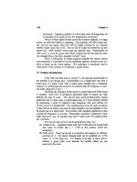
Light microscopic view of marine plankton.
womi_M 5/7/03 7:52 AM Page 365
Marshall, Barry J.
WORLD OF MICROBIOLOGY AND IMMUNOLOGY
366
•
•
3300 feet (1000 meters) in depth. The pressure on objects like
bacteria at increasing depths is enormous.
Some marine bacteria have adapted to the pressure of the
ocean depths and require the presence of the extreme pressure
in order to function. Such bacteria are barophilic if their require-
ment for pressure is absolute or barotrophic if they can tolerate
both extreme and near-atmospheric pressures. Similarly, many
marine bacteria have adapted to the cold growth temperatures.
Those which tolerate the temperatures are described as psy-
chrotrophic, while those bacteria that require the cold tempera-
tures are psychrophilic (“cold loving”).
Marine waters are elevated in certain ions such as
sodium. Not surprisingly, marine microbes like bacteria have
an absolute requirement for sodium, as well as for potassium
and magnesium ions. The bacteria have also adapted to grow
on very low concentrations of nutrients. In the ocean, most of
the organic material is located within 300 meters of the sur-
face. Very small amounts of usable nutrients reach the deep
ocean. The bacteria that inhabit these depths are in fact inhib-
ited by high concentrations of organic material.
The bacterial communication system known as
quorum
sensing was first discovered in the marine bacterium Vibrio
fischeri. An inhibitor of the quorum sensing mechanism has
also been uncovered in a type of marine algae.
Marine microbiology has become the subject of much
commercial interest. Compounds with commercial potential
as nutritional additives and antimicrobials are being discov-
ered from marine bacteria, actinomycetes and
fungi. For
example the burgeoning marine nutraceuticals market repre-
sents millions of dollars annually, and the industry is still in its
infancy. As relatively little is still known of the marine micro-
bial world, as compared to terrestrial microbiology, many
more commercial and medically relevant compounds
undoubtedly remain to be discovered.
See also Bacterial kingdoms; Bacterial movement;
Biodegradable substances; Biogeochemical cycles
MARSHALL, BARRY J. (1951- )
Marshall, Barry J.
Australian physician
Barry Marshall was born in Perth, Australia. He is a physician
with a clinical and research interest in gastroenterology. He is
internationally recognized for his discovery that the bacterium
Helicobacter pylori is the major cause of stomach ulcers.
Marshall studied medicine at the University of Western
Australia from 1969 to 1974. While studying for his medical
degree, Marshall decided to pursue medical research. He
undertook research in the laboratory of Dr. Robin Warren, who
had observations of a helical
bacteria in the stomach of people
suffering from ulcers.
Marshall and Warren succeeded in culturing the bac-
terium, which they named Helicobacter pylori. Despite their
evidence that the organism was the cause of stomach ulcera-
tion, the medical community of the time was not convinced
that a bacterium could survive the harsh acidic conditions of
the stomach yet alone cause tissue damage in this environ-
ment. In order to illustrate the relevance of the bacterium to
the disease, Marshall performed an experiment that has earned
him international renown. In July of 1984, he swallowed a
solution of the bacterium, developed the infection, including
inflammation of the stomach, and cured himself of both the
infection and the stomach inflammation by antibiotic therapy.
By 1994, Marshall’s theory of Helicobacter involve-
ment in stomach ulcers was accepted, when the United States
National Institutes of Health endorsed
antibiotics s the stan-
dard treatment for stomach ulcers.
Since Marshall’s discovery, Helicobacter pylori has
been shown to be the leading cause of stomach and intestinal
ulcers, gastritis and stomach cancer. Many thousands of ulcer
patients around the world have been successfully treated by
strategies designed to attack
bacterial infection. Marshall’s
finding was one of the first indications that human disease
thought to be due to biochemical or genetic defects were in
fact due to bacterial infections.
From Australia, Marshall spent a decade at the
University of Virginia, where he founded and directed the
Center for Study of Diseases due to H. pylori. While at
Virginia, he developed an enzyme-based rapid test for the
presence of the bacterium that tests patient’s breath. The test is
commercially available.
Currently, he is a clinician and researcher at the Sir
Charles Gairdner Hospital in Perth, Australia.
Marshall’s discovery has been recognized internation-
ally. He has received the Warren Alpert Prize from the Harvard
Medical School, which recognizes work that has most bene-
fited clinical practice. Also, he has won the
Paul Ehrlich Prize
(Germany) and the Lasker Prize (United States).
See also Bacteria and bacterial infection; Helicobacteriosis
MASTIGOPHORA
Mastigophora
Mastigophora is a division of single-celled protozoans. There
are approximately 1,500 species of Mastigophora. Their habi-
tat includes fresh and marine waters. Most of these species are
capable of self-propelled movement through the motion of one
or several flagella. The possession of flagella is a hallmark of
the Mastigophora.
In addition to their flagella, some mastigophora are able
to extend their interior contents (that is known as
cytoplasm)
outward in an arm-like protrusion. These protrusions, which
are called pseudopodia, are temporary structures that serve to
entrap and direct food into the microorganism. The cytoplas-
mic extensions are flexible and capable of collapsing back to
form the bulk of the wall that bounds the microorganism.
Mastigophora replicate typically by the internal dupli-
cation of their contents flowed by a splitting of the microbes
to form two daughter cells. This process, which is called
binary fission, is analogous to the division process in
bacteria.
In addition to replicating by binary fission, some
mastigophora can reproduce sexually, by the combining of
genetic material from two mastigophora. This process is
referred to as syngamy.
womi_M 5/7/03 7:52 AM Page 366
McCarty, Maclyn
WORLD OF MICROBIOLOGY AND IMMUNOLOGY
367
•
•
The mastigophora are noteworthy mainly because of the
presence in the division of several disease-causing species.
Some mastigophora are
parasites, which depend on the infec-
tion of a host for the completion of their life cycle. These par-
asites cause disease in humans and other animals. One
example is the Trypanosomes, which cause African
sleeping
sickness and Chaga’s disease. Another example is Giardia
lamblia. This microorganism is the agent that causes an intes-
tinal malady called giardiasis. The condition has also been
popularly dubbed “beaver fever,” reflecting its presence in the
natural habitat, where it is a resident of the intestinal tract of
warm-blooded animals.
Giardia lamblia is an important contaminant of drink-
ing water. The microorganism is resistant to the disinfectant
action of chlorine, which is the most common chemical for the
treatment of drinking water. In addition, a dormant form of the
microorganism called a cyst is small enough that it can elude
the filtration step in water treatment plants. The microbe is
increasingly becoming a concern in drinking waters all over
the world, even in industrialized countries with state of the art
water treatment infrastructure.
See also Protozoa
MATIN, A. C. (1941- )
Matin, A. C.
Indian American microbiologist
A. C. Matin is a Professor of Microbiology and Immunology
at Stanford University in Stanford, California. He has made
pioneering contributions to microbiology in a number of
areas; these include his notable research into the ways in
which
bacteria like Escherichia coli adapt and survive periods
of nutrient starvation. His studies have been important in com-
bating infections and the remediation of wastes.
Matin was born in Delhi, India. He attended the
University of Karachi, where he received his B.S. in microbi-
ology and zoology in 1960 and his M.S. in microbiology in
1962. From 1962 until 1964 he was a lecturer in microbiology
at St. Joseph’s College for Women in Karachi. He then moved
to the United States to attend the University of California at
Los Angeles, from which he received a Ph.D. in microbiology
(with distinction) in 1969. From 1969 until 1971 he was a
postdoctoral research associate at the State University of The
Netherlands. He then became a Scientific Officer, First Class,
in the Department of Microbiology at the same institution, a
post he held until 1975. That year Matin returned to the United
States to accept a position at Stanford University, the institu-
tion with which he remains affiliated.
Matin has made fundamental contributions to the bio-
chemical and molecular biological study of the bacterial stress
response—that is, how bacteria adapt to stresses in parameters
such as temperature,
pH (a measure of the acidity and alkalinity
of a solution), and food availability. Matin and his colleagues
provided much of the early data on the behavior of bacteria
when their nutrients begin to become exhausted and waste prod-
ucts accumulate. This phase of growth, termed the stationary
phase, has since been shown to have great relevance to the
growth conditions that disease-causing bacteria face in the
body, and which bacteria can face in the natural environment.
Matin has also made important contributions to the
study of multidrug resistance in the bacterium Escherichia
coli, specifically the use of a protein pump to exclude a vari-
ety of antibacterial drugs, and to the
antibiotic resistance of
Staphylococcus aureus.
Matin has published over 70 major papers and over 30
book chapters and articles. He has consulted widely among
industries concerned with bacterial drug resistance and bacte-
rial behavior.
For his scientific contributions Matin has received
numerous awards and honors. These include his appointment
as a Fulbright Scholar from 1964 until 1971, election to the
American Academy of Microbiology, and inclusion in publi-
cations such as Who’s Who in the Frontiers of Science and
Outstanding People of the 20th Century.
See also Antibiotic resistance, tests for; Bacterial adaptation
MCCARTY, MACLYN (1911- )
McCarty, Maclyn
American bacteriologist
Maclyn McCarty is a distinguished bacteriologist who has
done important work on the biology of
Streptococci and the
origins of rheumatic fever, but he is best known for his
involvement in early experiments which established the func-
tion of
DNA. In collaboration with Oswald Avery and Colin
Munro MacLeod, McCarty identified DNA as the substance
which controls heredity in living cells. The three men pub-
lished an article describing their experiment in 1944, and their
work opened the way for further studies in bacteriological
physiology, the most important of which was the demonstra-
tion of the chemical structure of DNA by James Watson and
Francis Crick in 1953.
McCarty was born in South Bend, Indiana. His father
worked for the Studebaker Corporation and the family moved
often, with McCarty attending five schools in three different
cities by the time he reached the sixth grade. In his autobio-
graphical book, The Transforming Principle, McCarty
recalled the experience as positive, believing that moving so
often made him an inquisitive and alert child. He spent a year
at Culver Academy in Indiana from 1925 to 1926, and he fin-
ished high school in Kenosha, Wisconsin. His family moved
to Portland, Oregon, and McCarty attended Stanford
University in California. He majored in
biochemistry under
James Murray Luck, who was then launching the Annual
Review of Biochemistry. McCarty presented public seminars
on topics derived from articles submitted to this publication,
and he graduated with a B.A. in 1933.
Although Luck asked him to remain at Stanford,
McCarty entered medical school at Johns Hopkins in
Baltimore in 1933. He was married during medical school
days, and he spent a summer of research at the Mayo Clinic in
Minnesota. After graduation, McCarty spent three years work-
ing in pediatric medicine at the Johns Hopkins Hospital. Even
in the decade before
penicillin, new chemotherapeutic agents
womi_M 5/7/03 7:52 AM Page 367
Measles
WORLD OF MICROBIOLOGY AND IMMUNOLOGY
368
•
•
had begun to change infectious disease therapy. McCarty
treated children suffering from Pneumococcal
pneumonia, and
he was able to save a child suffering from a Streptococcal
infection, then almost uniformly fatal, by the use of the newly
available sulfonamide antibacterials. Both of these groups of
bacteria, Streptococcus and the Pneumococcus, would play
important roles throughout the remainder of McCarty’s career.
McCarty spent his first full year of medical research at
New York University in 1940, in the laboratory of W. S.
Tillett. In 1941, McCarty was awarded a National Research
Council grant, and Tillett recommended him for a position
with Oswald Avery at the Rockefeller Institute, which was one
of the most important centers of biomedical research in the
United States. For many years, Avery had been working with
Colin Munro MacLeod on Pneumococci. In 1928, the British
microbiologist Frederick Griffith had discovered what he
called a “transforming principle” in Pneumococci. In a series
of experiments now considered a turning point in the history
of genetics, Griffith had established that living individuals of
one strain or variety of Pneumococci could be changed into
another, with different characteristics, by the application of
material taken from dead individuals of a second strain. When
McCarty joined Avery and MacLeod, the chemical nature of
this transforming material was not known, and this was what
their experiments were designed to discover.
In an effort to determine the chemical nature of
Griffith’s transforming principle, McCarty began as more of a
lab assistant than an equal partner. Avery and MacLeod had
decided that the material belonged to one of two classes of
organic compounds: it was either a protein or a nucleic acid.
They were predisposed to think it was a protein, or possibly
RNA, and their experimental work was based on efforts to
selectively disable the ability of this material to transform
strains of Pneumococci. Evidence that came to light during
1942 indicated that the material was not a protein but a nucleic
acid, and it began to seem increasingly possible that DNA was
the molecule for which they were searching. McCarty’s most
important contribution was the preparation of a deoxyribonu-
clease which disabled the transforming power of the material
and established that it was DNA. They achieved these results
by May of 1943, but Avery remained cautious, and their work
was not published until 1944.
In 1946, McCarty was named head of a laboratory at the
Rockefeller Institute which was dedicated to the study of the
Streptococci. A relative of Pneumococci, Streptococci is a
cause of rheumatic fever. McCarty’s research established the
important role played by the outer cellular covering of this
bacteria. Using some of the same techniques he had used in his
work on DNA, McCarty was able to isolate the cell wall of the
Streptococcus and analyze its structure.
McCarty became a member of the Rockefeller Institute
in 1950; he served as vice president of the institution from
1965 to 1978, and as physician in chief from 1965 to 1974. For
his work as co-discoverer of the nature of the transforming
principle, he won the Eli Lilly Award in Microbiology and
Immunology in 1946 and was elected to the National Academy
of Sciences in 1963. He won the first Waterford Biomedical
Science Award of the Scripps Clinic and Research Foundation
in 1977 and received honorary doctorates from Columbia
University in 1976 and the University of Florida in 1977.
See also Microbial genetics; Microbiology, clinical;
Streptococci and streptococcal infections
MEASLES
Measles
Measles is an infectious disease caused by a virus of the
paramyxovirus group. It infects only man and the infection
results in life-long
immunity to the disease. It is one of several
exanthematous (rash-producing) diseases of childhood, the
others being rubella (German measles), chicken pox, and the
now rare scarlet fever. The disease is particularly common in
both pre-school and young school children.
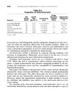
The measles virus mainly infects mucous membranes of
the respiratory tract and the skin. The symptoms include high
fever, headache, hacking cough, conjunctivitis, and a rash that
usually begins inside the mouth on the buccal mucosa as white
spots, (called Koplik’s spots) and progresses to a red rash that
spreads to face, neck, trunk and extremities. The incubation
period varies but is usually 10 to 12 days until symptoms
appear. Four to five days before the onset of the rash, the child
has fever or malaise and then may develop a sore throat and
cough. The duration of the rash is usually five days. The child
is infectious throughout the prodromal (early) period and for
up to four days after the first appearance of the rash. The virus
is highly contagious and is transmitted through respiratory
droplets or though direct contact. Measles is also sometimes
called rubeola or the nine-day measles.
Although certain complications can arise, in the vast
majority of cases, children make a full recovery from
measles. Acute local complications can occur if there is a sec-
ondary infection, for example
pneumonia due to bacteria
such as staphylococci, Streptococcus pyogene, pneumococci,
or caused by the virus itself. Also, ear infections and second-
ary bacterial otitis media can seriously aggravate the disease.
Central nervous system (CNS) complications include post-
measles encephalitis, which occurs about 10 days after the
illness with a significant mortality rate. Also, sub-acute scle-
rosing panencephalitis (SSPE), a rare fatal complication,
presents several years after the original measles infection.
Because hemorrhagic skin lesions, viraemia, and severe res-
piratory tract infection are particularly likely in malnourished
infants, measles is still frequently a life-threatening infection
in Africa and other underdeveloped regions of the world. The
microbiological diagnosis of measles is not normally required
because the symptoms are characteristic. However, if an acute
CNS complication is suspected, paired sera are usually sent
for the estimation of
complement fixing antibodies to
measles. If SSPE is suspected, the measles
antibody titres in
the CSF (determining the level of antibodies present) are also
estimated.
Epidemiological studies have shown that there is a
good correlation between the size of a population and the
number of cases of measles. A population of at least 500,000
is required to provide sufficient susceptible individuals (i.e.
womi_M 5/7/03 7:52 AM Page 368
Medawar, Peter Brian
WORLD OF MICROBIOLOGY AND IMMUNOLOGY
369
•
•
births) to maintain the virus within the population. Below that
level, the virus will eventually die out unless it is re-intro-
duced from an outside source. On the geological time-scale,
man has evolved recently and has only existed in large popu-
lations in comparatively modern times. In the past, when
human beings lived in small populations, it is concluded that
the measles virus could not exist in its present form. It may
have had another strategy of infection such as to persist in
some form and infect the occasional susceptible passer-by,
but this remains unproven. It has been suggested that the
modern measles virus evolved from an ancestral animal virus,
which is also common to the modern canine distemper and
the cattle disease rinderpest. This theory is based on the sim-
ilarities between these
viruses, and on the fact that these ani-
mals have been commensal (living in close proximity) with
man since his nomadic days. The ancestral virus is thought to
have evolved into the modern measles virus when changes in
the social behavior of man gave rise to populations large
enough to maintain infection. This evolutionary event would
have occurred within the last 6000 years when the river val-
ley civilizations of the Tigris and Euphrates were established.
To our knowledge, measles was first described as a disease in
ninth century when a Persian physician, Rhazes, was the first
to differentiate between measles and
smallpox. The physician
Rhazes also made the observation that the fever accompany-
ing the disease is a bodily defense and not the disease itself.
His writings on the subject were translated into English and
published in 1847.
The measles virus itself was first discovered in 1930,
and
John F. Enders of the Children’s Hospital in Boston suc-
cessfully isolated the measles virus in 1954. Enders then
began looking for an attenuated strain, which might be suit-
able for a live-virus
vaccine. A successful immunization pro-
gram for measles was begun soon after. Today measles is
controlled in the United States with a
vaccination that confers
immunity against measles,
mumps, and rubella and is com-
monly called the MMR vaccine. Following a series of measles
epidemics occurring in the teenage population, a second
MMR shot is now sometimes required by many school-age
children as it was found that one vaccination appeared not to
confer life-long immunity.
In October 1978, the Department of Health, Education,
and Welfare announced their intention of eliminating the
measles virus from the U.S.A. This idea was inspired by the
apparently successful global elimination of smallpox by the
World Health Organization vaccination program, which
recorded its last smallpox case in 1977.
Death from measles due to respiratory or neurological
causes occurs in about 1 out of every 1000 cases and
encephalitis also occurs at this frequency, with survivors of the
latter often having permanent brain damage. Measles virus
meets all the currently held criteria for successful elimination.
It only multiplies in man; there is a good live vaccine (95 %
effective) and only one sero-type of the virus is known.
Usually measles virus causes an acute infection but, rarely (1
out of every million cases), the virus persists and reappears
some 2-6 years causing SSPE. However, measles virus can
only be recovered with difficulty from infected tissue and
SSPE is a non-transmissible disease. To successfully eliminate
measles, it would be necessary to achieve a high immunization
level, especially in children.
See also Antibody-antigen, biochemical and molecular reac-
tions; History of immunology; History of public health;
Immunity, active, passive and delayed; Immunology;
Varicella; Viruses and responses to viral infection
MEDAWAR
, PETER BRIAN (1915-1987)
Medawar, Peter Brian
English biologist
Peter Brian Medawar made major contributions to the study of
immunology and was awarded the Nobel Prize in physiology
or medicine in 1960. Working extensively with skin grafts, he
and his collaborators proved that the
immune system learns to
distinguish between “self” and “non-self.” During his career,
Medawar also became a prolific author, penning books such as
The Uniqueness of the Individual and Advice to a Young
Scientist.
Measles rash on a child’s back.
womi_M 5/7/03 7:52 AM Page 369
Medawar, Peter Brian
WORLD OF MICROBIOLOGY AND IMMUNOLOGY
370
•
•
Medawar was born on February 28, 1915, in Rio de
Janeiro, Brazil, to Nicholas Medawar and the former Edith
Muriel Dowling. When he was a young boy, his family moved
to England, which he thereafter called home. Medawar
attended secondary school at Marlborough College, where he
first became interested in biology. The biology master encour-
aged Medawar to pursue the science under the tutelage of one
of his former students, John Young, at Magdalen College.
Medawar followed this advice and enrolled at Magdalen in
1932 as a zoology student.
Medawar earned his bachelor’s degree from Magdalen
in 1935, the same year he accepted an appointment as
Christopher Welch Scholar and Senior Demonstrator at
Magdalen College. He followed Young’s recommendation that
he work with pathologist Howard Florey, who was undertak-
ing a study of
penicillin, work for which he would later
become well known. Medawar leaned toward experimental
embryology and tissue cultures. While at Magdalen, he met
and married a fellow zoology student. Medawar and his wife
had four children.
In 1938, Medawar, by examination, became a fellow of
Magdalen College and received the Edward Chapman
Research Prize. A year later, he received his master’s from
Oxford. When World War II broke out in Europe, the Medical
Research Council asked Medawar to concentrate his research
on tissue transplants, primarily skin grafts. While this took
him away from his initial research studies into embryology,
his work with the military would come to drive his future
research and eventually lead to a Nobel Prize.
During the war, Medawar developed a concentrated
form of fibrinogen, a component of the blood. This substance
acted as a glue to reattach severed nerves, and found a place in
the treatment of skin grafts and in other operations. More
importantly to Medawar’s future research, however, were his
studies at the Burns Unit of the Glasgow Royal Infirmary in
Scotland. His task was to determine why patients rejected
donor skin grafts. He observed that the rejection time for
donor grafts was noticeably longer for initial grafts, compared
to those grafts that were transplanted for a second time.
Medawar noted the similarity between this reaction and the
body’s reaction to an invading virus or
bacteria. He formed the
opinion that the body’s rejection of skin grafts was immuno-
logical in nature; the body built up an
immunity to the first
graft and then called on that already-built-up immunity to
quickly reject a second graft.
Upon his return from the Burns Unit to Oxford, he
began his studies of immunology in the laboratory. In 1944, he
became a senior research fellow of St. John’s College, Oxford,
and university demonstrator in zoology and comparative
anatomy. Although he qualified for and passed his examina-
tions for a doctorate in philosophy while at Oxford, Medawar
opted against accepting it because it would cost more than he
could afford. In his autobiography, Memoir of a Thinking
Radish, he wrote, “The degree served no useful purpose and
cost, I learned, as much as it cost in those days to have an
appendectomy. Having just had the latter as a matter of
urgency, I thought that to have both would border on self-
indulgence, so I remained a plain mister until I became a
prof.” He continued as researcher at Oxford University
through 1947.
During that year Medawar accepted an appointment as
Mason professor of zoology at the University of Birmingham.
He brought with him one of his best graduate students at
Oxford, Rupert Everett “Bill” Billingham. Another graduate
student, Leslie Brent, soon joined them and the three began
what was to become a very productive collaboration that
spanned several years. Their research progressed through
Medawar’s appointment as dean of science, through his sev-
eral-month-long trip to the Rockefeller Institute in New York
in 1949—the same year he received the title of fellow from the
Royal Society—and even a relocation to another college. In
1951, Medawar accepted a position as Jodrell Professor of
Zoology and Comparative Anatomy at University College,
London. Billingham and Brent followed him.
Their most important discovery had its experimental
root in a promise Medawar made at the International Congress
of Genetics at Stockholm in 1948. He told another investiga-
tor, Hugh Donald, that he could formulate a foolproof method
for distinguishing identical from fraternal twin calves. He and
Billingham felt they could easily tell the twins apart by trans-
planting a skin graft from one twin to the other. They reasoned
that a calf of an identical pair would accept a skin graft from
its twin because the two originated from the same egg,
whereas a calf would reject a graft from its fraternal twin
because they came from two separate eggs. The results did not
bear this out, however. The calves accepted skin grafts from
their twins regardless of their status as identical or fraternal.
Puzzled, they repeated the experiment, but received the same
results.
They found their error when they became aware of work
done by Dr.
Frank Macfarlane Burnet of the University of
Melbourne, and Ray D. Owen of the California Institute of
Technology. Owen found that blood transfuses between twin
calves, both fraternal and identical. Burnet believed that an
individual’s immunological framework developed before
birth, and felt Owen’s finding demonstrated this by showing
that the immune system tolerates those tissues that are made
known to it before a certain age. In other words, the body does
not recognize donated tissue as alien if it has had some expo-
sure to it at an early age. Burnet predicted that this immuno-
logical tolerance for non-native tissue could be reproduced in
a lab. Medawar, Billingham, and Brent set out to test Burnet’s
hypothesis.
The three-scientist team worked closely together, inoc-
ulating embryos from mice of one strain with tissue cells from
donor mice of another strain. When the mice had matured, the
trio grafted skin from the donor mice to the inoculated mice.
Normally, mice reject skin grafts from other mice, but the
inoculated mice in their experiment accepted the donor skin
grafts. They did not develop an immunological reaction. The
prenatal encounter had given the inoculated mice an acquired
immunological tolerance. They had proven Burnet’s hypothe-
sis. They published their findings in a 1953 article in Nature.
Although their research had no applications to transplants
among humans, it showed that transplants were possible.
womi_M 5/7/03 7:52 AM Page 370
Medical training and careers in microbiology
WORLD OF MICROBIOLOGY AND IMMUNOLOGY
371
•
•
In the years following publication of the research,
Medawar accepted several honors, including the Royal Medal
from the Royal Society in 1959. A year later, he and Burnet
accepted the Nobel Prize for Physiology or Medicine for their
discovery of acquired immunological tolerance: Burnet devel-
oped the theory and Medawar proved it. Medawar shared the
prize money with Billingham and Brent.
Medawar’s scientific concerns extended beyond
immunology, even during the years of his work toward
acquired immunological tolerance. While at Birmingham, he
and Billingham also investigated pigment spread, a phenome-
non seen in some guinea pigs and cattle where the dark spots
spread into the light areas of the skin. “Thus if a dark skin graft
were transplanted into the middle of a pale area of skin it
would soon come to be surrounded by a progressively widen-
ing ring of dark skin,” Medawar asserted in his autobiography.
The team conducted a variety of experiments, hoping to show
that the dark pigment cells were somehow “infecting” the pale
pigment cells. The tests never panned out.
Medawar also delved into animal behavior at
Birmingham. He edited a book on the subject by noted scien-
tist Nikolaas Tinbergen, who ultimately netted a Nobel Prize
in 1973. In 1957, Medawar also became a book author with his
first offering, The Uniqueness of the Individual, which was
actually a collection of essays. In 1959, his second book, The
Future of Man, was issued, containing a compilation of a
series of broadcasts he read for British Broadcasting
Corporation (BBC) radio. The series examined the impacts of
evolution on man.
Medawar remained at University College until 1962
when he took the post of director of the National Institute for
Medical Research in London, where he continued his study of
transplants and immunology. While there, he continued writ-
ing with mainly philosophical themes. The Art of the Soluble,
published in 1967, is an assembly of essays, while his 1969
book, Induction and Intuition in Scientific Thought, is a
sequence of lectures examining the thought processes of sci-
entists. In 1969 Medawar, then president of the British
Association for the Advancement of Science, experienced the
first of a series of strokes while speaking at the group’s annual
meeting. He finally retired from his position as director of the
National Institute for Medical Research in 1971. In spite of his
physical limitations, he went ahead with scientific research in
his lab at the clinical research center of the Medical Research
Council. There he began studying cancer.
Through the 1970s and 1980s, Medawar produced sev-
eral other books—some with his wife as co-author—in addi-
tion to his many essays on growth, aging, immunity, and
cellular transformations. In one of his most well-known
books, Advice to a Young Scientist, Medawar asserted that for
scientists, curiosity was more important that genius.
See also Antibody and antigen; Antibody-antigen, biochemical
and molecular reactions; Antibody formation and kinetics;
Antibody, monoclonal; Immunity, active, passive and delayed;
Immunity, cell mediated; Immunity, humoral regulation;
Immunochemistry; Immunogenetics; Major histocompatibility
complex (MHC); Transplantation genetics and immunology
M
EDICAL TRAINING AND CAREERS IN
MICROBIOLOGY
Medical training and careers in microbiology
The world of microbiology overlaps the world of medicine. As
a result, trained microbiologists find a diversity of career paths
and opportunity in medicine.
Research in medical microbiology can involve clinical
or basic science.
Clinical microbiology focuses on the micro-
biological basis of various diseases and how to alleviate the
suffering caused by the infectious microorganism. Basic med-
ical research is concerned more with the molecular events
associated with infectious diseases or illnesses.
Both medical training and microbiology contain many
different areas of study. Medical microbiology is likewise an
area of many specialties. A medical bacteriologist can study
how
bacteria can infect humans and cause disease, and how
these disease processes can be dealt with. A medical mycolo-
gist can study pathogenic (disease-causing)
fungi, molds and
yeast to find out how they cause disease. A parasitologist is
concerned with how parasitic
microorganisms (those that
require a host in order to live) cause disease. A medical virol-
ogist can study the diseases attributed to infection by a virus,
such as the hemorrhagic fever caused by the
Ebola virus.
The paths to these varied disciplines of study are also
varied. One route that a student can take to incorporate both
research training and medical education is the combined M.D
PhD. program. In several years of rigorous study, students
become physician-scientists. Often, graduates develop a clini-
cal practice combined with basic research. The experience
gained at the bedside can provide research ideas. Conversely,
laboratory techniques can be brought to bear on unraveling the
basis of human disease. The M.D.–PhD. training exemplifies
what is known as the transdisciplinary approach. Incorporating
different approaches to an issue can suggest treatment or
research strategies that might otherwise not be evident if an
issue were addressed from only one perspective.
The training for a career in the area of medicine and
medical microbiology begins in high school. Courses in the
sciences lay the foundation for the more in-depth training that
will follow in university or technical institution. With under-
graduate level training, career paths can include research
assistant, providing key technical support to a research team,
quality assurance in the food, industrial or environmental
microbiology areas, and medical technology.
Medical microbiology training at the undergraduate and
graduate levels, in the absence of simultaneous medical train-
ing, can also lead to a career as a clinical microbiologist. Such
scientists are employed in universities, hospitals and in the
public sector. For example, the United Kingdom has an exten-
sive Public Health Laboratory Service. The PHLS employs
clinical microbiologists in reference laboratories, to develop
or augment test methods, and as epidemiologists. The latter
are involved in determining the underlying causes of disease
outbreaks and in uncovering potential microbiological health
threats. Training in medical microbiology can be at the
Baccalaureate level, and in research that leads to a Masters or
a Doctoral degree. The latter is usually undertaken if the aim
womi_M 5/7/03 7:52 AM Page 371
Medical training and careers in microbiology
WORLD OF MICROBIOLOGY AND IMMUNOLOGY
372
•
•
is to do original and independent research, teach undergradu-
ate and graduate students, or to assume an executive position.
Medical technologists are involved in carrying out the
myriad of microbiological tests that are performed on samples
such as urine, blood and other body fluids to distinguish path-
ogenic microorganisms from the normal flora of the body.
This can be very much akin to detective work, involving the
testing of samples by various means to resolve he identity of
an organism based on the various biochemical behaviors.
Increasingly, such work is done in conjunction with automated
equipment. Medical technologists must be skilled at schedul-
ing tests efficiently, independently and as part of a team.
Training as a medical technologist is typically at a community
college or technical institution and usually requires two years.
As in the other disciplines of medical microbiology,
medical technology is a specialized field. Histopathology is
the examination of body cells or tissues to detect or rule out
disease. This speciality involves knowledge of light and
elec-
tron microscopic examination
of samples. Cytology is the
study of cells for abnormalities that might be indicative of
infection or other malady, such as cancer. Medical
immunol-
ogy
studies the response of the host to infection. A medical
immunologist is skilled at identifying those immune cells that
active in combating an infection. Medical technology also
encompasses the area of clinical
biochemistry, where cells and
body fluids are analyzed for the presence of components
related to disease. Of course the study of microorganism
involvement in disease requires medical technologists who are
specialized microbiologists and virologists, as two examples.
Medical microbiologists also can find a rewarding
career path in industry. Specifically, the knowledge of the sus-
ceptibility or resistance of microorganisms to antimicrobial
Working as a specialist in a medical microbiology laboratory is one of many careers available in the field.
womi_M 5/7/03 7:52 AM Page 372
Membrane fluidity
WORLD OF MICROBIOLOGY AND IMMUNOLOGY
373
•
•
drugs is crucial to the development of new drugs. Work can be
at the research and development level, in the manufacture of
drugs, in the regulation and licensing of new antimicrobial
agents, and even in the sale of drugs. For example, the sale of
a product can be facilitated by the interaction of the sales asso-
ciate and physician client on an equal footing in terms of
knowledge of antimicrobial therapy or disease processes.
Following the acquisition of a graduate or medical
degree, specialization in a chosen area can involve years of
post-graduate or medical residence. The road to a university
lab or the operating room requires dedication and over a
decade of intensive study.
Careers in medical science and medical microbiology
need not be focused at the patient bedside or at the lab bench.
Increasingly, the medical and infectious disease fields are ben-
efiting from the advice of consultants and those who are able
to direct programs. Medical or microbiological training com-
bined with experience or training in areas such as law or busi-
ness administration present an attractive career combination.
See also Bioinformatics and computational biology; Food
safety; History of public health; Hygiene; World Health
Organization
MEMBRANE FLUIDITY
Membrane fluidity
The membranes of bacteria function to give the bacterium its
shape, allow the passage of molecules from the outside in and
from the inside out, and to prevent the internal contents from
leaking out. Gram-negative bacteria have two membranes that
make up their cell wall, whereas Gram-positive bacteria have
a single membrane as a component of their cell wall. Yeasts
and
fungi have another specialized nuclear membrane that
compartmentalizes the genetic material of the cell.
For all these functions, the membrane must be fluid. For
example, if the interior of a bacterial membrane was crys-
talline, the movement of molecules across the membrane
would be extremely difficult and the bacterium would not sur-
vive.
Membrane fluidity is assured by the construction of a
typical membrane. This construction can be described by the
fluid mosaic model. The mosaic consists of objects, such as
proteins, which are embedded in a supporting—but mobile—
structure of lipid.
The fluid mosaic model for membrane construction was
proposed in 1972 by S. J. Singer of the University of
California at San Diego and G. L. Nicolson of the Salk
Institute. Since that time, the evidence in support of a fluid
membrane has become irrefutable.
In a fluid membrane, proteins may be exposed on the
inner surface of the membrane, the outer surface, or at both
surfaces. Depending on their association with neighbouring
molecules, the proteins may be held in place or may capable
of a slow drifting movement within the membrane. Some pro-
teins associate together to form pores through which mole-
cules can pass in a regulated fashion (such as by the charge or
size of the molecule).
The fluid nature of the membrane rest with the support-
ing structure of the lipids. Membrane lipids of
microorgan-
isms
tend to be a type of lipid termed phospholipid. A
phospholipid consists of fatty acid chains that terminate at one
end in a phosphate group. The fatty acid chains are not
charged, and so do not tend to associate with water. In other
words they are
hydrophobic. On the other hand, the charged
phosphate head group does tend to associate with water. In
other words they are hydrophilic. The way to reconcile these
chemistry differences in the membrane are to orient the
phos-
pholipids with the water-phobic tails pointing inside and the
water-phyllic heads oriented to the watery external environ-
ment. This creates two so-called leaflets, or a bilayer, of phos-
pholipid. Essentially the membrane is a two dimensional fluid
that is made mostly of phospholipids. The consistency of the
membrane is about that of olive oil.
Regions of the membrane will consist solely of the lipid
bilayer. Molecules that are more hydrophobic will tend to dis-
solve into these regions, and so can move across the mem-
brane passively. Additionally, some of the proteins embedded
in the bilayer will have a transport function, to actively pump
or move molecules across the membrane.
The fluidity of microbial membranes also allows the
constituent proteins to adopt new configurations, as happens
when molecules bind to receptor portions of the protein. These
configurational changes are an important mechanism of sig-
naling other proteins and initiating a response to, for example,
the presence of a food source. For example, a protein that
binds a molecule may rotate, carrying the molecule across the
membrane and releasing the molecule on the other side. In
bacteria, the membrane proteins tend to be located more in one
leaflet of the membrane than the other. This asymmetric
arrangement largely drives the various transport and other
functions that the membrane can perform.
The phospholipids are capable of a drifting movement
laterally on whatever side of the membrane they happen to
be. Measurements of this movement have shown that the
drifting can actually be quite rapid. A flip-flop motion across
to the other side of the membrane is rare. The fluid motion of
the phospholipids increases if the hydrophobic tail portion
contains more double bonds, which cause the tail to be
kinked instead of straight. Such alteration of the phospho-
lipid tails can occur in response to temperature change. For
example if the temperature decreases, a bacterium may alter
the phospholipid chemistry so as to increase the fluidity of
the membrane.
See also Bacterial membranes and cell wall
M
EMBRANE TRANSPORT, EUKARYOTIC
•
see C
ELL MEMBRANE TRANSPORT
M
EMBRANE TRANSPORT, PROKARYOTIC
•
see P
ROKARYOTIC MEMBRANE TRANSPORT
womi_M 5/7/03 7:52 AM Page 373
Meningitis, bacterial and viral
WORLD OF MICROBIOLOGY AND IMMUNOLOGY
374
•
•
M
ENINGITIS, BACTERIAL AND VIRAL
Meningitis, bacterial and viral
Meningitis is a potentially fatal inflammation of the meninges,
the thin, membranous covering of the brain and the spinal
cord. Meningitis is most commonly caused by infection (by
bacteria, viruses, or fungi), although it can also be caused by
bleeding into the meninges, cancer, or diseases of the
immune
system
.
The meninges are three separate membranes, layered
together, which serve to encase the brain and spinal cord. The
dura is the toughest, outermost layer, and is closely attached to
the inside of the skull. The middle layer, the arachnoid, is
important in the normal flow of the cerebrospinal fluid (CSF),
a lubricating fluid that bathes both the brain and the spinal
cord. The innermost layer, the pia, helps direct brain blood
vessels into the brain. The space between the arachnoid and
the pia contains CSF, which serves to help insulate the brain
from trauma. Through this space course many blood vessels.
CSF, produced within specialized chambers deep inside the
brain, flows over the surface of the brain and spinal cord. This
fluid serves to cushion these relatively delicate structures, as
well as supplying important nutrients for brain cells. CSF is
reabsorbed by blood vessels that are located within the
meninges.
The cells lining the brain’s capillaries (tiny blood ves-
sels) are specifically designed to prevent many substances
from passing into brain tissue. This is commonly referred to as
the blood-brain barrier. The blood-brain barrier prevents vari-
ous toxins (substances which could be poisonous to brain tis-
sue), as well as many agents of infection, from crossing from
the blood stream into the brain tissue. While this barrier obvi-
ously is an important protective feature for the brain, it also
serves to complicate therapy in the case of an infection, by
making it difficult for medications to pass out of the blood and
into the brain tissue where the infection resides.
The most common infectious causes of meningitis vary
according to an individual host’s age, habits and living envi-
ronment, and health status. In newborns, the most common
agents of meningitis are those that are contracted from the
newborn’s mother, including Group B Streptococci (becoming
an increasingly common infecting organism in the newborn
period), Escherichia coli, and Listeria monocytogenes. Older
children are more frequently infected by Haemophilus
influenzae, Neisseria meningitidis, and Streptococcus pneu-
moniae, while adults are infected by S. pneumoniae and N.
meningitidis. N. meningitidis is the only organism that can
cause
epidemics of meningitis. These have occurred in partic-
ular when a child in a crowded day-care situation, a college
student in a dormitory, or a military recruit in a crowded train-
ing camp has fallen ill with N. meningitidis meningitis.
Viral causes of meningitis include the
herpes simplex
viruses,
mumps and measles viruses (against which most
children are protected due to mass
immunization programs),
the virus that causes chicken pox, the
rabies virus, and a num-
ber of viruses that are acquired through the bite of infected
mosquitoes. Patients with
AIDS (Acquired Immune Deficiency
Syndrome) are more susceptible to certain infectious causes of
meningitis, including by certain fungal agents, as well as by
the agent that causes tuberculosis. Patients who have had their
spleens removed, or whose spleens are no longer functional
(as in the case of patients with sickle cell disease) are more
susceptible to certain infections, including those caused by N.
meningitidis and S. pneumoniae.
The majority of meningitis infections are acquired by
blood-borne spread. An individual may have another type of
infection (of the lungs, throat, or tissues of the heart) caused
by an organism that can also cause meningitis. The organism
multiplies, finds its way into the blood stream, and is delivered
in sufficient quantities to invade past the blood-brain barrier.
Direct spread occurs when an already resident infec-
tious agent spreads from infected tissue next to or very near
the meninges, for example from an ear or sinus infection.
Patients who suffer from skull fractures provide openings to
the sinuses, nasal passages, and middle ears. Organisms that
frequently live in the human respiratory system can then pass
through these openings to reach the meninges and cause infec-
tion. Similarly, patients who undergo surgical procedures or
who have had foreign bodies surgically placed within their
skulls (such as tubes to drain abnormal amounts of accumu-
lated CSF) have an increased risk of the organisms causing
meningitis being introduced to the meninges.
The most classic symptoms of meningitis (particularly
of bacterial meningitis) include fever, headache, vomiting,
photophobia (sensitivity to light), irritability, lethargy (severe
fatigue), and stiff neck. The disease progresses with seizures,
confusion, and eventually coma.
Damage due to meningitis occurs from a variety of phe-
nomena. The action of infectious agents on the brain tissue is
one direct cause of damage. Other types of damage may be
due to mechanical effects of swelling of brain tissue, and com-
pression against the bony surface of the skull. Swelling of the
meninges may interfere with the normal absorption of CSF by
blood vessels, causing accumulation of CSF and damage due
to resulting pressure on the brain. Interference with the brain’s
carefully regulated chemical environment may cause damag-
ing amounts of normally present substances (carbon dioxide,
potassium) to accumulate. Inflammation may cause the blood-
brain barrier to become less effective at preventing the passage
of toxic substances into brain tissue.
Antibiotic medications (forms of penicillins and
cephalosporins, for example) are the most important element
of treatment against bacterial agents of meningitis. Because of
the effectiveness of the blood-brain barrier in preventing pas-
sage of substances into the brain, medications must be deliv-
ered directly into the patient’s veins (intravenous or IV) at
very high doses. Antiviral medications (acyclovir) may be
helpful in the case of viral meningitis, and antifungal medica-
tions are available as well.
Other treatment for meningitis involves decreasing
inflammation (with steroid preparations) and paying careful
attention to the balance of fluids, glucose, sodium, potassium,
oxygen, and carbon dioxide in the patient’s system. Patients
who develop seizures will require medications to halt the
seizures and prevent their return.
A series of immunizations against Haemophilus influen-
zae, started at two months of age, has greatly reduced the inci-
womi_M 5/7/03 7:52 AM Page 374
Meselson, Matthew Stanley
WORLD OF MICROBIOLOGY AND IMMUNOLOGY
375
•
•
dence of that form of meningitis. Vaccines also exist against
Neisseria meningitidis and Streptococcus pneumoniae bacte-
ria, but these vaccines are only recommended for those people
who have particular susceptibility to those organisms, due to
certain immune deficiencies, lack of a spleen, or sickle cell
anemia.
Because N. meningitidis is known to cause epidemics of
disease, close contacts of patients with such meningitis are
treated prophylactically, often with the antibiotic Rifampin.
This measure generally prevents spread of the disease.
See also Bacteria and bacterial infection; Viruses and
responses to viral infection
MESELSON
, M
ATTHEW STANLEY
(1930- )
Meselson, Matthew Stanley
American molecular biologist
Matthew Meselson, in collaboration with biologist Franklin
W. Stahl, showed experimentally that the replication of
deoxyribonucleic acid (DNA) in bacteria is semiconservative.
Semiconservative replication occurs in a double stranded
DNA molecule when the two strands are separated and a new
strand is copied from the parental strand to produce two new
double stranded DNA molecules. The new double stranded
DNA molecule is semiconservative because only one strand is
conserved from the parent; the other strand is a new copy.
(Conservative replication occurs when one offspring of a mol-
ecule contains both parent strands and the other molecule off-
spring contains newly replicated strands) The classical
experiment revealing semiconservative replication in bacteria
was central to the understanding of the living cell and to mod-
ern
molecular biology.
Matthew Stanley Meselson was born May 24, 1930, in
Denver, Colorado. After graduating in 1951 with a Ph.D. in
liberal arts from the University of Chicago, he continued his
education with graduate studies at the California Institute of
Technology in the field of chemistry. Meselson graduated with
a Ph.D. in 1957, and remained at Cal Tech as a research fel-
low. He acquired the position of assistant professor of chem-
istry at Cal Tech in 1958. In 1960, Meselson moved to
Cambridge, Massachusetts to fill the position of associate pro-
fessor of natural sciences at Harvard University. In 1964, he
was awarded professor of biology, which he held until 1976.
He was appointed the title of Thomas Dudley Cabot professor
of natural sciences in 1976. From that time on, Meselson held
a concurrent appointment on the council of the Smithsonian
Institute in Washington, DC.
After graduating from the University of Chicago,
Meselson continued his education in chemistry at the
California Institute of Technology. It was during his final year
at Cal Tech that Meselson collaborated with Franklin Stahl on
the classical experiment of semiconservative replication of
DNA. Meselson and Stahl wanted to design and perform an
experiment that would show the nature of DNA replication
from parent to offspring using the
bacteriophage T4 (a virus
that destroys other cells, also called a phage). The idea was to
use an isotope to mark the cells and centrifuge to separate par-
ticles that could be identified by their DNA and measure
changes in the new generations of DNA. Meselson, Stahl, and
Jerome Vinograd originally designed this technique of isolat-
ing phage samples. The phage samples isolated would contain
various amounts of the isotope based on the rate of DNA repli-
cation. The amount of isotope incorporated in the new DNA
strands, they hoped, would be large enough to determine quan-
titatively. The experiments, however, were not successful.
After further contemplation, Meselson and Stahl decided to
abandon the use of bacteriophage T4 and the isotope and use
instead the bacteria Escherichia coli (E. coli) and the heavy
nitrogen isotope 15N as the labeling substance. This time
when the same experimental steps were repeated, the analysis
showed three distinct types of bacterial DNA, two from the
original parent strands and one from the offspring. Analysis of
this offspring showed each strand of DNA came from a differ-
ent parent. Thus the theory of semiconservative replication of
DNA had been proven. With this notable start to his scientific
career Meselson embarked on another collaboration, this time
with biologists
Sydney Brenner, from the Medical Research
Council’s Division of Molecular Biology in Cambridge,
England, and
François Jacob from the Pasteur Institute
Laboratories in Paris, France. Together, Meselson, Brenner,
and Jacob performed a series of experiments in which they
showed that when the bacteriophage T4 enters a bacterial cell,
the phage DNA incorporates into the cellular DNA and causes
the release of messenger
RNA. Messenger RNA instructs the
cell to manufacture phage proteins instead of the bacterial cell
proteins that are normally produced. These experiments led to
the discovery of the role of messenger RNA as the instructions
that the bacterial cell reads to produce the desired protein
products. These experiments also showed that the bacterial
cell could produce proteins from messenger RNA that are not
native to the cell in which it occurs.
In his own laboratory at Harvard University, Meselson
and a postdoctoral fellow, Robert Yuan, were developing and
purifying one of the first of many known
restriction enzymes
commonly used in molecular biological analyses. Restriction
enzymes are developed by cultivating bacterial strains with
phages. Bacterial strains that have the ability to restrict foreign
DNA produce a protein called an enzyme that actually chews
up or degrades the foreign DNA. This enzyme is able to break
up the foreign DNA sequences into a number of small seg-
ments by breaking the double stranded DNA at particular loca-
tions. Purification of these enzymes allowed mapping of
various DNA sequences to be accomplished. The use of puri-
fied restriction enzymes became a common practice in the
field of molecular biology to map and determine contents of
many DNA sequences.
After many years working with the bacteria E. coli,
Meselson decided to investigate the fundamentals of DNA
replication and repair in other organisms. He chose to work on
the fruit fly called Drosophila melanogaster. Meselson dis-
covered that the fruit fly contained particular DNA sequences
that would be transcribed only when induced by heat shock or
stress conditions. These particular heat shock genes required a
womi_M 5/7/03 7:52 AM Page 375
Mesophilic bacteria
WORLD OF MICROBIOLOGY AND IMMUNOLOGY
376
•
•
specific setup of DNA bases upstream of the initiation site in
order for
transcription to occur. If the number of bases was
increased or reduced from what was required, the
gene would
not be transcribed. Meselson also found that there were par-
ticular DNA sequences that could be recombined or moved
around within the entire chromosome of DNA. These move-
able segments are termed
transposons. Transposons, when
inserted into particular sites within the sequence, can either
turn on or turn off expression of the gene that is near it, caus-
ing
mutations within the fly. These studies contributed to the
identity of particular regulatory and structural features of the
fruit fly as well as to the overall understanding of the proper-
ties of DNA.
Throughout his career as a scientist, Meselson has writ-
ten over 50 papers published in major scientific journals and
received many honors and awards for his contributions to the
field of molecular biology. In 1963, Meselson received the
National Academy of Science Prize for Molecular Biology,
followed by the Eli Lilly Award for Microbiology and
Immunology in 1964. He was awarded the Lehman Award in
1975 and the Presidential award in 1983, both from the New
York Academy of Sciences. In 1990, Meselson received the
Science Freedom and Responsibility Award from the
American Association for the Advancement of Science.
Meselson has also delved into political issues, particularly on
government proposals for worldwide chemical and biological
weapon disarmament.
See also Microbial genetics; Transposition
MESOPHILIC BACTERIA
Mesophilic bacteria
Mesophiles are microorganisms such as some species of
Bacteria, Fungi, and even some Archaea that are best active at
median temperatures. For instance, bacterial species involved
in biodegradation (i.e., digestion and decomposition of organic
matter), which are more active in temperatures ranging from
approximately 70° - 90°F (approx. 15°–40°C), are termed
mesophilic bacteria. They take part in the web of micro-organic
activity that form the humus layer in forests and other fertile
soils, by decomposing both vegetable and animal matter.
At the beginning of the decomposition process, another
group of bacteria, psychrophylic bacteria, start the process
because they are active in lower temperatures up to 55°F
(from below zero up to 20°C), and generate heat in the
process. When the temperature inside the decomposing layer
reaches 50–100°F, it attracts mesophilic bacteria to continue
the biodegradation. The peak of reproductive and activity of
mesophilic bacteria is reached between 86–99°F (30–37°C),
and further increases the temperature in the soil environment.
Between 104–170°F (40–85°C, or even higher), another group
of bacteria (thermophyllic bacteria) takes up the process that
will eventually result in organic soil, or humus. Several
species of fungi also take part in each decomposing step.
Mesophilic bacteria are also involved in food
contami-
nation
and degradation, such as in bread, grains, dairies, and
meats. Examples of common mesophilic bacteria are Listeria
monocytogenes, Pesudomonas maltophilia, Thiobacillus nov-
ellus, Staphylococcus aureus, Streptococcus pyrogenes,
Streptococcus pneumoniae, Escherichia coli, and Clostridium
kluyveri. Bacterial infections in humans are mostly caused by
mesophilic bacteria that find their optimum growth tempera-
ture around 37°C (98.6°F), the normal human body tempera-
ture. Beneficial bacteria found in human intestinal flora are
also mesophiles, such as dietary Lactobacillus acidophilus.
See also Archaeobacteria; Bacteria and bacterial infection;
Biodegradable substances; Composting, microbiological
aspects; Extremophiles
METABOLISM
Metabolism
Metabolism is the sum total of chemical changes that occur in
living organisms and which are fundamental to life. All
prokaryotic and eukaryotic cells are metabolically active. The
sole exception is
viruses, but even viruses require a metaboli-
cally active host for their replication.
Metabolism involves the use of compounds. Nutrients
from the environment are used in two ways by
microorgan-
isms. They can be the building blocks of various components
of the microorganism (assimilation or anabolism). Or, nutri-
ents can be degraded to yield energy (dissimilation or catabo-
lism). Some so-called amphibolic biochemical pathways can
serve both purposes. The continual processes of breakdown
and re-synthesis are in a balance that is referred to as turnover.
Metabolism is an open system. That is, there are constant
inputs and outputs. A chain of metabolic reactions is said to be
in a steady state when the concentration of all intermediates
remains constant, despite the net flow of material through the
system. That means the concentration of intermediates
remains constant, while a product is formed at the expense of
the substrate.
Primary metabolism comprises those metabolic
processes that are basically similar in all living cells and are
necessary for cellular maintenance and survival. They include
the fundamental processes of growth (e.g., the synthesis of
biopolymers and the macromolecular structures of cells and
organelles), energy production (glycolysis and the tricar-
boxylic acid cycle) and the turnover of cell constituents.
Secondary metabolism refers to the production of substances,
such as bile pigments from porphyrins in humans, which only
occur in certain eukaryotic tissues and are distinct from the
primary metabolic pathways.
Metabolic control processes that occur inside cells
include regulation of
gene expression and metabolic feedback
or feed-forward processes. The triggers of differential gene
expression may be chemical, physical (e.g., bacterial cell den-
sity), or environmental (e.g., light). Differential gene expres-
sion is responsible for the regulation, at the molecular level, of
differentiation and development, as well as the maintenance of
numerous cellular “house-keeping” reactions, which are
essential for the day-to-day functioning of a microorganism.
In many metabolic pathways, the metabolites (substances pro-
duced or consumed by metabolism) themselves can act
womi_M 5/7/03 7:52 AM Page 376
Metchnikoff, Élie
WORLD OF MICROBIOLOGY AND IMMUNOLOGY
377
•
•
directly as signals in the control of their own breakdown and
synthesis. Feedback control can be negative or positive.
Negative feedback results in the inhibition by an end product,
of the activity or synthesis of an enzyme or several
enzymes
in a reaction chain. The inhibition of the synthesis of enzymes
is called enzyme repression. Inhibition of the activity of an
enzyme by an end product is an allosteric effect and this type
of feedback control is well known in many metabolic path-
ways (e.g., lactose
operon). In positive feedback, an endprod-
uct activates an enzyme responsible for its own production.
Many reactions in metabolism are cyclic. A metabolic
cycle is a catalytic series of reactions, in which the product of
one bimolecular (involving two molecules) reaction is regen-
erated as follows: A + B → C + A. Thus, A acts catalytically
and is required only in small amounts and A can be regarded
as carrier of B. The catalytic function of A and other members
of the metabolic cycle ensure economic conversion of B to C.
B is the substrate of the metabolic cycle and C is the product.
If intermediates are withdrawn from the metabolic cycle, e.g.,
for biosynthesis, the stationary concentrations of the metabolic
cycle intermediates must be maintained by synthesis.
Replenishment of depleted metabolic cycle intermediates is
called anaplerosis. Anaplerosis may be served by a single
reaction, which converts a common metabolite into an inter-
mediate of the metabolic cycle. An example of this is pyruvate
to oxaloacetate reaction in the tricarboxylic acid cycle.
Alternatively, it may involve a metabolic sequence of reac-
tions, i.e., an anaplerotic sequence. An example of this is the
glycerate pathway which provides phosphoenol pyruvate for
anaplerosis of the tricarboxylic acid cycle.
Prokaryotes exhibit a great diversity of metabolic
options, even in a single organism. For example, Escherichia
coli can produce energy by
respiration or fermentation.
Respiration can be under aerobic conditions (e.g., using O
2
as
the final electron acceptor) or anaerobically (e.g., using some-
thing other than oxygen as the final electron acceptor).
Compounds like lactose or glucose can be used as the only
source of carbon. Other
bacteria have other metabolic capa-
bilities including the use of sunlight for energy.
Some of these mechanisms are also utilized by eukary-
otic cells. In addition, prokaryotes have a number of energy-
generating mechanisms that do not exist in eukaryotic cells.
Prokaryotic fermentation can be uniquely done via the phos-
phoketolase and Enter-Doudoroff pathways. Anaerobic respi-
ration is unique to prokaryotes, as is the use of inorganic
compounds as energy sources or as carbon sources during bac-
terial
photosynthesis. Archaebacteria possess metabolic path-
ways that use H
2
as the energy source with the production of
methane, and a nonphotosynthetic metabolism that can con-
vert light energy into chemical energy.
In bacteria, metabolic processes are coupled to the syn-
thesis of adenosine triphosphate (ATP), the principle fuel
source of the cell, through a series of membrane-bound pro-
teins that constituent the
electron transport system. The
movement of protons from the inside to the outside of the
membrane during the operation of the electron transport sys-
tem can be used to drive many processes in a bacterium, such
as the movement of the flagella used to power the bacterium
along, and the synthesis of ATP in the process called oxidative
phosphorylation.
The fermentative metabolism that is unique to some
bacteria is evolutionarily ancient. This is consistent with the
early appearance of bacteria on Earth, relative to eukaryotic
organisms. But bacteria can also ferment sugars in the same
way that brewing
yeast (i.e., Saccharomyces cerevesiae fer-
ment sugars to produce ethanol and carbon dioxide. This fer-
mentation, via the so-called Embden Myerhoff pathway, can
lead to different ends products in bacteria, such as lactic acid
(e.g., Lactobacillus), a mixture of acids (Enterobacteriacaeae,
butanediol (e.g., Klebsiella, and propionic acid (e.g.,
Propionibacterium).
See also Bacterial growth and division; Biochemistry
METCHNIKOFF, ÉLIE (1845-1916)
Metchnikoff, Élie
Russian immunologist
Élie Metchnikoff was a pioneer in the field of immunology and
won the 1908 Nobel Prize in physiology or medicine for his
discoveries of how the body protects itself from disease-caus-
ing organisms. Later in life, he became interested in the effects
of nutrition on aging and health, which led him to advocate
some controversial diet practices.
Metchnikoff, the youngest of five children, was born in
the Ukrainian village of Ivanovka on May 16, 1845, to Emilia
Nevahovna, daughter of a wealthy writer, and Ilya Ivanovich,
an officer of the Imperial Guard in St. Petersburg. He enrolled
at the Kharkov Lycee in 1856, where he developed an espe-
cially strong interest in biology. At age 16, he published a
paper in a Moscow journal criticizing a geology textbook.
After graduating from secondary school in 1862, he entered
the University of Kharkov, where he completed a four-year
program in two years. He also became an advocate of the the-
ory of
evolution by natural selection after reading Charles
Darwin’s On the Origin of Species by Means of Natural
Selection.
In 1864, Metchnikoff traveled to Germany to study,
where his work with nematodes (a species of worm) led to the
surprising conclusion that the organism alternates between
sexual and asexual generations. His studies at Kharkov, cou-
pled with his interest in Darwin’s theory, convinced him that
highly evolved animals should show structural similarities to
more primitive animals. He pursued his studies of inverte-
brates in Naples, Italy, where he collaborated with Russian
zoologist Alexander Kovalevsky. They demonstrated the
homology (similarity of structure) between the germ layers—
embryonic sheets of cells that give rise to specific tissue—in
different multicellular animals. For this work, the scientists
were awarded the Karl Ernst von Baer Prize.
Metchnikoff was only twenty-two when he received the
prize and had a promising career ahead of himself. However,
he soon developed severe eye strain, a condition that ham-
pered his work and prevented him from using the
microscope
for the next fifteen years. Nevertheless, in 1867, he completed
his doctorate at the University of St. Petersburg with a thesis
womi_M 5/7/03 7:52 AM Page 377
Methane oxidizing and producing bacteria
WORLD OF MICROBIOLOGY AND IMMUNOLOGY
378
•
•
on the embryonic development of fish and crustaceans. He
taught at the university for the next six years before moving to
the University of Odessa on the Black Sea where he studied
marine animals.
During the summer of 1880, he spent a vacation on a
farm where a beetle infection was destroying crops. In an
attempt to curtail the devastation, Metchnikoff injected a fun-
gus from a dead fly into a beetle to see if he could kill the pest.
Metchnikoff carried this interest in infection with him when he
left Odessa for Italy, following the assassination of Czar
Alexander II in 1884. A zoologist up to that point, Metchnikoff
began to focus more on pathology, or the study of diseases.
This
transformation was due primarily to his study of
the larva of the Bipinniara starfish. While studying this larva,
which is transparent and can be easily observed under the
microscope, Metchnikoff saw special cells surrounding and
engulfing foreign bodies, similar to the actions of white blood
cells in humans that were present in areas of
inflammation.
During a similar study of the water flea Daphniae, he
observed white blood cells attacking needle-shaped spores
that had invaded the insect’s body. He called these cells
phagocytes, from the Greek word phagein, meaning, to eat.
While scientists thought that human phagocytes merely
transported foreign material throughout the body, and there-
fore spread disease, Metchnikoff realized they performed a
protective function. He recognized that the human white blood
cells and the starfish phagocytes were embryologically homol-
ogous, both being derived from the mesoderm layer of cells.
He concluded that the human cells cleared the body of disease-
causing organisms. In 1884, he injected infected blood under
the skin of a frog and demonstrated that white blood cells in
higher animals served a similar function as those in starfish
larvae. The scientific community, however, still did not accept
his idea that phagocytic cells fought off infections.
Metchnikoff returned to Odessa in 1886 and became the
director of the Bacteriological Institute. He continued his
research on phagocytes in animals and pursued vaccines for
chicken cholera and sheep
anthrax. Hounded by scientists and
the press because of his lack of medical training, Metchnikoff
fled Russia a year later. A chance meeting with French scien-
tist
Louis Pasteur led to a position as the director of a new lab-
oratory at the Pasteur Institute in Paris. There, he continued his
study of
phagocytosis for the next twenty-eight years.
But conflict with his fellow scientists continued to fol-
low him. Many scientists asserted that antibodies triggered the
body’s immune response to infection. Metchnikoff accepted
the existence of antibodies but insisted that phagocytic cells
represented another important arm of the
immune system. His
work at the Pasteur Institute led to many fundamental discov-
eries about the immune response, and one of his students,
Jules Bordet, contributed important insights into the nature of
complement, a system of antimicrobial enzymes triggered by
antibodies. Metchnikoff received the Nobel Prize for physiol-
ogy and medicine in 1908 jointly with
Paul Ehrlich for their
work in initiating the study of immunology and greatly influ-
encing its development.
Metchnikoff’s interest in
immunity led to writings on
aging and death. His book The Nature of Man, published in
1903, extolled the health virtues of “right living,” which for
him included consuming large amounts of fermented milk or
yogurt made with a Bulgarian bacillus. In fact, his own name
became associated with a popular commercial preparation of
yogurt, although he received no royalties. With the exception
of yogurt, Metchnikoff warned of eating uncooked foods,
claiming that the
bacteria present on them could cause cancer.
Metchnikoff claimed he even plunged bananas into boiling
water after unpeeling them and passed his silverware through
flames before using it.
On July 15, 1916, after a series of heart attacks,
Metchnikoff died in Paris at the age of 71. He was a member
of the French Academy of Medicine, the Swedish Medical
Society, and the Royal Society of London, from which he
received the Copley Medal. He also received an honorary doc-
torate from Cambridge University.
See also Phagocyte and phagocytosis
METHANE OXIDIZING AND PRODUCING
BACTERIA
Methane oxidizing and producing bacteria
Methane is a chemical compound that consists of a carbon
atom to which are bound four hydrogen atoms. The gas is a
major constituent of oxygen-free mud and water, marshes, the
rumen of cattle and other animals, and the intestinal tract of
mammals. In oxygen-free (anaerobic) environments, methane
can be produced by a type of
bacteria known as methanogenic
bacteria. Methane can also be used as an energy source by
other bacteria that grow in the presence of oxygen (aerobic
bacteria), which break down the compound into carbon diox-
ide and water. These bacteria are known as methane oxidizing
bacteria.
Bacteria from a number of genera are able to oxidize
methane. These include Methylosinus, Methylocystis,
Methanomonas, Methylomonas, Methanobacter, and
Methylococcus. A characteristic feature of methane-oxidizing
bacteria is the presence of an extensive system of membranes
inside the bacterial cell. The membranes house the
enzymes
and other biochemical machinery needed to deal with the se of
methane as an energy source.
The oxidation of methane by bacteria requires oxygen.
The end result is the production of carbon dioxide and water.
Methane oxidation is restricted to prokaryotes. Eukaryotic
microorganisms such as algae and fungi do not oxidize
methane.
The production of methane is a feature of anaerobic
bacteria. Examples of methane producing genera are
Methanobacterium, Methanosarcina, Methanococcus, and
Methanospirillum. Methanogenic bacteria are widespread in
nature, and are found in mud, sewage, and sludge and in the
rumen of sheep and cattle. Some methanogenic bacteria have
adapted to live in extreme environments. For example,
Methanococcus jannaschii has an optimum growth tempera-
ture of 85° C (185° F), which is achieved in hot springs and
thermal vents in the ocean. Such anaerobic bacteria are among
womi_M 5/7/03 7:52 AM Page 378
Microbial flora of the oral cavity, dental caries
WORLD OF MICROBIOLOGY AND IMMUNOLOGY
379
•
•
the oldest life forms on Earth. They evolved long before the
presence of photosynthetic green plants, and so existed in an
oxygen-free world.
In the rumen, the methane-producing bacteria occupy a
central role in regulating the anaerobic breakdown (
fermenta-
tion
) of food. The bacteria remove hydrogen gas through the se
of the gas in the reduction of carbon dioxide to form methane.
By producing methane, the concentration of hydrogen is kept
at a low level that allows other bacterial species to grow. This
microbial diversity makes fermentation more efficient.
The bacterial production of methane is of economic
importance. “Biogas” obtained from digesters can be a com-
mercial and domestic energy source, although more economic
sources of energy currently limit this use. In large-scale live-
stock operations, the use of methane producing bacteria is
being increasing popular as a means of odor-control.
As on Earth, methane producing bacteria may be one of
the earliest forms of life on other planets. Experiments that
duplicate the atmosphere of the planet Mars have been suc-
cessful in growing methane producing bacteria. Aside from its
fundamental scientific importance, the discovery might be
exploited in future manned missions to Mars. Methane is
described as being a greenhouse gas, which means it can warm
the surface atmosphere. On a small-scale, methane production
might create a more hospitable atmosphere on the surface of
Mars. Additionally, the combustible nature of methane, uti-
lized on Earth as a biogas, could someday provide rocket fuel
for spacecraft.
See also Biogeochemical cycles; Chemoautotrophic and
chemolithitrophic bacteria; Extremophiles
MICRO ARRAYS
• see DNA CHIPS AND MICROARRAYS
MICROBIAL FLORA OF THE ORAL
CAVITY
, DENTAL CARIES
Microbial flora of the oral cavity, dental caries
The microbial flora of the oral cavity are rich and extremely
diverse. This reflects the abundant nutrients and moisture, and
hospitable temperature, and the availability of surfaces on
which bacterial populations can develop. The presence of a
myriad of
microorganisms is a natural part of proper oral
health. However, an imbalance in the microbial flora can lead
to the production of acidic compounds by some microorgan-
isms that can damage the teeth and gums. Damage to the teeth
is referred to a dental caries.
Microbes can adhere to surfaces throughout the oral
cavity. These include the tongue, epithelial cells lining the roof
of the mouth and the cheeks, and the hard enamel of the teeth.
In particular, the microbial communities that exist on the sur-
face of the teeth are known as dental
plaque. The adherent
communities also represent a biofilm. Oral biofilms develop
over time into exceedingly complex communities. Hundreds
of species of
bacteria have been identified in such biofilms.
Development of the adherent populations of microor-
ganisms in the oral cavity begins with the association and irre-
versible adhesion of certain bacteria to the tooth surface.
Components of the host oral cavity, such as proteins and gly-
coproteins from the saliva, also adhere. This early coating is
referred to as the conditioning film. The conditioning film
alters the chemistry of the tooth surface, encouraging the adhe-
sion of other microbial species. Over time, as the biofilm thick-
ens, gradients develop within the biofilm. For example, oxygen
may be relatively plentiful at the outer extremity of the biofilm,
with the core of the biofilm being essentially oxygen-free. Such
environmental alterations promote the development of differ-
ent types of bacteria in different regions of the biofilm.
This changing pattern represents what is termed bacter-
ial succession. Examples of some bacteria that are typically
present as primary colonizers include Streptococcus,
Actinomyces, Neisseria, and Veillonella. Examples of second-
ary colonizers include Fusobacterium nucleatum, Prevotella
intermedia, and Capnocytophaga species. With further time,
another group of bacteria can become associated with the
adherent community. Examples of these bacteria include
Campylobacter rectus, Eikenella corrodens, Actinobacillus
actinomycetemcomitans, and the oral
spirochetes of the genus
Treponema.
Under normal circumstances, the microbial flora in the
oral cavity reaches equilibrium, where the chemical by-prod-
ucts of growth of some microbes are utilized by other
microbes for their growth. Furthermore, the metabolic activi-
ties of some bacteria can use up oxygen, creating conditions
that are favorable for the growth of those bacteria that require
oxygen-free conditions.
This equilibrium can break down. An example is when
the diet is high in sugars that can be readily used by bacteria.
The
pH in the adherent community is lowered, which selects
for the predominance of acid-loving bacteria, principally
Streptococcus mutans and Lactobacillus species. These
species can produce acidic products. The resulting condition is
termed dental caries. Dental caries is the second most common
of all maladies in humans, next only to the common
cold. It is
the most important cause of tooth loss in people under 10
years of age.
Dental caries typically proceeds in stages. Discoloration
and loosening of the hard enamel covering of the tooth precedes
the formation of a microscopic hole in the enamel. The hole
subsequently widens and damage to the interior of the tooth
usually results. If damage occurs to the core of the tooth, a
region containing what is termed pulp, and the roots anchoring
the tooth to the jaw, the tooth is usually beyond saving.
Removal of the tooth is necessary to prevent accumulation of
bacterial products that could pose further adverse health effects.
Dental caries can be lessened or even prevented by coat-
ing the surface of the tooth with a protective sealant. This is
usually done as soon as a child acquires the second set of
teeth. Another strategy to thwart the development of dental
caries is the inclusion of a chemical called fluoride in drinking
water. Evidence supports the use of fluoride to lessen the pre-
dominance of acid-producing bacteria in the oral cavity.
Finally, good oral
hygiene is of paramount importance in den-
womi_M 5/7/03 7:52 AM Page 379
Microbial flora of the skin
WORLD OF MICROBIOLOGY AND IMMUNOLOGY
380
•
•
tal heath. Regular brushing of the teeth and the avoidance of
excessive quantities of sugary foods are very prudent steps to
maintaining the beneficial equilibrium microbial equilibrium
in the oral cavity.
See also Bacteria and bacterial infection
MICROBIAL FLORA OF THE SKIN
Microbial flora of the skin
The skin is the primary external coating of the human body. In
adults, skin occupies approximately 2.4 square yards (approx-
imately two square meters). Because it is exposed to the envi-
ronment, the skin is inhabited by a number of
bacteria. Over
much of the body there are hundreds of bacteria per square
inch of skin. In more moisture-laden regions, such as the
armpit, groin, and in between the toes, bacteria can number
upwards of one hundred thousand per square inch.
The majority of the skin microbes are found in the first
few layers of the epidermis (the outermost layer of skin)
and in the upper regions of the hair follicles. The bacteria
found here are mostly Staphylococcus epidermidis and
species of Corynebacteria, Micrococcus, Mycobacterium,
and Pityrosporum. These species are described as being
commensal; that is, the association is beneficial for one
organism (in this case the microbe) and not harmful to the
other organism (the human). They are part of the natural
environment of the skin and as such are generally benign.
The skin microflora can also be a protective mecha-
nism. By colonizing the skin, the commensal microbes can
restrict the colonization by other, hostile
microorganisms.
This phenomenon is referred to as competitive exclusion. The
environment of the skin also predisposes the skin to selective
colonization. Glands of the skin secrete compounds called
fatty acids. Many organisms will not tolerate these fatty acids.
But, the normal microflora of the skin is able to tolerate and
grow in the presence of the fatty acids. As well, sweat contains
a natural antibiotic known as dermicidin. The normal flora
seems to be more tolerant to dermicidin than are invading
microbes. Thus, their presence of a normal population of
microorganisms on the skin is encouraged by the normal phys-
iological conditions of the body.
Newborn babies do not have established skin microor-
ganisms. Colonization occurs within hours of birth, especially
following contact with parents and siblings. The resulting
competitive exclusion of more hostile microbes is especially
important in the newborn, whose
immune system is not yet
fully developed.
In contrast to the protection they bestow, skin microor-
ganisms can cause infections if they gain entry to other parts
of the body, such as through cut or during a surgical proce-
dure, or because of a malfunctioning immune system. Bacteria
and other microbes that are normal residents of the skin cause
some six to ten percent of common hospital-acquired infec-
tions. For example, the
yeast Candida albicans can cause a
urinary tract infection. In another example, if the sweat glands
malfunction, the resident Proprionibacterium acnes can be
encouraged to undergo explosive growth. The resulting block-
age of the sweat glands and inflammation can produce skin
irritation and sores. As a final example, the Corynebacterium
can cause infection of wounds and heart valve infections if
they gain entry to deeper regions of the body.
Other microorganisms that are transient members of the
skin population can be a problem. Escherichia coli, normally
a resident of the intestinal tract, can be acquired due to poor
personal
hygiene. Another bacterial species, Staphylococcus
aureus, can be picked up from infected patients in a hospital
setting. One on the skin, these disease-causing bacteria can be
passed on by touch to someone else directly or to a surface.
Fortunately, these problematic bacteria can be easily removed
by normal handwashing with ordinary soap. Unfortunately,
this routine procedure is sometimes not as widely practiced as
it should be. Organizations such as the American Society for
Microbiology have mounted campaigns to increase awareness
of the benefits of hand washing.
However, handwashing is not totally benign.
Particularly harsh soaps, or very frequent hand washing (for
example, 20–30 times a day) can increase the acidity of the
skin, which can counteract some of the protective fatty acid
secretions. Also the physical act of washing will shed skin
cells. If washing is excessive, the protective microflora will be
removed, leaving the newly exposed skin susceptible to colo-
nization by another, potentially harmful microorganism.
Health care workers, who scrub their hands frequently, are
prone to
skin infections and damage.
See also Acne, microbial basis of; Bacterial growth and divi-
sion; Colony and colony formation; Fatty acids; structures and
functions; Infection and resistance; Infection control;
Microbial flora of the oral cavity, dental caries; Microbial
flora of the stomach and gastrointestinal tract
MICROBIAL FLORA OF THE STOMACH
AND GASTROINTESTINAL TRACT
Microbial flora of the stomach and gastrointestinal tract
The stomach and gastrointestinal tract are not sterile and are
colonized by
microorganisms that perform functions benefi-
cial to the host, including the manufacture of essential vita-
mins, and the prevention of colonization by undesirable
microbes.
The benefits of the close relationship between the
microorganisms and the host also extends to the microbes.
Microorganisms are provided with a protected place to live
and their environment—rich in nutrients—and is relatively
free from predators.
This mutually beneficial association is always present.
At human birth, the stomach and gastrointestinal tract are usu-
ally sterile. But, with the first intake of food, colonization by
bacteria commences. For example, in breast-fed babies, most
of the intestinal flora consists of bacteria known as bifidobac-
teria. As breast milk gives way to bottled milk, the intestinal
flora changes to include enteric bacteria, bacteroides, entero-
cocci, lactobacilli, and clostridia.
womi_M 5/7/03 7:52 AM Page 380
Microbial genetics
WORLD OF MICROBIOLOGY AND IMMUNOLOGY
381
•
•
The flora of the gastrointestinal tract in animals has
been studied intensively. These studies have demonstrated that
bacteria are the most numerous microbes present in the stom-
ach and gastrointestinal tract. The composition of the bacterial
populations varies from animal to animal, even within a
species. Sometimes the diet of an animal can select for the
dominance of one or a few bacteria over other species. The sit-
uation is similar in humans. Other factors that influence the
bacterial make up of the human stomach and gastrointestinal
tract include age, cultural conditions, and the use of
antibi-
otics
. In particular, the use of antibiotics can greatly change
the composition of the gastrointestinal flora.
Despite the variation in bacterial flora, the following
bacteria tend to be present in the gastrointestinal tract of
humans and many animals: Escherichia coli, Clostridium per-
fringens, Enterococci, Lactobacilli, and Bacteroides.
The esophagus is considered to be part of the gastroin-
testinal tract. In this region, the bacteria present are usually
those that have been swallowed with the food. These bacteria
do not normally survive the journey through the highly acidic
stomach. Only bacteria that can tolerate strongly acidic envi-
ronments are able to survive in the stomach. One bacterium
that has been shown to be present in the stomach of many peo-
ple is Helicobacter pylori. This bacterium is now known to be
the leading cause of stomach ulcers. In addition, very con-
vincing evidence is mounting that links the bacterium to the
development of stomach and intestinal cancers.
In humans, the small intestine contains low numbers of
bacteria, some 100,000 to 10 million bacteria per milliliter of
fluid. To put these numbers into perspective, a laboratory liq-
uid
culture that has attained maximum bacterial numbers will
contain 100 million to one billion bacteria per milliliter. The
bacterial flora of this region consists mostly of lactobacilli and
Enterococcus faecalis. The lower regions of the small intestine
contain more bacteria and a wider variety of species, includ-
ing coliform bacteria such as Escherichia coli.
In the large intestine, the bacterial numbers can reach 100
billion per milliliter of fluid. The predominant species are anaer-
obic bacteria, which do not grow in the presence of oxygen.
These include anaerobic
lactic acid bacteria, Bacteroides, and
Bifidobacterium bifidum. The bacteria numbers and composi-
tion in the large intestine is effectively that of fecal material.
The massive numbers of bacteria in the large intestine
creates a great special variation in the flora. Sampling the
intestinal wall at different locations will reveal differences in
the species of bacteria present. As well, sampling any given
point in the intestine will reveal differences in the bacterial
population at various depths in the adherent growth on the
intestinal wall.
Some bacteria specifically associate with certain cells in
the gastrointestinal tract. Gram-positive bacteria such as
strep-
tococci
and lactobacilli often adhere to cells by means of cap-
sules surrounding the bacteria. Gram-negative bacteria such as
Escherichia coli can adhere to receptors on the intestinal
epithelial cells by means of the bacterial appendage called
fimbriae.
The importance of the microbial flora of the stomach
and gastrointestinal tract has been demonstrated by compari-
son of the structure and function of the digestive tracts of nor-
mal animals and notobiotic animals. The latter animals lack
bacteria. The altered structure of the intestinal tract in the
notobiotic animals is less efficient in terms of processing food
and absorbing nutrients. Additionally, in animals like cows
that consume cellulose, the
fermentation activity of intestinal
microorganisms is vital to digestion. Thus, the flora of the
stomach and intestinal tract is very important to the health of
animals including humans.
See also Enterobacteriaceae; Probiotics; Salmonella food
poisoning
MICROBIAL GENETICS
Microbial genetics
Microbial genetics is a branch of genetics concerned with the
transmission of hereditary characters in
microorganisms.
Within the usual definition, microorganisms include prokary-
otes like
bacteria, unicellular or mycelial eukaryotes e.g.,
yeasts and other
fungi, and viruses, notably bacterial viruses
(bacteriophages). Microbial genetics has played a unique role
in developing the fields of molecular and cell biology and also
has found applications in medicine, agriculture, and the food
and pharmaceutical industries.
Because of their relative simplicity, microbes are ideally
suited for combined biochemical and genetic studies, and have
been successful in providing information on the
genetic code
and the regulation of gene activity. The operon model formu-
lated by French biologists
François Jacob (1920– ) and
Jacques Monod (1910–1976) in 1961, is one well known
example. Based on studies on the induction of
enzymes of lac-
tose catabolism in the bacterium Escherichia coli, the operon
has provided the groundwork for studies on gene expression
and regulation, even up to the present day. The many applica-
tions of microbial genetics in medicine and the pharmaceuti-
cal industry emerge from the fact that microbes are both the
causes of disease and the producers of
antibiotics. Genetic
studies have been used to understand variation in pathogenic
microbes and also to increase the yield of antibiotics from
other microbes.
Hereditary processes in microorganisms are analogous
to those in multicellular organisms. In both prokaryotic and
eukaryotic microbes, the genetic material is
DNA; the only
known exceptions to this rule are the
RNA viruses. Mutations,
heritable changes in the DNA, occur spontaneously and the
rate of mutation can be increased by mutagenic agents. In
practice, the susceptibility of bacteria to mutagenic agents has
been used to identify potentially hazardous chemicals in the
environment. For example, the Ames test was developed to
evaluate the mutagenicity of a chemical in the following way.
Plates containing a medium lacking in, for example, the nutri-
ent histidine are inolculated with a histidine requiring strain of
the bacterium Salmonella typhimurium. Thus, only cells that
revert back to the wild type can grow on the medium. If plates
are exposed to a mutagenic agent, the increase in the number
of
mutants compared with unexposed plates can be observed
and a large number of revertants would indicate a strong muta-
womi_M 5/7/03 7:52 AM Page 381