WORLD OF MICROBIOLOGY AND IMMUNOLOGY VOL 2 - PART 7 pot
Bạn đang xem bản rút gọn của tài liệu. Xem và tải ngay bản đầy đủ của tài liệu tại đây (680.05 KB, 34 trang )
Urey, Harold
WORLD OF MICROBIOLOGY AND IMMUNOLOGY
564
•
•
At the end of the war, Urey returned to Montana State
University where he began teaching chemistry. In 1921 he
decided to resume his college education and enrolled in the
doctoral program in physical chemistry at the University of
California at Berkeley. His faculty advisor at Berkeley was
the great physical chemist Gilbert Newton Lewis. Urey
received his doctorate in 1923 for research on the calculation
of heat capacities and entropies (the degree of randomness in
a system) of gases, based on information obtained through the
use of a spectroscope. He then left for a year of postdoctoral
study at the Institute for Theoretical Physics at the University
of Copenhagen where Niels Bohr, a Danish physicist, was
researching the structure of the atom. Urey’s interest in
Bohr’s research had been cultivated while studying with
Lewis, who had proposed many early theories on the nature
of chemical bonding.
Upon his return to the United States in 1925, Urey
accepted an appointment as an associate in chemistry at the
Johns Hopkins University in Baltimore, a post he held until
1929. He interrupted his work at Johns Hopkins briefly to
marry Frieda Daum in Lawrence, Kansas, in 1926. Daum was
a bacteriologist and daughter of a prominent Lawrence educa-
tor. The Ureys later had four children.
In 1929, Urey left Johns Hopkins to become associate
professor of chemistry at Columbia University, and in 1930,
he published his first book, Atoms, Molecules, and Quanta,
written with A. E. Ruark. Writing in the Dictionary of
Scientific Biography, Joseph N. Tatarewicz called this work
“the first comprehensive English language textbook on atomic
structure and a major bridge between the new quantum
physics and the field of chemistry.” At this time he also began
his search for an isotope of hydrogen. Since Frederick Soddy,
an English chemist, discovered isotopes in 1913, scientists had
been looking for isotopes of a number of elements. Urey
believed that if an isotope of heavy hydrogen existed, one way
to separate it from the ordinary hydrogen isotope would be
through the vaporization of liquid hydrogen. Urey’s subse-
quent isolation of deuterium made Urey famous in the scien-
tific world, and only three years later he was awarded the
Nobel Prize in chemistry for his discovery.
During the latter part of the 1930s, Urey extended his
work on isotopes to other elements besides hydrogen. Urey
found that the mass differences in isotopes can result in mod-
est differences in their reaction rates
The practical consequences of this discovery became
apparent during World War II. In 1939, word reached the
United States about the discovery of nuclear fission by the
German scientists Otto Hahn and Fritz Strassmann. The mili-
tary consequences of the Hahn-Strassmann discovery were
apparent to many scientists, including Urey. He was one of the
first, therefore, to become involved in the U.S. effort to build
a nuclear weapon, recognizing the threat posed by such a
weapon in the hands of Nazi Germany. However, Urey was
deeply concerned about the potential destructiveness of a fis-
sion weapon. Actively involved in political topics during the
1930s, Urey was a member of the Committee to Defend
America by Aiding the Allies and worked vigorously against
the fascist regimes in Germany, Italy, and Spain. He explained
the importance of his political activism by saying that “no dic-
tator knows enough to tell scientists what to do. Only in dem-
ocratic nations can science flourish.”
Urey worked on the Manhattan Project to build the
nation’s first atomic bomb. As a leading expert on the separa-
tion of isotopes, Urey made critical contributions to the solu-
tion of the Manhattan Project’s single most difficult problem,
the isolation of
235
uranium.
At the conclusion of World War II, Urey left Columbia
to join the Enrico Fermi Institute of Nuclear Studies at the
University of Chicago where Urey continued to work on new
applications of his isotope research. During the late 1940s and
early 1950s, he explored the relationship between the isotopes
of oxygen and past planetary climates. Since isotopes differ in
the rate of chemical reactions, Urey said that the amount of
each oxygen isotope in an organism is a result of atmospheric
temperatures. During periods when the earth was warmer than
normal, organisms would take in more of a lighter isotope of
oxygen and less of a heavier isotope. During cool periods, the
differences among isotopic concentrations would not be as
great. Over a period of time, Urey was able to develop a scale,
or an “oxygen thermometer,” that related the relative concen-
trations of oxygen isotopes in the shells of sea animals with
atmospheric temperatures. Some of those studies continue to
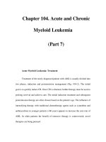
Harold Urey won the 1934 Nobel Prize in Chemistry for his discovery
of heavy hydrogen (deuterium).
womi_U 5/7/03 9:41 AM Page 564
Urey, Harold
WORLD OF MICROBIOLOGY AND IMMUNOLOGY
565
•
•
be highly relevant in current research on the possibilities of
global climate change.
In the early 1950s, Urey became interested in yet
another subject: the chemistry of the universe and of the for-
mation of the planets, including Earth. One of his first papers
on this topic attempted to provide an estimate of the relative
abundance of the elements in the universe. Although these
estimates have now been improved, they were remarkably
close to the values modern chemists now accept.
In 1958, Urey left the University of Chicago to become
Professor at Large at the University of California in San Diego
at La Jolla. At La Jolla, his interests shifted from original sci-
entific research to national scientific policy. He became
extremely involved in the U.S. space program, serving as the
first chairman of the Committee on Chemistry of Space and
Exploration of the Moon and Planets of the National Academy
of Science’s Space Sciences Board. Even late in life, Urey
continued to receive honors and awards from a grateful nation
and admiring colleagues.
See also Cell cycle and cell division; Evolution and evolution-
ary mechanisms; Evolutionary origin of bacteria and viruses
womi_U 5/7/03 9:41 AM Page 565
V
567
•
•
VACCINATION
Vaccination
Vaccination refers to a procedure in which the presence of an
antigen stimulates the formation of antibodies. The antibodies
act to protect the host from future exposure to the antigen.
Vaccination is protective against infection without the need of
suffering through a bout of a disease. In this artificial process
an individual receives the antibody-stimulating compound
either by injection or orally.
The technique of vaccination has been practiced since at
least the early decades of the eighteenth century. Then, a com-
mon practice in Istanbul was to retrieve material from the sur-
face sores of a
smallpox sufferer and rub the material into a cut
on another person. In most cases, the recipient was spared the
ravages of smallpox. The technique was refined by
Edward
Jenner
into a vaccine for cowpox in 1796.
Since Jenner’s time, vaccines for a variety of bacterial
and viral maladies have been developed. The material used for
vaccination is one of four types. Some vaccines consist of liv-
ing but weakened
viruses. These are called attenuated vac-
cines. The weakened virus does not cause an infection but
does illicit an immune response. An example of a vaccination
with attenuated material is the
measles, mumps, and rubella
(MMR) vaccine. Secondly, vaccination can involve killed
viruses or
bacteria. The biological material must be killed
such that the surface is not altered, in order to preserve the true
antigenic nature of the immune response. Also, the vaccina-
tion utilizes agents, such as alum, that act to enhance the
immune response to the killed target. Current thought is that
such agents operate by “presenting” the antigen to the
immune
system
in a more constant way. The immune system “sees”
the target longer, and so can mount a more concerted response
to it. A third type of vaccination involves an inactivated form
of a toxin produced by the target bacterium. Examples of such
so-called toxoid vaccines are the
diphtheria and tetanus vac-
cines. Lastly, vaccination can also utilize a synthetic conjugate
compound constructed from portions of two antigens. The Hib
vaccine is an example of such a biosynthetic vaccine.
During an infant’s first two years of life, a series of vac-
cinations is recommended to develop protection against a
number of viral and bacterial diseases. These are hepatitis B,
polio, measles, mumps, rubella (also called German measles),
pertussis (also called whooping cough), diphtheriae, tetanus
(lockjaw), Haemophilus influenzae type b, pneumococcal
infections, and chickenpox. Typically, vaccination against a
specific microorganism or groups of organisms is repeated
three or more times at regularly scheduled intervals. For
example, vaccination against diphtheria, tetanus, and pertussis
is typically administered at two months of age, four months,
six months, 15–18 months, and finally at four to six years of
age.
Often, a single vaccination will not suffice to develop
immunity to a given target antigen. For immunity to develop it
usually takes several doses over several months or years. A
series of vaccinations triggers a greater production of
antibody
by the immune system, and primes the antibody producing cells
such that they retain the memory (a form of protein coding and
antibody formation) of the stimulating antigen for along time.
For some diseases, this memory can last for a lifetime follow-
ing the vaccination schedule. For other diseases, such as tetanus,
adults should be vaccinated every ten years in order to keep
their body primed to fight the tetanus microorganism. This peri-
odic vaccination is also referred to as a booster shot. The use of
booster vaccinations produces a long lasting immunity.
Vaccination acts on the lymphocyte component of the
immune system. Prior to vaccination there are a myriad of lym-
phocytes. Each one recognizes only a single protein or bit of the
protein. No other lymphocyte recognizes the same site. When
vaccination occurs, a lymphocyte will be presented with a rec-
ognizable protein target. The lymphocyte will be stimulated to
divide and some of the daughter cells will begin to produce anti-
body to the protein target. With time, there will be many daugh-
ter lymphocytes and much antibody circulating in the body.
With the passage of more time, the antibody production
ceases. But the lymphocytes that have been produced still
retain the memory of the target protein. When the target is pre-
womi_V 5/7/03 10:59 AM Page 567
Vaccine
WORLD OF MICROBIOLOGY AND IMMUNOLOGY
568
•
•
sented again to the lymphocytes, as happens in the second vac-
cination in a series, the many lymphocytes are stimulated to
divide into daughter cells, which in turn form antibodies.
Thus, the second time around, a great deal more antibody is
produced. The antibody response also becomes highly specific
for the target. For example, if the target is a virus that causes
polio, then a subsequent entry of the virus into the body will
trigger a highly specific and prompt immune response, which
is designed to quell the invader.
Most vaccinations involve the injection of the immune
stimulant. However, oral vaccination has also proven effective
and beneficial. The most obvious example is the oral vaccine
to polio devised by
Albert Sabin. Oral vaccination is often lim-
ited by the passage of the vaccine through the highly acidic
stomach. In the future it is hoped that the bundling of the vac-
cine in a protective casing will negate the damage caused by
passage trough the stomach. Experiments using bags made out
of lipid molecules (liposomes) have demonstrated both pro-
tection of the vaccine and the ability to tailor the liposome
release of the vaccine.
The nature of vaccination, with the use of living or dead
material that stimulates the immune system, holds the poten-
tial for side effects. For some vaccines, the side effects are
minor. For example, a person may develop a slight ache and
redness at the site of injection. In some very rare cases, how-
ever, more severe reactions can occur, such as convulsions and
high fever. However, while there will always be a risk of an
adverse reaction from any vaccination, the risk of developing
disease is usually far greater than the probability of experi-
encing severe side effects.
See also Adjuvant; Anti-adhesion methods; Immune stimula-
tion, as a vaccine
VACCINE
Vaccine
A vaccine is a medical preparation given to provide immunity
from a disease. Vaccines use a variety of different substances
ranging from dead
microorganisms to genetically engineered
antigens to defend the body against potentially harmful
microorganisms. Effective vaccines change the
immune sys-
tem
by promoting the development of antibodies that can
quickly and effectively attack a disease causing microorgan-
ism when it enters the body, preventing disease development.
The development of vaccines against diseases ranging
from polio and
smallpox to tetanus and measles is considered
among one of the great accomplishments of medical science.
Vaccination via injection.
womi_V 5/7/03 10:59 AM Page 568
Vaccine
WORLD OF MICROBIOLOGY AND IMMUNOLOGY
569
•
•
Contemporary researchers are continually attempting to
develop new vaccinations against such diseases as Acquired
Immune Deficiency Syndrome (
AIDS), cancer, influenza, and
other diseases.
Physicians have long observed that individuals who
were exposed to an infectious disease and survived were
somehow protected against that disease in the future. Prior to
the invention of vaccines, however, infectious diseases swept
through towns, villages, and cities with a horrifying
vengeance.
The first effective vaccine was developed against small-
pox, an international peril that killed thousands of its victims
and left thousands of others permanently disfigured. The dis-
ease was so common in ancient China that newborns were not
named until they survived the disease. The development of the
vaccine in the late 1700s followed centuries of innovative
efforts to fight smallpox.
The ancient Chinese were the first to develop an effec-
tive measure against smallpox. A snuff made from powdered
smallpox scabs was blown into the nostrils of uninfected indi-
viduals. Some individuals died from the therapy; however, in
most cases, the mild infection produced offered protection
from later, more serious infection.
By the late 1600s, some European peasants employed a
similar method of immunizing themselves against smallpox.
In a practice referred to as “buying the smallpox,” peasants in
Poland, Scotland, and Denmark reportedly injected the small-
pox virus into the skin to obtain immunity. At the time, con-
ventional medical doctors in Europe relied solely on isolation
and quarantine of people with the disease.
Changes in these practices took place, in part, through
the vigorous effort of Lady
Mary Wortley Montague, the wife
of the British ambassador to Turkey in the early 1700s.
Montague said the Turks injected a preparation of small pox
scabs into the veins of susceptible individuals. Those injected
generally developed a mild case of smallpox from which they
recovered rapidly, Montague wrote.
Upon her return to Great Britain, Montague helped con-
vince King George I to allow trials of the technique on inmates
in Newgate Prison. Success of the trials cleared the way for
variolation, or the direct injection of smallpox, to become
accepted medical practice in England until a
vaccination was
developed later in the century. Variolation also was credited
with protecting United States soldiers from smallpox during
the Revolutionary War.
Regardless, doubts remained about the practice.
Individuals were known to die after receiving the smallpox
injections.
The next leap in the battle against smallpox occurred
when
Edward Jenner (1749–1823) acted on a hunch. Jenner
observed that people who were in contact with cows often
developed
cowpox, which caused pox but was not life threat-
ening. Those people did not develop smallpox. In 1796, Jenner
decided to test his hypothesis that cowpox could be used to
protect humans against smallpox. Jenner injected a healthy
eight-year-old boy with cowpox obtained from a milkmaid’s
sore. The boy was moderately ill and recovered. Jenner then
injected the boy twice with the smallpox virus, and the boy did
not get sick.
Jenner’s discovery launched a new era in medicine, one
in which the intricacies of the immune system would become
increasingly important. Contemporary knowledge suggests
that cowpox was similar enough to smallpox that the
antigen
included in the vaccine stimulated an immune response to
smallpox. Exposure to cowpox antigen transformed the boy’s
immune system, generating cells that would remember the
original antigen. The smallpox vaccine, like the many others
that would follow, carved a protective pattern in the immune
system, one that conditioned the immune system to move faster
and more efficiently against future infection by smallpox.
The term vaccination, taken from the Latin for cow
(vacca) was developed by
Louis Pasteur (1822–1895) a cen-
tury later to define Jenner’s discovery. The term also drew
from the word vaccinia, the virus drawn from cowpox and
developed in the laboratory for use in the smallpox vaccine. In
spite of Jenner’s successful report, critics questioned the wis-
dom of using the vaccine, with some worrying that people
injected with cowpox would develop animal characteristics,
such as women growing animal hair. Nonetheless, the vaccine
gained popularity, and replaced the more risky direct inocula-
tion with smallpox. In 1979, following a major cooperative
effort between nations and several international organizations,
world health authorities declared smallpox the only infectious
disease to be completely eliminated.
The concerns expressed by Jenner’s contemporaries
about the side effects of vaccines would continue to follow the
pioneers of vaccine development. Virtually all vaccinations
continue to have side effects, with some of these effects due to
the inherent nature of the vaccine, some due to the potential
for impurities in a manufactured product, and some due to the
potential for human error in administering the vaccine.
Virtually all vaccines would also continue to attract
intense public interest. This was demonstrated in 1885 when
Louis Pasteur (1822–1895) saved the life of Joseph Meister, a
nine year old who had been attacked by a rabid dog. Pasteur’s
series of experimental
rabies vaccinations on the boy proved
the effectiveness of the new vaccine.
Until development of the rabies vaccine, Pasteur had
been criticized by the public, though his great discoveries
included the development of the
food preservation process
called
pasteurization. With the discovery of a rabies vaccine,
Pasteur became an honored figure. In France, his birthday
declared a national holiday, and streets renamed after him.
Pasteur’s rabies vaccine, the first human vaccine cre-
ated in a laboratory, was made of an extract gathered from the
spinal cords of rabies-infected rabbits. The live virus was
weakened by drying over potash. The new vaccination was far
from perfect, causing occasional fatalities and temporary
paralysis. Individuals had to be injected 14–21 times.
The rabies vaccine has been refined many times. In the
1950s, a vaccine grown in duck embryos replaced the use of
live virus, and in 1980, a vaccine developed in cultured human
cells was produced. In 1998, the newest vaccine technology—
genetically engineered vaccines—was applied to rabies. The
new
DNA vaccine cost a fraction of the regular vaccine. While
womi_V 5/7/03 10:59 AM Page 569
Vaccine
WORLD OF MICROBIOLOGY AND IMMUNOLOGY
570
•
•
only a few people die of rabies each year in the United States,
more than 40,000 die worldwide, particularly in Asia and
Africa. The less expensive vaccine will make vaccination far
more available to people in less developed nations.
The story of the most celebrated vaccine in modern
times, the polio vaccine, is one of discovery and revision.
While the
viruses that cause polio appear to have been present
for centuries, the disease emerged to an unusual extent in the
early 1900s. At the peak of the epidemic, in 1952, polio killed
3,000 Americans and 58,000 new cases of polio were reported.
The crippling disease caused an epidemic of fear and illness as
Americans—and the world—searched for an explanation of
how the disease worked and how to protect their families.
The creation of a vaccine for
poliomyelitis by Jonas Salk
(1914–1995) in 1955 concluded decades of a drive to find a
cure. The Salk vaccine, a killed virus type, contained the three
types of polio virus which had been identified in the 1940s.
In 1955, the first year the vaccine was distributed, dis-
aster struck. Dozens of cases were reported in individuals who
had received the vaccine or had contact with individuals who
had been vaccinated. The culprit was an impure batch of vac-
cine that had not been completely inactivated. By the end of
the incident, more than 200 cases had developed and 11 peo-
ple had died.
Production problems with the Salk vaccine were over-
come following the 1955 disaster. Then in 1961, an oral polio
vaccine developed by Albert B. Sabin (1906–1993) was
licensed in the United States. The continuing controversy over
the virtues of the Sabin and Salk vaccines is a reminder of the
many complexities in evaluating the risks versus the benefits
of vaccines.
The Sabin vaccine, which used weakened, live polio
virus, quickly overtook the Salk vaccine in popularity in the
United States, and is currently administered to all healthy chil-
dren. Because it is taken orally, the Sabin vaccine is more con-
venient and less expensive to administer than the Salk vaccine.
Advocates of the Salk vaccine, which is still used exten-
sively in Canada and many other countries, contend that it is
safer than the Sabin oral vaccine. No individuals have devel-
oped polio from the Salk vaccine since the 1955 incident. In
contrast, the Sabin vaccine has a very small but significant rate
of complications, including the development of polio.
However, there has not been one new case of polio in the
United States since 1975, or in the Western Hemisphere since
1991. Though polio has not been completely eradicated, there
were only 144 confirmed cases worldwide in 1999.
Effective vaccines have limited many of the life-threat-
ening infectious diseases. In the United States, children starting
kindergarten are required to be immunized against polio,
diph-
theria
, tetanus, and several other diseases. Other vaccinations
are used only by populations at risk, individuals exposed to dis-
ease, or when exposure to a disease is likely to occur due to
travel to an area where the disease is common. These include
influenza,
yellow fever, typhoid, cholera, and Hepatitis Aand B.
The influenza virus is one of the more problematic dis-
eases because the viruses constantly change, making develop-
ment of vaccines difficult. Scientists grapple with predicting
what particular influenza strain will predominate in a given
year. When the prediction is accurate, the vaccine is effective.
When they are not, the vaccine is often of little help.
The classic methods for producing vaccines use biolog-
ical products obtained directly from a virus or a
bacteria.
Depending on the vaccination, the virus or bacteria is either
used in a weakened form, as in the Sabin oral polio vaccine;
killed, as in the Salk polio vaccine; or taken apart so that a
piece of the microorganism can be used. For example, the vac-
cine for Streptococcus pneumoniae uses bacterial polysaccha-
rides, carbohydrates found in bacteria which contain large
numbers of monosaccharides, a simple sugar. These classical
methods vary in safety and efficiency. In general, vaccines that
use live bacterial or viral products are extremely effective
when they work, but carry a greater risk of causing disease.
This is most threatening to individuals whose immune systems
are weakened, such as individuals with leukemia. Children
with leukemia are advised not to take the oral polio vaccine
because they are at greater risk of developing the disease.
Vaccines which do not include a live virus or bacteria tend to
be safer, but their protection may not be as great.
The classical types of vaccines are all limited in their
dependence on biological products, which often must be kept
cold, may have a limited life, and can be difficult to produce.
The development of recombinant vaccines—those using chro-
mosomal parts (or DNA) from a different organism—has gen-
erated hope for a new generation of man-made vaccines. The
hepatitis B vaccine, one of the first recombinant vaccines to be
approved for human use, is made using recombinant
yeast
cells genetically engineered to include the gene coding for the
hepatitis B antigen. Because the vaccine contains the antigen,
it is capable of stimulating
antibody production against hepa-
titis B without the risk that live hepatitis B vaccine carries by
introducing the virus into the blood stream.
As medical knowledge has increased—particularly in the
field of DNA vaccines—researchers have set their sights on a
wealth of possible new vaccines for cancer, melanoma, AIDS,
influenza, and numerous others. Since 1980, many improved
vaccines have been approved, including several genetically
engineered (recombinant) types which first developed during an
experiment in 1990. These recombinant vaccines involve the
use of so-called “naked DNA.” Microscopic portions of a
viruses’ DNA are injected into the patient. The patient’s own
cells then adopt that DNA, which is then duplicated when the
cell divides, becoming part of each new cell. Researchers have
reported success using this method in laboratory trials against
influenza and
malaria. These DNA vaccines work from inside
the cell, not just from the cell’s surface, as other vaccines do,
allowing a stronger cell-mediated fight against the disease.
Also, because the influenza virus constantly changes its surface
proteins, the immune system or vaccines cannot change quickly
enough to fight each new strain. However, DNA vaccines work
on a core protein, which researchers believe should not be
affected by these surface changes.
Since the emergence of AIDS in the early 1980s, a
worldwide search against the disease has resulted in clinical
trials for more than 25 experimental vaccines. These range
from whole-inactivated viruses to genetically engineered
types. Some have focused on a therapeutic approach to help
womi_V 5/7/03 10:59 AM Page 570
Vaccine
WORLD OF MICROBIOLOGY AND IMMUNOLOGY
571
•
•
infected individuals to fend off further illness by stimulating
components of the immune system; others have genetically
engineered a protein on the surface of
HIV to prompt immune
response against the virus; and yet others attempted to protect
uninfected individuals. The challenges in developing a protec-
tive vaccine include the fact that HIV appears to have multiple
viral strains and mutates quickly.
In January 1999, a promising study was reported in
Science magazine of a new AIDS vaccine created by injecting
a healthy cell with DNA from a protein in the AIDS virus that
is involved in the infection process. This cell was then injected
with genetic material from cells involved in the immune
response. Once injected into the individual, this vaccine
“catches the AIDS virus in the act,” exposing it to the immune
system and triggering an immune response. This discovery
offers considerable hope for development of an effective vac-
cine. As of June 2002, a proven vaccine for AIDS had not yet
been proven in clinical trials.
Stimulating the immune system is also considered key
by many researchers seeking a vaccine for cancer. Currently
numerous clinical trials for cancer vaccines are in progress,
with researchers developing experimental vaccines against
cancer of the breast, colon, and lung, among other areas.
Promising studies of vaccines made from the patient’s own
tumor cells and genetically engineered vaccines have been
reported. Other experimental techniques attempt to penetrate
the body in ways that could stimulate vigorous immune
responses. These include using bacteria or viruses, both
known to be efficient travelers in the body, as carriers of vac-
cine antigens. Such bacteria or viruses would be treated or
engineered to make them incapable of causing illness.
Current research also focuses on developing better vac-
cines. The Children’s Vaccine Initiative, supported by the
World Health Organization, the United Nation’s Children’s
Fund, and other organizations, are working diligently to make
vaccines easier to distribute in developing countries. Although
more than 80% of the world’s children were immunized by
1990, no new vaccines have been introduced extensively since
then. More than four million people, mostly children, die
needlessly every year from preventable diseases. Annually,
measles kills 1.1 million children worldwide; whooping cough
(
pertussis) kills 350,000; hepatitis B 800,000; Haemophilus
influenzae type b (Hib) 500,000; tetanus 500,000; rubella
300,000; and yellow fever 30,000. Another 8 million die from
diseases for which vaccines are still being developed. These
include pneumococcal
pneumonia (1.2 million); acute respira-
tory virus infections (400,000), malaria (2 million); AIDS (2.3
million); and rotavirus (800,000). In August, 1998, the Food
and Drug Administration approved the first vaccine to prevent
rotavirus—a severe diarrhea and vomiting infection.
The measles epidemic of 1989 was a graphic display of
the failure of many Americans to be properly immunized. A
total of 18,000 people were infected, including 41 children
who died after developing measles, an infectious, viral illness
whose complications include pneumonia and encephalitis. The
epidemic was particularly troubling because an effective, safe
vaccine against measles has been widely distributed in the
United States since the late 1960s. By 1991, the number of
new measles cases had started to decrease, but health officials
warned that measles remained a threat.
This outbreak reflected the limited reach of vaccination
programs. Only 15% of the children between the ages of 16
and 59 months who developed measles between 1989 and
1991 had received the recommended measles vaccination. In
many cases parent’s erroneously reasoned that they could
avoid even the minimal risk of vaccine side effects “because
all other children were vaccinated.”
Nearly all children are immunized properly by the time
they start school. However, very young children are far less
likely to receive the proper vaccinations. Problems behind the
lack of
immunization range from the limited health care
received by many Americans to the increasing cost of vacci-
nations. Health experts also contend that keeping up with a
vaccine schedule, which requires repeated visits, may be too
challenging for Americans who do not have a regular doctor or
health provider.
Internationally, the challenge of vaccinating large num-
bers of people has also proven to be immense. Also, the reluc-
tance of some parents to vaccinate their children due to
potential side effects has limited vaccination use. Parents in
Vaccines stimulate the production of antibodies that provide immunity
from disease.
womi_V 5/7/03 10:59 AM Page 571
Varicella
WORLD OF MICROBIOLOGY AND IMMUNOLOGY
572
•
•
the United States and several European countries have balked
at vaccinating their children with the pertussis vaccine due to
the development of neurological complications in a small
number of children given the vaccine. Because of incomplete
immunization, whooping cough remains common in the
United States, with 30,000 cases and about 25 deaths due to
complications annually. One response to such concerns has
been testing in the United States of a new pertussis vaccine
that has fewer side effects.
Researchers look to genetic engineering, gene discovery,
and other innovative technologies to produce new vaccines.
See also AIDS, recent advances in research and treatment;
Antibody formation and kinetics; Bacteria and bacterial infec-
tion; Bioterrorism, protective measures; Immune stimulation,
as a vaccine; Immunity, active, passive and delayed;
Immunity, cell mediated; Immunity, humoral regulation;
Immunochemistry; Immunogenetics; Immunologic therapies;
Immunology; Interferon actions; Poliomyelitis and polio;
Smallpox, eradication, storage, and potential use as a bacteri-
ological weapon
VARICELLA
Varicella
Varicella, commonly known as chickenpox, is a disease char-
acterized by skin lesions and low-grade fever, and is common
in the United States and other countries located in areas with
temperate climates. The incidence of varicella is extremely
high; almost everyone living in the United States is exposed to
the disease, usually during childhood, but sometimes in adult-
hood. In the United States, about 3.9 million people a year
contract varicella. A highly contagious disease, varicella is
caused by Varicella-Zoster virus (VZV), the same virus that
causes the skin disease shingles. For most cases of varicella,
no treatment besides comfort measures and management of
itching and fever is necessary. In some cases, however, vari-
cella may evolve into more serious conditions, such as
bacte-
rial infection
of the skin lesions or pneumonia. These
complications tend to occur in persons with weakened
immune systems, such as children receiving
chemotherapy for
cancer, or people with Acquired Immune Deficiency
Syndrome (
AIDS). A vaccine for varicella is now receiving
widespread use.
There are two possible origins for the colloquialism
“chickenpox.” Some think that “chicken” comes from the
French word chiche (chick-pea) because at one stage of the
disease, the lesions may resemble chick-peas. Others think
that “chicken” may have evolved from the Old English word
gigan (to itch). Interestingly, the term “varicella” is a diminu-
tive form of the term “variola,” the Latin word for
smallpox.
Although both varicella and smallpox are viral diseases that
cause skin lesions, smallpox is more deadly and its lesions
cause severe scarring.
Varicella is spread by breathing in respiratory droplets
spread through the air by a cough or sneeze of an infected indi-
vidual. Contact with the fluid from skin lesions can also
spread the virus. The incubation period, or the time from expo-
sure to VZV to the onset of the disease, is about 14–15 days.
The most contagious period is just prior to the appearance of
the rash, and early in the illness, when fresh vesicles are still
appearing. The first sign of varicella in children is often the
appearance of the varicella rash. Adults and some children
may have a prodrome, or series of warning symptoms. This
prodrome is typical of the flu, and includes headache, fatigue,
backache, and a fever. The onset of the rash is quite rapid.
First, a diffuse, small, red dot-like rash appears on the skin.
Soon, a vesicle containing clear fluid appears in the center of
the dots. The vesicle rapidly dries, forming a crust. This cycle,
from the appearance of the dot to the formation of the crust,
can take place within eight to 12 hours. As the crust dries, it
falls off, leaving a slight depression that eventually recedes.
Significant scarring from varicella is rare.
Over the course of a case of varicella, an individual may
develop between 250 and 500 skin lesions. The lesions occur
in waves, with the first set of lesions drying up just as succes-
sive waves appear. The waves appear over two to four days.
The entire disease runs its course in about a week, but the
lesions continue to heal for about two to three weeks. The
lesions first appear on the scalp and trunk. Most of the lesions
in varicella are found at the center of the body; few lesions
form on the soles and palms. Lesions are also found on the
mucous membranes, such as the respiratory tract, the gas-
trointestinal tract, and the urogenital tract. Researchers think
that the lesions on the respiratory tract may help transmit the
disease. If a person with respiratory lesions coughs, they may
spray some of the vesicle fluid into the atmosphere, to be
breathed by other susceptible persons.
Although the lesions may appear alarming, varicella in
children is usually a mild disease with few complications and
a low fever. Occasionally, if the rash is severe, the fever may
be higher. Varicells is more serious in adults, who usually have
a higher fever and general malaise. The most common com-
plaint about varicella from both children and adults is the itch-
ing caused by the lesions. It is important not to scratch the
lesions, as scratching may cause scarring.
Because varicella is usually a mild disease, no drug treat-
ment is normally prescribed. For pain or fever relief associated
with varicella, physicians recommended avoiding salicylate, or
aspirin. Salicylate may contribute to Reye’s syndrome, a seri-
ous neurological condition that is especially associated with
aspirin intake and varicella; in fact, 20–30% of the total cases
of Reye’s syndrome occur in children with varicella.
Varicella, although not deadly for most people, can be
quite serious in those who have weakened immune systems,
and drug therapy is recommended for these cases.
Antiviral
drugs
(such as acyclovir) have been shown to lessen the sever-
ity and duration of the disease, although some of the side
effects, such as gastrointestinal upset, can be problematic.
If the lesions are severe and the person has scratched
them, bacterial infection of the lesions can result. This com-
plication is managed with antibiotic treatment. A more serious
complication is pneumonia. Pneumonia is rare in otherwise
healthy children and is more often seen in older patients or in
children who already have a serious disease, such as cancer.
Pneumonia is also treated with
antibiotics. Another complica-
womi_V 5/7/03 10:59 AM Page 572
Varicella zoster virus
WORLD OF MICROBIOLOGY AND IMMUNOLOGY
573
•
•
tion of varicella is shingles. Shingles are painful outbreaks of
skin lesions that occur some years after a bout with varicella.
Shingles are caused by VZV left behind in the body that even-
tually reactivates. Shingles causes skin lesions and burning
pain along the region served by a specific nerve. It is not clear
why VZV is reactivated in some people and not in others, but
many people with compromised immune systems can develop
severe, even life-threatening cases of shingles.
Pregnant women are more susceptible to varicella,
which also poses a threat to both prenatal and newborn chil-
dren. If a woman contracts varicella in the first trimester (first
three months) of pregnancy, the fetus may be at increased risk
for birth defects such as eye damage. A newborn may contract
varicella in the uterus if the mother has varicella five days
before birth. Newborns can also contract varicella if the
mother has the disease up to two days after birth. Varicella can
be a deadly disease for newborns; the fatality rate from vari-
cella in newborns up to five days old approaches 30%. For this
reason, women contemplating pregnancy may opt to be vacci-
nated with the new VZV vaccine prior to conception if they
have never had the disease. If this has not been done, and a
pregnant woman contracts varicella, an injection of varicella-
zoster immunoglobulin can lessen the chance of complications
to the fetus.
Researchers have long noted the seasonality of vari-
cella. According to their research, varicella cases occur at their
lowest rate during September. Numbers of cases increase
throughout the autumn, peak in March and April, and then fall
sharply once summer begins. This cycle corresponds to the
typical school year in the United States. When children go
back to school in the fall, they begin to spread the disease;
when summer comes and school ends, cases of varicella
diminish. Varicella can spread quickly within a school when
one child contracts varicella. This child rapidly infects other
susceptible children. Soon, all the children who had not had
varicella contract the disease within two or three cycles of
transmission. It is not uncommon for high numbers of children
to be infected during a localized outbreak; one school with 69
children reported that the disease struck 67 of these students.
Contrary to popular belief, it is possible to get varicella
a second time. If a person had a mild case during childhood,
his or her
immunity to the virus may be weaker than that of
someone who had a severe childhood case. In order to prevent
varicella, especially in already-ill children and immunocom-
promised patients, researchers have devised a VZV vaccine,
consisting of live, attenuated (modified) VZV.
Immunization
recommendations of the American Academy of Pediatrics
state that children between 12 and 18 months of age who have
not yet had varicella should receive the vaccine. Immunization
can be accomplished with a single dose. Children up to the age
of 13 who have had neither varicella nor the immunization,
should also receive a single dose of the vaccine. Children
older than age 13 who have never had either varicella or the
vaccine should be immunized with two separate doses, given
about a month apart. The vaccine provokes immunity against
the virus. Although some side effects have been noted, includ-
ing a mild rash and the reactivation of shingles, the vaccine is
considered safe and effective.
See also Immunity, active, passive and delayed; Immunity,
cell mediated; Viruses and responses to viral infection
VARICELLA ZOSTER VIRUS
Varicella zoster virus
Varicella zoster virus is a member of the alphaherpesvirus
group and is the cause of both chickenpox (also known as vari-
cella) and shingles (
herpes zoster).
The virus is surrounded by a covering, or envelope, that
is made of lipid. As such, the envelope dissolves readily in sol-
vents such as alcohol. Wiping surfaces with alcohol is thus an
effective means of inactivating the virus and preventing spread
of chickenpox. Inside the lipid envelope is a protein shell that
houses the
deoxyribonucleic acid.
Varicella zoster virus is related to Herpes Simplex
viruses types 1 and 2. Indeed, nucleic acid analysis has
revealed that the genetic material of the three viruses is highly
similar, both in the genes present and in the arrangement of
the genes.
Chickenpox is the result of a person’s first infection
with the virus. Typically, chickenpox occurs most often in
children. From 75% to 90% of the cases of chickenpox occur
in children under five years old. Acquisition of the virus is
usually via inhalation of droplets containing the virus. From
the lung the virus migrates to the blood stream. Initially a sore
throat leads to a blister-like rash that appears on the skin and
the mucous membranes, as the virus is carried through the
blood stream to the skin. The extent of the rash varies, from
minimal to all over the body. The latter is also accompanied by
fever, itching, abdominal pain, and a general feeling of tired-
ness. Recovery is usually complete within a week or two and
immunity to another bout of chickenpox is life-long.
In terms of a health threat, childhood chickenpox is
advantageous. The life-long immunity conferred to the child
prevents adult onset infections that are generally more severe.
However, chickenpox can be dangerous in infants, whose
immune systems are undeveloped. Also chickenpox carries the
threat of the development of sudden and dangerous liver and
brain damage. This condition, called Reye’s Syndrome, seems
related to the use of aspirin to combat the fever associated with
chickenpox (as well as other childhood viruses). When adults
acquire chickenpox, the symptoms can be much more severe
than those experienced by a child. In immunocompromised
people, or those suffering from leukemia, chickenpox can be
fatal. The disease can be problematic in pregnant women in
terms of birth defects and the development of
pneumonia.
Treatment for chickenpox is available. A drug called
acyclovir can slow the replication of the virus. Topical lotions
can ease the itching associated with the disease. However, in
mild to moderate cases, intervention is unnecessary, other
than keeping the affected person comfortable. The life-long
immunity conferred by a bout of chickenpox is worth the
temporary inconvenience of the malady. The situation is dif-
ferent for adults. Fortunately for adults, a
vaccine to chicken-
pox exists for those who have not contracted chickenpox in
their childhood.
womi_V 5/7/03 10:59 AM Page 573
Variola virus
WORLD OF MICROBIOLOGY AND IMMUNOLOGY
574
•
•
Naturally acquired immunity to chickenpox does not
prevent individuals from contracting shingles years, even
decades later. Shingles occurs in between 10% and 20% of
those who have had chickenpox. In the United States, upwards
of 800,000 people are afflicted with shingles each year. The
annual number of shingles sufferers worldwide is in the mil-
lions. The disease occurs most commonly in those who are
over 50 years of age.
As the symptoms of chickenpox fade, varicella zoster
virus is not eliminated from the body. Rather, the virus lies
dormant in nerve tissue, particularly in the face and the body.
The roots of sensory nerves in the spinal cord are also a site of
virus hibernation. The virus is stirred to replicate by triggers
that are as yet unclear. Impairment of the
immune system
seems to be involved, whether from immunodeficiency dis-
eases
or from cancers, the effect of drugs, or a generalized
debilitation of the body with age. Whatever forces of the
immune system that normally operate to hold the hibernating
virus in check are abrogated.
Reactivation of the virus causes pain and a rash in the
region that is served by the affected nerves. The affected areas
are referred to as dermatomes. These areas appear as a rash or
blistering of the skin. This can be quite painful during the one
to two weeks they persist. Other complications can develop.
For example, shingles on the face can lead to an eye infection
causing temporary or even permanent blindness. A condition of
muscle weakness or paralysis, known as Guillan-Barre
Syndrome, can last for months after a bout of shingles. Another
condition known as postherpetic neuralgia can extend the pain
of shingles long after the visible symptoms have abated.
See also Immunity, active, passive and delayed; Infection and
resistance; Latent viruses and diseases
VARIOLA VIRUS
Variola virus
Variola virus (or variola major virus) is the virus that causes
smallpox. The virus is one of the members of the poxvirus
group (Family Poxviridae). The virus particle is brick shaped
and contains a double strand of
deoxyribonucleic acid. The
variola virus is among the most dangerous of all the potential
biological weapons.
Variola virus infects only humans. The virus can be eas-
ily transmitted from person to person via the air. Inhalation of
only a few virus particles is sufficient to establish an infection.
Transmission of the virus is also possible if items such as con-
taminated linen are handled. The various common symptoms
of smallpox include chills, high fever, extreme tiredness,
headache, backache, vomiting, sore throat with a cough, and
sores on mucus membranes and on the skin. As the sores burst
and release pus, the afflicted person can experience great pain.
Males and females of all ages are equally susceptible to infec-
tion. At the time of smallpox eradication approximately one
third of patients died—usually within a period of two to three
weeks following appearance of symptoms.
The origin of the variola virus in not clear. However, the
similarity of the virus and
cowpox virus has prompted the sug-
gestion that the variola virus is a mutated version of the cow-
pox virus. The mutation allowed to virus to infect humans. If
such a mutation did occur, then the adoption of farming activ-
ities by people, instead of the formally nomadic existence,
would have been a selective pressure for a virus to adopt the
capability to infect humans.
Vaccination to prevent infection with the variola virus is
long established. In the 1700s, English socialite and
public
health advocate Lady Mary Wortley Montague popularized the
practice of injection with the pus obtained from smallpox
sores as a protection against the disease. This technique
became known as variolation. Late in the same century,
Edward Jenner successfully prevented the occurrence of
smallpox by an injection of pus from cowpox sores. This rep-
resented the start of vaccination.
Vaccination has been very successful in dealing with
variola virus outbreaks of smallpox. Indeed, after two decades
of worldwide vaccination programs, the virus has been virtu-
ally eliminated from the natural environment. The last
recorded case of smallpox infection was in 1977 and vaccina-
tion against smallpox is not practiced anymore.
In the late 1990s, a resolution was passed at the World
Health Assembly that the remaining stocks of variola virus be
destroyed, to prevent the re-emergence of smallpox and the
misuse of the virus as a biological weapon. At the time only
two high-security laboratories were thought to contain variola
virus stock (
Centers for Disease Control and Prevention in
Atlanta, Georgia, and the Russian State Centre for Research
on
Virology and Biotechnology, Koltsovo, Russia). However,
this decision was postponed until 2002, and now the United
States government has indicated its unwillingness to comply
with the resolution for security issues related to potential
bioterrorism. Destruction of the stocks of variola virus would
deprive countries of the material needed to prepare
vaccine in
the event of the deliberate use of the virus as a biological
weapon. This scenario has gained more credence in the past
decade, as terrorist groups have demonstrated the resolve to
use biological weapons, including smallpox. In addition, intel-
ligence agencies in several Western European countries issued
opinions that additional stocks of the variola virus exist in
other than the previously authorized locations.
See also Bioterrorism, protective measures; Bioterrorism;
Centers for Disease Control (CDC); Smallpox, eradication,
storage, and potential use as a bacteriological weapon; Viral
genetics; Virology; Virus replication; Viruses and responses to
viral infection
VENTER, JOHN CRAIG (1946- )
Venter, John Craig
American molecular biologist
John Craig Venter, who until January 2002 was the President
and Chief Executive Officer of Celera Genomics, is one of the
central figures in the Human Genome Project. Venter co-
founded Celera in 1998, and he directed its research and oper-
ations while he and the company’s other scientists completed
a draft of the human genome. Using a fast sequencing tech-
womi_V 5/7/03 10:59 AM Page 574
Veterinary microbiology
WORLD OF MICROBIOLOGY AND IMMUNOLOGY
575
•
•
nique, Venter and his colleagues were able to sequence the
human genome, and the genomes of other organisms, includ-
ing the bacterium Haemophilus infuenzae,.
Venter was born in Salt Lake City, Utah. After high
school he seemed destined for a career as a surfer rather than
as a molecular biologist. But a tour of duty in Vietnam as a
hospital corpsman precipitated a change in the direction of his
life. He returned from Vietnam and entered university, earning
a doctorate in physiology and pharmacology from the
University of California at San Diego. After graduation he
took a research position at the National Institutes of Health.
While at NIH, Venter became frustrated at the then slow pace
of identifying and sequencing genes. He began to utilize a
technology that decodes only a portion of the
DNA from nor-
mal copies of genes made by living cells. These partial tran-
scripts, called expressed sequence tags, could then be used to
identify the gene-coding regions on the DNA from which they
came. The result was to speed up the identification of genes.
Hundreds of genes could be discovered in only weeks using
the method.
Supported by venture capital, Venter started a nonprofit
company called
The Institute for Genomic Research (TIGR) in
the mid-1990s. TIGR produced thousands of the expressed
sequence tag probes to the human genome.
Venter’s success and technical insight attracted the
interest of PE Biosystems, makers of automated DNA
sequencers. With financial and equipment backing from PE
Biosystems, Venter left TIGR and formed a private for-profit
company, Celera (meaning ‘swift’ in Latin). The aim was
to decode the human genome faster than the government
effort that was underway. Celera commenced operations in
May 1998.
Another of Venter’s accomplishments was to use a non-
traditional approach to quickly sequence DNA. At that time,
DNA was typically sequenced by dividing it into several large
pieces and then decoding each piece. Venter devised the so-
called shotgun method, in which a genome was blown apart
into many small bits and then to sequence them without regard
to their position. Following sequencing, supercomputer power
would reassemble the bits of sequence into the intact genome
sequence. The technique, which was extremely controversial,
was tried first on the genome of the fruit fly Drosophila. In
only a year the fruit fly genome sequence was obtained. The
sequencing of the genome of the bacterium H. influenzae fol-
lowed this.
Although the privatization of human genome sequence
data remains highly controversial, Venter’s accomplishments
are considerable, both technically and as a force within the sci-
entific community to spur genome sequencing.
See also DNA (Deoxyribonucleic acid); DNA hybridization;
Economic uses and benefits of microorganisms; Genetic code;
Genetic identification of microorganisms; Genetic mapping;
Genetic regulation of eukaryotic cells; Genetic regulation of
prokaryotic cells; Genotype and phenotype; Immunogenetics;
Molecular biology and molecular genetics
V
ETERINARY MICROBIOLOGY
Veterinary microbiology
Veterinary microbiology is concerned with the microorgan-
isms
, both beneficial and disease causing, to non-human ani-
mal life. For a small animal veterinarian, the typical animals
of concern are domesticated animals, such as dogs, cats,
birds, fish, and reptiles. Large animal veterinarians focus on
animals of economic importance, such as horses, cows,
sheep, and poultry.
The dogs and cats that are such a familiar part of the
household environment are subject to a variety of microbio-
logical origin ailments. As with humans,
vaccination of young
dogs and cats is a wise precaution to avoid microbiological
diseases later in life.
Cats can be infected by a number of
viruses and bacte-
ria
that cause respiratory tract infections. For example the bac-
terium Bordetella pertussis, the common cause of kennel
cough in dogs, also infects cats, causing the same persistent
cough. Another bacteria called Chlamydia causes another res-
piratory disease, although most of the symptoms are apparent
in the eyes.
Inflammation of the mucous covering of the eye-
lids (conjunctivitis) can be so severe that the eyes swell shut.
Cats are prone to viral infections. Coronavirus is com-
mon in environments such as animal shelters, where numbers
of cats live in close quarters. The virus causes an infection of
the intestinal tract. Feline panleukopenia is a very contagious
viral disease that causes a malaise and a decrease in the num-
ber of white blood cells. The immune disruption can leave the
cat vulnerable to other infections and can be lethal.
Fortunately, a protective
vaccine exists. Like humans, cats are
also prone to
herpes virus infections. In cats the infection is in
the respiratory tract and eyes. Severe infections can produce
blindness. Another respiratory disease, reminiscent of a
cold
in humans, is caused by a calicivirus. Pneumonia can develop
and is frequently lethal. Finally the feline leukemia virus
causes cancer of the blood. The highly contagious nature of
this virus makes vaccination prudent for young kittens.
Dogs are likewise susceptible to bacterial and viral
infections. A virus known as parainfluenzae virus also causes
kennel cough. Dogs are also susceptible to coronavirus.
Members of the bacterial genus called Leptospira can infect
the kidneys. This infection can be passed to humans and to
other animals. A very contagious viral infection, which typi-
cally accompanies bacterial infections, is called canine dis-
temper. Distemper attacks many organs in the body and can
leave the survivor permanently disabled. A vaccine against
distemper exists, but must be administered periodically
throughout the dog’s life to maintain the protection. Another
virus called parvovirus produces a highly contagious, often
fatal, infection. Once again, vaccination needs to be at regular
(usually yearly) intervals. Like humans, dogs are susceptible
to
hepatitis, a destructive viral disease of the liver. In dogs that
have not been vaccinated, the liver infection can be debilitat-
ing. Finally, dogs are also susceptible to the viral agent of
rabies. The virus, often passed to the dog via the bite of
another rabid animal, can in turn be passed onto humans.
Fortunately again, vaccination can eliminated the risk of
acquiring rabies.
womi_V 5/7/03 10:59 AM Page 575
Veterinary microbiology
WORLD OF MICROBIOLOGY AND IMMUNOLOGY
576
•
•
Microbiological infections of farm animals and poul-
try is common. For example, studies have shown that well
over half the poultry entering processing plants are infected
with the bacterium Campylobacter jejuni. Infection with
members of the bacterial genus Salmonella are almost as
common. Fecal
contamination of poultry held in close quar-
ters is responsible. Similarly the intestinal bacterium
Escherichia coli is spread from bird to bird. Improper pro-
cessing can pass on these bacteria to humans, where they
cause intestinal maladies.
Chickens and turkeys are also susceptible to a bacterial
respiratory disease caused by Mycoplasma spp. The “air sac
disease” causes lethargy, weight loss, and decreased egg pro-
duction. Poultry can also acquire a form of cholera, which is
caused by Pasteurella multocida. Examples of some other
bacteria of note in poultry are species of Clostridium (intes-
tinal tract infection and destruction of tissue), Salmonella pul-
lorum (intestinal infection that disseminates widely
throughout the body), Salmonella gallinarum (typhoid), and
Clostridium botulinum (
botulism).
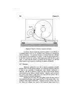
Two veterinarians treat a dog with an infected leg.
womi_V 5/7/03 10:59 AM Page 576
Viral genetics
WORLD OF MICROBIOLOGY AND IMMUNOLOGY
577
•
•
Cattle and sheep are also susceptible to microbiological
ailments. Foot and mouth disease is a prominent example.
This contagious and fatal disease can sweep through cattle and
sheep populations, causing financial ruin for ranchers.
Moreover, there is now evidence that bovine spongiform
encephalopathy, a disease caused by an infectious agent
termed a prion, may be transmissible to humans, where it is
manifest as the always lethal brain deterioration called
Creutzfeld-Jacob disease.
See also Zoonoses
V
IABLE BUT NONCULTURABLE BACTERIA
Viable but nonculturable bacteria
Viable but nonculturable bacteria are bacteria that are alive,
but which are not growing or dividing. Their metabolic activ-
ity is almost nonexistent.
This state was recognized initially by microbial ecolo-
gists examining bacterial populations in natural sediments.
Measurements of the total bacterial count, which counts both
living and dead bacteria, are often far higher than the count of
the living bacteria. At certain times of year, generally when
nutrients are plentiful, the total and living numbers match more
closely. These observations are not the result of seasonal “die-
off,” but reflect the adoption of an almost dormant mode of
existence by a sizable proportion of some bacterial populations.
A viable but nonculturable bacteria cannot be cultured
on conventional laboratory growth media but can be demon-
strated to be alive by other means, such as the uptake and
metabolism of radioactively labeled nutrients. Additionally,
the microscopic examination of populations shows the bacte-
ria to be intact. When bacteria die they often lyse, due to the
release of
enzymes that disrupt the interior and the cell wall of
the bacteria.
The viable but nonculturable state is reversible.
Bacterial that do not form spores can enter the state when con-
ditions become lethal for their continued growth. The state is
a means of bacterial survival to stresses that include elevated
salt concentration, depletion of nutrients, depletion of oxygen,
and exposure to certain wavelengths of light. When the stress
is removed, bacteria can revive and resume normal growth.
The shift to the nonculturable state triggers the expres-
sion of some 40 genes in bacteria. As well, the composition of
the cell wall changes, becoming enhanced in fatty acid con-
stituents, and the genetic material becomes coiled more tightly.
The entry of a bacterium into the nonculturable state
varies from days to months. Younger bacterial cells are capa-
ble of a more rapid transition than are older cells. In general,
however, the transition to a nonculturable state seems to be in
response to a more gradual change in the environment than
other bacterial stress responses, (e.g., spore formation,
heat
shock response
).
In contrast to the prolonged entry into the quiescent
phase, the exit from the viable but nonculturable state is quite
rapid (within hours for Vibrio vulnifucus). Other bacteria, such
as Legionella pneumophila, the causative agent of
Legionnaires’ disease, revive much more slowly. The adoption
of this mode of survival by disease-causing bacteria further
complicates strategies to detect and eradicate them.
See also Bacteria and bacterial infection; Bacterial adaptation
VIRAL EPIDEMICS
• see EPIDEMICS, VIRAL
VIRAL GENETICS
Viral genetics
Viral genetics, the study of the genetic mechanisms that oper-
ate during the life cycle of
viruses, utilizes biophysical, bio-
logical, and genetic analyses to study the viral genome and its
variation. The virus genome consists of only one type of
nucleic acid, which could be a single or double stranded
DNA
or RNA. Single stranded RNA viruses could contain positive-
sense (+RNA), which serves directly as mRNA or negative-
sense RNA (–RNA) that must use an RNA polymerase to
synthesize a complementary positive strand to serve as
mRNA. Viruses are obligate
parasites that are completely
dependant on the host cell for the replication and
transcription
of their genomes as well as the translation of the mRNA tran-
scripts into proteins. Viral proteins usually have a structural
function, making up a shell around the genome, but may con-
tain some
enzymes that are necessary for the virus replication
and life cycle in the host cell. Both bacterial virus (bacterio-
phages) and animal viruses play an important role as tools in
molecular and cellular biology research.
Viruses are classified in two families depending on
whether they have RNA or DNA genomes and whether these
genomes are double or single stranded. Further subdivision into
types takes into account whether the genome consists of a sin-
gle RNA molecule or many molecules as in the case of seg-
mented viruses. Four types of bacteriophages are widely used in
biochemical and genetic research. These are the T phages, the
temperate phages typified by
bacteriophage lambda, the small
DNA phages like M13, and the RNAphages. Animal viruses are
subdivided in many classes and types. Class I viruses contain a
single molecule of double stranded DNA and are exemplified
by adenovirus, simian virus 40 (SV40),
herpes viruses and
human papilloma viruses. Class II viruses are also called par-
voviruses and are made of single stranded DNA that is copied
in to double stranded DNA before transcription in the host cell.
Class III viruses are double stranded RNA viruses that have seg-
mented genomes which means that they contain 10–12 separate
double stranded RNA molecules. The negative strands serve as
template for mRNA synthesis. Class IV viruses, typified by
poliovirus, have single plus strand genomic RNA that serves as
the mRNA. Class V viruses contain a single negative strand
RNA which serves as the template for the production of mRNA
by specific virus enzymes. Class VI viruses are also known as
retroviruses and contain double stranded RNA genome. These
viruses have an enzyme called reverse transcriptase that can
both copy minus strand DNA from genomic RNA catalyze the
synthesis of a complementary plus DNA strand. The resulting
double stranded DNA is integrated in the host chromosome and
womi_V 5/7/03 10:59 AM Page 577
Viral vectors in gene therapy
WORLD OF MICROBIOLOGY AND IMMUNOLOGY
578
•
•
is transcribed by the host own machinery. The resulting tran-
scripts are either used to synthesize proteins or produce new
viral particles. These new viruses are released by budding, usu-
ally without killing the host cell. Both
HIV and HTLV viruses
belong to this class of viruses.
Virus genetics are studied by either investigating genome
mutations or exchange of genetic material during the life cycle
of the virus. The frequency and types of genetic variations in the
virus are influenced by the nature of the viral genome and its
structure. Especially important are the type of the nucleic acid
that influence the potential for the viral genome to integrate in
the host, and the segmentation that influence exchange of
genetic information through assortment and
recombination.
Mutations in the virus genome could either occur spon-
taneously or be induced by physical and chemical means.
Spontaneous mutations that arise naturally as a result of viral
replication are either due to a defect in the genome replication
machinery or to the incorporation of an analogous base instead
of the normal one. Induced virus
mutants are obtained by either
using chemical mutants like nitrous oxide that acts directly on
bases and modify them or by incorporating already modified
bases in the virus genome by adding these bases as substrates
during virus replication. Physical agents such as ultra-violet
light and x rays can also be used in inducing mutations.
Genotypically, the induced mutations are usually point muta-
tions, deletions, and rarely insertions. The
phenotype of the
induced mutants is usually varied. Some mutants are condi-
tional lethal mutants. These could differ from the wild type
virus by being sensitive to high or low temperature. A low tem-
perature mutant would for example grow at 88°F (31°C) but
not at 100°F (38°C), while the wild type will grow at both tem-
peratures. A mutant could also be obtained that grows better at
elevated temperatures than the wild type virus. These mutants
are called hot mutants and may be more dangerous for the host
because fever, which usually slows the growth of wild type
virus, is ineffective in controlling them. Other mutants that are
usually generated are those that show drug resistance, enzyme
deficiency, or an altered pathogenicity or host range. Some of
these mutants cause milder symptoms compared to the parental
virulent virus and usually have potential in
vaccine develop-
ment as exemplified by some types of
influenza vaccines.
Besides mutation, new genetic variants of viruses also
arise through exchange of genetic material by recombination
and reassortment. Classical recombination involves the break-
ing of covalent bonds within the virus nucleic acid and
exchange of some DNA segments followed by rejoining of the
DNA break. This type of recombination is almost exclusively
reserved to DNA viruses and retroviruses. RNAviruses that do
not have a DNA phase rarely use this mechanism.
Recombination usually enables a virus to pick up genetic
material from similar viruses and even from unrelated viruses
and the eukaryotic host cells. Exchange of genetic material
with the host is especially common with retroviruses.
Reassortment is a non-classical kind of recombination that
occurs if two variants of a segmented virus infect the same
cell. The resulting progeny virions may get some segments
from one parent and some from the other. All known seg-
mented virus that infect humans are RNA viruses. The process
of reassortment is very efficient in the exchange of genetic
material and is used in the generation of viral vaccines espe-
cially in the case of influenza live vaccines. The ability of
viruses to exchange genetic information through recombina-
tion is the basis for virus-based vectors in recombinant DNA
technology and hold great promises in the development of
gene therapy. Viruses are attractive as vectors in gene therapy
because they can be targeted to specific tissues in the organs
that the virus usually infect and because viruses do not need
special chemical reagents called transfectants that are used to
target a plasmid vector to the genome of the host.
Genetic variants generated through mutations, recom-
bination or reassortment could interact with each other if
they infected the same host cell and prevent the appearance
of any
phenotype. This phenomenon, where each mutant
provide the missing function of the other while both are still
genotypically mutant, is known as complementation. It is
used as an efficient tool to determine if mutations are in
unique or in different genes and to reveal the minimum num-
ber of genes affecting a function. Temperature sensitive
mutants that have the same mutation in the same gene will
for example not be able to
complement each other. It is
important to distinguish complementation from multiplicity
reactivation where a higher dose of inactivated mutants will
be reactivated and infect a cell because these inactivated
viruses cooperate in a poorly understood process. This reac-
tivation probably involves both a complementation step that
allows defective viruses to replicate and a recombination
step resulting in new genotypes and sometimes regeneration
of the wild type. The viruses that need complementation to
achieve an infectious cycle are usually referred to as defec-
tive mutants and the complementing virus is the helper virus.
In some cases, the defective virus may interfere with and
reduce the infectivity of the helper virus by competing with
it for some factors that are involved in the viral life cycle.
These defective viruses called “defective interfering” are
sometimes involved in modulating natural infections.
Different wild type viruses that infect the same cell may
exchange coat components without any exchange of genetic
material. This phenomenon, known as phenotypic mixing is
usually restricted to related viruses and may change both the
morphology of the packaged virus and the tropism or tissue
specificity of these infectious agents.
See also Viral vectors in gene therapy; Virology; Virus repli-
cation; Viruses and responses to viral infection
VIRAL INFECTIONS
• see VIRUSES AND RESPONSES
TO VIRAL INFECTION
VIRAL VECTORS IN GENE THERAPY
Viral vectors in gene therapy
Gene therapy is the introduction of a gene into cells to reverse
a functional defect caused by a defect in a host genome (the
set of genes present in an organism).
womi_V 5/7/03 10:59 AM Page 578
Virology, viral classification, types of viruses
WORLD OF MICROBIOLOGY AND IMMUNOLOGY
579
•
•
The use of viruses quickly became an attractive possi-
bility once the possibility of gene therapy became apparent.
Viruses require other cells for their replication. Indeed, an
essential feature of a
virus replication cycle is the transfer of
their genetic material (
deoxyribonucleic acid, DNA; or ribonu-
cleic acid
, RNA) into the host cell, and the replication of that
material in the host cell. By incorporating other DNA or RNA
into the virus genome, the virus then becomes a vector for the
transmission of that additional genetic material. Finally, if the
inserted genetic material is the same as a sequence in the host
cell that is defective, then the expression of the inserted gene
will provide the product that the defective host genome does
not. As a result, host defective host genetic function and the
consequences of the defects can be reduced or corrected.
Retroviruses contain RNA as the genetic material. A
viral enzyme called reverse transcriptase functions to manu-
facture DNA from the RNA, and the DNA can then become
incorporated into the host DNA. Despite the known involve-
ment of some retroviruses in cancer, these viruses are attrac-
tive for gene therapy because of their pronounced tendency to
integrate the viral DNA into the host genome. Retroviruses
used as gene vectors also have had the potential cancer-caus-
ing genetic information deleted. The most common retrovirus
that has been used in experimental gene therapy is the
Moloney murine leukaemia virus. This virus can infect cells of
both mice and humans. This makes the results obtained from
mouse studies more relevant to humans.
Adenoviruses are another potential gene vector. Once
they have infected the host cell, many rounds of DNA replica-
tion can occur. This is advantageous, as much of the therapeu-
tic product could be produced. However, because integration
of the virally transported gene does not occur, the expression
of the gene only occurs for a relatively short time. To produce
levels of the gene product that would have a substantial effect
on a patient, the virus vector needs to administered repeatedly.
As for retroviruses, the adenoviruses used as vectors need to
be crippled so as to prevent the production of new viruses.
Adenovirus vector has been used to correct
mutations
the gene that is defective in cystic fibrosis. However, as of
May 2002, the success rate in human trials remained low. In
addition, the immune response to the high levels of the vector
that are needed can be problematic.
Another important aspect of gene therapy concerns the
target of the viral vectors. The viruses need to be targeted at
host cells that are actively dividing, because only in cells in
which DNA replication is occurring will the inserted viral
genetic material be replicated. This is one reason why cancers
are a conceptually attractive target of virus-mediated gene
therapy, as cancerous cells are dangerous by virtue of their
rapid and uncontrolled division.
Cancerous cells arise by some form of mutation.
Therefore, therapy to replace defective genes with functional
genes holds promise for cancer researchers. The target of gene
therapy can vary, as many cancers have mutations that direct a
normal cell towards acquiring the potential to become cancer-
ous, and other mutations that inactivate mechanisms that func-
tion to regulate growth control. Furthermore, gene therapy can
be directed at the
immune system rather than directly at the
cancerous cell. An example of this strategy is known as
immunopotentiation (the enhancement of the immune
response to cancers).
A risk of viral gene therapy, in those viruses that oper-
ate by integrating genetic material into the host genome, is the
possibility of damage to the host DNA by the insertion.
Alteration of some other host gene could have unforeseen and
undesirable side effects. The elimination of this possibility
will require further technical refinements. Adenoviruses are
advantageous in this regard as the replication of their DNA in
the host cell does not involve insertion of the viral DNA into
the host DNA. Accordingly, the possibility of mutations due to
insertion do not exist.
The September 1999 death of an 18 year old patient
with a rare metabolic condition, who died while receiving
viral gene therapy, considerably slowed progress on clinical
applications of viral gene therapy.
See also Biotechnology
VIROLOGY, VIRAL CLASSIFICATION,
TYPES OF VIRUSES
Virology, viral classification, types of viruses
Virology is the discipline of microbiology that is concerned
with the study of
viruses. Viruses are essentially nonliving
repositories of nucleic acid that require the presence of a liv-
ing prokaryotic or eukaryotic cell for the replication of the
nucleic acid.
Scientists who make virology their field of study are
known as virologists. Not all virologists study the same things,
as viruses can exist in a variety of hosts. Viruses can infect ani-
mals (including humans), plants,
fungi, birds, aquatic organ-
isms,
protozoa, bacteria, and insects. Some viruses are able to
infect several of these hosts, while other viruses are exclusive
to one host.
All viruses share the need for a host in order to replicate
their
deoxyribonucleic acid (DNA) or ribonucleic acid (RNA).
The virus commandeers the host’s existing molecules for the
nucleic acid replication process. There are a number of differ-
ent viruses. The differences include the disease symptoms
they cause, their antigenic composition, type of nucleic acid
residing in the virus particle, the way the nucleic acid is
arranged, the shape of the virus, and the fate of the replicated
DNA. These differences are used to classify the viruses and
have often been the basis on which the various types of viruses
were named.
The classification of viruses operates by use of the same
structure that governs the classification of bacteria. The
International Committee on Taxonomy of Viruses established
the viral classification scheme in 1966. From the broadest to
the narrowest level of classification, the viral scheme is:
Order, Family, Subfamily, Genus, Species, and Strain/type. To
use an example, the virus that was responsible for an outbreak
of Ebola hemorrhagic fever in a region of Africa called Kikwit
is classified as Order Mononegavirales, Family Filoviridae,
Genus Filovirus, and Species
Ebola virus Zaire.
womi_V 5/7/03 10:59 AM Page 579
Virology, viral classification, types of viruses
WORLD OF MICROBIOLOGY AND IMMUNOLOGY
580
•
•
In the viral classification scheme, all families end in the
suffix
viridae, for example Picornaviridae. Genera have the
suffix
virus. For example, in the family Picornaviridae there
are five genera: enterovirus, cardiovirus, rhinovirus,
apthovirus, and hepatovirus. The names of the genera typically
derive from the preferred location of the virus in the body (for
those viral genera that infect humans). As examples, rhi-
novirus is localized in the nasal and throat passages, and hepa-
tovirus is localized in the liver. Finally, within each genera
there can be several species.
As noted above, there are a number of criteria by which
members of one grouping of viruses can be distinguished from
those in another group. For the purposes of classification,
however, three criteria are paramount. These criteria are the
host organism or organisms that the virus utilizes, the shape of
the virus particle, and the type and arrangement of the viral
nucleic acid.
An important means of classifying viruses concerns the
type and arrangement of nucleic acid in the virus particle.
Some viruses have two strands of DNA, analogous to the dou-
ble helix of DNA that is present in prokaryotes such as bacte-
ria and in eukaryotic cells. Some viruses, such as the
Adenoviruses, replicate in the nucleus of the host using the
replication machinery of the host. Other viruses, such as the
poxviruses, do not integrate in the host genome, but replicate
in the cytoplasm of the host. Another example of a double-
stranded DNA virus are the Herpesviruses.
Other viruses only have a single strand of DNA. An
example is the Parvoviruses. Viruses such as the Parvoviruses
replicate their DNA in the host’s nucleus. The replication
involves the formation of what is termed a negative-sense strand
of DNA, which is a blueprint for the subsequent formation of
the RNA and DNA used to manufacture the new virus particles.
The genome of other viruses, such as Reoviruses and
Birnaviruses, is comprised of double-stranded RNA. Portions
of the RNA function independently in the production of a
number of so-called messenger RNAs, each of which pro-
duces a protein that is used in the production of new viruses.
Still other viruses contain a single strand of RNA. In
some of the single-stranded RNA viruses, such as
Picornaviruses, Togaviruses, and the
Hepatitis A virus, the
RNA is read in a direction that is termed “+ sense.” The sense
strand is used to make the protein products that form the new
virus particles. Other single-stranded RNA viruses contain
what is termed a negative-sense strand. Examples are the
Orthomyxoviruses and the Rhabdoviruses. The negative strand
is the blueprint for the formation of the messenger RNAs that
are required for production of the various viral proteins.
Still another group of viruses have + sense RNA that is
used to make a DNA intermediate. The intermediate is used to
Thin section electron micrograph of adenoviruses.
womi_V 5/7/03 11:00 AM Page 580
Virus replication
WORLD OF MICROBIOLOGY AND IMMUNOLOGY
581
•
•
manufacture the RNA that is eventually packaged into the new
virus particles. The main example is the
Retroviruses (the
Human
Immunodeficiency Viruses belong here). Finally, a
group of viruses consist of double-stranded DNA that is used
to produce a RNA intermediate. An example is the
Hepadnaviruses.
An aspect of virology is the identification of viruses.
Often, the diagnosis of a viral illness relies, at least initially, on
the visual detection of the virus. For this analysis, samples are
prepared for electron microscopy using a technique called
negative staining, which highlights surface detail of the virus
particles. For this analysis, the shape of the virus is an impor-
tant feature.
A particular virus will have a particular shape. For
example, viruses that specifically infect bacteria, the so-called
bacteriophages, look similar to the Apollo lunar landing space-
craft. A head region containing the nucleic acid is supported
on a number of spider-like legs. Upon encountering a suitable
bacterial surface, the virus acts like a syringe, to introduce the
nucleic acid into the cytoplasm of the bacterium.
Other viruses have different shapes. These include
spheres, ovals, worm-like forms, and even irregular (pleomor-
phic) arrangements. Some viruses, such as the
influenza virus,
have projections sticking out from the surface of the virus.
These are crucial to the infectious process.
As new species of eukaryotic and prokaryotic organ-
isms are discovered, no doubt the list of viral species will con-
tinue to grow.
See also Viral genetics; Virus replication
VIRULENCE
• see MICROBIOLOGY, CLINICAL
VIRUS REPLICATION
Virus replication
Viral replication refers to the means by which virus particles
make new copies of themselves.
Viruses cannot replicate by themselves. They require
the participation of the replication equipment of the host cell
that they infect in order to replicate. The molecular means by
which this replication takes place varies, depending upon the
type of virus.
Viral replication can be divided up into three phases:
initiation, replication, and release.
The initiation phase occurs when the virus particle
attaches to the surface of the host cell, penetrates into the cell
and undergoes a process known as uncoating, where the viral
genetic material is released from the virus into the host cell’s
cytoplasm. The attachment typically involves the recognition
of some host surface molecules by a corresponding molecule
on the surface of the virus. These two molecules can associate
tightly with one another, binding the virus particle to the sur-
face. A well-studied example is the haemagglutinin receptor of
the influenzae virus. The receptors of many other viruses have
also been characterized.
A virus particle may have more than one receptor mol-
ecule, to permit the recognition of different host molecules, or
of different regions of a single host molecule. The molecules
on the host surface that are recognized tend to be those that are
known as glycoproteins. For example, the
human immunode-
ficiency virus
recognizes a host glycoprotein called CD4.
Cells lacking CD4 cannot, for example, bind the
HIV particle.
Penetration of the bound virus into the host interior
requires energy. Accordingly, penetration is an active step, not
a passive process. The penetration process can occur by sev-
eral means. For some viruses, the entire particle is engulfed by
a membrane-enclosed bag produced by the host (a vesicle) and
is drawn into the cell. This process is called endocytosis. Polio
virus and orthomyxovirus enters a cell via this route. A second
method of penetration involves the fusion of the viral mem-
brane with the host membrane. Then the viral contents are
directly released into the host. HIV, paramyxoviruses, and
her-
pes
viruses use this route. Finally, but more rarely, a virus par-
ticle can be transported across the host membrane. For
example, poliovirus can cause the formation of a pore through
the host membrane. The viral
DNA is then released into the
pore and passes across to the inside of the host cell.
Once inside the host, the viruses that have entered via
endocytosis or transport across the host membrane need to
release their genetic material. With poxvirus, viral proteins
made after the entry of the virus into the host are needed for
uncoating. Other viruses, such as
adenoviruses, her-
pesviruses, and papovaviruses associate with the host mem-
brane that surrounds the
nucleus prior to uncoating. They are
guided to the nuclear membrane by the presence of so-called
nuclear localization signals, which are highly charged viral
proteins. The viral genetic material then enters the nucleus via
pores in the membrane. The precise molecular details of this
process remains unclear for many viruses.
For animal viruses, the uncoating phase is also referred
to as the eclipse phase. No infectious virus particles can be
detected during that 10–12 hour period of time.
In the replication, or synthetic, phase the viral genetic
material is converted to
deoxyribonucleic acid (DNA), if the
material originally present in the viral particle is
ribonucleic
acid
(RNA). This so-called reverse transcription process needs
to occur in
retroviruses, such as HIV. The DNA is imported
into the host nucleus where the production of new DNA,
RNA, and protein can occur. The replication phase varies
greatly from virus type to virus type. However, in general, pro-
teins are manufactured to ensure that the cell’s replication
machinery is harnessed to permit replication of the viral
genetic material, to ensure that this replication of the genetic
material does indeed occur, and to ensure that this newly made
material is properly packaged into new virus particles.
Replication of the viral material can be a complicated
process, with different stretches of the genetic material being
transcribed simultaneously, with some of these
gene products
required for the transcription of other viral genes. Also repli-
cation can occur along a straight stretch of DNA, or when the
DNA is circular (the so-called “rolling circle” form). RNA-
containing viruses must also undergo a reverse transcription
womi_V 5/7/03 11:00 AM Page 581
Viruses and responses to viral infection
WORLD OF MICROBIOLOGY AND IMMUNOLOGY
582
•
•
from DNA to RNA prior to packaging of the genetic material
into the new virus particles.
In the final stage, the viral particles are assembled and
exit the host cell. The assembly process can involve helper pro-
teins, made by the virus or the host. These are also called
chap-
erones
. Other viruses, such as tobacco mosaic virus, do not
need these helper chaperones, as the proteins that form the build-
ing blocks of the new particles spontaneously self-assemble. In
most cases, the assembly of viruses is symmetrical; that is, the
structure is the same throughout the viral particle. For example,
in the tobacco mosaic virus, the proteins constituents associate
with each other at a slight angle, producing a symmetrical helix.
Addition of more particles causes the helix to coil “upward”
forming a particle. An exception to the symmetrical assembly is
the
bacteriophage. These viruses have a head region that is sup-
ported by legs that are very different in structure. Bacteriophage
assembly is very highly coordinated, involving the separate
manufacture of the component parts and the direct fitting
together of the components in a sequential fashion.
Release of viruses can occur by a process called bud-
ding. A membrane “bleb” containing the virus particle is
formed at the surface of the cell and is pinched off. For herpes
virus this is in fact how the viral membrane is acquired. In
other words, the viral membrane is a host-derived membrane.
Other viruses, such as bacteriophage, may burst the host cell,
spewing out the many progeny virus particles. But many
viruses do not adopt such a host destructive process, as it lim-
its the time of an infection due to destruction of the host cells
needed for future replication.
See also Herpes and herpes virus; Human immunodeficiency
virus (HIV); Invasiveness and intracellular infection
VIRUSES AND RESPONSES TO VIRAL
INFECTION
Viruses and responses to viral infection
There are a number of different viruses that challenge the
human
immune system and that may produce disease in
humans. In common, viruses are small, infectious agents that
consist of a core of genetic material—either
deoxyribonucleic
acid
(DNA) or ribonucleic acid (RNA)—surrounded by a shell
of protein. Although precise mechanisms vary, viruses cause
disease by infecting a host cell and commandeering the host
cell’s synthetic capabilities to produce more viruses. The
newly made viruses then leave the host cell, sometimes killing
it in the process, and proceed to infect other cells within the
host. Because viruses invade cells, drug therapies have not yet
been designed to kill viruses, although some have been devel-
oped to inhibit their growth. The human immune system is the
main defense against a viral disease.
Bacterial viruses, called bacteriophages, infect a variety
of
bacteria, such as Escherichia coli, a bacteria commonly
found in the human digestive tract. Animal viruses cause a
variety of fatal diseases.
Acquired Immunodeficiency
Syndrome
(AIDS) is caused by the Human Immunodeficiency
Virus
(HIV); hepatitis and rabies are viral diseases; and hem-
orrhagic fevers
, which are characterized by severe internal
bleeding, are caused by filoviruses. Other animal viruses
cause some of the most common human diseases. Often these
diseases strike in childhood.
Measles, mumps, and chickenpox
are viral diseases. The common
cold and influenza are also
caused by viruses. Finally, some viruses can cause cancer and
tumors. One such virus,
Human T-cell Leukemia Virus (HTLV),
was only recently discovered and its role in the development
of a particular kind of leukemia is still being elucidated.
Although viral structure varies considerably between
the different
types of viruses, all viruses share some common
characteristics. All viruses contain either
RNA or DNA sur-
rounded by a protective protein shell called a capsid. Some
viruses have a double strand of DNA, others a single strand
of DNA. Other viruses have a double strand of RNA or a sin-
gle strand of RNA. The size of the genetic material of viruses
is often quite small. Compared to the 100,000 genes that exist
within human DNA, viral genes number from 10 to about 200
genes.
Viruses contain such small amounts of genetic material
because the only activity that they perform independently of a
host cell is the synthesis of the protein capsid. In order to
reproduce, a virus must infect a host cell and take over the host
cell’s synthetic machinery. This aspect of viruses—that the
virus does not appear to be “alive” until it infects a host cell—
has led to controversy in describing the nature of viruses. Are
they living or non-living? When viruses are not inside a host
cell, they do not appear to carry out many of the functions
ascribed to living things, such as reproduction,
metabolism,
and movement. When they infect a host cell, they acquire
these capabilities. Thus, viruses are both living and non-living.
It was once acceptable to describe viruses as agents that exist
on the boundary between living and non-living; however, a
more accurate description of viruses is that they are either
active or inactive, a description that leaves the question of life
behind altogether.
All viruses consist of genetic material surrounded by a
capsid; but variations exist within this basic structure.
Studding the envelope of these viruses are protein “spikes.”
These spikes are clearly visible on some viruses, such as the
Growth of virus causes clearing (plaques) in lawn of Escherichia coli
culture on agar.
womi_V 5/7/03 11:00 AM Page 582
Viruses and responses to viral infection
WORLD OF MICROBIOLOGY AND IMMUNOLOGY
583
•
•
influenza viruses; on other enveloped viruses, the spikes are
extremely difficult to see. The spikes help the virus invade
host cells. The influenza virus, for instance, has two types of
spikes. One type, composed of
hemagglutinin protein (HA),
fuses with the host cell membrane, allowing the virus particle
to enter the cell. The other type of spike, composed of the pro-
tein neuraminidase (NA), helps the newly formed virus parti-
cles to bud out from the host cell membrane.
The capsid of viruses is relatively simple in structure,
owing to the few genes that the virus contains to encode the
capsid. Most viral capsids consist of a few repeating protein
subunits. The capsid serves two functions: it protects the viral
genetic material and it helps the virus introduce itself into the
host cell. Many viruses are extremely specific, targeting only
certain cells within the plant or animal body. HIV, for instance,
targets a specific immune cell, the T helper cell. The cold virus
targets respiratory cells, leaving the other cells in the body
alone. How does a virus “know” which cells to target? The
viral capsid has special receptors that match receptors on their
targeted host cells. When the virus encounters the correct
receptors on a host cell, it “docks” with this host cell and
begins the process of infection and replication.
Most viruses are rod-shaped or roughly sphere-shaped.
Rod-shaped viruses include
tobacco mosaic virus and the
filoviruses. Although they look like rods under a
microscope,
these viral capsids are actually composed of protein molecules
arranged in a helix. Other viruses are shaped somewhat like
spheres, although many viruses are not actual spheres. The
capsid of the adenovirus, which infects the respiratory tract of
animals, consists of 20 triangular faces. This shape is called an
icosahedron. HIV is a true sphere, as is the influenza virus.
Some viruses are neither rod- nor sphere-shaped. The
poxviruses are rectangular, looking somewhat like bricks.
Parapoxviruses are ovoid. Bacteriophages are the most unusu-
ally shaped of all viruses. A
bacteriophage consists of a head
region attached to a sheath. Protruding from the sheath are tail
fibers that dock with the host bacterium. Bacteriophage struc-
ture is eminently suited to the way it infects cells. Instead of
the entire virus entering the bacterium, the bacteriophage
injects its genetic material into the cell, leaving an empty cap-
sid on the surface of the bacterium.
Viruses are obligate intracellular
parasites, meaning
that in order to replicate, they need to be inside a host cell.
Viruses lack the machinery and
enzymes necessary to repro-
duce; the only synthetic activity they perform on their own is
to synthesize their capsids.
The infection cycle of most viruses follows a basic pat-
tern. Bacteriophages are unusual in that they can infect a bac-
terium in two ways (although other viruses may replicate in
these two ways as well). In the lytic cycle of replication, the
bacteriophage destroys the bacterium it infects. In the lyso-
genic cycle, however, the bacteriophage coexists with its bac-
terial host and remains inside the bacterium throughout its life,
reproducing only when the bacterium itself reproduces.
An example of a bacteriophage that undergoes lytic
replication inside a bacterial host is the T4 bacteriophage,
which infects E. coli. T4 begins the infection cycle by docking
with an E. coli bacterium. The tail fibers of the bacteriophage
make contact with the cell wall of the bacterium, and the bac-
teriophage then injects its genetic material into the bacterium.
Inside the bacterium, the viral genes are transcribed. One of
the first products produced from the viral genes is an enzyme
that destroys the bacterium’s own genetic material. Now the
virus can proceed in its replication unhampered by the bacter-
ial genes. Parts of new bacteriophages are produced and
assembled. The bacterium then bursts, and the new bacterio-
phages are freed to infect other bacteria. This entire process
takes only 20–30 minutes.
In the lysogenic cycle, the bacteriophage reproduces its
genetic material but does not destroy the host’s genetic mate-
rial. The bacteriophage called lambda, another E. coli-infect-
ing virus, is an example of a bacteriophage that undergoes
lysogenic replication within a bacterial host. After the viral
DNA has been injected into the bacterial host, it assumes a cir-
cular shape. At this point the replication cycle can become
either lytic or lysogenic. In a lysogenic cycle the circular DNA
attaches to the host cell genome at a specific place. This com-
bination host-viral genome is called a prophage. Most of the
viral genes within the prophage are repressed by a special
repressor protein, so they do not encode the production of new
bacteriophages. However, each time the bacterium divides, the
viral genes are replicated along with the host genes. The bac-
terial progeny are thus lysogenically infected with viral genes.
Interestingly, bacteria that contain prophages can be
destroyed when the viral DNA is suddenly triggered to
undergo lytic replication. Radiation and chemicals are often
the triggers that initiate lytic replication. Another interesting
aspect of prophages is the role they play in human diseases.
The bacteria that cause
diphtheria and botulism both harbor
viruses. The viral genes encode powerful toxins that have dev-
astating effects on the human body. Without the infecting
viruses, these bacteria may well be innocuous. It is the pres-
ence of viruses that makes these bacterial diseases so lethal.
Scientists have classified viruses according to the type
of genetic material they contain. Broad categories of viruses
include double-stranded DNA viruses, single-stranded DNA
viruses, double-stranded RNA viruses, and single stranded
RNA viruses. For the description of virus types that follows,
however, these categories are not used. Rather, viruses are
described by the type of disease they cause.
Poxviruses are the most complex kind of viruses
known. They have large amounts of genetic material and fib-
rils anchored to the outside of the viral capsid that assist in
attachment to the host cell. Poxviruses contain a double strand
of DNA.
Viruses cause a variety of human diseases, including
smallpox and cowpox. Because of worldwide vaccination
efforts, smallpox has virtually disappeared from the world,
with the last known case appearing in Somalia in 1977. The
only places on Earth where smallpox virus currently exists are
two labs: the
Centers for Disease Control in Atlanta and the
Research Institute for Viral Preparation in Moscow. Prior to
the eradication efforts begun by the
World Health Organization
in 1966, smallpox was one of the most devastating of human
diseases. In 1707, for instance, an outbreak of smallpox killed
18,000 of Iceland’s 50,000 residents. In Boston in 1721,
womi_V 5/7/03 11:00 AM Page 583
Viruses and responses to viral infection
WORLD OF MICROBIOLOGY AND IMMUNOLOGY
584
•
•
smallpox struck 5,889 of the city’s 12,000 inhabitants, killing
15% of those infected.
Edward Jenner (1749–1823) is credited with developing
the first successful
vaccine against a viral disease, and that dis-
ease was smallpox. A vaccine works by eliciting an immune
response. During this immune response, specific immune
cells, called memory cells, are produced that remain in the
body long after the foreign microbe present in a vaccine has
been destroyed. When the body again encounters the same
kind of microbe, the memory cells quickly destroy the
microbe. Vaccines contain either a live, altered version of a
virus or bacteria, or they contain only parts of a virus or bac-
teria, enough to elicit an immune response.
In 1797, Jenner developed his smallpox vaccine by tak-
ing infected material from a cowpox lesion on the hand of a
milkmaid. Cowpox was a common disease of the era, trans-
mitted through contact with an infected cow. Unlike smallpox,
however, cowpox is a much milder disease. Using the cowpox
pus, he inoculated an eight-year-old boy. Jenner continued his
vaccination efforts through his lifetime. Until 1976, children
were vaccinated with the smallpox vaccine, called vaccinia.
Reactions to the introduction of the vaccine ranged from a
mild fever to severe complications, including (although very
rarely) death. In 1976, with the eradication of smallpox com-
plete, vaccinia vaccinations for children were discontinued,
although vaccinia continues to be used as a carrier for recom-
binant DNA techniques. In these techniques, foreign DNA is
inserted in cells. Efforts to produce a vaccine for HIV, for
instance, have used vaccinia as the vehicle that carries specific
parts of HIV.
Herpesviruses are enveloped, double-stranded DNA
viruses. Of the more than 50
herpes viruses that exist, only
eight cause disease in humans. These include the human her-
pes virus types 1 and 2 that cause cold sores and genital her-
pes; human herpes virus 3, or varicella-zoster virus (VZV),
that causes chickenpox and shingles; cytomegalovirus
(CMV), a virus that in some individuals attacks the cells of the
eye and leads to blindness; human herpes virus 4, or
Epstein-
Barr virus
(EBV), which has been implicated in a cancer
called Burkitt’s lymphoma; and human herpes virus types 6
and 7, newly discovered viruses that infect white blood cells.
In addition, herpes B virus is a virus that infects monkeys and
can be transmitted to humans by handling infected monkeys.
Adenoviruses are viruses that attack respiratory, intes-
tinal, and eye cells in animals. More than 40 kinds of human
adenoviruses have been identified. Adenoviruses contain dou-
ble-stranded DNA within a 20-faceted capsid. Adenoviruses
that target respiratory cells cause bronchitis,
pneumonia, and
tonsillitis. Gastrointestinal illnesses caused by adenoviruses
are usually characterized by diarrhea and are often accompa-
nied by respiratory symptoms. Some forms of appendicitis are
also caused by adenoviruses. Eye illnesses caused by aden-
oviruses include conjunctivitis, an infection of the eye tissues,
as well as a disease called pharyngoconjunctival fever, a dis-
ease in which the virus is transmitted in poorly chlorinated
swimming pools.
Human papoviruses include two groups: the papilloma
viruses and the polyomaviruses. Human papilloma viruses
(HPV) are the smallest double-stranded DNA viruses. They
replicate within cells through both the lytic and the lysogenic
replication cycles. Because of their lysogenic capabilities,
HPV-containing cells can be produced through the replication
of those cells that HPV initially infects. In this way, HPV
infects epithelial cells, such as the cells of the skin. HPVs cause
several kinds of benign (non-cancerous) warts, including plan-
tar warts (those that form on the soles of the feet) and genital
warts. However, HPVs have also been implicated in a form of
cervical cancer that accounts for 7% of all female cancers.
HPV is believed to contain oncogenes, or genes that
encode for growth factors that initiate the uncontrolled growth
of cells. This uncontrolled proliferation of cells is called can-
cer. When the HPV oncogenes within an epithelial cell are acti-
vated, they cause the epithelial cell to proliferate. In the cervix
(the opening of the uterus), the cell proliferation manifests first
as a condition called cervical neoplasia. In this condition, the
cervical cells proliferate and begin to crowd together.
Eventually, cervical neoplasia can lead to full-blown cancer.
Polyomaviruses are somewhat mysterious viruses.
Studies of blood have revealed that 80% of children aged five
to none years have antibodies to these viruses, indicating that
they have at some point been exposed to polyomaviruses.
However, it is not clear what disease this virus causes. Some
evidence exists that a mild respiratory illness is present when
the first antibodies to the virus are evident. The only disease
that is certainly caused by polyomavirses is called progressive
multifocal leukoencephalopathy (PML), a disease in which the
virus infects specific brain cells called the oligodendrocytes.
PML is a debilitating disease that is usually fatal, and is
marked by progressive neurological degeneration. It usually
occurs in people with suppressed immune systems, such as
cancer patients and people with AIDS.
The
hepadnaviruses cause several diseases, including
hepatitis B. Hepatitis B is a chronic, debilitating disease of the
liver and immune system. The disease is much more serious
than hepatitis A for several reasons: it is chronic and long-last-
ing; it can cause cirrhosis and cancer of the liver; and many
people who contract the disease become carriers of the virus,
able to transmit the virus through body fluids such as blood,
semen, and vaginal secretions.
The hepatitis B virus (HBV) infects liver cells and has
one of the smallest viral genomes. A double-stranded DNA
virus, HBV is able to integrate its genome into the host cell’s
genome. When this integration occurs, the viral genome is
replicated each time the cell divides. Individuals who have
integrated HBV into their cells become carriers of the disease.
Recently, a vaccine against HBV was developed. The vaccine
is especially recommended for health care workers who
through exposure to patient’s body fluids are at high risk for
infection.
Parvoviruses are icosahedral, single-stranded DNA
viruses that infect a wide variety of mammals. Each type of
parvovirus has its own host. For instance, one type of par-
vovirus causes disease in humans; another type causes dis-
ease in cats; while still another type causes disease in dogs.
The disease caused by parvovirus in humans is called ery-
thremia infectiosum, a disease of the red blood cells that is
womi_V 5/7/03 11:00 AM Page 584
Vozrozhdeniye island
WORLD OF MICROBIOLOGY AND IMMUNOLOGY
585
•
•
relatively rare except for individuals who have the inherited
disorder sickle cell anemia. Canine and feline parvovirus
infections are fatal, but a vaccine against parvovirus is avail-
able for dogs and cats.
Orthomyxoviruses cause influenza (“flu”). This highly
contagious viral infection can quickly assume epidemic pro-
portions, given the right environmental conditions. An
influenza outbreak is considered an epidemic when more than
10% of the population is infected. Antibodies that are made
against one type of rhinovirus are often ineffective against
other types of viruses. For this reason, most people are sus-
ceptible to colds from season to season.
These helical, enveloped, single-stranded RNA viruses
cause pneumonia, croup, measles, and mumps in children. A
vaccine against measles and mumps has greatly reduced the
incidence of these diseases in the United States. In addition, a
paramyxovirus called respiratory syncytial virus (RSV)
causes bronchiolitis (an infection of the bronchioles) and
pneumonia.
Flaviviruses (from the Latin word meaning “yellow”)
cause insect-carried diseases including
yellow fever, an often-
fatal disease characterized by high fever and internal bleeding.
Flaviviruses are single-stranded RNA viruses.
The two filoviruses,
Ebola virus and Marburg virus, are
among the most lethal of all human viruses. Both cause severe
fevers accompanied by internal bleeding, which eventually
kills the victim. The fatality rate of Marburg is about 60%,
while the fatality rate of Ebola virus approaches 90%. Both are
transmitted through contact with body fluids. Marburg and
Ebola also infect primates.
Rhabdoviruses are bullet-shaped, single-stranded RNA
viruses. They are responsible for rabies, a fatal disease that
affects dogs, rodents, and humans.
Retroviruses are unique viruses. They are double-
stranded RNA viruses that contain an enzyme called reverse
transcriptase. Within the host cell, the virus uses reverse tran-
scriptase to make a DNA copy from its RNA genome. In all
other organisms, RNA is synthesized from DNA. Cells
infected with retroviruses are the only living things that
reverse this process.
The first retroviruses discovered were viruses that
infect chickens. The Rous sarcoma virus, discovered in the
1950s by
Peyton Rous (1879–1970), was also the first virus
that was linked to cancer. However, it was not until 1980 that
the first human retrovirus was discovered. Called Human T-
cell Leukemia Virus (HTLV), this virus causes a form of
leukemia called adult T-cell leukemia. In 1983, another
human retrovirus, Human
Immunodeficiency Virus, the virus
responsible for AIDS, was discovered independently by
two researchers. Both HIV and HTLV are transmitted in
body fluids.
See also Bacteria and bacterial infection; Epidemics, viral;
Immune stimulation, as a vaccine; Immunity, active, passive,
and delayed; Immunology; Virology; Virus replication
VITAL STAINS
• see LABORATORY TECHNIQUES IN
MICROBIOLOGY
VOZROZHDENIYE ISLAND
Vozrozhdeniye island
Vozrozhdeniye island is located in the Aral Sea approximately
1,300 miles (2,092 km) to the east of Moscow. The island was
used as biological weapons test site for the former Soviet
Union. Now decommissioned, the island has served for
decades as the repository of a large quantity of spores of
Bacillus anthracis, the bacterial agent of
anthrax, and other
disease-causing
bacteria and viruses.
Vozrozhdeniye island translates as Renaissance island.
The island was used for open-air testing of bioweapons. The
sparse vegetation on the island, remote location, and summer
temperatures that reach 140°F (60°C) reduced the chances that
escaping bioweapons would survive. Besides the testing of
anthrax bioweapons, Soviet archives indicate that the micro-
bial agents of
tularemia, plague, typhoid, and possibly small-
pox
were used for experimentation.
The
biological warfare agents buried on the island were
supposed to have been destroyed following the signing of a
treaty with the Soviet Union banning the manufacture and use
of such weapons. Similar weapons manufactured for the same
reason by the United States were reportedly destroyed in 1972.
The bioweapons were manufactured by Soviet Union as part
of their Cold War–inspired biological warfare program. They
were buried on the island in 1988. The island has been aban-
doned since 1991 by the Russian government.
Vozrozhdeniye island has remained unguarded since
that time. The main reason has been the isolated location of the
facility in the middle of the Aral Sea. Over the past two
decades, irrigation demands for water have depleted the fresh-
water sea to such an extent that the sea is becoming smaller.
Many scientists now fear that Vozrozhdeniye Island might
soon be directly connected to the mainland, making the stock-
piled weapons more vulnerable to bioterrorist theft.
Additionally, indications are that some of the buried
bioweapons are migrating towards the surface. Once exposed,
some of the materials could be aerosolized and spread by the
wind, or transported by birds.
The anthrax buried on the island was designed espe-
cially for the lethal use on humans in the time of war. The
powder is a freeze-dried form of the bacteria called a spore.
The spore is a dormant form of the bacterium that allows the
persistence of the genetic material for very long periods of
time. Resuscitation of the spore requires only suspension in
growth media having the appropriate nutrients and incubation
of the suspension at a temperature that is hospitable for the
bacterial growth. Direct inhalation of the spores produces a
lethal form of anthrax.
See also Bioterrorism, protective measures; Containment and
release prevention protocol
womi_V 5/7/03 11:00 AM Page 585
W
587
•
•
WAKSMAN, SELMAN ABRAHAM
(1888-1973)
Waksman, Selman Abraham
Russian-born American microbiologist
Selman Waksman discovered life-saving antibacterial com-
pounds and his investigations spawned further studies for
other disease-curing drugs. Waksman isolated streptomycin,
the first chemical agent effective against
tuberculosis. Prior to
Waksman’s discovery, tuberculosis was often a lifelong debil-
itating disease, and was fatal in some forms. Streptomycin
effected a powerful and wide-ranging cure, and for this dis-
covery, Waksman received the 1952 Nobel Prize in physiology
or medicine. In pioneering the field of antibiotic research,
Waksman had an inestimable impact on human health.
The only son of a Jewish furniture textile weaver,
Selman Abraham Waksman was born in the tiny Russian vil-
lage of Novaya Priluka on July 22, 1888. Life was hard in late-
nineteenth-century Russia. Waksman’s only sister died from
diphtheria when he was nine. There were particular tribula-
tions for members of a persecuted ethnic minority. As a teen
during the Russian revolution, Waksman helped organize an
armed Jewish youth defense group to counteract oppression.
He also set up a school for underprivileged children and
formed a group to care for the sick. These activities prefaced
his later role as a standard-bearer for social responsibility.
Several factors led to Waksman’s immigration to the
United States. He had received his diploma from the
Gymnasium in Odessa and was poised to attend university, but
he doubtless recognized the very limited options he held as a
Jew in Russia. At the same time, in 1910, his mother died, and
cousins who had immigrated to New Jersey urged him to fol-
low their lead. Waksman did so, and his move to a farm there,
where he learned the basics of scientific farming from his
cousin, likely had a pivotal influence on Waksman’s later
choice of field of study.
In 1911 Waksman enrolled in nearby Rutgers College
(later University) of Agriculture, following the advice of fel-
low Russian immigrant Jacob Lipman, who led the college’s
bacteriology department. He worked with Lipman, developing
a fascination with the bacteria of soil, and graduated with a
B.S. in 1915. The next year he earned his M.S. degree. Around
this time, he also became a naturalized United States citizen
and changed the spelling of his first name from Zolman to
Selman. Waksman married Bertha Deborah Mitnik, a child-
hood sweetheart and the sister of one of his childhood friends,
in 1916. Deborah Mitnik had come to the United States in
1913, and in 1919 she bore their only child, Byron Halsted
Waksman, who eventually went on to a distinguished career at
Yale University as a pathology professor.
Waksman’s intellect and industry enabled him to earn
his Ph.D. in less than two years at the University of California,
Berkeley. His 1918 dissertation focused on proteolytic
enzymes (special proteins that break down proteins) in fungi.
Throughout his schooling, Waksman supported himself
through various scholarships and jobs. Among the latter were
ranch work, caretaker, night watchman, and tutor of English
and science.
Waksman’s former advisor invited him to join Rutgers
as a lecturer in soil bacteriology in 1918. He was to stay at
Rutgers for his entire professional career. When Waksman
took up the post, however, he found his pay too low to support
his family. Thus, in his early years at Rutgers he also worked
at the nearby Takamine Laboratory, where he produced
enzymes and ran toxicity tests.
In the 1920s Waksman’s work gained recognition in sci-
entific circles. Others sought out his keen mind, and his prolific
output earned him a well-deserved reputation. He wrote two
major books during this decade. Enzymes: Properties,
Distribution, Methods, and Applications, coauthored with
Wilburt C. Davison, was published in 1926, and in 1927 his
thousand-page Principles of Soil Microbiology appeared. This
latter volume became a classic among soil bacteriologists. His
laboratory produced more than just books. One of Waksman’s
students during this period was
René Dubos, who would later
discover the antibiotic gramicidin, the first chemotherapeutic
womi_W 5/7/03 10:17 AM Page 587
Waksman, Selman Abraham
WORLD OF MICROBIOLOGY AND IMMUNOLOGY
588
•
•
agent effective against gram-positive bacteria (bacteria that hold
dye in a stain test named for Danish bacteriologist Hans Gram).
Waksman became an associate professor at Rutgers in the
mid–1920s and advanced to the rank of full professor in 1930.
During the 1930s Waksman systematically investigated
the complex web of microbial life in soil, humus, and peat. He
was recognized as a leader in the field of soil microbiology,
and his work stimulated an ever-growing group of graduate
students and postdoctoral assistants. He continued to publish
widely, and he established many professional relationships
with industrial firms that utilized products of microbes. These
companies that produced enzymes, pharmaceuticals, vitamins,
and other products were later to prove valuable in Waksman’s
researches, mass-producing and distributing the products he
developed. Among his other accomplishments during this
period was the founding of the division of Marine
Bacteriology at Woods Hole Oceanographic Institution in
1931. For the next decade he spent summers there and even-
tually became a trustee, a post he filled until his death.
In 1939, Waksman was appointed chair of the U.S. War
Committee on Bacteriology. He derived practical applications
from his earlier studies on soil
microorganisms, developing
antifungal agents to protect soldiers and their equipment. He
also worked with the Navy on the problem of bacteria that
attacked ship hulls. Early that same year Dubos announced his
finding of two antibacterial substances, tyrocidine, and gram-
icidin, derived from a soil bacterium (Bacillus brevis). The lat-
ter compound, effective against gram-positive bacteria,
proved too toxic for human use but did find widespread
employment against various bacterial infections in veterinary
medicine. The discovery of gramicidin also evidently inspired
Waksman to dedicate himself to focus on the medicinal uses
of antibacterial soil microbes. It was in this period that he
began rigorously investigating the antibiotic properties of a
wide range of soil fungi.
Waksman set up a team of about 50 graduate students
and assistants to undertake a systematic study of thousands of
different soil fungi and other microorganisms. The rediscovery
at this time of the power of
penicillin against gram-positive
bacteria likely provided further incentive to Waksman to find
an antibiotic effective against gram-negative bacteria, which
include the kind that causes tuberculosis.
In 1940, Waksman became head of Rutgers’ department
of microbiology. In that year too, with the help of Boyd
Woodruff, he isolated the antibiotic actinomycin. Named for
the actinomycetes (rod- or filament-shaped bacteria) from
which it was isolated, this compound also proved too toxic for
human use, but its discovery led to the subsequent finding of
variant forms (actinomycin A, B, C, and D), several of which
were found to have potent anti-cancer effects. Over the next
decade Waksman isolated 10 distinct
antibiotics. It is
Waksman who first applied the term antibiotic, which literally
means against life, to such drugs.
Among these discoveries, Waksman’s finding of strepto-
mycin had the largest and most immediate impact. Not only did
streptomycin appear nontoxic to humans, however, it was
highly effective against gram-negative bacteria. (Prior to this
time, the antibiotics available for human use had been active
only against the gram-positive strains.) The importance of strep-
tomycin was soon realized. Clinical trials showed it to be effec-
tive against a wide range of diseases, most notably tuberculosis.
At the time of streptomycin’s discovery, tuberculosis
was the most resistant and irreversible of all the major infec-
tious diseases. It could only be treated with a regime of rest
and nutritious diet. The tuberculosis bacillus consigned its vic-
tims to a lifetime of invalidism and, when it invaded organs
other than the lungs, often killed. Sanatoriums around the
country were filled with persons suffering the ravages of
tuberculosis, and little could be done for them.
Streptomycin changed all of that. From the time of its
first clinical trials in 1944, it proved to be remarkably effective
against tuberculosis, literally snatching sufferers back from
the jaws of death. By 1950, streptomycin was used against
seventy different germs that were not treatable with penicillin.
Among the diseases treated by streptomycin were bacterial
meningitis (an inflammation of membranes enveloping the
brain and spinal cord), endocarditis (an inflammation of the
lining of the heart and its valves), pulmonary and urinary tract
infections,
leprosy, typhoid fever, bacillary dysentery,
cholera, and
bubonic plague.
Waksman arranged to have streptomycin produced by a
number of pharmaceutical companies, since demand for it
soon skyrocketed beyond the capacity of any single company.
Manufacture of the drug became a $50-million-per-year
industry. Thanks to Waksman and streptomycin, Rutgers
received millions of dollars of income from the royalties.
Waksman donated much of his own share to the establishment
of an Institute of Microbiology there. He summarized his early
researches on the drug in Streptomycin: Nature and Practical
Applications (1949). Streptomycin ultimately proved to have
some human toxicity and was supplanted by other antibiotics,
Selman Waksman won the 1952 Nobel prize in Physiology or Medicine
for his discovery of streptomycin, the first antibiotic effective against
the bacterium that causes tuberculosis.
womi_W 5/7/03 10:17 AM Page 588
Wasserman test
WORLD OF MICROBIOLOGY AND IMMUNOLOGY
589
•
•
but its discovery changed the course of modern medicine. Not
only did it directly save countless lives, but its development
stimulated scientists around the globe to search the microbial
world for other antibiotics and medicines.
In 1949, Waksman isolated neomycin, which proved
effective against bacteria that had become resistant to strepto-
mycin. Neomycin also found a broad niche as a topical antibi-
otic. Other antibiotics soon came forth from his Institute of
Microbiology. These included streptocin, framicidin, erlichin,
candidin, and others. Waksman himself discovered eighteen
antibiotics during the course of his career.
Waksman served as director of the Institute for
Microbiology until his retirement in 1958. Even after that
time, he continued to supervise research there. He also lec-
tured widely and continued to write at the frenetic pace estab-
lished early in his career. He eventually published more than
twenty-five books, among them the autobiography My Life
with the Microbes, and hundreds of articles. He was author of
popular pamphlets on the use of thermophilic (heat-loving)
microorganisms in composting and on the enzymes involved
in jelly-making. He wrote biographies of several noted micro-
biologists, including his own mentor, Jacob Lipman. These
works are in addition to his numerous publications in the
research literature.
On August 16, 1973, Waksman died suddenly in
Hyannis, Massachusetts, of a cerebral hemorrhage. He was
buried near the institute to which he had contributed so much
over the years. Waksman’s honors over his professional career
were many and varied. In addition to the 1952 Nobel Prize,
Waksman received the French Legion of Honor, a Lasker
award for basic medical science, elected a fellow of the
American Association for the Advancement of Science, and
received numerous commendations from academies and
scholarly societies around the world.
See also Antibiotic resistance, tests for; Bacteria and bacterial
infection; Streptococci and streptococcal infections
VON WASSERMAN, AUGUST PAUL
(1866-1925)
von Wasserman, August Paul
German bacteriologist
August Paul von Wasserman was a German physician and
bacteriologist. He is most noteworthy in the
history of micro-
biology
for his invention of the first test for the sexually trans-
mitted disease of
syphilis. The test is known as the
Wasserman test.
Wasserman was born in 1866 in Bamberg, Germany. His
entire education was received in that country. Wasserman
received his undergraduate bacteriology degree and medical
training at the universities of Erlanger, Vienna, Munich, and
Strasbourg. He graduated from Strasbourg in 1888. Beginning
in 1890, Wasserman joined
Robert Koch at the latter’s Institute
for Infectious Diseases in Berlin. He became head of the insti-
tute’s Department of Therapeutics and Serum research in 1907.
In 1913, Wasserman left the Koch institute and joined the fac-
ulty at the Kaiser Wilhelm Institute, where he served as the
Director of Experimental Therapeutics until his death in 1925.
Wasserman is remembered for a number of bacteriolog-
ical accomplishments. He devised a test for
tuberculosis and
developed an antitoxin that was active against
diphtheria. But
his most noteworthy accomplishment occurred while he was
still at the Institute for Infectious Diseases. In 1906, he devel-
oped a test for the presence of Treponema pallidum in humans.
The bacterium is a spirochaete and is the cause of syphilis. The
test became known as the Wasserman test.
The basis of the test is the production of antibodies to
the syphilis bacterium and the ability of those antibodies to
combine with known antigens in a solution. The antibody-
antigen combination prevents a component called
complement
from subsequently destroying red blood cells. Clearing of the
test solution (e.g., destruction of the red blood cells) is diag-
nostic for the absence of antibodies to Treponema pallidum.
The Wasserman test represents the first so-called com-
plement test. In the decades since its introduction the
Wasserman’s test for syphilis has been largely superseded by
other methods. But, the test is still reliable enough to be per-
formed even to the present day in the diagnosis of syphilis.
See also Complement; Sexually transmitted diseases
WASSERMAN TEST
Wasserman test
The Wasserman test is used to diagnose the illness known as
syphilis. The test is named after its developer, the German
bacteriologist August Wasserman (1866–1925). The
Wasserman test was devised in 1906.
The Wasserman test is used to detect the presence of the
bacterium that causes syphilis, the spirochete (spiral-shaped
microorganism) Treponema pallidum. The basis of the test is
the reaction of the
immune system to the presence of the bac-
terium. Specifically, the test determines the presence or
absence of an
antibody that is produced in response to the pres-
ence of a constituent of the membrane of Treponema pallidum.
The particular constituent is the membrane phospholipid.
The Wasserman test represents one of the earliest appli-
cations of an immunological reaction that is termed
comple-
ment
fixation. In the test, a patient’s serum is heated to destroy
a molecule called complement. A known amount of comple-
ment (typically from a guinea pig) is then added to the
patient’s serum. Next, the
antigen (the bacterial phospholipid)
is added along with red blood cells from sheep. The natural
action of complement is to bind to the red blood cells and
cause them to lyse (burst). Visually, this is evident as a clear-
ing of the red-colored suspension. However, if the added anti-
gen has bound to antibody that is present in the suspension, the
complement becomes associated with the antigen-antibody
complex. In technical terms, the complement is described
being “fixed.” Thus, if lysis of the red blood cells does not
occur, then antibody to Treponema pallidum is present in the
patient’s serum, and allows a positive diagnosis for syphilis.
The Wasserman test is still used in the diagnosis of
syphilis. However, the test has been found to be limiting, as
womi_W 5/7/03 10:17 AM Page 589
Wastewater treatment
WORLD OF MICROBIOLOGY AND IMMUNOLOGY
590
•
•
antibodies to the bacterium are not prevalent in the early
stages of the disease. Thus, a patient who had contracted
syphilis—but who is in the earliest stages of infection—could
produce a negative Wasserman test. This can compromise
patient health and treatment, as syphilis becomes more serious
as the disease progresses with time.
See also Bacteria and bacterial infection; Laboratory tech-
niques in immunology
W
ASTEWATER TREATMENT
Wastewater treatment
Wastewater includes the sewage-bearing water that is flushed
down toilets as well as the water used to wash dishes and for
bathing. Processing plants use water to wash raw material and
in other stages of the wastewater treatment production
process. The treatment of water that exits households, pro-
cessing plants and other institutions is a standard, even man-
dated, practice in many countries around the world. The
purpose of the treatment if to remove compounds and
microor-
ganisms that could pollute the water to which the wastewater
is discharged. Particularly with respect to microorganisms, the
sewage entering a treatment plant contains extremely high
numbers of
bacteria, viruses, and protozoa that can cause dis-
ease if present in drinking water. Wastewater treatment lowers
the numbers of such disease-causing microbes to levels that
are deemed to be acceptable from a health standpoint. As well,
organic matter, solids, and other pollutants are removed.
Wastewater treatment is typically a multi-stage process.
Typically, the first step is known as the preliminary treatment.
This step removes or grinds up large material that would oth-
erwise clog up the tanks and equipment further on in the treat-
ment process. Large matter can be retained by screens or
ground up by passage through a grinder. Examples of items
that are removed at this stage are rags, sand, plastic objects,
and sticks.
The next step is known as primary treatment. The
wastewater is held for a period of time in a tank. Solids in the
water settle out while grease, which does not mix with water,
floats to the surface. Skimmers can pass along the top and bot-
tom of the holding tank to remove the solids and the grease.
The clarified water passes to the next treatment stage, which is
known as secondary treatment.
During secondary treatment, the action of microorgan-
isms comes into play. There are three versions of secondary
treatment. One version, which was developed in the mid-nine-
teenth century, is called the fixed film system. The fixed film
in such a system is a film of microorganisms that has devel-
oped on a support such as rocks, sand, or plastic. If the film is
in the form of a sheet, the wastewater can be overlaid on the
fixed film. The domestic septic system represents such a type
of fixed film. Alternatively, the sheets can be positioned on a
rotating arm, which can slowly sweep the microbial films
through the tank of wastewater. The microorganisms are able
to extract organic and inorganic material from the wastewater
to use as nutrients for growth and reproduction. As the micro-
bial film thickens and matures, the metabolic activity of the
film increases. In this way, much of the organic and inorganic
load in the wastewater can be removed.
Another version of secondary treatment is called the
suspended film. Instead of being fixed on a support, microor-
ganisms are suspended in the wastewater. As the microbes
acquire nutrients and grow, they form aggregates that settle
out. The settled material is referred to as sludge. The sludge
can be scrapped up and removed. As well, some of the sludge
is added back to the wastewater. This is analogous to inocu-
lating growth media with microorganisms. The microbes in
the sludge now have a source of nutrients to support more
growth, which further depletes the wastewater of the organic
waste. This cycle can be repeated a number of times on the
same volume of water.
Sludge can be digested and the methane that has been
formed by bacterial
fermentation can be collected. Burning of
the methane can be used to produce electricity. The sludge can
also be dried and processed for use as compost.
A third version of secondary treatment utilizes a spe-
cially constructed lagoon. Wastewater is added to a lagoon and
the sewage is naturally degraded over the course of a few
months. The algae and bacteria in the lagoon consume nutri-
ents such as phosphorus and nitrogen. Bacterial activity pro-
duces carbon dioxide. Algae can utilize this gas, and the
resulting algal activity produces oxygen that fuels bacterial
activity. A cycle of microbiological activity is established.
Bacteria and other microorganisms are removed from
the wastewater during the last treatment step. Basically, the
final treatment involves the addition of disinfectants, such as
chlorine compounds or ozone, to the water, passage of the
water past ultraviolet lamps, or passage of the water under
pressure through membranes whose very small pore size
impedes the passage of the microbes. In the case of ultraviolet
irradiation, the wavelength of the lamplight is lethally disrup-
tive to the genetic material of the microorganisms. In the case
of disinfectants, neutralization of the high concentration of the
chemical might be necessary prior to discharge of the treated
water to a river, stream, lake, or other body of water. For
example, chlorinated water can be treated with sulfur dioxide.
Chlorination remains the standard method for the final
treatment of wastewater. However, the use of the other sys-
tems is becoming more popular. Ozone treatment is popular in
Europe, and membrane-based or ultraviolet treatments are
increasingly used as a supplement to chlorination.
Within the past several decades, the use of sequential
treatments that rely on the presence of living material such as
plants to treat wastewater by filtration or metabolic use of the
pollutants has become more popular. These systems have been
popularly dubbed “living machines.” Restoration of waste-
water to near drinking
water quality is possible.
Wastewater treatment is usually subject to local and
national standards of operational performance and quality in
order to ensure that the treated water is of sufficient quality so
as to pose no threat to aquatic life or settlements downstream
that draw the water for drinking.
See also Biodegradable substances; Biofilm formation and
dynamic behavior; Disinfection and disinfectants; Disposal of
womi_W 5/7/03 10:17 AM Page 590