Báo cáo y học: " HIV-1 resistance conferred by siRNA cosuppression of CXCR4 and CCR5 coreceptors by a bispecific lentiviral vector" ppt
Bạn đang xem bản rút gọn của tài liệu. Xem và tải ngay bản đầy đủ của tài liệu tại đây (576.14 KB, 12 trang )
BioMed Central
Page 1 of 12
(page number not for citation purposes)
AIDS Research and Therapy
Open Access
Research
HIV-1 resistance conferred by siRNA cosuppression of CXCR4 and
CCR5 coreceptors by a bispecific lentiviral vector
Joseph Anderson and Ramesh Akkina*
Address: Dept Microbiology, Immunology and Pathology, Colorado State University, Fort Collins, Colorado 80523, USA
Email: Joseph Anderson - ; Ramesh Akkina* -
* Corresponding author
HIV/AIDS gene therapyHIV-1 co-receptorsCCR5 siRNACXCR4 siRNABispecific Lentiviral vector
Abstract
Background: RNA interference (RNAi) mediated by small interfering RNAs (siRNAs) has proved
to be a highly effective gene silencing mechanism with great potential for HIV/AIDS gene therapy.
Previous work with siRNAs against cellular coreceptors CXCR4 and CCR5 had shown that down
regulation of these surface molecules could prevent HIV-1 entry and confer viral resistance. Since
monospecific siRNAs targeting individual coreceptors are inadequate in protecting against both T
cell tropic (X4) and monocyte tropic (R5) viral strains simultaneously, bispecific constructs with
dual specificity are required. For effective long range therapy, the bispecific constructs need to be
stably transduced into HIV-1 target cells via integrating viral vectors.
Results: To achieve this goal, lentiviral vectors incorporating both CXCR4 and CCR5 siRNAs of
short hairpin design were constructed. The CXCR4 siRNA was driven by a U6 promoter whereas
the CCR5 siRNA was driven by an H1 promoter. A CMV promoter driven EGFP reporter gene is
also incorporated in the bispecific construct. High efficiency transduction into coreceptor
expressing Magi and Ghost cell lines with a concomitant down regulation of respective coreceptors
was achieved with lentiviral vectors. When the siRNA expressing transduced cells were challenged
with X4 and R5 tropic HIV-1, they demonstrated marked viral resistance. HIV-1 resistance was also
observed in bispecific lentiviral vector transduced primary PBMCs.
Conclusions: Both CXCR4 and CCR5 coreceptors could be simultaneously targeted for down
regulation by a single combinatorial lentiviral vector incorporating respective anti-coreceptor
siRNAs. Stable down regulation of both the coreceptors protects cells against infection by both X4
and R5 tropic HIV-1. Stable down regulation of cellular molecules that aid in HIV-1 infection will
be an effective strategy for long range HIV gene therapy.
Background
HIV/AIDS continues to be a major public health problem
worldwide with millions of people currently infected and
new infections being on the rise. As no effective vaccines
are currently available for prevention, new and innovative
therapies need to be developed. Although combinatorial
therapies such as HAART have proven to be effective in
prolonging life, they do not afford a complete cure. Other
Published: 13 January 2005
AIDS Research and Therapy 2005, 2:1 doi:10.1186/1742-6405-2-1
Received: 26 October 2004
Accepted: 13 January 2005
This article is available from: />© 2005 Anderson and Akkina; licensee BioMed Central Ltd.
This is an Open Access article distributed under the terms of the Creative Commons Attribution License ( />),
which permits unrestricted use, distribution, and reproduction in any medium, provided the original work is properly cited.
AIDS Research and Therapy 2005, 2:1 />Page 2 of 12
(page number not for citation purposes)
constraints with HAART therapy are the development of
drug resistant viral mutants and toxicity after prolonged
therapy. Intracellular immunization by gene therapy strat-
egies offers a promising alternative approach for control-
ling and managing HIV disease. A number of previous
approaches that involved the use of transdominant pro-
teins [1-3], decoys [3-7], and ribozymes [5,8-12] had
shown initial promise but fell short of practical utility in
providing adequate protection. With the discovery that
the RNA interference phenomenon operates in mamma-
lian cells and is highly effective in selective gene silencing,
new potent small interfering RNA (siRNA) molecules
have become available to add to the anti-HIV arsenal [13].
RNAi is a highly potent mechanism of post-transcrip-
tional gene silencing. Mediated by sequence specific siR-
NAs, it can effectively down regulate expression of either
viral or cellular RNA target molecules by selective degrada-
tion of mRNAs [13-16]. Mechanism of destruction
involves an endonuclease present in the RISC complex
which is guided by the antisense component of the siRNA
for target recognition. A number of reports have shown
that delivery of siRNAs by transfection of presynthesized
or plasmids encoding siRNAs into cultured cells can effec-
tively inhibit HIV-1 infections [17-26]. Antiviral effects of
these delivery methods are only transient due to eventual
degradation and dilution of siRNAs during cell division.
For HIV gene therapy strategies to succeed in long range,
it is necessary that siRNA coding transgenes be main-
tained and expressed long term in a virus susceptible tar-
get cell. In this regard, lentiviral vectors have proven to be
highly effective in high efficiency gene transduction and
sustained gene expression.
A number of previous approaches using either synthetic
siRNAs or plasmid expressed constructs have successfully
targeted viral transcripts and achieved effective viral inhi-
bition. Of these, some anti-HIV-1 siRNAs, such as siRNAs
against tat, tat-rev had been introduced into lentiviral vec-
tors and their efficacy was demonstrated both in cell lines
and primary T cells and macrophages [27,28]. Promising
data was also obtained in experiments showing that anti-
rev siRNAs against HIV-1 were functional in conferring
viral resistance in differentiated T cells and macrophages
derived from lentiviral transduced CD34+ hematopoietic
progenitor cells [29].
In addition to targeting viral transcripts, many studies
including ours also investigated the efficacy of siRNAs in
down regulating host cell molecules necessary for HIV-1
infection [18,21,23,24,30,31]. An advantage in targeting
cellular molecules is that efficacy will be more broad spec-
trum against all the clades of the virus and the frequency
of escape mutants will be lower. Down regulation of the
primary cell surface receptor CD4 and consequent inhibi-
tion of HIV-1 infection was shown using synthetic siR-
NAs. However, since CD4 is an essential cell surface
molecule for immunological function, it is not a practical
target for HIV gene therapy. Chemokine receptors CCR5
and CXCR4 play critical roles as coreceptors for viral entry
during infection with macrophage tropic R5 and T cell
tropic X4 HIV-1 viral strains respectively [32,33]. Thus
they are suitable targets for siRNA mediated down regula-
tion. Since both R5 and X4 viral strains are involved in dis-
ease pathogenesis, it is important to consider blocking of
both respective coreceptors when developing effective
therapeutics. In a segment of the human population, a
naturally occurring 32-bp deletion in the CCR5 gene
results in the loss of this coreceptor thus conferring signif-
icant resistance to HIV infection [34-36]. Homozygous or
heterozygous individuals for this mutation remain physi-
ologically normal. With regard to the CXCR4 coreceptor,
it was found to be dispensable for T cell development and
maturation in murine studies [37]. These findings suggest
that CCR5 and CXCR4 are promising targets for HIV
therapies.
Based on this rationale, recent work with synthetic siRNAs
demonstrated that down regulating either CXCR4 or
CCR5 will protect cells from X4 or R5 HIV-1 strains
respectively at the level of viral entry [18,21,23,24].
Although stable expression of an anti-CCR5 siRNA was
achieved using a lentiviral vector in one study, down reg-
ulating CCR5 alone in the face of an HIV-1 infection is
insufficient [31]. Therefore, we recently experimented
with synthetic bispecific combinatorial constructs tar-
geted to both CXCR4 and CCR5 and have shown their
efficacy in cultured cells [24]. To make further progress,
our present studies are directed towards constructing a
single bispecific lentiviral vector expressing both CXCR4
and CCR5 siRNAs. Using this combinatorial construct,
here we show high efficiency transduction, simultaneous
down regulation of both coreceptors resulting in HIV-1
resistance.
Results and Discussion
Coreceptor down regulation by a bispecific lentiviral
vector
Our major goal in these studies is to introduce both
CXCR4 and CCR5 siRNAs into a single lentiviral construct
to achieve their stable expression in transduced cells. Len-
tiviral vectors offer advantages over conventional retrovi-
ral vector systems since they can transduce dividing as well
as nondividing cells and are less prone to transgene silenc-
ing [44-47]. The transfer vector HIV-7-GFP-XHR (referred
to as XHR) contained a short hairpin type anti-CXCR4
siRNA driven by a Pol-III U6 promoter followed by a short
hairpin anti-CCR5 siRNA driven by a different Pol-III pro-
moter, H1. Downstream, the reporter gene, EGFP is driven
AIDS Research and Therapy 2005, 2:1 />Page 3 of 12
(page number not for citation purposes)
by a CMV promoter. The control GFP-alone vector, HIV-
7-GFP, contained only the reporter gene EGFP (Fig 1).
Magi-CXCR4 cells constitutively expressing CXCR4 on the
cell surface when transduced with the control vector or
XHR vector had shown 97% and 83% EGFP expression
respectively as measured by FACS analysis indicating high
efficiency of transduction (Fig 2A and 2C). To determine
if CXCR4 was down regulated by the respective siRNA in
the XHR construct, the transduced cells were analyzed for
CXCR4 surface expression. The surface levels of CXCR4
were reduced significantly in XHR transduced cells (73%
lower) compared to the cells transduced with control vec-
tor (Fig 2B and 2D) indicating the efficacy of the CXCR4
siRNA on its target. Similarly, to determine the activity of
the anti-CCR5 siRNA in the XHR vector, transduced Ghost
R5 cells that constitutively express CCR5 were evaluated.
As seen in Fig 3A and 3C, high levels of transduction (84%
and 83%) were seen in Ghost-R5 cells with either the con-
trol vector or XHR vector, respectively. When the trans-
duced cells were analyzed for CCR5 expression, a
dramatic decrease in CCR5 expression was seen in XHR
cells (72%) compared to control vector transduced cells
(Fig 3B and 3D). These results had shown that the bispe-
cific lentiviral vector XHR efficiently down regulates both
CXCR4 and CCR5 targets in respective cells.
Expression of siRNAs and down regulation of CXCR4 and
CCR5 transcripts
To confirm that the down regulation of both CXCR4 and
CCR5 coreceptors as seen by FACS analysis is due to
reduced levels of the corresponding mRNAs, vector trans-
duced cells were analyzed by RT-PCR. As an internal con-
trol, GAPDH mRNA was also analyzed. XHR vector
transduced cells showed considerable reduction in tran-
script levels for both CXCR4 and CCR5 as compared to
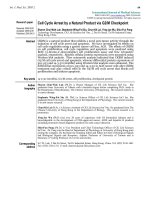
Bispecific lentiviral vector (XHR) encoding anti-CXCR4 and CCR5 siRNAsFigure 1
Bispecific lentiviral vector (XHR) encoding anti-CXCR4 and CCR5 siRNAs.
A) Control transfer vector pHIV-7-GFP encoding a
CMV promoter driven EGFP reporter gene. B) To derive the bispecific vector pHIV-XHR-GFP, a U6 promoter driven short
hairpin CXCR4 siRNA cassette was cloned into the BamHI site upstream to the CMV-EGFP cassette. The H1-CCR5 siRNA
cassette was inserted into an MluI site downstream to the U6-CXCR4 siRNA cassette.
AIDS Research and Therapy 2005, 2:1 />Page 4 of 12
(page number not for citation purposes)
non-transduced and control GFP vector transduced cells.
The levels of GAPDH control mRNA remained unchanged
in all samples (Fig 4). To validate the expression of indi-
vidual siRNAs in transduced Magi-CXCR4 and Ghost R5
cells, cellular RNA was analyzed by northern analysis for
their presence. As internal controls, the presence of consti-
tutively expressed miRNA-16 RNAs were also analyzed in
parallel. As expected, comparable levels of miRNA-16
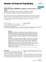
Cell surface down regulation of CXCR4 in XHR transduced Magi-CXCR4 cellsFigure 2
Cell surface down regulation of CXCR4 in XHR transduced Magi-CXCR4 cells.
Magi-CXCR4 cells that constitutively express
CXCR4 were transduced with control GFP or XHR vectors. Cells were stained with PECy5-conjugated antibodies to CXCR4
and analyzed by FACS 72 hours post-transduction. Levels of CXCR4 in non-transduced cells are superimposed (unshaded
areas). Transduction efficiency was determined by FACS for EGFP expression. Levels of EGFP in control GFP-alone vector (A)
and XHR vector (C) transduced cells. Levels of CXCR4 expression in GFP-alone (B) and XHR (D) vector transduced cells.
Percent positive cells are indicated.
AIDS Research and Therapy 2005, 2:1 />Page 5 of 12
(page number not for citation purposes)
RNAs (22 bp in length)were detected in GFP control vec-
tor transduced as well as in XHR vector transduced cells
(Fig 5A). RNAs corresponding to CXCR4 and CCR5 shR-
NAs (representing the 21nt antisense strand of each
shRNA) were seen in XHR transduced but not in GFP con-
trol vector transduced cells (Fig 5B).
Bispecific siRNA vector does not induce interferon
Double stranded RNA molecules longer than ~30 bp are
known to induce the interferon pathway in response to
viral infections. As siRNAs are generally comprised of 19–
24 bp in length, they are not expected to activate such a
response that mediates a non-specific down regulation of
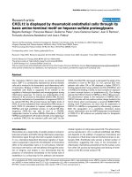
Cell surface down regulation of CCR5 in XHR transduced Ghost-R5 cellsFigure 3
Cell surface down regulation of CCR5 in XHR transduced Ghost-R5 cells.
Ghost-R5 cells that constitutively express CCR5
were transduced with GFP-alone or XHR vectors. Cells were stained with PECy5-conjugated antibodies to CCR5 and analyzed
by FACS 72 hours post-transduction. Levels of CCR5 in non-transduced cells are superimposed (unshaded areas). Transduc-
tion efficiency was measured by FACS for EGFP expression. Levels of EGFP in control GFP-alone vector (A) and XHR vector
(C) transduced cells. Levels of CCR5 expression in GFP-alone (B) and XHR (D) vector transduced cells. Percent positive cells
are indicated.
AIDS Research and Therapy 2005, 2:1 />Page 6 of 12
(page number not for citation purposes)
cellular or viral mRNAs. However, recent data had shown
that in some circumstances, certain siRNAs might induce
variable levels of interferon activation [48-50]. To rule out
such a possibility with the present siRNAs, we looked for
upregulation of phosphorylated-PKR by western blot
analysis. PKR is a protein kinase that becomes activated
through phosphorylation in the presence of dsRNA and is
involved during the interferon response. Our results have
shown that the levels of phosphorylated PKR remain
unchanged in XHR transduced cells similar to mock and
GFP vector transduced cells. In contrast, elevated levels of
phosphorylated PKR could be seen in poly I:C transfected
cells used as positive controls (Fig 6). These data exclude
the possibility of non-specific interferon activation by the
combinatorial lentiviral construct.
Resistance of siRNA transduced cells to HIV-1 infection
To determine if down regulation of the essential corecep-
tors, CXCR4 and CCR5, translated to virus resistance,
transduced Magi-CXCR4 and Ghost R5 cells were chal-
lenged with X4 (NL4-3) and R5 (BaL1)-tropic strains of
HIV-1 respectively. Viral p24 antigen levels at different
days post-challenge were determined by ELISA to quantify
levels of HIV-1 resistance. Over a 10-fold reduction in
viral antigen levels was seen with both XHR transduced
Magi-CXCR4 and Ghost-R5 cells as compared to non-
transduced and GFP-alone vector transduced cells (Fig 7).
There was a slight increase in viral production in XHR
transduced cells on days 5 to 7. This could be due to non-
transduced and/or low siRNA expressing cells producing
the virus. We next wanted to determine if the XHR vector
expressing CXCR4 and CCR5 siRNAs is effective in physi-
ologically relevant cells for gene therapy. Accordingly,
PBMCs transduced with vectors were challenged in the
same manner as above. A 3-fold level of inhibition was
seen on days 3, 5, and 7 (Fig 8). These results established
that the XHR vector is also effective in primary cells in
inhibiting HIV-1. Although clearly significant, the levels
of virus inhibition were not as dramatic as seen with Magi
and Ghost cell lines. The observed levels of viral inhibi-
tion in primary PBMC are similar to those observed in a
recent report [31]. Lower levels of protection in PBMCs
were likely due to the lower levels of transduction. Future
studies that are aimed at increasing transduction efficien-
cies into primary lymphocytes and macrophages are likely
to overcome this hurdle.
In summary, our studies have shown for the first time that
a single lentiviral vector could be used to stably deliver
two different siRNAs targeted to two different cell surface
co-receptor molecules and achieve protection against
both X4 and R5 tropic HIV-1 viral strains. The short hair-
pin design permitted use of a single promoter to tran-
scribe both the sense and anti-sense strands of each of the
siRNAs. No promoter interference was observed between
the U6 promoter driving the transcription of CXCR4
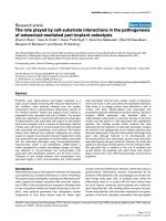
RT-PCR detection of CXCR4 and CCR5 mRNA down regulationFigure 4
RT-PCR detection of CXCR4 and CCR5 mRNA down regulation.
Total RNA was extracted from vector transduced cells and
one-step RT-PCR was performed. PCR products of 450 bp were amplified to detect the coreceptor transcripts. A) Levels of
CXCR4 mRNA in non-transduced (lane 1), GFP-alone (lane 2), and XHR (lane 3) vector transduced Magi-X4 cells. B) CCR5
transcript levels in non-transduced (lane 1), GFP-alone (lane 2), and XHR vector transduced Ghost-R5 cells. GAPDH transcript
levels were used as internal controls (PCR product size ~550 bp).
AIDS Research and Therapy 2005, 2:1 />Page 7 of 12
(page number not for citation purposes)
siRNA and the H1 promoter driving the CCR5 siRNA
since comparable amounts of both the siRNAs could be
seen in transduced cells. Furthermore, possible interferon
induction by the combinatorial construct was also ruled
out.
A major advantage in using a combinatorial lentiviral con-
struct targeted to both the coreceptors is that infection
with either of the viral strains could be prevented at the
entry step thus eliminating the possibility of proviral inte-
gration and viral latency. Given the success with the cur-
rent bispecific construct, other novel constructs could be
designed and experimented with that incorporate siRNAs
targeted to both the cellular as well as viral targets. Based
on the design employed here, it is possible to introduce
more than two siRNAs in a single construct in the future.
However caution should be exercised while incorporating
multiple siRNAs in a single construct because the possibil-
ity exists that over expression of foreign siRNAs in a cell
may have undesirable effects such as saturating the endog-
enous RISC complex and consequent toxicity. Such a pos-
sibility needs to be tested in long range experiments in
vivo. We previously have introduced a monospecific
siRNA targeted to HIV-1 rev into CD34 hematopoietic
progenitor cells via lentiviral vectors and derived trans-
genic macrophages in vitro and T cells in vivo [29]. The
transgenic cells were found to be apparently normal while
markedly resistant to HIV-1 infection.
No deleterious effects are expected by the stable knock
down of the CCR5 coreceptor in vivo since individuals har-
boring a 32 bp deletion in the corresponding gene are
physiologically normal [34,35]. Although CXCR4 down
regulation in circulating mature T cells in the periphery
may not have any insurmountable ill effects, this may
have possible drawbacks in a stem cell setting due to its
role in cell homing into bone marrow [51,52]. Addition-
ally, recent gene expression profiling studies indicated
Northern analysis to detect siRNA expression in transduced cellsFigure 5
Northern analysis to detect siRNA expression in transduced cells.
Small RNAs (<200 nt) were extracted from transduced cells
and probed with specific primers to detect the expression of siRNAs as described in materials and methods. A) Northern blot
to detect the presence of miRNA-16 (~22 bp) as an internal control in GFP-alone vector transduced (lane 2) and XHR trans-
duced Magi-X4 (lane 3) and Ghost-R5 (lane 4) cells. B) siRNA (~21 bp) detection in GFP-alone vector transduced (lane 2) and
XHR transduced Magi-X4 (lane 3) and Ghost-R5 (lane 4) cells. Decade markers (lanes A1 and B1).
AIDS Research and Therapy 2005, 2:1 />Page 8 of 12
(page number not for citation purposes)
some off-target effects by siRNAs [53]. Therefore, the
present combinatorial construct targeted to both CXCR4
and CCR5 coreceptor molecules need to be thoroughly
tested in an in vivo system such as the SCID-hu mouse
model to evaluate its efficacy and possible toxicity in
differentiated cells before it can be used for gene therapy
in human subjects. Such experiments are currently
underway.
Conclusions
For HIV/AIDS gene therapy strategies to succeed, novel
molecules need to be harnessed. In this regard, siRNAs
offer great potential. Exploitation of these promising can-
didates to down regulate essential cellular coreceptors via
the use of lentiviral vectors facilitates long term derivation
of resistant T cells and macrophages which are the main
targets for the virus. Our results showed for the first time
that expression of both CXCR4 and CCR5 siRNAs in com-
bination is possible by the use of lentiviral vectors. Core-
ceptor specific siRNAs stably transduced with the
bispecific lentiviral vector showed marked resistance
against both T cell tropic and monocyte tropic HIV-1
infection in cell lines and primary PBMCs. The newly
developed bispecific vector shows promise for potential in
vivo application.
Materials and Methods
Plasmid and lentiviral vector construction
Previously characterized siRNAs against CXCR4 and
CCR5 were used in generating the bispecific lentiviral vec-
tor [23,24,30]. A third generation lentiviral vector back-
bone was employed to derive the bispecific constructs.
The two cis-acting elements, namely, the central DNA flap
consisting of cPPT and CTS (to facilitate the nuclear
import of the viral preintegration complex) and the WPRE
(to promote nuclear export of transcripts and/or increase
the efficiency of polyadenylation of transcripts), are engi-
neered to enhance the performance of the vector [38,39].
An siRNA expression cassette targeting CXCR4 under the
control of the Pol-III U6 promoter was PCR amplified
from the plasmid pTZ-U6+1 as described by Castanotto et
al [40]. This cassette was cloned into pHIV-7-GFP transfer
vector in the BamHI site immediately upstream of the
CMV-EGFP gene. This cassette contained a MluI restriction
site downstream from the CXCR4 siRNA sequence for
subsequent cloning of the H1 promoter driven CCR5
Lack of interferon induction in siRNA transduced cellsFigure 6
Lack of interferon induction in siRNA transduced cells.
To detect interferon induction in siRNA vector transduced cells, west-
ern blot analysis was performed to detect elevated levels of phophorylated PKR. Poly I:C was used to induce interferon as a
positive control. Transduced cell extracts were run on 10% SDS-PAGE gels, transferred, and probed with an anti-phospho-
PKR antibody. Positive control poly I:C transfected (lanes 1 and 2), non-transduced (lane 3), GFP-alone vector (lane 4), and
XHR transduced (lane 5) Magi-X4 cells and XHR transduced Ghost-R5 cells (lane 6). An anti-actin antibody was used as an
internal control.
AIDS Research and Therapy 2005, 2:1 />Page 9 of 12
(page number not for citation purposes)
siRNA cassette. The H1-CCR5 siRNA expression cassette
was also generated as described above using the plasmid
pSUPER (Oligoengine, Seattle, WA). Sequencing and con-
firmation of candidate clones was performed by Laragen
Inc. (Los Angeles, CA). The transfer vector containing the
inserts U6-X4 siRNA and H1-CCR5 siRNA is termed
pHIV-XHR-GFP.
Cell culture and vector production
293T cells and PBMCs were maintained in DMEM media
supplemented with 10% FBS. Magi-CXCR4 cells obtained
from the AIDS Reference and Reagent Program were
maintained in media as previously described [41,42].
Ghost-R5 cells obtained from the AIDS Reference and
Reagent Program were maintained in media as previously
described [43]. To generate lentiviral vectors, fifteen
micrograms of transfer vector with either GFP-alone or
XHR were transfected along with 15 ug pCHGP-2, 5 ug
pCMV-Rev, and 5 ug pCMV-VSVG into 293T cells at 60%
confluency in 100 mm culture dishes using a calcium
phosphate transfection kit (Sigma-Aldrich, St. Louis,
MO). Six hours after transfection, fresh medium was
exchanged. Cell culture supernatants containing the vec-
tor were collected at 24, 36, 48, and 60 hours post trans-
fection and pooled. Vector supernatants were
concentrated by ultracentrifugation and later titrated on
293T cells using FACS analysis for GFP expression.
Lentiviral vector transduction and FACS analysis
Magi-CXCR4 and Ghost-CCR5 cells were seeded in 6-well
plates 24 hours prior to transduction, 5 × 10
5
cells per
well. Cells were transduced with lentiviral vectors at an
m.o.i. of 10 in the presence of 4 ug/ml polybrene for 2
hours. For transduction of PBMCs, cells were first isolated
from whole blood by Histopaque
®
-1077 (Sigma-Aldrich),
and then cultured in CD3 and CD28 antibody coated
plates. Three days after stimulation, PBMCs were trans-
duced at an m.o.i of 20 in the presence of 4 ug/ml poly-
brene. PBMC transduction was repeated the following
day. Seventy-two hours post transduction with siRNA
containing lentiviral vectors, FACS analysis was per-
formed to determine the levels of cell surface expression
of CXCR4 and CCR5. Non-transduced and transduced
cells were stained with appropriate antibodies conjugated
with PE-Cy 5 (Pharmingen, San Diego, CA) namely, anti-
CXCR4 for Magi-CXCR4 cells and anti-CCR5 for Ghost-
CCR5 cells. Transduction efficiency was determined by
assaying for EGFP expression. FACS analysis was per-
formed on the Beckman Coulter Epics XL using ADC soft-
ware for analysis.
Northern analysis for shRNA expression
Total RNA was extracted from non-transduced and trans-
duced Magi-CXCR4 and Ghost-CCR5 cells using the RNA-
STAT-60 reagent (Tel-Test, Friendswood, TX). Small
HIV-1 challenge of XHR transduced Magi-X4 and Ghost-R5 cellsFigure 7
HIV-1 challenge of XHR transduced Magi-X4 and Ghost-R5 cells.
Vector transduced cells were challenged with either X4
tropic or R5 tropic viruses at an m.o.i of 0.01. Culture supernatants were collected at different days post challenge and p24
antigen was assayed by ELISA. A) Transduced Magi-X4 cells challenged with X4 tropic HIV-1 NL4-3. B) Transduced Ghost-R5
cells challenged with R5 tropic HIV-1 BaL-1. Data presented is from triplicate experiments.
AIDS Research and Therapy 2005, 2:1 />Page 10 of 12
(page number not for citation purposes)
RNAs, <200 nt, were separated and concentrated using the
mirVana™ miRNA Isolation Kit (Ambion, Austin, TX).
Twenty micrograms of small RNAs were hybridized over-
night at 37°C using the mirVana™ miRNA Detection Kit
(Ambion) with γ-
32
P labeled probes made using the mir-
Vana™ Probe & Marker Kit (Ambion). Probes were
complementary to the antisense strands of CXCR4 and
CCR5 siRNAs. Hybridization reactions were processed
according to the manufacturer's protocol and run on 15%
polyacrylamide TBE-Urea gels. Gels were then exposed to
X-ray film. A probe complementary to miRNA-16
supplied with the miRNA detection kit was used as an
internal control.
Western Blot analysis of phosphorylated PKR
Cell lysates of non-transduced and transduced cells were
run on 10%-polyacrylamide-SDS TBE gels. Proteins were
immunoblotted onto Immobilon™-P membranes (Milli-
pore, Bedford, MA) and incubated with antibody specific
for phosphorylated-PKR (Sigma-Aldrich), while anti-actin
antibody (Sigma-Aldrich) was used to detect cellular actin
as an internal control. A secondary antibody, goat anti-
rabbit IgG conjugated with alkaline phophatase
(Promega, Madison, WI), was then added. An alkaline
phophatase substrate reagent, Western Blue (Promega),
was used to visualize the bands.
RT-PCR
Total RNA was extracted from non-transduced and trans-
duced cells. Primers specific for CXCR4 (forward: 5'-ggag-
gggatcagtatatacacttc and reverse: 5'-cgccaacatagaccaccttttc)
and CCR5 (forward: 5'-caaaaagaaggtcttcattacacc and
reverse: 5'-cttgctcgctcgggagcctc) (IDT, Coralsville, IA) were
used to determine transcript levels while GAPDH (for-
ward: 5'-ctgagaacgggaagcttgtcatcaa and reverse: 5'-gcctgct-
tcaccaccttcttgatg) primers were used as an internal control.
One-step RT-PCR reactions were performed using the
Superscript™ III One-Step RT-PCR kit (Invitrogen,
Carlsbad, CA). Reactions were run on 1% agarose gels and
appropriate bands were visualized with UV light.
HIV-1 Challenge
To determine if down-regulation of CXCR4 and CCR5
transcript levels and cell surface expression inhibited HIV-
1 infection, non-transduced and transduced cells were
challenged with NL4-3 (X4-tropic) and BaL-1 (R5-tropic)
strains of HIV-1, at an m.o.i of 0.01, as previously
described [24]. Viral supernatants were collected daily
HIV-1 challenge of XHR transduced PBMCsFigure 8
HIV-1 challenge of XHR transduced PBMCs.
Vector transduced PBMCs were challenged with either X4 tropic or R5 tropic
viruses. Culture supernatants were collected at different days post challenge and p24 antigen was assayed by ELISA. Trans-
duced PBMCs challenged with either HIV-1 NL4-3 (A) or BaL-1 (B). Data presented is from triplicate experiments.
AIDS Research and Therapy 2005, 2:1 />Page 11 of 12
(page number not for citation purposes)
from infected Magi-CXCR4 and Ghost-CCR5 cells for p24
assay. ELISA was used to determine p24 values employing
a Coulter-p24 kit (Beckman Coulter, Fullerton, CA). For
PBMC challenge experiments, non-transduced and trans-
duced cells were infected with NL4-3 and Bal-1 strains and
cell culture supernatants were collected on days 1, 3, 5,
and 7 post-infection to measure p24 levels.
Competing interests
The author(s) declare that they have no competing
interests.
Author's contributions
JA carried out all of the experiments. RA was responsible
for the overall experimental design and implementation
of the project.
Acknowledgements
Work reported here was supported by NIH grants AI50492 and AI057066
to R.A. This work has also been facilitated by the infrastructure and
resources provided by the Colorado Center for AIDS Research Grant P30
AI054907. We thank Karen Helms for help with FACS analysis and William
Wheat for critically reading the manuscript. We thank NIH AIDS Research
and Reference Reagents Program for providing many reagents and cell lines
used in this work.
References
1. Malim MH, Freimuth WW, Liu J, Boyle TJ, Lyerly HK, Cullen BR,
Nabel GJ: Stable expression of transdominant rev protein in
human T cells inhibits Human Immunodeficiency Virus
replication. J Exp Med 1992, 176:1197-1201.
2. Bonyhadi ML, Moss K, Voytovich A, Auten J, Kalfoglou C, Plavec I,
Forestell S, Su L, Bohnlein E, Kaneshima H: RevM10-expressing T
cells derived in vivo from transduced human hematopoietic
stem-progenitor cells inhibit human immunodeficiency virus
replication. J Virol 1997, 71:4707-4716.
3. Ding SF, Lombardi R, Nazari R, Joshi S: A combination anti-HIV-1
gene therapy approach using a single transcription unit that
expresses antisense, decoy, and sense RNAs, and transdom-
inant negative mutant Gag and Env proteins. Front Biosci 2002,
7:a15-28.
4. Michienzi A, Li S, Zaia JA, Rossi J: A nucleolar TAR decoy inhibi-
tor of HIV-1 replication. Proc Natl Acad Sci 2002, 99:14047-14052.
5. Akkina R, Banerjea A, bai J, Anderson J, Li MJ, Rossi J: siRNAs,
ribozymes, and RNA decoys in modeling stem cell-based
gene therapy for HIV/AIDS. Anticancer Res 2003, 23:1997-2006.
6. Bahner I, Kearns K, Hao QL, Smogorzewska EM, Kohn DB: Trans-
duction of human CD34+ hematopoietic progenitor cells by
a retroviral vector expressing an RRE decoy inhibits human
immunodeficiency virus type 1 replication in myelomono-
cytic cells produced in long-term culture. J Virol 1996,
70:4352-4360.
7. Lisziewicz J, Sun D, Smythie J, Lusso P, Lori F, Louie A, Markham P,
Rossi J, Reitz M, Gallo RC: Inhibition of human immunodefi-
ciency virus type 1 replication by regulated expression of a
polymeric tat activation response RNA decoy as a strategy
for gene therapy in AIDS. Proc Natl Acad Sci USA 1993,
90:8000-8004.
8. Bai J, Gorantla S, Banda N, Cagnon L, Rossi J, Akkina R: Character-
ization of anti-CCR5 ribozyme-transduced CD34+ hemat-
opoietic progenitor cells in vitro and in a SCID-hu mouse
model in vivo. Mol Ther 2000, 1:244-254.
9. Bai J, Rossi J, Akkina R: Multivalent anti-CCR5 ribozymes for
stem cell-based HIV type 1 gene therapy. AIDS Res Hum
Retroviruses 2001, 17:385-399.
10. Bai J, Banda N, Lee NS, Rossi J, Akkina R: RNA-based anti-HIV-1
gene therapeutic constructs in SCID-hu mouse model. Mol
Ther 2002, 6:770-782.
11. Cagnon L, Rossi J: Downregulation of the CCR5 beta-chemok-
ine receptor and inhibition of HIV-1 infection by stable VA1-
ribozyme chimeric transcripts. Antisense Nucleic Acid Drug Dev
2000, 10:251-261.
12. Feng Y, Leavitt M, Tritz R, Duarte E, Kang D, Mamounas M, Gilles P,
Wong-Staal F, Kennedy S, Merson J, Yu M, Barber JR: Inhibition of
CCR5-dependent HIV-1 infection by hairpin ribozyme gene
therapy against CC-chemokine receptor 5. Virology 2000,
276:271-278.
13. Elbashir SM, Harborth J, Lendeckel W, Yalcin A, Weber K, Tuschl T:
Duplexes of 21-nucleotide RNAs mediate RNA interference
in cultured mammalian cells. Nature 2001, 411:494-498.
14. Fire A, xu S, Montgomery MK, Kostas SA, Driver SE, Mello CC:
Potent and specific genetic interference by double-stranded
RNA in Caenorhabditis elegans. Nature 1998, 391:806-811.
15. Hannon GJ: RNA Interference. Nature 2002, 418:244-251.
16. Sharp P: RNA interference-2001. Genes Dev 2001, 15:485-490.
17. Lee NS, Dohjima T, Bauer G, Li H, Li M, Ehsani A, Salvaterra P, Rossi
J: Expression of small interfering RNAs targeted against HIV-
1 rev transcripts in human cells. Nat Biotechnol 2002, 20:500-505.
18. Song E, Lee S, Dykxhoorn DM, Novina C, Zhang D, Crawford K,
Cerny J, Sharp PA, Leiberman J, Manjunath N, Shankar P: Sustained
small interfering RNA-mediated human immunodeficiency
virus type 1 inhibition in primary macrophages. J Virol 2003,
77:7174-7181.
19. Novina CD, Murray MF, Dykxhoorn DM, Beresford PJ, Riess J, Lee S,
Collman RG, Lieberman J, Shankar P, Sharp PA: siRNA-directed
inhibition of HIV-1 infection. Nat Med 2002, 8:681-686.
20. Jacque J, Triques K, Stevenson M: Modulation of HIV-1 replica-
tion by RNA interference. Nature 2002, 418:435-438.
21. Martinez MA, Gutierrez A, Armand-Ugon M, Blanco J, Parera M,
Gomez J, Clotet B, Este JA: Suppression of chemokine receptor
expression by RNA interference allows for inhibition of HIV-
1 replication. AIDS 2002, 16:2385-2390.
22. Coburn GA, Cullen BR: Potent and specific inhibition of human
immunodeficiency virus type-1 replication by RNA
interference. J Virol 2002, 76:9225-9231.
23. Anderson J, Banerjea A, Planelles V, Akkina R: Potent suppression
of HIV type 1 infection by a short hairpin anti-CXCR4 siRNA.
AIDS Res Hum Retroviruses 2003, 19:699-706.
24. Anderson J, Banerjea A, Akkina R: Bispecific short hairpin siRNA
constructs targeted to CD4, CXCR4, and CCR5 confer HIV-
1 resistance. Oligonucleotides 2003, 13:303-312.
25. Capodici J, Kariko K, Weissman D: Inhibition of HIV-1 infection
by small interfering RNA-mediated RNA interference. J
Immunol 2002, 169:5196-5201.
26. Haasnoot PCJ, Cupac D, Berkhout B: Inhibition of virus replica-
tion by RNA interference. J Biomed Sci 2003, 10:607-616.
27. Lee MM, Coburn G, McClure MO, Cullen BR: Inhibition of human
immunodeficiency virus type 1 replication in primary macro-
phages by using tat- or CCR5-specific small interfering RNAs
expressed from a lentivirus vector. J Virol 2003, 77:11964-11972.
28. Li M, Bauer G, Michienzi A, Yee J, Lee NS, Kim J, Li S, Castanotto D,
Zaia J, Rossi J: Inhibition of HIV-1 infection by lentiviral vectors
expressing Pol III-promoted anti-HIV RNAs. Mol Ther 2003,
8:196-206.
29. Banerjea A, Li M, Bauer G, Remling L, Lee NS, Rossi J, Akkina R: Inhi-
bition of HIV-1 by lentiviral vector-transduced siRNAs in
lymphocytes diferentiated in SCID-hu mice and CD34+ pro-
genitor cell-derived macrophages. Mol Ther 2003, 8:62-71.
30. Butticaz C, Ciuffi A, Munoz M, Thomas J, Bridge A, Pebernard S, Iggo
R, Meylan P, Telenti A: Protection from HIV-1 infection of pri-
mary CD4 T cells by CCR5 silencing is effective for the full
spetrum of CCR5 expression. Antiviral Therapy 2003, 8:373-377.
31. Qin X, An DS, Chen ISY, Baltimore D: Inhibiting HIV-1 infection
in human T cells by lentiviral-mediated delivery of small
interfering RNA against CCR5. Proc Natl Acad Sci USA 2003,
100:183-188.
32. Bieniasz PD, Cullen BR: Chemokine receptors and Human
Immunodeficiency Virus infection. Front Biosci 1998, 3:44-58.
33. Berger EA, Murphy PM, Farber JM: Chemokine receptors as HIV-
1 coreceptors: Roles in viral entry, tropism, and disease. Annu
Rev Immunol 1999, 17:657-700.
Publish with Bio Med Central and every
scientist can read your work free of charge
"BioMed Central will be the most significant development for
disseminating the results of biomedical research in our lifetime."
Sir Paul Nurse, Cancer Research UK
Your research papers will be:
available free of charge to the entire biomedical community
peer reviewed and published immediately upon acceptance
cited in PubMed and archived on PubMed Central
yours — you keep the copyright
Submit your manuscript here:
/>BioMedcentral
AIDS Research and Therapy 2005, 2:1 />Page 12 of 12
(page number not for citation purposes)
34. Liu R, Paxton W, Choe S, Ceradini D, Martin S, Horuk R, MacDonald
M, Stuhlman H, Koup R, Landau N: Homozygous defect in HIV-1
coreceptor accounts for resistance of some multiply
exposed individuals to HIV-1 infection. Cell 1996, 86:367-377.
35. Huang Y, Paxton WA, Wolinsky SM, Neumann AU, Zhang L, He T,
Kang S, Ceradini D, Jin Z, Yazdanbakhsh K, Kunstman K, Erickson D,
Dragon E, Landau NR, Phair J, Ho DD, Koup RA: The role of a
mutant CCR5 allele in HIV-1 transmission and disease
progression. Nat Med 1996, 2:1240-1243.
36. Naif HM, Cunningham AL, Alali M, Li S, Nasr N, Buhler MM, Schols
D, Clercq E, tewart G: A human immunodeficiency virus type 1
isolate from an infected person homozygous for CCR5∆32
exhibits dual tropism by infecting macrophages and MT2
cells via CXCR4. J Virol 2002, 76:3114-3124.
37. Ma Q, Jones D, Borghesani PR, Segal RA, Nagasawa T, Kishimoto T,
Bronson RT, Springer TA: Impaired B-lymphopoiesis, myelopoi-
esis, and derailed cerebellar neuron migration in CXCR4-
and SDF-1-deficient mice. Proc Natl Acad Sci USA 1998,
95:9448-9453.
38. Yam P, Li S, Wu J, Hu J, Zaia J, Yee J: Design of HIV-1 vectors for
efficient gene delivery into human hematopoietic cells. Mol
Ther 2002, 6:770-782.
39. Ailles LE, Naldini L: HIV-1 Derived Lentiviral Vectors. In Lentiviral
Vectors Edited by: Trono D. Berlin: Springer-Verlag; 2002:31-48.
40. Castanotto D, Li H, Rossi J: Functional siRNA expression from
transfected PCR products. RNA 2002, 8:1454-1460.
41. Kimpton J, Emerman M: Detection of replication-competent
and pseudotyped human immunodeficiency virus with a sen-
sitive cell line on the basis of activation of an integrated β-
galactosidase gene. J Virol 1992, 66:2232-2239.
42. Vodicka MA, Goh WC, Wu LI, Rogel ME, Bartz SR, Schweickart VL,
Raport CJ, Emerman M: Indicator cell lines for detection of pri-
mary strains of human and simian immunodeficiency
viruses. Virology 1997, 233:193-198.
43. Morner A, Bjorndal A, KewalRamani V, Littman DR, Inoue R,
Thorstensson R, Fenyo EM, Bjorling E: Primary human immuno-
deficiency virus type 2 (HIV-2) isolates, like HIV-1 isolates,
frequently use CCR5 but show promiscuity in coreceptor
usage. J Virol 1999, 73:2343-2349.
44. VandenDriessche T, Naldini L, Collen D, Chuah MKL: Oncoretrovi-
ral and lentiviral vector-mediated gene therapy. Methods in
Enzymol 2002, 346:573-589.
45. Ketteler R, Glaser S, Sandra O, Martens UM, Klingmuller U:
Enhanced trnsgene expression in primitive hematopoietic
progenitor cells and embryonic stem cells efficiently trns-
duced by optimized retroviral hybrid vectors. Gene Therapy
2002, 9:477-487.
46. An DS, Koyanagi Y, Zhao J, Akkina R, Bristol G, Yamamoto N, Zack
JA, Chen ISY: High-efficiency transduction of huma lymphoid
progenitor cells and expression in differentiated T cells. J Virol
1997, 71:1397-1404.
47. Mautino MR, Morgan RA: Gene therapy of HIV-1 infection using
lentiviral vectors expressing anti-HIV-1 genes. AIDS Patient
Care and STDs 2002, 16:11-26.
48. Moss EG, Taylor JM: Small-interfering RNAs in the radar of the
interferon system. Nat Cell Biol 2003, 5:771-772.
49. Pebernard S, Iggo RD: Determinants of interferon-stimulated
gene induction by RNAi vectors. Differentiation 2004,
72:103-111.
50. Scacheri PC, Rozenblatt-Rosen O, Caplen NJ, Wolfsberg TG,
Umayam L, Lee JC, Hughes CM, Shanmugam KS, Bhattacharjee A,
Meyerson M, Collins FS: Short interfering RNAs can induce
unexpected and divergent changes in the levels of untar-
geted proteins in mammalian cells. Proc Natl Acad Sci USA 2004,
101:1892-1897.
51. Molyneaux KA, Zinszner H, Kunwar PS, Schaible K, Stebler J, Sun-
shine MJ, O'Brien W, Raz E, Littman D, Wylie C, Lehmann R: The
chemokine SDF1/CXCL12 and its receptor CXCR4 regulate
mouse germ cell migration and survival. Development 2002,
130:4279-4286.
52. Coffield VM, Jiang Q, Su L: A genetic approach to inactivating
chemokine receptors using a modified viral protein. Nat
Biotechnol 2003, 21:1321-1327.
53. Jackson AL, Bartz SR, Shelter J, Kobayashi SV, Burchard J, Mao M, Li
B, Cavet G, Linsley PS: Expression profiling reveals off-target
gene regulation by RNAi. Nat Biotechnol 2003, 21:635-637.