Caldesmon regulates the motility of vascular smooth muscle cells by modulating the actin cytoskeleton stability doc
Bạn đang xem bản rút gọn của tài liệu. Xem và tải ngay bản đầy đủ của tài liệu tại đây (2.65 MB, 12 trang )
Jiang et al. Journal of Biomedical Science 2010, 17:6
/>The cost of publication in Journal of Biomedical Science
is bourne by the National Science Council, Taiwan.
Open Access
RESEARCH
© 2010 Jiang et al; licensee BioMed Central Ltd. This is an Open Access article distributed under the terms of the Creative Commons
Attribution License ( which permits unrestricted use, distribution, and reproduction in
any medium, provided the original work is properly cited.
Research
Caldesmon regulates the motility of vascular
smooth muscle cells by modulating the actin
cytoskeleton stability
Qifeng Jiang1,2, Renjian Huang
2
, Shaoxi Cai*
1
and Chih-Lueh A Wang*
2
Abstract
Background: Migration of vascular smooth muscle cells (SMCs) from the media to intima constitutes a critical step in
the development of proliferative vascular diseases. To elucidate the regulatory mechanism of vacular SMC motility, the
roles of caldesmon (CaD) and its phosphorylation were investigated.
Methods: We have performed Transwell migration assays, immunofluorescence microscopy, traction microscopy and
cell rounding assays using A7r5 cells transfected with EGFP (control), EGFP-wtCaD or phosphomimetic CaD mutants,
including EGFP-A1A2 (the two PAK sites Ser452 and Ser482 converted to Ala), EGFP-A3A4 (the two Erk sites Ser497 and
Ser527 converted to Ala), EGFP-A1234 (both PAK- and Erk-sites converted to Ala) and EGFP-D1234 (both PAK- and Erk-
sites converted to Asp).
Results: We found that cells transfected with wtCaD, A1A2 or A3A4 mutants of CaD migrated at a rate approximately
50% more slowly than those EGFP-transfected cells. The migration activity for A1234 cells was only about 13% of
control cells. Thus it seems both MAPK and PAK contribute to the motility of A7r5 cells and the effects are comparable
and additive. The A1234 mutant also gave rise to highest strain energy and lowest rate of cell rounding. The migratory
and contractile properties of these cells are consistent with stabilized actin cytoskeletal structures. Indeed, the A1234
mutant cells exhibited most robust stress fibers, whereas cells transfected with wtCaD or A3A4 (and A1A2) had
moderately reinforced actin cytoskeleton. The control cells (transfected with EGFP alone) exhibited actin cytoskeleton
that was similar to that in untransfected cells, and also migrated at about the same speed as the untransfected cells.
Conclusions: These results suggest that both the expression level and the level of MAPK- and/or PAK-mediated
phosphorylation of CaD play key roles in regulating the cell motility by modulating the actin cytoskeleton stability in
dedifferentiated vascular SMCs such as A7r5.
Background
Migration of vascular smooth muscle cells (SMCs) from
media to intima is a critical step in the development of pro-
liferative vascular diseases such as atherosclerosis, and in
response to vascular injuries such as angioplasty and organ
transplatation. Fully differentiated SMCs normally do not
proliferate nor migrate. Upon stimulation, however, SMCs
can dedifferentiate and change from contractile to synthetic
phenotypes, which enable cell proliferation and migration.
During this process SMCs undergo cellular remodelling
and a number of smooth muscle-specific contractile pro-
teins are converted to non-muscle isoforms. One of such
signature proteins is caldesmon (CaD).
CaD is an actin-binding protein that also interacts with
myosin, tropomyosin and calmodulin [1]. The two alterna-
tively spliced isoforms of CaD derive from a single gene
[2]: the heavy caldesmon (h-CaD), found exclusively in dif-
ferentiated SMCs, and the light isoform (l-CaD), present in
nearly all types of vertebrate cells. Unlike visceral smooth
muscles, which only express h-CaD, vascular smooth mus-
cles contain both h- (>75%) and l-CaD (<25%) [3]. How-
ever, upon dedifferentiation, h-CaD is rapidly degraded in
vascular SMCs, and only l-CaD is expressed. Therefore, l-
* Correspondence: ,
1
Key Laboratory of Biorheological Science and Technology, Ministry of
Education, Bioengineering College, Chongqing University, Chongqing, 400044,
China
2
Boston Biomedical Research Institute, 64 Grove St, Watertown, MA 02472,
USA
Jiang et al. Journal of Biomedical Science 2010, 17:6
/>Page 2 of 12
CaD is the form that is closely associated with the synthetic
type of smooth muscle organs.
Both h- and l-CaD bind actin filaments and stabilize the
filamentous structure. In SMCs h-CaD, together with tropo-
myosin, modulates the actomyosin ATPase activity by
reversibly and cooperatively inhibiting myosin binding to
actin [4]. The inhibitory effect of h-CaD on muscle contrac-
tility has been demonstrated by peptide intervention [5-8]
and antisense knockdown [9] experiments. Reversal of this
inhibition is accompanied by phosphorylation of h-CaD at
MAPK-specific sites [10], which partially dissociates h-
CaD from actin filaments and allows myosin to bind [11].
The C-terminal region of h-CaD can also be phosphorylated
by PAK [12] in vitro, although it is less clear whether or not
the in vivo modification occurs in SMCs.
In non-muscle cells l-CaD appears to have more diverse
functions. l-CaD has been reported to be involved in cell
division [13], migration [14], adhesion [15], postmitotic
spreading [16], apoptosis [17], and intracellular granule
movement [18]. Like that of h-CaD, the action of l-CaD is
also regulated via MAPK-mediated phosphorylation by
such enzymes as cdc2 kinase [13] and Erk1/2 [14]. We have
previously shown that, when cells are stimulated with phor-
bol ester, l-CaD is phosphorylated at the Erk sites and
moves from stress fibers in the cytosol to nascent focal con-
tacts at cell peripheries [16]. Phosphorylation of l-CaD by
PAK also affects the morphology and migratory properties
of non-muscle cells [19]. Consistently, l-CaD is present in
podosomes [20,21], where it regulates podosome dynamics
in a PAK-dependent manner [22].
The fact that the presumed functions of CaD are affected
by MAPK and PAK has inspired much interest. Both types
of kinases add phosphate groups to residues near the actin-
binding sites at the C-terminal region of CaD, thereby
decreasing its effectiveness of actin binding, as well as its
stabilizing action on the actin cytoskeleton. This may sug-
gest that CaD (particularly l-CaD) serves as a converging
point for the MAPK and PAK signaling under the stimula-
tion of a wide variety of agonists; it also raises the question
as how the two types of CaD phosphorylation relate to each
other. In this work we have analyzed the cellular conse-
quences of the MAPK and PAK actions, individually and in
combination, on l-CaD in a dedifferentiated SMC line
(A7r5). By using phosphomimetic mutagenesis, we aimed
to dissect the effect of MAPK- and PAK-mediated phos-
phorylation on CaD. Our results indicate that CaD phos-
phorylation is an obligatory step for cell motility, and that
MAPK and PAK work independently and additively toward
this process. Since only l-CaD is expressed in these cul-
tured cells, CaD refers to the non-muscle isoform exclu-
sively throughout this work except otherwise specified.
Methods
Cell Culture
Rat aorta smooth muscle cells A7r5 (ATCC# CRL-1444™)
were maintained in DMEM (Cellgro™) supplemented with
10% fetal bovine serum (Cellgro™) and 1% antibiotics
(Penicillin-Streptomycin, Cellgro™). Cells were cultured at
37°C under a 5% CO
2
atmosphere.
Plasmids
The pCB6 hx plasmid containing the cDNA of human l-
CaD (GeneBank #M64110
) was originally a gift from Dr.
Jim Lin (University of Iowa, Iowa City, IA). The insert was
subcloned into the mammalian expression vector pEGFPC1
(Clontech). Site-directed mutagenesis was performed as
previously described [16] with the two PAK sites Ser452
and Ser482 converted to Ala (EGFP-A1A2); the two Erk
sites Ser497 and Ser527 to Ala (EGFP-A3A4); both PAK-
and Erk-sites to Ala (EGFP-A1234); or both PAK- and Erk-
sites to Asp (EGFP-D1234).
Cell Transfection
A7r5 cells were plated at 60% confluence in 6-well cell cul-
ture plates. 18 h after plating, the cells were starved for 2 h
before transfection using the Lipofectamine™ reagent sup-
plemented with Plus™ reagent (Invitrogen). Briefly, 2 μg
DNA in 5 μl Plus™ reagent and in 4 μl Lipofectamine™
reagent were diluted, respectively, with 43 μl and 46 μl
serum-free DMEM medium. After 20 min incubation at
room temperature, the two solutions were mixed and incu-
bated for another 20 min. The mixture was then added to
the cells; after 5 h the transfection medium was replaced
with full medium.
Western Blot Analysis
The expression level of exogenous CaD induced by trans-
fection, and the extent of CaD phosphorylation in A7r5
cells were evaluated by Western blot analysis using a Odys-
sey Infrared Imaging System by Li-COR Biosciences (Lin-
coln, NE) [23,24] as described previously [16]. Cells
transfected with vehicle alone (EGFP), EGFP-wtCaD
(wtCaD), EGFP-A1A2 (A1A2), EGFP-A3A4 (A3A4),
EGFP-A1234 (A1234), or EGFP-D1234 (D1234) were
seeded at 10
5
cells/well on 6-well cell culture plates (Bec-
ton-Dickinson, Rutherford, NJ). After 24 h incubation, cul-
ture medium was removed, and cells were rinsed twice with
ice-cold PBS. Proteins were extracted by adding to each
well 150 μl of lysing buffer containing phenylmethylsulfo-
nyl fluoride 1 mM (Sigma), leupeptin 10 mg/ml (Sigma),
aprotinin 30 mg/ml (Sigma), and NaVO
3
1 mM (Sigma).
The plates were incubated on ice for 30 min and scraped.
Total cell extracts were separated on SDS-PAGE and
immunoblotted with lab-made polyclonal anti-CaD and
affinity purified polyclonal anti-pSer527 (Ser527 of l-CaD
is equivalent to Ser789 in h-CaD), as well as monoclonal
Jiang et al. Journal of Biomedical Science 2010, 17:6
/>Page 3 of 12
anti-β-actin (Sigma), followed by affinity purified anti-rab-
bit and anti-mouse secondary antibodies conjugated with
IRDyeTM 700 and 800, respectively. The digitized fluores-
cent bands were integrated, and the ratios (GFP-tagged
CaD to endogenous CaD) were calculated for both protein
level and phosphorylation of each pair after normalized
against the amount of β-actin, which was used as a loading
reference.
Fluorescence Microscopic Imaging
For fluorescence microscopy, cells transfected with EGFP,
wtCaD, A1A2, A3A4, A1234, or D1234 were seeded on
glass coverslips placed in a plastic culture dish and incu-
bated overnight, during which time the cell became well-
spread. Cells were then starved for 24 h and washed in PBS,
fixed for 15 min in freshly prepared 4% paraformaldehyde
(PFA) in PBS and permeabilized with 0.3% Triton X-100 in
4% PFA in PBS for 5 min. For all subsequent steps, solu-
tions were prepared in PBS. Cells were thoroughly rinsed in
PBS between steps and incubations were performed at
room temperature. F-actin was stained with rhodamine-
phalloidin and incubated for 1 h. Finally, cell-loaded cover-
slips were rinsed and mounted on glass slides in Mowiol
(Sigma). Images were obtained using a laser scanning sys-
tem BioRad Radiance 2000 equipped with the confocal
head attached to the Nikon Eclipse TE300 microscope.
Data were acquired and analyzed with Laser Sharp 2000
BioRad software. All images were collected through single-
section acquisition with scan performed from the top to the
bottom of the cell in two-color (green and red) channels in
parallel.
Cell Migration Assay
Cell migration was assayed with 24-well tissue culture
Transwell (Becton Dickinson) plates comprising a polycar-
bonate membrane with 8-μm pores. The inner and outer
chamber membranes were coated with 5 μg/ml of human
fibronectin (R&D, Minneapolis, MN) at 37°C for 2 h, and
then rinsed with PBS. A7r5 cells transfected with EGFP,
wtCaD, A1A2, A3A4, A1234, or D1234 were then seeded
on the inner chamber of the Transwell plate at a concentra-
tion of 2 × 10
4
cells/well in 200 μl serum-free DMEM. The
outer chamber was filled with 800 μl full culture medium
which contained 10% FBS and 50 ng/ml FGF (R&D, Min-
neapolis, MN), and incubated for 36 h at 37°C. The number
of total migrated cells and green (i.e., transfected) cells
were counted in fields randomly chosen from 9 equally
divided zones of the membrane in triplicates under phase-
contrast and fluorescence channel with the ZEISS-AXIO
fluorescence microscope system. The percentage of trans-
fected cells in the total migrated cells was determined for
each experiment. These numbers were then divided by the
transfection efficiency to obtain the motility of the trans-
fected cells relative to the untransfected cells.
Fourier-Transform Traction Microscopy
A7r5 cells transfected with EGFP, wtCaD, A1A2, A3A4,
A1234, or D1234 were seeded on the collagen-coated, fluo-
rescence mircobeads-embedded polyacrylamide gel, which
was prepared according to a previously described protocol
[25,26], with the cell density kept lower than 10
4
per dish.
After 24 h incubation, the cells were starved overnight prior
to measurements. With fluorescence channel first followed
by phase-contrast, the image of a single green fluorescence
cell and that of the fluorescent micropatterned beads were
recorded before and after trypsinization. The two images of
the micropatterned beads plus the phase-contrast cell image
were taken to calculate the displacement field of the gel
generated by the cell [27]. The projected cell area was also
calculated based on the cell contour determined from the
phase-contrast image obtained at the start of the experi-
ment. From the displacement field the traction field within
a 50 μm × 50 μm square was calculated as described by
Butler et al. [28]. The magnitude and the direction of the
vectors corresponding to the traction imposed on the gel
underneath the cell yielded a scalar measure of cell contrac-
tility (strain energy), which is the total energy (in pJ) trans-
ferred from the cell to the substratum.
Cell Rounding Assays
Cell rounding assays after treatment with trypsin were per-
formed as previously described [16].
Statistics
All measurements are expressed in terms of mean ± stan-
dard deviation (SD), except those of calculated total strain
energy, which are expressed in mean ± standard error (SE).
Comparisons between 2 samples of unequal variance were
performed by Student's t-test using 2-tailed distribution. P <
0.05 was considered as significant.
Results
Effect of phosphomimetic mutation of CaD on the
morphology of A7r5 cells
The inhibitory action of CaD on the actomyosin interaction
is known to be regulated by MAPK- [11] and PAK- [12,19]
mediated phosphorylation. To probe the significance of
such regulation, to dissect the effect of the two types of
phosphorylation, and to test their combined effect on the
actin cytoskeleton and motility of SMCs, we have designed
EGFP-tagged phosphomimetic mutants of CaD and force-
expressed them in A7r5 cells. Serine residues at positions
452 (designated as position #1) and 482 (position #2) are
taken as the "PAK-sites", whereas serines at positions 492
(position #3) and 527 (position #4) are taken as the "Erk-
sites", which may also be phosphorylated by other MAPKs.
Mutants include A1A2 (PAK-sites disabled), A3A4 (Erk-
sites disabled), A1234 (both PAK- and Erk-sites disabled)
and D1234 (both PAK- and Erk-sites phosphorylated).
Jiang et al. Journal of Biomedical Science 2010, 17:6
/>Page 4 of 12
Cells were also transfected with either EGFP-tagged wild-
type CaD (wtCaD) or the vehicle alone (EGFP), the latter
being used as controls. The cell viability was not apprecia-
bly affected by transient transfection. No sign of cell death
was detected within the time period of experimentation.
Among all constructs cells transfected with the A1234
mutant showed most robust cytoskeleton structure. The
majority of A1234-expressing cells exhibited thicker and
longer stress fibers than the untrasnfected cells and the
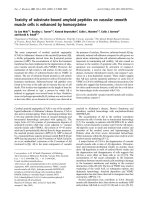
EGFP-expressing control cells. The green fluorescence in
these cells overlapped closely with the red phalloidin stain-
ing (Fig. 1-c), indicating that the expressed CaD mutant
binds to the actin cytoskeleton with augmented stability.
The wtCaD, A1A2 and A3A4 transfected cells also showed
prominent cytoskeleton structures with similar overlapping
distribution of EGFP-tagged CaD with stress fibers (Fig. 1-
a,b,f), but the difference between transfected cells and
untransfected cells was less striking than that for A1234. In
contrast, the D1234-transfected cells exhibited much
weaker stress fiber staining than cells transfected with the
A-mutants and wt-CaD. Although some faint stress fibers
were nevertheless detected in D1234-transfected cells, the
exogenous CaD primarily overlapped with the F-actin
staining at the cortical regions. Notably, the actin cytoskele-
ton in these cells exhibited little or no difference from that
in the untransfected cells, as seen in the control cells (Fig.
1-d,e).
Effect of phosphomimetic mutation of CaD on the motility
of A7r5 cells
To determine the effect of phosphomimetic mutation of
CaD on the motility of A7r5 cells, we have performed Boy-
den chamber migration assays using Transwell filters of 8-
μm pore size. Cells were stimulated by 50 ng/ml FGF and
10% serum to undergo chemotactic migration. The motility
of cells expressing different CaD mutants relative to the
normal, untransfected cells was evaluated after 36 hours.
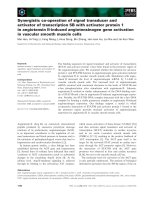
Among all constructs the A1234 mutant resulted in most
hindered motility. Taking untransfected A7r5 cells as a
standard, we found that the relative migration activity for
the A1234-transfected cells was only 0.113 ± 0.02 (n = 3;
same below), which was about 13% of the EGFP-trans-
fected control cells (0.85 ± 0.07; Fig. 2). Transfection with
wtCaD also slowed down the rate of cell migration, but to a
lesser extent (0.417 ± 0.01 of the control cells). The motil-
ity of D1234 transfected cells (0.65 ± 0.028) was higher
than the cells transfected with all other CaD variants, but
still about 24% lower than the control cells. It is clear that
CaD plays an important role in controlling the activity of
migration in A7r5 cells, and phosphorylation of CaD
appears to facilitate this activity. Interestingly, cells trans-
fected with A1A2 or A3A4 mutant of CaD migrated at a
rate (0.42 ± 0.06 and 0.40 ± 0.04, respectively) that is
approximately half way between the A1234 cells and the
control cells. Apparently both MAPK and PAK contribute
to the motility of A7r5 cells and these effects are compara-
ble and additive.
Effect of phosphomimetic mutation of CaD on the
contractility of A7r5 cells
One of the key steps during cell migration is contraction by
which the cell body is moved forward [29]. The observation
that the A1234 mutant of CaD slowed down the overall rate
of migration might lead to the prediction that the cell con-
tractility is also hampered. To test whether this is the case
and to decipher the origin of the observed effect mechanis-
tically, we wished to determine how phosphomimetic muta-
tions of CaD affect the contractility of A7r5 cells. Fourier-
transform traction microscopy at the single cell level was
used for this purpose. We have performed contractility
assays using A7r5 cells transfected with EGFP, wtCaD,
A1A2, A3A4, A1234 and D1234. Stationary, individual
cells were first plated on the fluorescent microbeads-imbed-
ded polyacrylamide gel slab and examined by fluorescence
microscopy, while the cell images were recorded before and
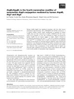
after trypsinization. From these images the displacement
field of the fluorescent beads was calculated (Fig. 3A-a).
The magnitude and the direction of the vectors correspond-
ing to the bead movement underneath the cell were then
used to compute the average strain energy (i.e., the energy
that the cell transfers to the substratum owing to the con-
tractile activity; in pJ) of the cell.
Contrary to our expectation, we found that the A1234
mutant transfected A7r5 cells that showed severely ham-
pered motility (see Fig. 2), exhibited most strengthened,
instead of compromised, contractility, among all CaD vari-
ant transfected cells (Fig. 3B). The A1234 mutant (3.08 ±
0.22 pJ) showed about 15-fold enhancement in the traction
force measurement compared to the control cells (0.21 ±
0.07 pJ). Both the A1A2 (1.81 ± 0.39 pJ) and A3A4 (1.60 ±
0.40 pJ) mutants resulted in about 8-fold enhancement in
total strain energy. The wtCaD transfected cells also had
significantly higher traction force than the control cells,
although not as high as the A1234 mutant transfected cells.
The increases in the contractility caused by the wtCaD
transfection were about 6-fold (1.23 ± 0.24 pJ). The D1234
mutant transfection showed little enhancement for cell con-
tractility, the measured force for the D1234 mutant trans-
fected cells (0.44 ± 0.13 pJ) was almost the same as that of
the control cells. It should be mentioned that because the
traction measurements were based on single cell experi-
ments, the scattering of the data was relatively large even
with multiple measurements for each set (n = 10). Never-
theless, from the trend of the data it is rather striking that
the effect of phosphomimetic CaD transfection on the con-
tractility is precisely reciprocal to that on the migratory
activity: The cells with the highest migratory activity (i.e.,
the EGFP-expressing cells) showed the lowest strain
Jiang et al. Journal of Biomedical Science 2010, 17:6
/>Page 5 of 12
e
Figure 1 (See figure legend on next page.)
Jiang et al. Journal of Biomedical Science 2010, 17:6
/>Page 6 of 12
nergy, whereas the cells with the slowest migration (i.e., the
A1234-expressing cells) had the strongest strain energy.
Like in the case of migration assays, the PAK- (A1A2) or
Erk- (A3A4) sites disabled mutants exhibited about 50% of
the change displayed by the mutant with both types of phos-
phorylation sites disabled (A1234). Phosphorylation of
CaD therefore must have similar but opposite effects on
these two events. Another important finding from these
data is that, since the strain energy results from actin-based
contractile force, the observed lower migration activity of
mutated A7r5 cells clearly was not owing to inhibited con-
tractility.
State of phosphorylation of CaD mutants by Western blot
analysis
The fact that cells expressing the A1A2 and the A3A4
mutants exhibited about 50% of the overall changes found
for the A1234 cells in both migration and contractile activi-
ties compared to those of the control cells implies: (a) PAK-
and MAPK-mediated phosphorylation of CaD contributes
equally and additively to these activities; and (b) the A1A2
and the A3A4 mutants might in fact be phosphorylated at
other available sites in these cells. To test the latter idea, we
have examined the phosphorylation status of the engineered
CaD mutants by Western blot analysis. A lab-prepared
polyclonal antibody against the Erk-site pSer527 (or
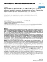
pSer789 in h-CaD) was used for this purpose. Indeed, as
shown in Fig. 4, the EGFP-tagged A1A2 and wtCaD, but
not the A3A4 and A1234, were found positive for MAPK-
mediated phosphorylation. Not surprisingly, the endoge-
nous CaD in all cell lines was also phosphorylated at this
Erk-site. Since we don't have the anti-PAK-sites antibody, a
similar experiment to verify the PAK-mediated phosphory-
(See figure on previous page.)
Figure 1 Fluorescence images of transfected A7r5 cells. All CaD constructs were EGFP-tagged (left panels); actin was stained with red (middle pan-
els). Merged images are shown on the right panels. Cells were transfected with EGFP (control, Row e), EGFP-wtCaD (wild-type CaD; Row f) and CaD
mutant, including EGFP-A1A2 (Row a), EGFP-A3A4 (Row b), EGFP-A1234 (Row c) and EGFP-D1234 (Row d). A1234 transfected cells had most robust
cytoskeleton structure, the wtCaD, A1A2 and A3A4 transfected cells also had more robust structure than D1234 and control cells (EGFP). The D1234
transfected cells exhibited similar cytoskeleton structure to the control cells. Scale bar, 100 μm.
Figure 2 Summary of Transwell migration assays. Cells transfected with EGFP (control), EGFP-tagged wtCaD, A1A2, A3A4, A1234 and D1234 were
subjected to migration assays, and the migration activity (see Methods) was compared to that of untransfected cells. The relative migration activity
for the A1234-transfected cells was about 13% of the control cells (EGFP), and the ratios of A1A2 and A3A4 were 49% and 47% of the control cells,
respectively. It seems that both Erk and PAK contributed equally to the motility of these two types of cells and this effects are additive. The D1234
mutant had a relatively weak inhibitory effect on the motility of A7r5 cells. The error bars represent the standard deviations (SD) of 3 independent
measurements. Single (*) and double (**) asterisks on the peaks denote P < 0.05 and P < 0.005, respectively.
Jiang et al. Journal of Biomedical Science 2010, 17:6
/>Page 7 of 12
l
Figure 3 (A) Fourier transform tranction microscopy measurements and the images of a representative A7r5 cell gathered in the measure-
ment process. (a) The single green cell was first identified under fluorescence channel and then the phase-contrast image (upper right) was recorded.
Based on beads movement, the traction field was calculated by MATLAB. The magnitude and the direction of the vectors indicate the bead move-
ment, which was used to compute the contractile moment. Scale bar: 50 μm. (b) The color coding for the magnitude of the bead movement. (B) Re-
sults of total strain energy measurements (in pJ) of A7r5 cells transfected with phosphomimetic mutants of CaD and EGFP alone (control). The error
bars represent the standard errors (SE) of 10 measurements. Single (*) and double (**) asterisks on the peaks denote P < 0.05 and P < 0.005, respectively.
Jiang et al. Journal of Biomedical Science 2010, 17:6
/>Page 8 of 12
ation on the EGFP-tagged CaD variants is not possible at
this time.
Effect of phosphomimetic mutation of CaD on the
detachment behavior of A7r5 cells upon trypsinization
Finally, in search of the cause for the decreased migration
activity, we wished to test whether CaD mutation affects
cell detachment, which constitutes another step critical to
cell migration. Cells undergo rapid retraction and rounding,
and eventual detachment from the substratum upon trypsin
treatment because of disengagement of focal adhesions and
partial disassembly of actin bundles. We used a simple
assay by quantifying the number of rounded cells (includ-
ing detached cells) as a function of time following trypsini-
zation to compare the detachment kinetics of A7r5 cells
transfected with either EGFP or CaD mutants. We found
that cells transfected with wtCaD, A1A2 or A3A4 all
showed delayed responses to trypsin digestion as compared
to the control cells (Fig. 5). Even more hampered rounding
was observed for the A1234 transfected cells, in agreement
with previous observations with rat aortic fibroblast cells
[16]. In contrast, the D1234 transfected cells showed simi-
lar kinetics of rounding up as the control cells. The detach-
ment behavior thus parallels the migration activity.
Discussion
CaD is known to bind actin and stabilize the filamentous
structure. Binding of CaD to actin also inhibits the actomy-
osin interaction, and results in inhibition on many cellular
processes such as migration, adhesion and proliferation
[30]. These inhibitory actions can be reversed by binding to
calmodulin in the presence of Ca
2+
, although it is more
likely that the in vivo function of CaD is regulated by phos-
phorylation. In vivo CaD phosphorylation was documented
not only in activated SMCs [31], but also in mitotic cells
[32] and migrating smooth muscle [14] or non-muscle cells
[16]. Because of these properties, CaD serves as a target for
manipulation of cellular behaviors. There have been plenty
of data in the literature using ectopic expression of CaD or
mutants to probe cell movement; however, the results are
not always consistent [16,17,20,33-38]. While most studies
found over-expressed CaD stabilizes stress fibers in the cell
and inhibits cell motility, one report [37] showed opposite
results, in which case transient transfection of CaD not only
disrupted stress fibers, but also disassembled focal adhe-
sions. Because of this controversy, the exact function of
CaD has not been settled, although the critical involvement
of CaD in cell motility is widely recognized.
Notably, in that earlier study the phosphorylation status of
CaD was not determined. In light of the findings that
unphosphorylated and phosphorylated CaD display quite
different actin-binding properties [11,39] and intracellular
distributions [16,21], variations in CaD phosphorylation
may have contributed to this apparent discrepancy. To bet-
ter understand the role of CaD phosphorylation in vascular
SMCs, we have force-expressed phosphomimetic mutants
of CaD in A7r5 cells, a model for remodelled or diseased
vascular SMCs, and examined the resulting cells in terms of
their morphology, migration activity, contractility and
detachment kinetics. We focused on Erk and PAK by sepa-
rately or simultaneously altering the residues of the respec-
tive modification sites, because both kinases have
previously been shown to affect CaD's affinity for actin fil-
aments. Our data indicated that in order for A7r5 cells to
attain motility, phosphorylation of CaD by either kinase is
an obligatory step, primarily through the modulation of the
actin cytoskeleton dynamics, rather than the contractile
machinery of the cell.
Figure 4 Western blot analysis of CaD phosphorylation in the transfected A7r5 cells. The total cell extracts from A7r5 cells transfected with var-
ious constructs were immunoblotted with polyclonal anti-pSer527 (Left Panel) and anti-CaD (Right Panel) antibodies. M: Molecular weight markers;
samples (Lanes 2-6) are, respectively, cells transfected with A1A2, A3A4, A1234, wtCaD, and EGFP alone (Control). The corresponding ratios of the dig-
itized intensity of the EGFP-tagged CaD variant (the upper red band; Right Panel) to that of the endogenous CaD (the lower red band; Right Panel)
are, respectively, 0.93, 1.71, 2.60, 0.43 and, 0 (EGFP); and the corresponding ratios of the digitized intensity of the phospho-EGFP-CaD variant (the upper
red band; Left Panel) to that of the phosphorylated endogenous CaD (the lower red band; Left Panel) are, respectively, 0.13 (A1A2), 0, 0, 0.10 (wtCaD)
and, 0. The apparent lower signal in the phosphorylation for the engineered variants (human) than the endogenous CaD (rat) may be partly due to
different immuno-reactivities of the antibodies. Moreover, since the transfection efficiency is ~35% in all cases, the actual ratios could be higher.
Jiang et al. Journal of Biomedical Science 2010, 17:6
/>Page 9 of 12
The phosphorylation-disabled CaD mutant, A1234, at
both PAK- and Erk-sites hampered the migration activity to
the greatest extent (to ~13% of the control cells; Fig. 2).
Cell migration is a multiple-step process, which includes
cell extension, attachment, contraction and rear detachment
[29]. The observed slower migration could have resulted
from one or more compromised steps in this process. How-
ever, when we examined the cell contractility by traction
microscopy, we found that the A1234-expressing cells
exhibited strongest traction force (Fig. 3B). Thus the over-
all cell motility must be dominated by step(s) other than
contraction. Indeed, the detachment assay showed that
A1234 mutant rendered A7r5 cells to round up and detach
from the substratum more slowly than other variants (Fig.
5). Consistent with this observation is the fact that cells
transfected with A1234 exhibited most robust stress fibers
(Fig. 1). It is conceivable that these structures may be hard
to disassemble. Together, these results suggest that it is the
less dynamic actin cytoskeleton, which is overly stabilized
by the unphosphorylatable CaD, that makes the cell more
resistant to shape changes, and thereby hampering the cell
motility. This interpretation agrees with Yamashiro's work
[40], and reinforces our understanding about the functional
role of CaD phosphorylation as a means to reverse the actin
stabilizing effect of CaD.
The fact that Ser-to-Ala mutation at only four positions
(amino acid residue-452, 482, 497 and 527) attains a reduc-
tion in cell motility of nearly 90% is quite remarkable. On
one hand, it means phosphorylation at these four residues is
necessary for cell migration, on the other hand, it also dem-
onstrates that other mechanisms including phosphorylation
at other positions only contribute no more than 13% of the
overall migration activity. Previously, it has been suggested
that Ca
2+
/calmodulin also regulates the CaD-imposed inhib-
Figure 5 Detachment of transfected A7r5 cells upon trypsinization. A1A2- (squares), A3A4- (circles), A1234- (triangles), D1234- (inverse triangles),
EGFP- (diamonds), wtCaD- (turned triangles) transfected cells were plated on 60 mm dishes. Cells from each plate were trypsinized and monitored
under the phase-contrast and fluorescence microscope for time-dependent retraction, rounding and detachment. Percentages of round cells at 2, 4,
6, 8 and 10 min were plotted for each type of cells. Each point was an average of 6 independent measurements; error bars represent standard devia-
tions.
Jiang et al. Journal of Biomedical Science 2010, 17:6
/>Page 10 of 12
itory effect [41]; our results argue that such an effect may
only play a minor role in A7r5 cells, particularly in the
absence of phosphorylation-mediated regulation. Transfec-
tion with A1A2 or A3A4 mutants of CaD inhibited approx-
imately 50% of the extent by the A1234 mutant in cell
migration when compared to the control cells (Fig. 2). This
intermediate activity could be due to phosphorylation at
residues that remain available. Indeed, Ser-527 (one of the
Erk-sites) of the A1A2 mutant was found to be phosphory-
lated (Fig. 4). Thus it seems both MAPK and PAK contrib-
uted equally and additively to the overall motility of
cultured aorta SMCs. The involvement of PAK, which is a
downstream effector of Rac signaling, in cell migration is
well established. That MAPK-mediated phosphorylation
also enables cell migration is consistent with previous
observations [14], and suggests the existence of cross-talks
between MAPK- and PAK-pathways.
Force-expression of wild-type CaD (in wtCaD cells)
decreased the motility of the A7r5 cells. This may be under-
stood by the assumption that the total CaD level in these
cells exceeds the capacity of the kinases and results in a net
increase of unphosphorylated CaD, which causes inhibition
of the cell motility. This situation is similar to that of the
A1A2 and A3A4 cells, where phosphorylation of CaD is
partially blocked. This interpretation is supported by the
observation that these three types of cells exhibited about
the same degree of suppression in motility. When we force-
expressed D1234 mutant, we expected an increase in the
motility, because the phospho-mimicking mutation might
represent a scenario opposite to the fully inhibited state
(e.g., A1234). Yet we found the D1234 mutant also sup-
pressed the motility of the cells, although to a lesser extent
(~24% inhibited). This could be attributed to the fact that
such modification was irreversible. Not being able to be
dephosphorylated, this CaD mutant disrupts the phosphory-
lation cycling of endogenous CaD and may thereby lead to
inhibition of cell motility [19]. In the meantime, since
D1234 mutant has a much weakened affinity for the actin
filament, it cannot stabilize the cytoskeleton as much as
A1234 or wt-CaD; so the D1234 mutant cells showed
weaker stress fiber structure and similar contractility and
detachment behaviors compared to the control cells.
Our data indicate that the migratory activity of cells
changes in a reciprocal manner as the contractility of the
cells, despite that contraction is an essential step during cell
migration. The contractile activity, as evaluated by our
assays based on the total strain energy the cell exerts on the
substratum, requires active actomyosin interactions. How-
ever, an equally important factor is a sturdy cytoskeleton
structure that provides a framework for such interactions
and supports the contractility. Without firm actin cables,
such as in the D1234-transfected cells, even a relatively
high actomyosin ATPase activity would not be able to gen-
erate force. The cell contractility, the cytoskeleton stability,
and the cell migration activity must therefore behave in a
coordinated manner. On the other hand, the A1234-express-
ing cells, which produced highest traction force because of
the stabilized actin cytoskeleton therein, must also have an
actomyosin activity that is not severely inhibited. This is
rather surprising in view of the overwhelming evidence that
unphosphorylated CaD (i.e., A1234) inhibits such activity.
One possible explanation is that the actomyosin interaction
is activated through the binding of calmodulin to CaD, as
the calmodulin-binding sites are still functional in these
mutants. However, this mechanism apparently fails to
restore the migration activity, since cells expressing A1234
were only 13% as motile as the control cells (Fig. 2).
Clearly, the cytoskeleton stability is a more dominant factor
than the actomyosin ATPase activity in the case of cell
migration, because multiple steps (i.e., extension, attach-
ment and detachment) in this process depend on the ability
of actin filaments to be assembled and disassembled
dynamically.
The finding that stabilization of the actin cytoskeleton by
CaD is critical for cell motility provides the rationale for
using CaD to curb SMC migration which in most cases is
pathogenic. In particular, transfection of CaD in mice has
been shown to suppress the growth of vascular SMCs and
inhibit neointimal formation after angioplasty [42]. More
recently it was reported that expression of CaD suppresses
the invasive activity of cancer cells [43]. Our study further
suggests that CaD with combined mutations at both PAK-
and Erk-sites (e.g., A1234) may serve as a more effective
therapeutic reagent in cancer and proliferative vascular dis-
eases such as atherosclerosis and restenosis.
Conclusions
In this study we showed that CaD suppresses the motility of
vascular SMCs by stabilizing the actin filamentous struc-
ture. Despite intensive studies over the past three decades,
the functional role of CaD in non-muscle cells remains elu-
sive. In this regard, our data shed light onto the following
aspects: (1) Phosphorylation of CaD at the Erk- and PAK-
sites near the actin-binding sites of CaD is required for cell
motility, because it reverses CaD's inhibitory effect and
allows dynamic changes of the actin cytoskeleton. (2) Both
Erk and PAK contribute equally and additively toward this
regulatory role in cell migration and contraction. (3) Other
mechanisms such as phosphorylation at residues other than
the four positions (residues 452, 482, 497 and 527) or inter-
action with calmodulin only play a relatively minor role in
modulating the motility of A7r5 cells. These new insights
not only help us to better understand how CaD works, but
also afford useful information on how cell motility is regu-
lated. For SMCs, in particular, our findings suggest that
mutations at CaD phosphorylation sites serve as a novel
therapeutic strategy to combat vascular diseases such as
atherosclerosis and restenosis.
Jiang et al. Journal of Biomedical Science 2010, 17:6
/>Page 11 of 12
List of abbreviation used
CaD: caldesmon; Erk: extracellular signal-regulated kinase;
FGF: fibroblast growth factor; h-CaD: heavy or smooth
muscle-specific caldesmon; l-CaD: light or non-muscle
caldesmon; MAPK: mitogen-activated protein kinase;
PAK: p21-activated protein kinase; PBS: phosphate buff-
ered saline; PFA: paraformaldehyde; SMC: smooth muscle
cell.
Competing interests
The authors declare that they have no competing interests.
Authors' contributions
QJ carried out the cell biology studies, participated in the mechanical mea-
surements and drafted the manuscript. RH carried out the immunoassays. SC
participated in the design of the study. CLAW conceived of the study, and par-
ticipated in its design and coordination of the experiments, and helped to
write the manuscript. All authors read and approved the final manuscript.
Acknowledgements
The authors wish to thank Ms. Lijia Zhu for technical assistance, Dr. Jolanta Kor-
dowska for helpful discussion at the early phase of this project, and Dr. Chan
Young Park and Professor Jeffrey Fredberg (of Harvard School of Public Health)
for their help in performing and analyzing the traction microscopic measure-
ments. This study was supported by a grant (No.10872224 to SC) from Chinese
National Natural Science Foundation and "111" project, and a grant (R01
HL92252 to CLAW) from NIH.
Author Details
1
Key Laboratory of Biorheological Science and Technology, Ministry of
Education, Bioengineering College, Chongqing University, Chongqing, 400044,
China and
2
Boston Biomedical Research Institute, 64 Grove St, Watertown, MA
02472, USA
References
1. Wang C-LA: Caldesmon and smooth-muscle regulation. Cell Biochem
Biophys 2001, 35(3):275-288.
2. Humphrey MB, Herrera-Sosa H, Gonzalez G, Lee R, Bryan J: Cloning of
cDNAs encoding human caldesmons. Gene 1992, 112(2):197-204.
3. Glukhova MA, Kabakov AE, Frid MG, Ornatsky OI, Belkin AM, Mukhin DN,
Orekhov AN, Koteliansky VE, Smirnov VN: Modulation of human aorta
smooth muscle cell phenotype: a study of muscle-specific variants of
vinculin, caldesmon, and actin expression. Proc Natl Acad Sci USA 1988,
85(24):9542-9546.
4. Wang CL: Caldesmon and the Regulation of Cytoskeletal Functions.
Adv Exp Med Biol 2008, 644:250-272.
5. Lajoie CA, Parker CA, Wang CL, Morgan KG: Loading of caldesmon
peptide (GS17C) into intact vascular smooth muscle by reversible
permeablization. Biophys J 1994, 66:A408.
6. Katsuyama H, Wang CL, Morgan KG: Regulation of vascular smooth
muscle tone by caldesmon. J Biol Chem 1992, 267(21):14555-14558.
7. Lee YH, Gallant C, Guo H, Li Y, Wang CL, Morgan KG: Regulation of
vascular smooth muscle tone by N-terminal region of caldesmon.
Possible role of tethering actin to myosin. J Biol Chem 2000,
275(5):3213-3220.
8. Hulvershorn J, Gallant C, Wang CL, Dessy C, Morgan KG: Calmodulin
levels are dynamically regulated in living vascular smooth muscle cells.
American Journal of Physiology - Heart & Circulatory Physiology 2001,
280(3):H1422-H1426.
9. Earley JJ, Su X, Moreland RS: Caldesmon inhibits active crossbridges in
unstimulated vascular smooth muscle: an antisense
oligodeoxynucleotide approach. Circ Res 1998, 83(6):661-667.
10. Adam LP, Gapinski CJ, Hathaway DR: Phosphorylation sequences in h-
caldesmon from phorbol ester-stimulated canine aortas. FEBS Lett
1992, 302(3):223-226.
11. Huang R, Li L, Guo H, Wang CL: Caldesmon binding to actin is regulated
by calmodulin and phosphorylation via different mechanisms.
Biochemistry 2003, 42(9):2513-2523.
12. Foster DB, Shen LH, Kelly J, Thibault P, Van Eyk JE, Mak AS:
Phosphorylation of caldesmon by p21-activated kinase. Implications
for the Ca(2+) sensitivity of smooth muscle contraction. J Biol Chem
2000, 275(3):1959-1965.
13. Yamashiro S, Yamakita Y, Hosoya H, Matsumura F: Phosphorylation of
non-muscle caldesmon by p34cdc2 kinase during mitosis. Nature
1991, 349(6305):169-172.
14. Yamboliev IA, Gerthoffer WT: Modulatory role of ERK MAPK-caldesmon
pathway in PDGF-stimulated migration of cultured pulmonary artery
SMCs. American Journal of Physiology - Cell Physiology 2001,
280(6):C1680-C1688.
15. Haxhinasto KB, Kamath AM, Blackwell K, Zabner J, Lin J, Moy AB: The
effects of caldesmon on fibroblast cell adhesion and motility. Mol Biol
Cell 2002, 13:314a.
16. Kordowska J, Hetrick T, Adam LP, Wang CL: Phosphorylated l-caldesmon
is involved in disassembly of actin stress fibers and postmitotic
spreading. Exp Cell Res 2006, 312(2):95-110.
17. Numaguchi Y, Huang S, Polte TR, Eichler GS, Wang N, Ingber DE:
Caldesmon-dependent switching between capillary endothelial cell
growth and apoptosis through modulation of cell shape and
contractility. Angiogenesis 2003, 6(1):55-64.
18. Hegmann TE, Schulte DL, Lin JL, Lin JJ: Inhibition of intracellular granule
movement by microinjection of monoclonal antibodies against
caldesmon. Cell Motil Cytoskeleton 1991, 20(2):109-120.
19. Eppinga RD, Li Y, Lin JL, Mak AS, Lin JJ: Requirement of reversible
caldesmon phosphorylation at P21-activated kinase-responsive sites
for lamellipodia extensions during cell migration. Cell Motil Cytoskeleton
2006, 63(9):543-562.
20. Eves R, Webb BA, Zhou S, Mak AS: Caldesmon is an integral component
of podosomes in smooth muscle cells. J Cell Sci 2006, 119(Pt
9):1691-1702.
21. Gu Z, Kordowska J, Williams GL, Wang CL, Hai CM: Erk1/2 MAPK and
caldesmon differentially regulate podosome dynamics in A7r5
vascular smooth muscle cells. Exp Cell Res 2007, 313(5):849-866.
22. Morita T, Mayanagi T, Yoshio T, Sobue K: Changes in the balance
between caldesmon regulated by p21-activated kinases and the Arp2/
3 complex govern podosome formation. J Biol Chem 2007,
282(11):8454-8463.
23. Snapyan M, Lecocq M, Guevel L, Arnaud MC, Ghochikyan A, Sakanyan V:
Dissecting DNA-protein and protein-protein interactions involved in
bacterial transcriptional regulation by a sensitive protein array method
combining a near-infrared fluorescence detection. Proteomics 2003,
3(5):647-657.
24. Grosch S, Tegeder I, Schilling K, Maier TJ, Niederberger E, Geisslinger G:
Activation of c-Jun-N-terminal-kinase is crucial for the induction of a
cell cycle arrest in human colon carcinoma cells caused by flurbiprofen
enantiomers. Faseb J 2003, 17(10):1316-1318.
25. Dembo M, Wang YL: Stresses at the cell-to-substrate interface during
locomotion of fibroblasts. Biophys J 1999, 76(4):2307-2316.
26. Wang N, Tolic-Norrelykke IM, Chen J, Mijailovich SM, Butler JP, Fredberg JJ,
Stamenovic D: Cell prestress. I. Stiffness and prestress are closely
associated in adherent contractile cells. Am J Physiol Cell Physiol 2002,
282(3):C606-616.
27. Tolic-Norrelykke IM, Butler JP, Chen J, Wang N: Spatial and temporal
traction response in human airway smooth muscle cells. American
journal of physiology 2002, 283(4):C1254-1266.
28. Butler JP, Tolic-Norrelykke IM, Fabry B, Fredberg JJ: Traction fields,
moments, and strain energy that cells exert on their surroundings. Am
J Physiol Cell Physiol 2002, 282(3):C595-605.
29. Condeelis J: The biochemistry of animal cell crawling. In Motion Analysis
of Living Cell New York: Wiley-Liss; 1998:85-100.
30. Lin JJ, Li Y, Eppinga RD, Wang Q, Jin JP: Chapter 1: roles of caldesmon in
cell motility and actin cytoskeleton remodeling. Int Rev Cell Mol Biol
2009, 274:1-68.
31. Adam LP, Haeberle JR, Hathaway DR: Phosphorylation of caldesmon in
arterial smooth muscle. J Biol Chem 1989, 264(13):7698-7703.
32. Yamashiro S, Yamakita Y, Ishikawa R, Matsumura F: Mitosis-specific
phosphorylation causes 83K non-muscle caldesmon to dissociate from
microfilaments. Nature 1990, 344(6267):675-678.
Received: 29 October 2009 Accepted: 3 February 2010
Published: 3 February 2010
This article is available from: 2010 Jiang et al; licensee BioMed Central Ltd. This is an Open Access article distributed under the terms of the Creative Commons Attribution License ( ), which permits unrestricted use, distribution, and reproduction in any medium, provided the original work is properly cited.Journal of Biomedical Science 2010, 17:6
Jiang et al. Journal of Biomedical Science 2010, 17:6
/>Page 12 of 12
33. Warren KS, Shutt DC, McDermott JP, Lin JL, Soll DR, Lin JJ: Overexpression
of microfilament-stabilizing human caldesmon fragment, CaD39,
affects cell attachment, spreading, and cytokinesis. Cell Motil
Cytoskeleton 1996, 34(3):215-229.
34. Li Y, Wessels D, Wang T, Lin JL, Soll DR, Lin JJ: Regulation of caldesmon
activity by Cdc2 kinase plays an important role in maintaining
membrane cortex integrity during cell division. Cell Mol Life Sci 2003,
60(1):198-211.
35. Surgucheva I, Bryan J: Over-expression of smooth muscle caldesmon in
mouse fibroblasts. Cell Motil Cytoskeleton 1995, 32(3):233-243.
36. Mirzapoiazova T, Kolosova IA, Romer L, Garcia JG, Verin AD: The role of
caldesmon in the regulation of endothelial cytoskeleton and
migration. J Cell Physiol 2005, 203(3):520-528.
37. Helfman DM, Levy ET, Berthier C, Shtutman M, Riveline D, Grosheva I,
Lachish-Zalait A, Elbaum M, Bershadsky AD: Caldesmon inhibits
nonmuscle cell contractility and interferes with the formation of focal
adhesions. Mol Biol Cell 1999, 10(10):3097-3112.
38. Grosheva I, Vittitow JL, Goichberg P, Gabelt BT, Kaufman PL, Borras T,
Geiger B, Bershadsky AD: Caldesmon effects on the actin cytoskeleton
and cell adhesion in cultured HTM cells. Exp Eye Res 2006, 82(6):945-958.
39. Foster DB, Huang R, Hatch V, Craig R, Graceffa P, Lehman W, Wang C-LA:
Modes of caldesmon binding to actin: sites of caldesmon contact and
modulation of interactions by phosphorylation. J Biol Chem 2004,
279(51):53387-53394.
40. Yamashiro S, Chern H, Yamakita Y, Matsumura F: Mutant caldesmon
lacking cdc2 phosphorylation sites delays M-phase entry and inhibits
cytokinesis. Molecular Biology of the Cell 2001, 12(1):239-250.
41. Li Y, Lin JL, Reiter RS, Daniels K, Soll DR, Lin JJ: Caldesmon mutant
defective in Ca2+-calmodulin binding interferes with assembly of
stress fibers and affects cell morphology, growth and motility. J Cell Sci
2004, 117(Pt 16):3593-604.
42. Yokouchi K, Numaguchi Y, Kubota R, Ishii M, Imai H, Murakami R, Ogawa Y,
Kondo T, Okumura K, Ingber DE, Murohara T: l-Caldesmon regulates
proliferation and migration of vascular smooth muscle cells and
inhibits neointimal formation after angioplasty. Arterioscler Thromb
Vasc Biol 2006, 26(10):2231-2237.
43. Yoshio T, Morita T, Kimura Y, Tsujii M, Hayashi N, Sobue K: Caldesmon
suppresses cancer cell invasion by regulating podosome/
invadopodium formation. FEBS Lett 2007, 581(20):3777-3782.
doi: 10.1186/1423-0127-17-6
Cite this article as: Jiang et al., Caldesmon regulates the motility of vascular
smooth muscle cells by modulating the actin cytoskeleton stability Journal of
Biomedical Science 2010, 17:6