Calcium-sensing receptors regulate cardiomyocyte Ca2+ signaling via the sarcoplasmic reticulum-mitochondrion interface during hypoxia/reoxygenation pdf
Bạn đang xem bản rút gọn của tài liệu. Xem và tải ngay bản đầy đủ của tài liệu tại đây (1.17 MB, 11 trang )
Lu et al. Journal of Biomedical Science 2010, 17:50
/>Open Access
RESEARCH
© 2010 Lu et al; licensee BioMed Central Ltd. This is an Open Access article distributed under the terms of the Creative Commons Attri-
bution License ( which permits unrestricted use, distribution, and reproduction in any
medium, provided the original work is properly cited.
Research
Calcium-sensing receptors regulate cardiomyocyte
Ca
2+
signaling via the sarcoplasmic
reticulum-mitochondrion interface during
hypoxia/reoxygenation
Fang-hao Lu
†1
, Zhiliang Tian
†2
, Wei-hua Zhang*
1,5
, Ya-jun Zhao
1
, Hu-lun Li
3
, Huan Ren
4
, Hui-shuang Zheng
1
,
Chong Liu
1
, Guang-xia Hu
1
, Ye Tian
1
, Bao-feng Yang
5
, Rui Wang
6
and Chang-qing Xu*
1,5
Abstract
Communication between the SR (sarcoplasmic reticulum, SR) and mitochondria is important for cell survival and
apoptosis. The SR supplies Ca
2+
directly to mitochondria via inositol 1,4,5-trisphosphate receptors (IP
3
Rs) at close
contacts between the two organelles referred to as mitochondrion-associated ER membrane (MAM). Although it has
been demonstrated that CaR (calcium sensing receptor) activation is involved in intracellular calcium overload during
hypoxia/reoxygenation (H/Re), the role of CaR activation in the cardiomyocyte apoptotic pathway remains unclear. We
postulated that CaR activation plays a role in the regulation of SR-mitochondrial inter-organelle Ca
2+
signaling, causing
apoptosis during H/Re. To investigate the above hypothesis, cultured cardiomyocytes were subjected to H/Re. We
examined the distribution of IP
3
Rs in cardiomyocytes via immunofluorescence and Western blotting and found that
type 3 IP
3
Rs were located in the SR. [Ca
2+
]i, [Ca
2+
]
m
and [Ca
2+
]
SR
were determined using Fluo-4, x-rhod-1 and Fluo 5N,
respectively, and the mitochondrial membrane potential was detected with JC-1 during reoxygenation using laser
confocal microscopy. We found that activation of CaR reduced [Ca
2+
]
SR
, increased [Ca
2+
]
i
and [Ca
2+
]
m
and decreased the
mitochondrial membrane potential during reoxygenation. We found that the activation of CaR caused the cleavage of
BAP31, thus generating the pro-apoptotic p20 fragment, which induced the release of cytochrome c from
mitochondria and the translocation of bak/bax to mitochondria. Taken together, these results reveal that CaR activation
causes Ca
2+
release from the SR into the mitochondria through IP
3
Rs and induces cardiomyocyte apoptosis during
hypoxia/reoxygenation.
Background
The mitochondrion is a fundamental organelle that is
intimately involved in many aspects of cellular physiol-
ogy, such as energy production, free radical production,
regulation of cytosolic Ca
2+
signaling pathways and apop-
tosis [1,2]. The mitochondrion also acts as a spatial Ca
2+
buffer that reduces cytosolic Ca
2+
overload and regulates
Ca
2+
-dependent signaling in the cytosol. Mitochondrial
Ca
2+
is taken up from the cytosol via a low-affinity Ca
2+
uniporter at mitochondrial membranes [3]. However, the
intracellular Ca
2+
concentration ([Ca
2+
]i) is not high
enough to initiate the uniporter under physiological con-
ditions. Therefore, it has been postulated that activation
of the inositol 1,4,5-trisphosphate receptors (IP
3
Rs) sig-
naling pathway could release Ca
2+
from the sarcoplasmic
reticulum (SR) to increase the microdomain Ca
2+
concen-
tration ([Ca
2+
]) at focal contacts, known as mitochon-
dria-associated ER membranes (MAM), between the SR
and mitochondria, and then activate the uniporter.
Recent studies have suggested that IP
3
Rs are highly com-
partmentalized at MAMs, providing direct mitochon-
drial Ca
2+
signaling. Cardiomyocytes contain an
* Correspondence: ,
1
Department of Pathophysiology, Harbin Medical University, Harbin 150086,
China
†
Contributed equally
Full list of author information is available at the end of the article
Lu et al. Journal of Biomedical Science 2010, 17:50
/>Page 2 of 11
abundance of mitochondria, many of which are in close
apposition to SR Ca
2+
release sites [4].
The SR is a multifunctional organelle that controls pro-
tein translation and Ca
2+
homeostasis. Under SR stress
(e.g., SR Ca
2+
depletion), SR chaperone proteins such as
Grp78 and Grp94 are up-regulated [5]. Prolonged SR
stress will initiate apoptotic signals in the SR, including
bax/bak-translocation to the SR to activate the release of
Ca
2+
from the SR, cleavage and activation of procaspase
12 and BAP31, and Ire 1-mediated activation of apoptosis
signal-regulating kinase 1 (ASK1)/c-Jun N-terminal
kinase (JNK) [6].
The calcium-sensing receptor (CaR) is a member of the
family of G protein-coupled receptors (GPCRs). One of
the effects of CaR signal transduction is the activation of
phospholipase C, which leads to the generation of the
secondary messengers diacylglycerol (DAG) and inositol
1,4,5 trisphosphate (IP
3
). IP
3
then mobilizes Ca
2+
from
intracellular stores via the activation of specific IP
3
recep-
tors [7]. Wang et al. and Tfelt-Hansen et al. reported that
CaR was functionally expressed in rat cardiac tissue and
rat neonatal ventricular cardiomyocytes, respectively
[8,9]. Later, Berra-Romani et al. showed that cardiac
microvascular endothelial cells express a functional CaR
[10]. Our group has demonstrated that CaR is involved in
apoptosis in isolated adult rat hearts and in rat neonatal
cardiomyocytes during ischemia/reperfusion [11].
Although it is known that CaR elevates the intracellular
calcium concentration and then induces apoptosis, the
in-depth mechanisms are still not known. The aim of this
study was to investigate whether [Ca
2+
]
SR
would change
with CaR activation in response to hypoxia/reoxygen-
ation in cardiomyocytes. We specifically focused on the
relationship between SR Ca
2+
depletion, mitochondrial
Ca
2+
uptake and cardiomyocyte apoptosis during
hypoxia/reoxygenation (H/Re).
Materials and methods
Isolation of neonatal rat cardiomyocytes and H/Re
experiments
Primary cultures of neonatal rat cardiomyocytes were
performed as previously described [12]. Newborn Wistar
rats (1-3 days) were used for this study. The rats were
handled in accordance with the Guide for the Care and
Use of Laboratory Animals published by the China
National Institutes of Health. Briefly, hearts from male
Wistar rats (1-3 days old) were minced and dissociated
with 0.25% trypsin. Dispersed cells were seeded at 2 × 10
5
cells/cm
2
in 60-mm culture dishes with Dulbecco's modi-
fied Eagle medium (DMEM) supplemented with 10%
fetal bovine serum (FBS) and then cultured in a 5% CO
2
incubator at 37°C. Hypoxic conditions were produced
using D-Hanks solution (mM: 5.37 KCl, 0.44 KH
2
PO
4
,
136.89 NaCl, 4.166 NaHCO
3
, 0.338 Na
2
HPO
4
, 5 D-glu-
cose, pH 7.3-7.4 at 37°C) saturated with 95% N
2
and 5%
CO
2
. The pH was adjusted to 6.8 with lactate to mimic
ischemic conditions. The dishes were put into a hypoxic
incubator that was equilibrated with 1% O
2
/5%CO
2
/
94%N
2
. After hypoxic treatment, the culture medium was
rapidly replaced with fresh DMEM with 10% FBS (10%
FBS/DMEM) to initiate reoxygenation [13].
Experimental protocols
At 72 h post-culturing with 10% FBS/DMEM, the cells
were randomly divided into six groups: (1) control group:
cells were continuously cultured for 9 h with 10% FBS-
DMEM; (2) H/Re: cells were placed in hypoxic culture
medium for 3 h and then reoxygenated for 6 h by replac-
ing hypoxic culture medium with fresh DMEM contain-
ing 10% FBS; (3) CaCl
2
+ NiCl
2
+ CdCl
2
-H/Re (Ca + Ni +
Cd-H/Re): neonatal rat cardiomyocytes were treated with
CaCl
2
(2.2 mM), NiCl
2
(1 mM) and CdCl
2
(200 μM) for 30
min in hypoxic medium and then reoxygenated for 6 h by
replacing hypoxic culture medium with fresh DMEM
containing 10% FBS (CaCl
2
is an activator of CaR, NiCl
2
is
an inhibitor of the Na
+
-Ca
2+
exchanger, CdCl
2
is a inhibi-
tor of the L-type calcium channel; these drugs do not
affect cardiomyocyte viability); (4) NPS-2390 + CaCl
2
+
NiCl
2
+ CdCl
2
-H/Re (NPS-2390 + Ca + Ni + Cd-H/Re):
neonatal rat cardiomyocytes were treated with NPS-2390
(10 μM) for 40 min, and the following steps were the
same as for group 3 (NPS-2390 is an allosteric antagonist
of the group 1 metabotropic glutamate receptors); (5) 2-
APB + CaCl
2
+ NiCl
2
+ CdCl
2
-H/Re (2-APB + Ca + Ni +
Cd-H/Re): neonatal rat cardiomyocytes were treated with
2-APB (3 μM) for 40 min, and then other steps were the
same as in group 3 (2-APB or 2- aminoethoxydiphenyl
borate is a membrane permeable IP
3
R inhibitor); (6)
Ruthenium red + CaCl
2
+ NiCl
2
+ CdCl
2
-H/Re (Ru + Ca +
Ni + Cd-H/Re): neonatal rat cardiomyocytes were treated
with Ruthenium red (10 μM) for 40 min, and then under-
went the same steps as in group 3 (Ruthenium red is an
inhibitor of mitochondrial calcium uniporter ).
Immunocytochemistry
Cardiomyocytes were fixed in 10% formaldehyde in phos-
phate-buffered saline (PBS) for 10 min, permeabilized
with 0.1% Triton X-100, washed three times in PBS and
blocked in PBS containing 5% bovine serum albumin, 5%
horse serum and 0.05% Triton X-100 for 1 h at room tem-
perature (RT). Specific subtype anti-IP
3
R rabbit poly-
clonal antibodies were incubated overnight at 4°C at
1:200 or 1:100 (Santa Cruz). FITC-conjugated anti-rabbit
IgG was used as a secondary antibody. As indicated, some
cells were stained with 4-6- diamidino-2-phenylindole
Lu et al. Journal of Biomedical Science 2010, 17:50
/>Page 3 of 11
(25 μg/ml) (DAPI, Roche) for 1 h. The results of immuno-
cytochemical staining were read and recorded with a
laser confocal scanning microscope (Olympus, LSM,
Japan).
3-(4,5-dimethyl thiazol-2yl)-2,5-diphenyltetrazolium
bromide(MTT) assay
In the current study, cardiomyocytes were planted in 96-
well plates. The MTT assay was performed as described
previously [10]. Briefly, MTT (Sigma) was added into the
cell cultures at a final concentration of 0.5 mg/mL and the
mixture was incubated for 4 h at 37°C. Subsequently, the
culture medium was removed and DMSO was added to
each well to dissolve the resulting formazan crystals. The
absorbance was measured at a wavelength of 570 nm
using a microplate reader (Bio-Tek Instruments Inc.,
Richmond, Va). Background absorbance of medium in
the absence of cells was subtracted [14]. Percent viability
was defined as the relative absorbance of treated versus
untreated control cells.
Hoechst staining
Apoptotic cells were identified by the distinctive con-
densed or fragmented nuclear structure in cells stained
with the chromatin dye Hoechst 33342 (Sigma). Cells
were fixed with 4% paraformaldehyde for 10 min at room
temperature and were washed twice with phosphate buf-
fer solution (PBS). Cells were then incubated with 5 μg/
mL Hoechst 33342 for 15 min. Next, the cells were
washed three times and photographed using fluorescence
microscope (Leica DFC500 System; Leica Microsystems,
Bannockburn, Ill). At least 500 nuclei from randomly
selected fields in each group were analyzed for each
experiment, and the percentage of apoptotic cells was cal-
culated as the ratio of the number of apoptotic cells ver-
sus the total cells counted.
Neonatal rat cardiomyocytes loaded with Fluo-4 AM, Fluo-
5N AM and X-rhod-1 AM and cell permeabilization
[Ca
2+
]i was determined as previously described [15].
Briefly, cells were seeded on the culture slides. After
experimentation, cells were loaded with fluo-4 AM in 1%
working solution at 37°C for 1 h, washed three times with
Ca
2+
-free PBS to remove extracellular fluo-4 AM, and
diluted to the required concentration. The reagents were
added in Ca
2+
-free solution (145 mM NaCl, 5 mM KCl,
1.0 mM EGTA, 1 mM MgCl
2
, 10 mM HEPES-Na, 5.6 mM
glucose, pH 7.4). Fluorescence measurement of Ca
2+
was
performed using a laser confocal scanning microscope
(Olympus, LSM, Japan) at an excitation wavelength of
485 nm for [Ca
2+
]i and an emission wavelength of 530 nm
for [Ca
2+
]i, using the equation [Ca
2+
]i = K
d
[(F -F
min
)/(F
max
- F)], where Kd is the dissociation constant (345 nM for
fluo-4), F is the fluorescence at intermediate Ca
2+
levels
(corrected from background fluorescence), Fmin is the
fluorescence intensity of the indicator in the absence of
Ca
2+
and is obtained by adding a solution of 10 mM EGTA
for 15 min, and F
max
is the fluorescence of the Ca
2+
-satu-
rated indicator and is obtained by adding a solution of 25
μM digitonin in 2.2 nM CaCl
2
for 15 min. Final values for
[Ca
2+
]i are expressed in nanomoles.
To determine [Ca
2+
]
SR
, cardiomyocytes were treated
with Fluo-5N acetoxymethylester (10 μM) for 2 h and
deesterified for 1.5 h. For intact myocytes, the super-
fusate contained (in mM) 140 NaCl, 4 KCl, 1 MgCl
2
, 2
CaCl
2
, 10 HEPES, and 10 glucose (pH 7.4, 23°C). For per-
meabilization, myocytes were exposed to solution (in
mM: 0.1 EGTA, 10 HEPES, 120 K-aspartate, 1 free MgCl
2
,
5 ATP, 10 reduced glutathione, and 5 phosphocreatine;
pH 7.4) and then permeabilized using saponin (50 μg/ml)
for 20 seconds. Excitation was set at 488 nm and emission
was measured at 530 nm at room temperature [15].
Images of fluorescence reflecting [Ca
2+
]
i
and [Ca
2+
]
SR
were recorded using a laser confocal scanning micro-
scope (Olympus, LSM, Japan). There were more than 10
cells to be analyzed in each view and quantified using the
analysis software for the microscope.
Recent study showed that the mitochondrial Ca
2+
con-
centration ([Ca
2+
]
m
) consistently increases during reoxy-
genation [12]. Therefore, [Ca
2+
]
m
was measured at 60 min
post-reoxygenation. [Ca
2+
]
m
was determined according to
the manufacturer's instructions (Molecular Probes). In
brief, the cultured cardiomyocytes (1 × 10
6
cells/sample)
were initially washed with HEPES buffer containing (in
mM) 130 NaCl, 4.7 KCl, 1.2 MgSO
4
, 1.2 KH
2
PO
4
, 10
HEPES, 11 glucose, and 0.2 CaCl
2
at pH 7.4 and then
stained with 5 μmol/L X-rhod-1 AM for 30 min at room
temperature. To avoid deesterification of intracellular X-
rhod-1 AM in the cytosolic compartment, which would
interfere with the detection of [Ca
2+
]
m
, the cardiomyo-
cytes were rinsed and incubated with 100 μM MnCl
2
-
HEPES for an additional 20 min to quench the cytosolic
Ca
2+
signal [16]. Fluorescence measurement was deter-
mined using a fluorescence plate reader (CytoFluor II;
PerSeptive Biosystems; Framingham, MA) at an excita-
tion wavelength of 580 nm and an emission wavelength of
645 nm for [Ca
2+
]
m
. To validate the measurement of
[Ca
2+
]
m
, the cultured cardiomyocytes were transferred
into a slide chamber after X-rhod-1 AM staining and
were placed on the stage of a fluorescence microscope
(×50 objective; Olympus). The images from the slides
were captured using a digital camera connected to Image-
Pro Plus software (Media Cybernetics; Silver Spring,
MD). There were more than 10 cells to be analyzed in
each view.
Lu et al. Journal of Biomedical Science 2010, 17:50
/>Page 4 of 11
Measurement of mitochondrial membrane potential
Mitochondrial membrane potential (nψ
m
) was measured
with a unique cationic dye of 5,5',6,6'-tetrachloro 1,1'3,3'-
tetraethylbenzimidazolcarbocyanine iodide (JC-1), as
previously described [12]. Briefly, cells were seeded on
culture slides and treated according to experimental pro-
tocols. Previous data demonstrated that [Ca
2+
]
m
might
continuously increase during the process of reoxygen-
ation and result in mitochondrial nψ
m
collapse [12], so
we detected nψ
m
at 1 h after reoxygenation. At the end of
the above-described treatments, cells were stained with
JC-1 (1 μg/ml) at 37°C for 15 min and then rinsed three
times with PBS. Observations were immediately made
using a laser confocal scanning microscope. In live cells,
the mitochondria appear red due to the aggregation of
accumulated JC-1, which has absorption/emission max-
ima of 585/590 nm (red). In apoptotic and dead cells, the
dye remains in its monomeric form, which has absorp-
tion/emission maxima of 510/530 nm (green). More than
100 areas were selected from each image. The average
intensity of red and green fluorescence was determined.
The ratio of JC-1 aggregate (red) to monomer (green)
intensity was calculated. A decrease in this ratio was
interpreted as a decrease in the nψ
m
, whereas an increase
in this ratio was interpreted as a gain in the nψ
m
.
Identification of bax/bak translocation to the mitochondria
and assay for cytochrome c release from mitochondria
Western blotting of cellular fractions was used to quan-
tify changes in cytochrome c, bax and bak distribution
within cells, as previously described [17]. Briefly, 1 × 10
7
rat cardiomyocytes were homogenized in ice-cold Tris-
sucrose buffer (in mM: 350 sucrose, 10 Tris-HCl, 1 ethyl-
enediaminetetraacetic acid, 0.5 dithiothreitol, and 0.1
phenylmethanesulfonylfluoride; pH 7.5). After 10 min of
incubation, cardiomyocyte homogenates were initially
centrifuged at 1000 × g for 5 min at 4°C, and the superna-
tant was further centrifuged at 40,000 × g for another 30
min at 4°C. The supernatant was saved as the cytosolic
fraction. The precipitate was re-suspended in the above
buffer (containing 0.5% v/v Nonidet P-40) and saved as
the mitochondrial fraction. The mitochondrial fractions
were blotted with a primary rat anti-bax, bak and cyto-
chrome c monoclonal antibody (Santa Cruz Inc.). The
volume of specific bands was measured using a Bio-Rad
Chemi EQ densitometer and Bio-Rad QuantityOne soft-
ware (Bio-Rad laboratories, Hercules, USA).
Western blotting
Western blot analyses were performed as previously
described [18]. In brief, the protein concentration of sam-
ples was first determined using the Bio-Rad DC protein
assay kit (Bio-Rad Laboratories, Hercules, CA). A total of
20 μg of protein was electrophoresed on a 12% SDS-poly-
acrylamide gel and transferred to nitrocellulose mem-
branes (Amersham International, Amersham, UK). The
membranes were blocked with 10% skim milk in TBST
buffer (10 mM Tris, pH 7.6, 150 mM NaCl, and 0.1%
Tween 20) for 1 h at room temperature and then incu-
bated with a rabbit anti-BAP31 polyclonal antibody
(1:500 dilution, sc-48766, Santa Cruz Biotechnology)
overnight at 4°C. HRP-conjugated anti-rabbit IgG (1:3000
dilution, Bio-Rad Laboratories) was used as a secondary
antibody. Specific bands were visualized with a chemilu-
minescent substrate (ECL kit, Amersham International).
Statistical analyses
Significance was evaluated using student's t-test, and p <
0.05 was considered statistically significant. Data are
expressed as mean ± standard error of the mean (S.E.M.)
and are representative of at least three independent
experiments. [Ca
2+
]
i
data were obtained from 2-3 experi-
ments, and 10-12 images were analyzed in each group.
Results
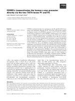
Asymmetric subcellular distribution of IP
3
R subtypes in
cardiomyocytes
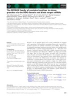
Western blot results showed that type 2 and 3 IP
3
Rs were
expressed in cardiomyocytes, while type 1 IP
3
R expres-
sion was undetectable (Fig. 1A). Similar to the results of
the Western blot analysis, type 3 IP
3
R was distributed in
the cytoplasm and intense perinuclear and intranuclear
staining was evident for type 2 IP
3
R in immunofluores-
cence study, while type 1 IP
3
R was undetectable.
Figure 1 Subcellular IP
3
Rs localization. (A) Immunocytochemical
staining of cardiomyocyte with specific antibodies for type 1, type 2
and type 3 IP3Rs. (B) Western blot analysis of cardiomyocyte lysates us-
ing antibodies specific for IP3R, type 1, type 2 and type 3, respectively.
DAPI and FITC to co-stain nuclei and type 3 IP
3
receptors and show the
spatial relation between the two structures.
Type 1 IP
3
R Ab Type 2 IP
3
R Ab Type 3 IP
3
R Ab
A
B
Lu et al. Journal of Biomedical Science 2010, 17:50
/>Page 5 of 11
Activation of CaR induces cardiomyocyte apoptosis by H/
Re
To confirm the role of CaR in cardiomyocyte apoptosis
evoked by H/Re, we examined whether activation of CaR
induced apoptosis in cultured cardiomyocytes of neona-
tal rats under our experimental conditions. We used two
CaR agonists, CaCl
2
and GdCl
3
, to demonstrate the role
of CaR in the induction of apoptosis during H/Re. When
cardiomyocytes were exposed to the activation of CaR by
H/Re, cell viability was shown to be reduced to 80.2 ±
4.8% (H/Re), 78.3 ± 6.8% (Ca + Ni + Cd-H/Re) and 77.6 ±
5.1% (Gd + Ni + Cd-H/Re), respectively, compared with
that of control cells using the MTT assay. Cell viability in
NPS-2390 + Ca + Ni + Cd-H/Re (91.7 ± 4.6%), NPS-2390
is an allosteric antagonist of group 1 metabotropic gluta-
mate receptors. 2-APB + Ca + Ni + Cd-H/Re (88.3 ±
5.2%, 2-APB is a selective inhibitor) and Ru + Ca + Ni +
Cd-H/Re (87.6 ± 5.6%, Ruthenium red is an inhibitor of
mitochondrial calcium uniporter) groups was more than
that of the H/Re, Ca + Ni + Cd-H/Re and Gd + Ni + Cd-
H/Re groups (Fig. 2).
To further determine whether the cell death induced by
H/Re and activation of CaR was mediated by apoptosis,
the nuclear morphology was analyzed using the Hoechst
staining assay. The apoptotic cells exhibited typical frag-
mented nuclei and condensed chromatin on staining with
Hoechst 33342 (Fig. 3). The percentage of apoptotic cells
relative to the total number of cells was increased to H/Re
(33 ± 6%), Ca + Ni + Cd-H/Re (31 ± 5%) and Gd + Ni +
Cd-H/Re (34 ± 3%) compared with the NPS-2390 + Ca +
Ni + Cd-H/Re (20 ± 4%), 2-APB + Ca + Ni + Cd-H/Re (18
± 4%) and Ru + Ca + Ni + Cd-H/Re (23 ± 5%) groups.
Therefore, these data show that the activation of CaR is
involved in H/Re - induced cardiomyocyte apoptosis.
CaR-mediated Ca
2+
release in cardiomyocytes during
hypoxia/reoxygenation
According to previous reports, the increase of [Ca
2+
]i in
cardiomyocytes occurs in the early phase of reoxygen-
ation, concomitant with the burst of calcium overload
[19]. In our study, we quantified [Ca
2+
]i during the first
hour after reoxygenation. [Ca
2+
]i was measured by fluo-4
AM staining (sensitive Ca
2+
probe). The calcium concen-
tration of the H/Re (346 ± 35 nM) and Ca + Ni + Cd-H/
Re (321 ± 29 nM) groups was significantly increased
compared to the control (81 ± 9 nM), NPS-2390 + Ca +
Ni + Cd-H/Re (163 ± 15 nM) and 2-APB + Ca + Ni + Cd-
H/Re (142 ± 11 nM) groups (Fig.4). The CaCl
2
-induced
increase in intracellular calcium was significantly attenu-
ated by NPS-2390, which was shown previously to modu-
late the effects of Ca
2+
in other CaR-expressing cells [16].
In our study, we also found similar results in neonatal
cardiomyocytes. Likewise, the CaCl
2
-induced increase in
[Ca
2+
]i was also significantly reduced by 2-APB com-
pared to the Ca + Ni + Cd-H/Re group (Fig. 4).These
results suggest that CaCl
2
may activate CaR that then
induces Ca
2+
release through a PLC-mediated/IP
3
-depen-
dent process.
Figure 2 Viability of cardiomyocytes was examined using the
MTT assay. The cell viability of the control was adjusted to 100%. The
data presented are expressed as the mean ± SEM. *p < 0.05 vs Control
group; †p < 0.05 vs Ca + Ni + Cd-H/Re .The experiment was repeated
three times with similar results.
0
20
40
60
80
100
cont rol
H
/R
e
C
a+Ni+
Cd
-H/R
e
NPS-
2
390+C
a+
Ni+Cd
-
H/Re
2-
A
PB+C
a+
Ni+Cd
-H
/Re
Ru+
Ca+
Ni+
Cd-
H/Re
Gd+Ni+Cd-H/Re
cell viability (% of control)
*
*
*
*
† † †
Figure 3 Hoechst-stained nuclei of apoptotic myocytes were an-
alyzed morphologically and were expressed as the percentage of
total nuclei. (magnification × 400). A: control group. B: H/Re group. C:
Ca + Ni + Cd-H/Re group. D: NPS-2390 + Ca + Ni + Cd-H/Re. E: 2-APB +
Ca + Ni + Cd-H/Re. F: Ru + Ca + Ni + Cd-H/Re group. G: Gd + Ca + Ni +
Cd-H/Re The cardiomyocytes were placed in hypoxic culture medium
for 3 h and then reoxygenated for 6 h by replacing hypoxic culture me-
dium with fresh DMEM containing 10% FBS, and were treated with dif-
ferent inhibitors, respectively. The data presented are expressed as the
mean ± SEM. *p < 0.05 vs Control group; †p < 0.05 vs Ca + Ni + Cd-H/
Re.
0
10
20
30
40
control
H/R
e
Ca+Ni+Cd-H/Re
N
P
S-2390+Ca
+
Ni+C
d
-H/
R
e
2-APB+Ca+
N
i+Cd
-
H/Re
Ru+Ca
+Ni
+C
d-
H/
R
e
G
d+Ni
+
Cd-H/R e
apoptotic rate (%)
*
*
*
*† *†
*†
Lu et al. Journal of Biomedical Science 2010, 17:50
/>Page 6 of 11
Activation of CaR depletes [Ca
2+
]
SR
during H/Re
We have demonstrated that CaCl
2
-activated CaR induces
the increase of [Ca
2+
]i, but the origin of intracellular cal-
cium remains unclear. We examined [Ca
2+
]
SR
by Fluo-5N
staining. Fluo-5N is a low-affinity Ca
2+
indicator (K
d
=
400 μmol/L) that is only bright where [Ca
2+
] is very high,
such as in the SR [15]. Rat neonatal cardiomyocytes were
loaded with Fluo-5N and permeabilized with saponin.
Irregularly distributed bright spots were seen in cardio-
myocytes. The Fluo-5N signal was stable at the beginning
of reperfusion (Fig. 5). At 60 min after reperfusion, the
Fluo-5N signal was detected in the SR. We found that the
fluorescence intensity in the SR in the Ca + Ni + Cd-H/Re
(376 ± 44) and H/Re (399 ± 42) groups was significantly
decreased compared to the control (648 ± 62), NPS-2390
+ Ca + Ni + Cd-H/Re (562 ± 64) and 2-APB + Ca + Ni +
Cd-H/Re (532 ± 51) groups. Luo et al. have previously
demonstrated that 3 μM 2-APB inhibited IP
3
Rs and pre-
vented PE-induced enhancement of Ca
2+
sparks in neo-
natal cardiomyocytes [20]. Our study also suggests that 3
μM 2-APB may decrease [Ca
2+
]i through the inhibition of
Ca
2+
release from the SR via IP
3
R. Thus, 2-APB treatment
could maintain the fluorescence intensity in the SR of
cardiomyocytes during reperfusion. These results sug-
gested that the activation of CaR by CaCl
2
or H/Re
induced SR release of Ca
2+
.
Activation of CaR increases [Ca
2+
]
m
and reduces the
mitochondrial membrane potential
Although CaCl
2
-activated CaR significantly reduced
[Ca
2+
]
SR
, the role of type 3 IP
3
Rs at the MAM in mediat-
ing Ca
2+
uptake to mitochondria is less clear. To address
this question, [Ca
2+
]
m
was measured at 60 minutes post-
reoxygenation by X-rhod-1 AM staining. The [Ca
2+
]
m
was
markedly low in the control group (108 ± 11 nM, Fig.
6.A). The [Ca
2+
]
m
was significantly greater in the H/Re
(626 ± 65 nM) and Ca + Ni + Cd-H/Re (589 ± 52 nM)
groups than in the NPS-2390 + Ca + Ni + Cd-H/Re (331
± 27 nM), 2-APB + Ca + Ni + Cd-H/Re (277 ± 29 nM), or
Ru + Ca + Ni + Cd-H/Re (233 ± 26 nM)groups.
The mitochondrial membrane potential was detected
with JC-1 staining (Fig. 6C). The ratio of JC-1 aggregates
(red) to monomer (green) intensity was reduced in the H/
Re (4.4 ± 0.7) and Ca + Ni + Cd-H/Re (3.8 ± 0.6) groups
compared with the control (18.1 ± 3.2), NPS-2390 + Ca +
Ni + Cd-H/Re (12.9 ± 2.7), 2-APB + Ca + Ni + Cd-H/Re
(16.4 ± 2.1) and Ru + Ca + Ni + Cd-H/Re (15.5 ± 2.4)
groups.
[Ca
2+
]
SR
depletion induced by CaR activation causes
apoptosis via a mitochondria-mediated pathway
BAP31, an integral membrane protein of the SR, is a cas-
pase-8 substrate [21]. It is cleaved into a p20 fragment fol-
lowing CaCl
2
treatment during H/Re (Fig.7). The p20
fragment expression was higher in the H/Re (4.57 ± 0.42)
and Ca + Ni + Cd-H/Re (5.28 ± 0.59) groups than in the
NPS-2390-+Ca + Ni + Cd-H/Re (2.16 ± 0.27) and 2-APB
+ Ca + Ni + Cd-H/Re (1.94 ± 0.21) groups.
The p20-BAP31 protein has been shown to direct pro-
apoptotic signals between the SR and the mitochondria,
resulting in the insertion of bax and bak into the outer
mitochondria membrane, homo-oligomerization and
release of cyt c from the mitochondria [22]. Our results
suggest that bax and bak translocation to the mitochon-
dria was significantly increased in the H/Re (3.52 ± 0.31,
3.22 ± 0.28) and Ca + Ni + Cd-H/Re (3.16 ± 0.33, 3.44 ±
0.41) groups compared with the NPS-2390 + Ca + Ni +
Cd-H/Re (1.86 ± 0.15, 1.77 ± 0.22) and Ru + Ca + Ni +
Cd-H/Re (1.29 ± 0.17, 1.4 ± 0.18) groups (Fig. 8). Next,
mitochondrial release of cytochrome c was analyzed to
prove the role of the mitochondrial apoptotic pathway. It
was found that cytochrome c from mitochondria in the
H/Re (0.3 ± 0.05) and Ca + Ni + Cd-H/Re (0.25 ± 0.04)
groups was significantly decreased compared with the
control (1.0 ± 0.1), NPS-2390- + Ca + Ni + Cd-H/Re (0.75
± 0.09) and Ru + Ca + Ni + Cd-H/Re (0.69 ± 0.08) groups
(Fig. 9).
Discussion
This study was designed to address the potential involve-
ment of the sarcoplasmic reticulum and mitochondria in
Figure 4 The measurement of [Ca
2+
] after hypoxia/reoxygen-
ation by laser confocal microscopy. (a) A: Control group. B: H/Re
group. C: Ca + Ni + Cd-H/Re group. D: NPS-2390 + Ca + Ni + Cd-H/Re.
E:2-APB + Ca + Ni + Cd-H/Re -H/Re. (b) Values represent the group
mean ± SEM of at least four independent experiments. *p < 0.05 vs
Control group; †p < 0.05 vs Ca + Ni + Cd-H/Re.
a
0
100
200
300
400
500
control H/Re Ca+Ni+Cd-H/Re NPS-
2390+Ca+Ni+Cd-
H/Re
2-
APB+Ca+Ni+Cd -
H/Re
[Ca
2+
]i(nM)
b
20μM
*
*
*†
*†
Lu et al. Journal of Biomedical Science 2010, 17:50
/>Page 7 of 11
regulating cardiomyocyte Ca
2+
signaling through MAM
subjected to CaR activation and H/Re. The main findings
of this study are as follows: (i) Activation of CaR induced
the release of Ca
2+
from the SR and, simultaneously, the
increase of Ca
2+
uptake into the mitochondria through
MAM during H/Re. (ii) The CaR activation increased the
expression of the p20-BAP31 fragment, the translocation
of bax/bak from the cytoplasm to the mitochondria and
the release of cytochrome c from the mitochondria dur-
ing H/Re.
The membrane receptor CaR couples to the enzyme
PLC, which liberates IP
3
from phosphatidylinositol 4,5-
bisphosphate (PIP
2
). The major function of IP
3
is to
induce endogenous Ca
2+
release through IP
3
Rs [23]. Ca
2+
is the primary agonist of CaRs. The EC50 for Ca
2+
activa-
tion of the CaR is 3-4 mM [24]. CaCl
2
was chosen as an
agonist to activate CaR, and was shown to increase the
expression of CaR (Additional file 1). NPS-2390 was cho-
sen as an antagonist of CaR. In previous study, NPS-2390
is an allosteric antagonist of the group 1 metabotropic
Figure 5 CaR activation induced Ca
2+
release from the ER during H/Re. (A) a images represent the beginning of reperfusion (0 min). a' images
represent 60 min after reperfusion. (B) Values represent the group mean ± SEM of at least four independent experiments. *p < 0.05 vs Control group;
†p < 0.05 vs Ca + Ni + Cd-H/Re . White bar represents reoxygenation 0 min; grey bar represents reoxygenation 60 min.
Control group. H/Re group. Ca+Ni + Cd-H/Re group.
NPS-2390+Ca + Ni + Cd-H/Re 2-APB+Ca + Ni + Cd-H/Re
B
A
20μM
0
250
500
750
c
o
ntro
l
H/R
e
Ca
+
N
i+
Cd
-H
/Re
NPS-23
9
0+Ca+Ni+ Cd-H/Re
2-
A
PB
+
C
a
+
N
i+C
d
-H
/Re
T
G+Ca+
N
i+Cd-H/Re
Fluorescence intensity of ER calcium
*
*
Lu et al. Journal of Biomedical Science 2010, 17:50
/>Page 8 of 11
Figure 6 The measurement of [Ca
2+
]m after 1 h of reoxygenation by laser confocal microscopy. A: control group. B: H/Re group. C: Ca + Ni +
Cd-H/Re group. D: NPS-2390 + Ca + Ni + Cd-H/Re E: 2-APB + Ca + Ni + Cd-H/Re -H/Re. F: Ru + Ca + Ni + Cd-H/Re group. (B) Value represents the group
mean ± SEM of at least four independent experiments. *p < 0.05 vs Control group; †p < 0.05 vs Ca + Ni + Cd-H/Re . (C) Effect of hypoxia/reoxygenation
and CaR activation on nψm in neonatal rat cardiomyocytes Summarized data for the relative changes of JC-1 fluorescence. Data are mean ± SEM. †p
< 0.05 vs sham control group *p < 0.05 vs Ca + Ni + Cd-H/Re group.
A
B
0
5
10
15
20
25
co
ntr
o
l
H
/
R
e
C
a
+N
i
+C
d
-
H
/
R
e
N
PS-2
39
0+C
a
+Ni+
C
d
-
H
/
Re
2-
APB+Ca+Ni
+C
d-
H
/R
e
Ru+C
a+Ni
+
Cd-
H
/
Re
JC-1 Aggregate/Monomer
C
0
250
500
750
control
H
/
R
e
C
a
+N i
+
Cd-H
/
R
e
NP
S
-23
9
0+Ca+N
i
+Cd-H/
Re
2-AP
B
+
C
a
+
Ni
+
C
d
-H/
R
e
Ru+Ca+Ni+Cd-H /R
e
[Ca
2+
]m(nM)
*
*
*† *†
*†
*
*
Lu et al. Journal of Biomedical Science 2010, 17:50
/>Page 9 of 11
glutamate receptors. Group 1 metabotropic glutamate
receptors are seven transmembrane domain G protein
coupled receptors that activate the Gaq class of G-pro-
teins and stimulate Phospholipase C, resulting in phos-
phoinositide(PI) hydrolysis and the formation of inositol
triphosphate and diacylglycerol.
IP
3
Rs are ligand-gated Ca
2+
channels that function to
release intracellular Ca
2+
(predominantly from the sarco-
plasmic reticulum) in response to IP
3
[5]. During reoxy-
genation, CaR activation caused a significant decrease in
the [Ca
2+
]
SR
, which could be reversed by either the CaR
inhibitor NPS-2390 or the IP
3
Rs inhibitor 2-APB. Fur-
thermore, the type 3 isoform of the IP
3
R localized to the
SR membranes. Taken together, these results suggest that
activation of CaR is involved in the release of Ca
2+
from
the SR through the IP
3
R during H/Re.
Rizzuto et al. have provided a structural basis for this
hypothesis by showing that mitochondria and ER form an
interconnected network in living cells with a restricted
number of close contacts [25]. It has been reported that
IP
3
Rs play an important role in establishing macromolec-
ular complexes on the surface of the SR membranes and
in modulating the linkage between the SR and mitochon-
drial membranes. Mitochondria respond rapidly to physi-
ological increases in [Ca
2+
]e, and stimulation with Gq-
coupled receptor agonists, which induce IP
3
production
and the subsequent release of Ca
2+
from ER, causes a
rapid rise in [Ca
2+
]
m
[26]. This effect has been detected in
many cells types: HeLa cells, fibroblasts, endothelial and
epithelial cells, cardiac and skeletal muscle cells, neurons
and pancreatic β cells [27,28]. CaR, as a Gq-coupled
receptor, could be involved in promoting Ca
2+
release
from ER and then in induced the [Ca
2+
]
m
rise. Our results
suggest that [Ca
2+
]
m
was elevated and mitochondrial
membrane potential collapsed in the Ca + Ni + Cd-H/Re
group, whereas [Ca
2+
]m and mitochondrial membrane
potentials were maintained in the 2-APB + Ca + Ni + Cd-
H/Re group. The rapid mitochondrial Ca
2+
uptake is
related to the low affinity of the Ca
2+
transport system.
Therefore, Ruthenium red, an inhibitor of the mitochon-
drial calcium transporter, was used in our experiment.
The results reveal that [Ca
2+
]
m
and mitochondrial poten-
tials were maintained in the Ru + Ca + Ni + Cd-H/Re
group. These results suggest that both the SR and the
Figure 7 The intact (A) and p20 (B) of BAP31 expression during H/
Re. A: sham control group. B: H/Re group. C: Ca + Ni + Cd-H/Re group.
D: NPS-2390 + Ca + Ni + Cd-H/Re. E: 2-APB + Ca + Ni + Cd-H/Re. The
fold change values were mean ± SEM n = 3-4.*p < 0.05 vs control
group †p < 0.05 vs H/Re (C)
0
2
4
6
con
t
r
ol
H
/
Re
Ca+Ni
+
Cd-H/
Re
NP
S
-2390+Ca+Ni
+C
d
-
H
/
Re
2-A
P
B+Ca+Ni+
C
d-
H
/R
p20-BAP31 fragment fold increase(compare to control)
(A)
(B)
*
*
*† *†
(C)
Figure 8 Bax (A) and bak (B) translocation to the mitochondrial
fractions in rat cardiomyocytes after H/Re. A: control group, B: H/Re
group, C: Ca + Ni + Cd-H/Re group, D: NPS-2390 + Ca + Ni + Cd-H/Re
group and E: Ru + Ca + Ni + Cd-H/Re group. The fold-change values are
mean ± SEM, n = 3-4, *p < 0.05 vs. control group †p < 0.05 vs. H/Re (C).
Black bar represented the fold change of bax; white bar represented
the fold change of bak.
(A)
(B)
(C)
0
1
2
3
4
con
t
rol
H/Re
Ca+
Ni+
Cd
-H/
Re
NP
S
-2
3
9
0
+
Ca+
N
i
+
Cd
-H/
Re
Ru+Ca
+
Ni+Cd-H/Re
bax/bak translocation to
mitochondria
*
*
*
*
*† *†
*† *†
Lu et al. Journal of Biomedical Science 2010, 17:50
/>Page 10 of 11
mitochondria orchestrate the regulation of Ca
2+
signaling
between these two organelles.
Although a role for the SR in the mitochondrial redis-
tribution of Ca
2+
has been implicated in many models of
apoptosis, a primary role for IP
3
generation and the acti-
vation of IP
3
Rs in this process has been examined in only
a few instances. Caspase-8 cleavage of BAP31 at the SR
leads to the generation of a p20 fragment, which directs
pro-apoptotic signals between the SR and mitochondria,
resulting in early discharge of Ca
2+
from the SR and its
concomitant uptake into the mitochondria. Early and
critical events in apoptosis occur in mitochondria and in
the ER, and the release of elements acting as caspase
cofactors, such as cytochrome c (from mitochondria) and
Ca
2+
(from the ER), into the cytosol are requisites for cell
death in many cases [29]. The mitochondrial pathway of
apoptosis is regulated by members of the Bcl-2 protein
family, subdivided into two groups: anti-apoptotic (Bcl-2)
and pro-apoptotic (Bax, Bak). The link between Bcl-2
(localized in several intracellular membranes including
those of mitochondria and the ER) and Ca
2+
homeostasis
has been established by showing that Bcl-2 reduces the
steady state Ca
2+
levels in the ER, thereby dampening the
apoptotic signal [30,31]. Jiang et al. showed that CaR was
involved in neonatal cardiomyocyte apoptosis in isch-
emia/reperfusion injury. They suggested that [Ca
2+
]i was
increased, inhibiting the expression of Bcl-2 and elevating
the expression of the pro-apoptotic protein caspase-3 in
cytoplasm [32]. However, the Ca
2+
-dependent model of
apoptosis was subsequently supported by a series of
observations with the pro-apoptotic Bcl-2 family mem-
bers Bax and Bak. Cells deriving from knockout mice
lacking Bax and Bak that are very resistant to apoptotic
death have a dramatic reduction in the [Ca
2+
] within the
ER and a drastic reduction in the transfer of Ca
2+
from
the ER to mitochondria [33].This change prompts mito-
chondrial fission and cytochrome c release into the cyto-
sol. Green et al. demonstrated that [Ca
2+
]
SR
depletion
caused bax- and bak-mediated permeability of the outer
mitochondrial membrane, thereby releasing pro-apop-
totic factors and particularly cytochrome c [34]. Our
present data show that CaR activation induced the cleav-
age of BAP31 with the formation of the pro-apoptotic p20
fragment, causing bax and bak translocation to the mito-
chondria and cytochrome c release from the mitochon-
dria during H/Re.
In conclusion, our results constitute the first report that
CaR plays an important role in the SR-mitochondrial
inter-organelle Ca
2+
signaling through the IP
3
Rs, which
are also involved in apoptosis during H/Re.
Additional material
Abbreviations
IP
3
Rs: inositol 1,4,5-trisphosphate receptors; MAM: mitochondrion-associated
ER membrane; H/Re: hypoxia/reoxygenation; CaR: calcium sensing receptor;
GPCR: G protein-coupled receptors; PIP
2
: phosphatidylinositol 4,5-bisphos-
phate; MTT: 3-(4,5-dimethyl thiazol-2yl)-2,5-diphenyltetrazolium bromide; JC-1:
5,5',6,6'-tetrachloro 1,1'3,3'-tetraethylbenzimidazolcarbocyanine iodide
Competing interests
The authors declare that they have no competing interests.
Authors' contributions
WZ and CX drafted the manuscript, FL and ZT participated in the design of the
study and did most of the experiments, YZ conceived of the study, HL, HR, HZ,
CL and GH participated in its design and coordination, YT, BY and RW revised
the paper and gave some suggestions. All authors read and approved the final
manuscript.
Acknowledgements
This study was supported by grants from the National Basic Research Program
of China (973 program No. 2007CB512000), the National Natural Science Foun-
dation of China (No. 30700288, 30770878, 30871012), the Harbin Medical Uni-
versity fund for younger scientists (No. 060015), from Harbin Medical University
fund for graduated Students (HCXB2009015) and from Hei Longjiang Province
fund for graduated Students (YJSCX209-223HLJ).
Author Details
1
Department of Pathophysiology, Harbin Medical University, Harbin 150086,
China,
2
Department of Pediatrics, the second affiliated Hospital of Harbin
Medical University, Harbin 150086, China,
3
Department of Neurobiology,
Harbin Medical University, Harbin 150086, China,
4
Department of
Immunology, Harbin Medical University, Harbin 150086, China,
5
Bio-
pharmaceutical Key Laboratory of Heilongjiang Province, Harbin Medical
University, Harbin 150086, China and
6
Department of Biology, Lakehead
University, Thunder Bay, Ontario, P7B5E1, Canada
Additional file 1 CaR inducing apoptosis via the sarcoplasmic reticu-
lum-mitochondrion crosstalk in hypoxia/reoxygenation.
Figure 9 The release of cytochrome-C from mitochondrial frac-
tions. A: control group. B: H/Re group. C: Ca + Ni + Cd-H/Re group. D:
NPS-2390 + Ca + Ni + Cd-H/Re group. E: Ru + Ca + Ni + Cd-H/Re group.
The fold change of cyt c values are mean ± SEM n = 3-4. *p < 0.05 vs
control group †p < 0.05 vs H/Re.
0
0.5
1
1.5
con
t
r
ol
H/Re
Ca+Ni
+
Cd-H/
Re
N
PS-
23
90+Ca+Ni+ Cd-H/
Re
Ru+Ca+Ni+Cd-
H
/Re
cyt c fold increase of mitochndrial fraction
* *
Lu et al. Journal of Biomedical Science 2010, 17:50
/>Page 11 of 11
References
1. Duchen MR: Role of mitochondria in health and disease. Diabetes 2004,
53(Suppl 1):S96-100.
2. Duchen MR: Mitochondria in health and disease: perspectives on a new
mitochondrial biology. Mol Aspects Med 2004, 25:365-451.
3. Belmonte S, Morad M: Shear fluid-induced Ca
2+
release and the role of
mitochondria in rat cardiomyocytes. Ann NY Acad Sci 2008, 1123:58-63.
4. Hayashi T, Su TP: Sigma-1 receptor chaperones at the ER-
mitochondrion interface regulate Ca
2+
signaling and cell survive. Cell
2007, 131:596-610.
5. Alzayady KJ, Wojicikiewicz RJ: The role of Ca
2+
in triggering inositol 1,4,5-
trisphophate receptor ubiquitination. Biochem J 2005, 392:601-606.
6. Puthalakath H, O'Reilly LA, Gunn P, Lee L, Kelly PN, Huntington ND,
Hughes PD, Michalak EM, McKimm-Breschkin J, Motoyama N, Gotoh T,
Akira S, Bouillet P, Strasser A: ER stress triggers apoptosis by activating
BH3-only protein Bim. Cell 2007, 129:1337-1349.
7. Brown EM, Macleod RJ: Extracellular calcium sensity and extracellular
calcium signaling. Physiol Rev 2001, 81:239-297.
8. Wang R, Xu C, Zhao W, Zhang J, Cao K, Yang B, Wu L: Calcium and
polyamine regulated calcium-sensing receptors in cardiac tissues. Eur
J Biochem 2003, 270:2680-2688.
9. Tfelt-Hansen J, Hansen JL, Smajilovic S, Terwilliger EF, Haunso S, Sheikh SP:
Calcium receptor is functionally expressed in rat neonatal ventricular
cardiomyocytes. Am J Physiol Heart Circ Physiol 2006, 290:H1165-71.
10. Berra Romani R, Raqeeb A, Laforenza U, Scaffino MF, Moccia F, Avelino-
Cruz JE, Oldani A, Coltrini D, Milesi V, Taglietti V, Tanzi F: Cardiac
microvascular endothelial cells express a functional Ca2+-sensing
receptor. J Vasc Res 2009, 46:73-82.
11. Zhang WH, Fu SB, Lu FH, Wu B, Gong DM, Pan ZW, Lv YJ, Zhao YJ, Li QF,
Wang R, Yang BF, Xu CQ: Involvement of calcium-sensing receptor in
ischemia/reperfusion-induced apoptosis in rat cardiomyocytes.
Biochem Biophys Res Commun 2006, 347:872-881.
12. Sun HY, Wang NP, Kerendi F, Halkos M, Kin H, Guyton RA, Vinten-Johansen
J, Zhao ZQ: Hypoxic postconditioning reduces cardiomyocyte loss by
inhibiting ROS generation and intracellular Ca
2+
overload. Am J Physiol
Heart Circ Physiol 2005, 288:H1900-H1908.
13. Ladilov Y, Schäfer C, Held A, Schäfer M, Noll T, Piper HM: Mechanism of
Ca(2+) overload in endothelial cells exposed to simulated ischemia.
Cardiovas Res 2000, 47:394-403.
14. Xu J, Lü XW, Huang Y, Zhu PL, Li J: Synergism of simvastatin with
losartan prevents angiotensinII-induced cardiomyocyte apoptosis in
vitro. J Pharm Pharmacol 2009, 61:503-510.
15. Kubalova Z, Györke I, Terentyeva R, Viatchenko-Karpinski S, Terentyev D,
Williams SC, Györke S: Modulation of cytosolic and intra-sarcoplasmic
reticulum calcium waves by calsequestrin in rat cardiac myocytes. J
Physiol 2004, 561:515-524.
16. Narayan P, Mentzer RM Jr, Lasley RD: Annexin V staining during
reperfusion detects cardiomyocytes with unique properties. Am J
Physiol Heart Circ Physiol 2001, 281:H1931-H1937.
17. Sun HY, Wang NP, Halkos ME, Kerendi F, Kin H, Wang RX, Guyton RA, Zhao
ZQ: Involvement of Na
+
/H
+
exchanger in hypoxia/re-oxygenation-
induced neonatal rat cardiomyocyte apoptosis. Eur J Pharmacol 2004,
486:121-131.
18. Tao J, Xu H, Yang C, Liu CN, Li S: Effect of urocortin on L-type calcium
currents in adult rat ventricular myocytes. J Pharmacol Res 2004,
50:471-476.
19. Di Lisa F, Blank PS, Colonna R, Gambassi G, Silverman HS, Stern MD,
Hansford RG: Mitochondrial membrane potential in sigle living adult rat
cardiac myocytes exposed to anoxia or metabolic inhibition. J Physiol
1995, 486:1-13.
20. Luo D, Yang D, Lan X, Li K, Li X, Chen J, Zhang Y, Xiao RP, Han Q, Cheng H:
Nuclear Ca
2+
sparks and waves mediated by inositol 1,4,5-
trisphosphate receptors in neonatal rat cardiomyocytes. Cell Calcium
2008, 43:165-174.
21. Patterson RL, Boehning D, Snyder SH: Inositol 1,4,5-trisphosphate
receptors as signal intergrators. Annu Rev Biochem 2004, 73:437-456.
22. Kockskämper J, Zima AV, Roderick HL, Pieske B, Blatter LA, Bootman MD:
Emerging roles of inositol 1,4,5- triphosphate signaling in cardiac
myocytes. J Mol Cell Cardiol 2008, 45:128-147.
23. Chang W, Chen TH, Pratt S, Shoback D: Amino acids in the second and
third intracellular loops of the parathyroid Ca
2+
-sensing receptor
mediate efficient coupling to phospholipase C. J Biol Chem 2000,
275:19955-19963.
24. Handlogten ME, Shiraishi N, Awata H, Huang C, Miller RT: Extracellular
Ca
2+
-sensing receptor is a promiscuous divalent cation sensing that
responds to lead. Am J Physiol Renal Physiol 2000, 279:F1083-F1091.
25. Rizzuto R, Pozzan T: Microdomains of intracellular Ca
2+
; molecular
determinants and functional consequences. Physiol Rev 2006,
86:369-408.
26. Roderick HL, Bootman MD: Bi-directional signaling from InsP
3
receptor:
regulation by calcium and accessory factors. Biochem Soc Trans 2003,
31:950-953.
27. Szado T, Vanderheyden V, Parys JB, De Smedt H, Rietdorf K, Kotelevets L,
Chastre E, Khan F, Landegren U, Söderberg O, Bootman MD, Roderick HL:
Phosphorylation of inositol 1,4,5-triphosphate receptors by protein
kinase B/Akt inhibits Ca
2+
release and apoptosis. Proc Natl Acad Sci USA
2008, 105:2427-32.
28. Missiaen L, De Smedt H, Parys JB, Casteels R: Co-actvation of inositol
triphosphate-induced Ca
2+
release by cytosolic Ca
2+
is loading-
dependent. J Biol Chem 1994, 269:7238-7242.
29. Bootman MD, Missiaen L, Parys JB, De Smedt H, Casteels R: Control of
inositol triphosphate-induced Ca
2+
release by cytosolic Ca
2+
. Biochem J
1995, 306:445-451.
30. Mackenzie L, Bootman MD, Laine M, Berridge MJ, Thuring J, Holmes A, Li
WH, Lipp P: The role of inositol 1,4,5- triphosphate receptors in Ca
2+
signaling and the generation arrhythmias in rat atrial myocytes. J
Physiol 2002, 87:457-409.
31. Foskett JK, White C, Cheung KH, Mak DO: Inositol triphosphate receptor
Ca
2+
release channels. Physiol Rev 2007, 87:593-658.
32. Jiang CM, Han LP, Li HZ, Qu YB, Zhang ZR, Wang R, Xu CQ, Li WM: Calcium
sensing receptor induces in cultured neonatal rat ventricular
cardiomyocyte apoptosis during simulated ischemia/reperfusion. Cell
Biol Int 2008, 38:792-800.
33. Sugimoto s, Iwashiro K, Monti F: The risk of myocardial stunning is
decreased concentration-dependent by K
AT P
channel activation with
nicorandil before high K
+
cardioplegia. Int J Cardiol 1995, 48:11-25.
34. Green DR: Apoptotic pathways: ten minutes to dead. Cell 1999,
121:671-674.
doi: 10.1186/1423-0127-17-50
Cite this article as: Lu et al., Calcium-sensing receptors regulate cardiomyo-
cyte Ca2+ signaling via the sarcoplasmic reticulum-mitochondrion interface
during hypoxia/reoxygenation Journal of Biomedical Science 2010, 17:50
Received: 16 August 2009 Accepted: 17 June 2010
Published: 17 June 2010
This article is available from: 2010 Lu et al; licensee BioMed Central L td. This is an Open Access article distributed under the terms of the Creative Commons Attribution License ( ), which permits unrestricted use, distribution, and reproduction in any medium, provided the original work is properly cited.Journal of Biomedical Science 2010, 17:50


![Báo cáo khoa học: Assignment of the [4Fe-4S] clusters of Ech hydrogenase from Methanosarcina barkeri to individual subunits via the characterization of site-directed mutants pdf](https://media.store123doc.com/images/document/14/rc/do/medium_dor1394202044.jpg)