A putative lytic transglycosylase tightly regulated and critical for the EHEC type three secretion ppt
Bạn đang xem bản rút gọn của tài liệu. Xem và tải ngay bản đầy đủ của tài liệu tại đây (978.73 KB, 9 trang )
Yu et al. Journal of Biomedical Science 2010, 17:52
/>Open Access
RESEARCH
© 2010 Yu et al; licensee BioMed Central Ltd. This is an Open Access article distributed under the terms of the Creative Commons Attri-
bution License ( which permits unrestricted use, distribution, and reproduction in any
medium, provided the original work is properly cited.
Research
A putative lytic transglycosylase tightly regulated
and critical for the EHEC type three secretion
Yen-Chi Yu
†1
, Ching-Nan Lin
†1
, Shao-Hung Wang
2
, Swee-Chuan Ng
1
, Wensi S Hu
3
and Wan-Jr Syu*
1
Abstract
Open reading frame l0045 in the pathogenic island of enterohemorrhagic Escherichia coli O157:H7 has been predicted
to encode a lytic transglycosylase that is homologous to two different gene products encoded by the same bacteria at
loci away from the island. To deduce the necessity of the presence in the island, we created an l0045-deleted strain of
EHEC and observed that both the level of cytosolic EspA and that of the other type III secreted proteins in the media
were affected. In a complementation assay, a low level-expressing L0045 appeared to recover efficiently the type III
secretion (TTS). On the other hand, when l0045 was driven to express robustly, the intracellular levels of representative
TTS proteins were severely suppressed. This suppression is apparently caused by the protein of L0045 per se since
introducing an early translational termination codon abolished the suppression. Intriguingly, the authentic L0045 was
hardly detected in all lysates of EHEC differently prepared while the same construct was expectedly expressed in the K-
12 strain. A unique network must exist in EHEC to tightly regulate the presence of L0045, and we found that a LEE
regulator (GrlA) is critically involved in this regulation.
Introduction
Enterohemorrhagic E. coli (EHEC) causes bloody diar-
rhea and forms typical histological lesions, called attach-
ing and effacing (A/E) lesions in the infected intestinal
tract. This pathogenic characteristic has been attributed
to that the bacteria attach to the epithelial cells and
employ a type III secretion system (TTSS) to deliver
effector proteins into the infected cells to result in rear-
rangement of cellular actin and formation of pedestal
structures [1,2]. TTSS has been found in many Gram
negative bacteria and is composed of a basal part, which
transverses the inner membrane, periplasmic region and
the outer membrane, and a filament part, which directly
connects bacteria to the infected cells.
In EHEC, EspA is the major component that polymer-
izes into the filamentous structure enclosing a channel of
25-Å diameter for translocating effector proteins EspF,
EspG, EspH, Map and the intimin receptor (Tir) into the
target cells [2]. Along with EspA, EspB and EspD are also
assembled into the filamentous needle but at tips, which
insert onto the cell membrane to facilitate the effectors'
translocation. These translocator proteins as well as
effectors are all type III secretion (TTS) proteins and
could be up-regulated and increasingly expressed when
bacteria are cultured in conditions mimicking a contact
with host cells. One of the simplest models is to switch
medium of bacterial culture from LB broth to M9 (in the
presence of 5% CO
2
); the activated TTS could then be
monitored by detection of representative proteins such as
EspA, EspB and Tir in the spent media [3,4].
The EHEC genes involved in TTSS and formation of A/
E lesion reside in a locus called enterocyte effacement
(LEE) island that is totally absent in the K-12 strains. LEE
contains 41 open reading frames organized mainly into
LEE1-5 [5]. Gene expressions from the LEE island are
hierarchically regulated. Several regulators are implicated
in the regulation and have been experimentally proven.
They are distributed outside as well as inside the LEE
island. Per, GadX, H-NS, IHF, EtrA and EivF [6] are
encoded by genes outside the LEE island whereas Ler
(LEE-encoded regulator) [7], GrlR (global regulator of
LEE repressor) [3,4] and GrlA (global regulator of LEE
activator) [3,8] are products encoded by genes within the
island. ler, which is the first gene of LEE1 operon, is
expressed right after the environmental stimuli and its
gene product activates LEE2-5 and grlRA [6,7,9] that is a
* Correspondence:
1
Institute of Microbiology and Immunology, National Yang-Ming University,
Taipei, Taiwan
†
Contributed equally
Full list of author information is available at the end of the article
Yu et al. Journal of Biomedical Science 2010, 17:52
/>Page 2 of 9
small operon located between LEE1 and LEE2 and
encodes GrlR and GrlA. While GrlA binds to LEE1 pro-
moter to further activate LEE1, GrlR interacts with GrlA
to counteract the action and tunes down the activation
[3,8,10].
l0045 is one of the less-well characterized genes in the
LEE island. It locates between LEE1 and the grlRA oper-
ons, and its transcription is in a direction opposite to that
of the adjacent operons (Fig. 1A). Comparative analysis
using BLAST shows that l0045 potentially encodes a lytic
transglycosylase (LT) domain. In a bioinfomatic analysis,
Pallen et al. [11] compared homologues among the TTS
systems and proposed to rename this gene as etgA (stand-
ing for E. coli transglycosylase). This family includes rorf3
of enteropathogenic E. coli (EPEC) and that of mouse
pathogen Citrobacter rodentium. Nevertheless, the activ-
ity of this LT family presumably is to enlarge the gap of
peptidoglycan so that an assembly of a large transmem-
brane complex could be efficiently carried out [12-14]. A
conserved glutamate residue at position 42 is thought to
be critical for the LT enzyme catalysis [12]. Experimen-
tally, a replacement of Glu with Gln at residue 42 resulted
in a complete abolishment of the transglycosylase activity
of IpgF, a L0045 homologue in Salmonella enterica [15].
In C. rodentium, when the l0045 homologue (rorf3) was
deleted, the mutant had a phenotype of attenuations with
the type III secretion, pedestal formation and in vivo vir-
ulence [3]. In EHEC, genes in addition to l0045 that
encodes the LT protein domain are found [12]. In an
attempt to better understand how important it is for
l0045 to exist in the LEE island and whether its expres-
sion is regulated by other components in LEE, we created
a strain of EHEC with l0045 deleted. We found that dele-
tion of l0045 affected the intracellular level of EspA and
the secretion of TTS proteins. And the expression of
exogenous L0045 in EHEC was tightly regulated but not
so in the laboratory K-12 strain. We further report that
the regulation of L0045 in EHEC is intriguingly linked to
the presence of the LEE-encoded GrlA.
Materials and methods
Bacterial culture
The EHEC strain (ATCC 43888) and E. coli K-12 strain
JM109 were routinely cultured in Luria-Bertani (LB)
broth. To induce TTS, EHEC was cultured in the minimal
M9 medium at 37°C in the presence of 5% CO
2
for 6 hr.
Ampicillin (100 μg/ml), cholorampenical (34 μg/ml), tet-
racycline (10 μg/ml) or kanamycin (50 μg/ml) was added
in the media when needed. Mutant strains Δtir, ΔespB
and ΔgrlA have been previously described [16].
Construction of the l0045 deletion mutant and
transformation
EHEC with a specific deletion at l0045 was created by a
one-step method described by Datsenko and Wanner
[17]. In brief, pKD4 containing a kanamycin resistant
gene (kan) was used as the template for PCR amplifica-
tion. A upper 50-base PCR primer is composed of 30 nt
from the l0045 upstream region and 20 nt of the P1 site
from pKD4 and has a sequence of 5'-
GCATATAACATAGATCCATTAATATTAAAATGTAG-
GCTGGAGCTGCTTCG-3'; a lower 50-base primer con-
tains 30 nt downstream to l0045 followed by 20 nt of the
P2 site in pKD4 and has a sequence of 5'-
TCGTATTGCGATAGACCTTGATTATTAATCCATAT-
GAATATCCT CCTTAG-3'. After amplification and puri-
fication, the linear PCR product was transformed by
electroporation into EHEC harboring pKD46 that
encodes a product with the ability to inhibit the degrada-
tion of the incoming PCR fragment. After selection with
kanamycin, strains with l0045 replaced with kan were
selected. Mutants were verified by PCR amplification and
identification of the expected fragments. Thereafter, the
kan gene was eliminated with the help of a FLP recombi-
nase-coding plasmid pCP20. As a result, in the so-
obtained strain Δl0045, l0045 was deleted (Fig. 1A) and
FRT, a scar containing the FLP-recognition target, was
left in-frame in the chromosome.
Transformation of EHEC was carried out by electropo-
ration. In brief, appropriate amounts of DNA were mixed
gently with competent cells that were in distilled water
and prepared from an early log-phase growth culture.
This mixture was transferred into an electroporation
cuvette (BTX, Model No. 610) and subjected to a high-
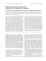
Figure 1 Illustration of the l0045-deleted mutant and genes
flanking l0045. (B) Diagrams representing L0045-related constructs
expressed from plasmids. Box indicates the open reading frame and
the filled area marks a putative signal peptide in the N-terminal region
of authentic L0045. In Δl0045, the segment encoding amino acid 37 to
the C-terminus of L0045 was deleted by a facilitated homologous re-
combination method; as a result, an FRT scar was left in the recombi-
nation spot. L45_K3stop represents that the construct engineered in
pQE_L45_K3stop has the third codon of l0045 (coding for Lys) mutated
to TAG so that the downstream translation was forced to stop.
L45_E42A is a construct where residue 42 at a predicted active site for
transglycosylase was mutated; residue marked is: E, an authentic Glu;
A, residue mutated to Ala. L45_NS, a non-secreted form of L0045 with
residues 2-18 spanning the putative signal peptide deleted.
Yu et al. Journal of Biomedical Science 2010, 17:52
/>Page 3 of 9
voltage electrical pulse (2500 V, 25 F, 200 ohms). Thereaf-
ter, 1-ml LB broth was added and gently mixed. After
being incubated at 37°C for 1 h, the bacteria were plated
on LB agar supplemented with appropriate selection anti-
biotics. Bacteria of K-12 strains were transformed rou-
tinely by chemical transformation.
Plasmid construction
Plasmid pQE60_L45 was constructed by PCR amplifica-
tion of l0045 from chromosomal DNA of EHEC followed
by insertion of the PCR product into NcoI and BglII sites
of pQE60 (Qiagen), a vector that provides an open-read-
ing frame with a six consecutive histidine-coding codons
before the translational stop site. By doing so, expressed
L0045 was tagged by His
x6
at the C-terminus as illustrated
in Fig. 1B. To express grlA [8] driven by T5 promoter
from pQE60, the corresponding fragment was PCR
amplified and inserted into SalI and HindIII sites of
pACYC184 to generate pACYC184_GrlA. To express
L0045 from pACYC184, PCR amplification of l0045 was
carried out with primers PL45_F_NcoI_2 (5'-TACCATG-
GTTTCATACTAACCTCACTC-3') and PL45_R_BglII
(5'-GTAAGATCTATCGATAATTTGCTCATTATTC-3').
The PCR product was inserted into NcoI/BglII-restricted
pACYC184 to result in pACYC184_L45, in which l0045 is
driven by its own promoter.
To construct plasmids expressing variants of L0045
(Fig. 1B), pQE60_L45 was used as the template. First, to
express the signal peptide-less L0045, primers
PL45_F_NcoI_NS (5'-TTACCATGGATTGTTTTGAAA
TTACAGG-3') and PL45_R_BglII were used to perform
PCR. The PCR product was then cloned into NcoI/BglII-
digested pQE60 (Qiagen) to generate pQE60_L45_NS. To
generate L45_K3stop fragment, in which a stop codon
was introduced at the third codon of l0045, the mutant
fragment was generated in two overlapping segments
with two primer pairs: PQE_F (5'-GGCGTATCACGAG-
GCCCTTTCG-3') paired with PL45_L3stop_R (5'-GCT-
CAGTATTATTTATTTCATGCCATGG-3'); PL45_L3
stop_F (5'-CATGGCATGAAATAAATAATACTGAGC-
3') paired with PQE_R (5'-CATTACTGGATCTATCAA-
CAGG-3'). These two PCR products were then mixed,
annealed, extended and then used as the template to gen-
erate the mutated fragment. Subsequently, the mutated
fragment was inserted into NcoI/BglII-restricted pQE60
to generate pQE60_L45_K3stop. With the same strategy,
pQE60_L45_E42A, in which residue Glu
42
of L0045 was
replaced with Ala, was generated similarly except for two
primer pairs: PL45_E42A_ F_ NsiI (5'-TTGAATGCAT-
CAAAATGCAAAAGCGGA-3') paired with
PL45_R_BglII; PL45_F_NcoI (5'-TACCATGGCAATGA
AAAAAATAATACTG-3') paired with PL45_E42A_ R_
NsiI (5'-TTTGATGCATTCCATGCAATTGCTTTT-3').
Immunoblotting
Bacterial cell lysates and the secreted proteins of EHEC
were prepared and analyzed by Western blotting as
described previously [18]. All the primary antibodies
were raised from rabbits. Species-specific secondary anti-
bodies with conjugation of horseradish peroxidase
(Sigma) were used to detect the primary antibody-bound
protein on blots. The blots were finally developed with
chemiluminescence reagent (20), of which signals were in
turn detected by exposing to X-ray film (Fuji).
L0045 induction by IPTG
Bacteria harboring T5 promoter-driven plasmids were
grown at 37°C overnight in 5-ml LB broth containing
ampicillin at 100 μg/ml. The culture was 1:50 diluted and
agitated at 37°C. With an interval of 1 h, 200 μl of culture
was sampled and its optical density at 600 nm was mea-
sured. After incubation for 3 h, isopropyl-thio-β-D-thio-
galactoside (IPTG) was added to a final concentration of
1 mM. After additional 3-h agitation, cells from 1 ml of
culture were harvested by centrifugation, dissolved in
SDS sample buffer and boiled.
Fractionation of bacterial proteins in different
compartments
The bacteria after appropriate cultivation were collected
by centrifugation, washed with Tris buffer (100 mM Tris,
pH 7.0) and suspended in a solution that were prepared
by mixing 10 ml 20% sucrose and 20 μl 500 mM EDTA.
Then, the bacteria cells were centrifuged and suspended
in 10 ml MgSO
4
followed by incubation at 4°C for 10 min.
After centrifugation, the supernatants were collected,
concentrated, and used as the periplasmic sample. The
cells were suspended in 6-ml Tris buffer and disrupted by
a French Press cell (SLM Amicon). After centrifugation,
the supernatants were collected and centrifuged again.
The supernatants were concentrated by centrifugation
filtration to obtain a sample that represented the cyto-
plasmic fraction. The pellets, which contain the mem-
brane proteins of the bacteria, were washed twice with
distilled H
2
O, suspended in 200 μl Sarkosyl buffer (100
mM NaCl, 10 mM Tris-HCl, pH 8.0, 1.0 mM PMSF, 0.5
μl/ml aprotinin, and 0.5% N-lauroylsarcosine) and incu-
bated at 4°C for 4 h. After ultracentrifugation, the super-
natants were collected as the inner membrane sample.
The remaining pellets were dissolved in 100 μl 0.1% SDS
and the resulting sample was defined as the outer mem-
brane fraction.
RT-PCR to monitor the l0045-specific mRNA in bacteria
RNA extraction from M9-cultivated EHEC was carried
out as previously described [19]. After ensuring no con-
tamination of chromosomal DNA, total RNA (2 μg) was
used to synthesize cDNA with a RevertAidTM First
Yu et al. Journal of Biomedical Science 2010, 17:52
/>Page 4 of 9
Strand cDNA synthesis kit (Fermentas). The obtained
cDNA was then primed with PL45_F_NcoI_1 (5'-TAC-
CATGGCAATGAAAAAAATAATACTG-3') and
PL45_R_BglII to PCR amplify the l0045 DNA fragment.
The same batch of cDNA was simultaneously amplified
for ompC with primers OMPCF (5'-GACGGCCTGCAC-
TATTTCTCTG-3') and OMPCR (5'-CTGCGAATGC-
CACACGGGTC-3'). As a result, the ompC fragment so
obtained was used as an internal comparison control.
Results
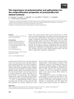
The effect of deleting l0045 from the LEE island of EHEC
was first examined by Western blotting. Fig. 2 shows that
representative LEE proteins were affected to different
degrees in the cellular lysates (compare lanes 1 and 2) but
all suppressed severely in the secreted portions (lanes 3
and 4). In the bacterial lysates, the EspA level decreased
most apparently (Fig. 2A, lanes 1 and 2). Complementa-
tion expressing L0045 from pACYC184_L45, where l0045
was driven by its upstream promoter, restored the cellular
level of EspA (Fig. 2B, lanes 1-3). At the same time, the
levels of Tir, EspB and EspA in the spent media were all
recovered to what have been seen with the parental strain
(Fig. 2B, lanes 4-6).
In an attempt to complement L0045 from a high-level
expression vector, i.e. pQE60_L45, we did not see a satis-
factory level of EspA detected in the transformant of
mutant Δl0045. Instead, these proteins (EspA and Tir in
particular) in the bacterial lysates were barely seen. In the
secreted portion, none of EspA, EspB, and Tir was well
detected (data not shown). The result that mutant Δl0045
was poorly complemented by pQE60_L45 was contrary
to what has been seen above with pACYC184_L45.
Therefore, we speculated that the opposite dosage effect
of L0045 might arise from the necessity of L0045 but in a
minute amount, a phenomenon similar to that has been
seen previously with L0036 [20]. A hypothesis is, then,
that L0045 might suppress the LEE protein expressions
when robustly induced. To test this, we used the parental
wild-type EHEC strain instead of mutant Δl0045 for the
transformation. Fig. 3 shows that the levels of Tir and
EspA were decreasingly seen in the bacterial lysates when
the wild-type EHEC received pQE60_L45, as compared
to that harbors the vector pQE60 control (lanes 1 and 2).
Seen in the same experiments was that the EspB level was
also perturbed but to a less distinct level. In the spent
media, however, all levels of Tir, EspA, and EspB were
profoundly reduced (Fig. 3, lanes 5 and 6), an observation
Figure 2 Effect of deleting l0045 on the levels of representative
TTS proteins. (A) Comparison of representative TTS proteins detected
in the bacterial lysates and the culture media. (B) Similar comparison of
the representative proteins as in (A) except that bacteria were trans-
formed with the specified plasmids. Bacteria were cultivated in M9 for
6 h in the presence of 5% CO
2
, harvested by centrifugation and disrupt-
ed by suspending in SDS sample buffer whereas the spent media were
filtered and then concentrated by TCA precipitation. Protein samples
were run in SDS-PAGE and examined by Western blotting. Note:
pACYC184_L45 was derived from pACYC184 by inserting fragment
containing l0045 with its own upstream promoter. OmpC from the
bacterial outer membrane was also detected to assure a comparable
sample loading.
Figure 3 Repression of representative TTS proteins' expressions
by robust induction of l0045. Plasmids were transformed into the
wild-type (WT) EHEC strain and the expression was induced by adding
IPTG. Assays of proteins present in the cell lysates and spent media
were done similar to that described in legend to Fig. 2. Note: the ex-
pression strength of pQE60_L45 is higher than that of pACYC184_L45
used in Fig. 2. No repression of both synthesis and secretion of the LEE
proteins was seen when an early termination was introduced at the 3
rd
codon of l0045. Repression was readily relieved with L45_E42A whose
transglycosylase was inactivated due to a mutation created at the pu-
tative active site.
Yu et al. Journal of Biomedical Science 2010, 17:52
/>Page 5 of 9
consistent with the notion that pQE60_L45 causes a sup-
pression effect on LEE.
To examine whether mRNA transcribed alone exerts
the same suppression, a stop codon was introduced into
the third position of l0045. By doing so, mRNA was nor-
mally transcribed from l0045 while translation from
mRNA would be aberrantly terminated, as illustrated in
Fig. 1B (L45K3stop). In Western blotting analyses, Fig. 3
shows that the bacteria transformed with
pQE60_L45K3stop yielded a normal expression of target
proteins in both cell lysates and spent media, as if it were
the wild-type strain harboring the control vector (com-
pare lanes 1 and 3 as well as lanes 5 and 7). Therefore, the
mRNA from the L0045-expressing plasmid unlikely is the
cause to suppress the synthesis and secretion of the
EHEC TTS proteins. Accordingly, the protein of L0045
per se is likely to play a major role in the above suppres-
sion.
Next is to address whether an inactive version of L0045
is able to suppress the cellular levels and secretion of the
EHEC TTS proteins. To do so, residue 42 of L0045, which
is conserved among LT domains and presumably
involved in the catalysis of the transglycosylases [12], was
changed from Glu to Ala (Fig. 1B, L45_E42A). The con-
struct expressed from pQE60_L45_E42A was similarly
examined for the effect on the representative LEE pro-
teins' expression in EHEC. Fig. 3 shows that the so-con-
structed variant of L0045 failed to show the strength seen
with the authentic L0045; no apparent repression was
observed with the expression and secretion of Tir, EspB
and EspA (compare lanes 1 and 4 as well as lanes 5 and 8
in Fig. 3). The above data altogether suggest that driving
l0045 toward a highly active expression would induce the
suppression of the TTS proteins and this suppression
readily requires an active construct of L0045.
It was puzzling that no Western blotting signals of
L0045 were detected in all bacterial lysates prepared,
including that from the parental EHEC strain, which was
analyzed with anti-L0045, and that from strain Δl0045
harboring either pACYC184_L45 or pQE60_L45, which
was detected with anti-His
x6
. However, the presence of
L0045 in the bacteria was evidently proven by fraction-
ation of the bacterial lysates. When fractionated proteins
were concentrated, L0045 was detectable mainly in the
periplasmic fraction (data not shown). Therefore, L0045
must be expressed, but regulated, to a low level. To
address why exogenously expressed L0045 was difficult to
detect in EHEC, the K-12 strain harboring pQE60_L45
was similarly analyzed. Fig. 4A shows that the growth of
JM109 carrying pQE60_L45 stopped immediately after
receiving IPTG and, then, the bacterial density declined
gradually. In contrast, IPTG-added EHEC continued to
grow, and the growth curve was similar to that of the bac-
teria harboring a control plasmid (pQE60). An explana-
tion for this is that actively expressed L0045 could have
generated a stress against JM109 so that bacterial growth
stopped and subsequently deteriorated. Unlike JM109,
EHEC continued to grow, a fact suggesting that the
expression of L0045 could have been restricted so that no
stress is generated. This notion was fully supported by the
Western blotting results in Fig. 4B that shows L0045 was
abundantly expressed in JM109 but hardly detected in the
EHEC strain (compare lanes 2 and 4).
To understand how the EHEC strain restricts L0045
from being expressed to a detectable level, we first exam-
ined whether EHEC senses any signal present in the mol-
Figure 4 Tight regulation of l0045 in EHEC. (A) Comparison of
growth curves of plasmid-transformed bacteria. Upper panel: K-12
(JM109); lower panel: wild-type EHEC. IPTG was added to the media as
marked during the cultivation. (B) L0045 detected in the total lysates of
bacteria harvested from (A) after 3-h IPTG induction. Loadings of bac-
terial lysates were comparable as seen with the internal control of
OmpC.
Yu et al. Journal of Biomedical Science 2010, 17:52
/>Page 6 of 9
ecule of L0045. The full-length but functionally inactive
L45_E42A described above and L45_NS with a trunca-
tion at the N-terminal putative signal peptide (Fig. 1B)
were examined. In JM109, these two constructs under the
pQE60 expression system were readily expressed and
their protein levels were similar to that of the authentic
L0045 (compare lanes 2 to 4 in Fig. 5B). However, only
L45_NS was equally well detected in both EHEC and
JM109 (lane 3 in Fig. 5A and 5B) and L45_E42A was not
detected in EHEC, as it were an authentic molecule (com-
pare lanes 2 and 4 in Fig. 5A). Therefore, deleting the
putative sec-dependent signal of L0045 removed the sig-
nal that suppresses L0045 from being highly expressed in
EHEC. Apparently, abolishing the activity of L0045 did
not turn off the suppression signal.
To address whether authentic L0045 not seen well in
EHEC was simply due to a lack of specific transcription,
the bacterial mRNAs were extracted and examined for
the relative abundances of the l0045-specific mRNA by
comparative RT-PCR. Fig. 5C shows that the DNA frag-
ments with an expected size were amplified from the
wild-type EHEC strain (lane 1). Similarly, mutant strain
Δl0045 transformed with the L0045-expressing plasmids
(lanes 3 and 4) gave the same results whereas transforma-
tion with the control vector (lane 2) yielded no signal (Fig.
5C), a fact suggesting the specificity of RT-PCR. It is
worth noting that strain Δl0045 harboring pQE60_L45
gave the strongest signal (compare lanes 1, 3 and 4). This
observation is consistent with the expectation that,
among the three positive expression settings, pQE60_L45
would have the highest expression strength. Given so,
L0045 directly from a total bacterial lysate remained
hardly detected in all these circumstances (data not
shown). Therefore, this fact suggests that the suppression
of L0045 in the EHEC might be a post-transcriptional
regulation.
To explore whether EHEC could resemble the K-12
strain to produce detectable L0045 under certain circum-
stances, a few mutants of EHEC were transformed with
pQE60_L45. The resulting transformants were analyzed
for the expression of L0045 by Western blotting (Fig. 6A).
Neither deleting Tir (in Δtir) nor EspB (in ΔespB) made
the mutant strains express detectable L0045 (lanes 3-6).
In contrast, deleting grlA yielded a difference. The His
x6
-
tagged L0045 was well detected in the lysate of mutant
ΔgrlA (Fig. 6A, compare lanes 1 and 2). The doublet
appearance of L0045 was presumably due to the molecu-
lar weight difference between the authentic molecule and
the signal peptide-processed product; the supporting evi-
dence was from the observation that the lower band
remained detected while the upper band disappeared
when the signal peptide was molecularly deleted (lane 3
Figure 5 Analyzing molecular determinant that causes L0045 dif-
ferently seen between EHEC and JM109. (A) & (B), total proteins
from bacteria examined for the plasmid-encoded L0045 by SDS-PAGE
in conjunction with Western blotting using anti-His
x6
. OmpC was de-
tected in parallel by anti-OmpC for the purpose of comparable loading
control. (C) Comparative RT-PCR to detect l0045-mRNA in EHEC strains
that carry the specified plasmids. OmpC, as internal control to normal-
ize the amount of mRNA.
Yu et al. Journal of Biomedical Science 2010, 17:52
/>Page 7 of 9
in Fig. 5A and 5B with the construct encoded by
pQE60_L45_NS). Anyway, complementation using
pACYC184_GrlA to express GrlA ectopically in ΔgrlA
did see a reversion and the expression of L0045 from
pQE60_L45 was repressed (Fig. 6B, lanes 1-3). Incom-
plete suppression is partly due to the fact that the two
plasmids co-transformed into strain ΔgrlA are compati-
ble but not necessarily expressed to appropriate levels.
Nevertheless, this repression phenomenon was repeat-
edly seen in strain ΔgrlA and it was not seen when exper-
iments were similarly carried out in JM109 (Fig. 6B, lanes
4-6). Therefore, these results suggest that GrlA in EHEC
is involved in sensing the level of L0045, a way likely
through an indirect effect since co-expressing GrlA and
L0045 in the K-12 JM109 strain gave no apparent sup-
pression (Fig. 6B, lanes 5 and 6). When the l0045 mRNA
was examined by comparative RT-PCR as described
above, no apparent difference was found in EHEC
between the parental strain and the grlA-disrupted strain
(data not shown). This result again suggests that the
L0045 regulation is at the post-transcription level.
Discussion
The phenotypes of the l0045-deleted strain were charac-
terized by a noticeable defect on TTS and a decreasing
level of EspA in the bacterial lysate. These phenotypic
changes could be reverted by complementation with
expressing L0045 from pACYC184_L45. Therefore, the
phenotypes observed could be strongly associated with
the gene deleted. The family homologues of L0045
includes rOrf3s of EPEC and C. rodentium, IagB of Sal-
monella, IpgF of Shigella, HrpH of Pseudomonas and
Hpa2 of Xanthomonas. These proteins all associate with
systems of TTS. IagB, IpgF and Hpa2 have been proven
with lytic activity against the bacterial cell wall and serve
as specialized LT in TTSS [15,21]. When comparing the
amino acid sequences, L0045 is 98% identical to rOrf3
and homologous to IagB with identity at 36.8% (or simi-
larity at 52.1%). In motif, they all share a conserved
domain of LT. Therefore, it is conceivable that L0045 rep-
resents the specialized LT in the EHEC LEE island to pro-
mote the assembly of TTSS.
Peptidoglycan, located between the inner and outer
membranes of Gram-negative bacteria, is composed of
glycan chains formed by N-acetyl-muraic acid (MurNAc)
and N-acetyl-glucosamine (GlcNAc). After cross-linking
with peptides, the resulted peptidoglycan forms a mesh-
work structure that maintains the shape of the bacteria
and provides protection against mechanical forces. On
the other hand, the peptidoglycan structures must be
constantly in dynamics to fit into the need of bacterial
growth and daughter cell divisions. Also, as to the need of
responses to different environmental changes, bacteria
may have to assemble some trans-envelope protein com-
plexes across the peptidoglycan. It is then reasonable to
believe that bacteria need specialized enzymes to reverse
timely the assembled peptidoglycan. Thus, LTs could
interrupt the glycan chains, help the reorganization of
peptidoglycan and facilitate the formation of large trans-
membrane structures, such as flagella, pili, and TTSS [12-
14]. With the case of rorf3 in C. rodentium, deletion of the
gene down-regulates TTS, attenuates pedestal formation
and decreases bacterial virulence in mice [3]. On the con-
trary, no obvious phenotypic difference in virulence has
been observed between the wild-type Shigella spp and
the ipgF mutant [22]. The latter has been attributed to the
redundancy of LTs in the bacteria. In EHEC, there are
three enzymes found in this family: flgJ for constructing
flagellum [23], pilT for the assembly of pilus [24] and
l0045 in the LEE island. Apparently, the redundancy of
LTs in EHEC provides limited compensation to the dele-
tion of l0045 as revealed by the decreasing secretion of
Tir, EspB and EspA when compared to that of the paren-
tal strain (Fig. 2A, lanes 3 and 4). In the experiments with
C. rodentium [3], the levels of intracellular Tir and EspB
were not apparently affected by the rorf3 deletion and we
had similar results in EHEC. However, in our analysis
with strain Δl0045, an apparent reduction was seen with
the intracellular level of EspA, of which data were absent
in the work with C. rodentium [3]. Since EspA constitutes
Figure 6 Effect on the L0045 expression in EHEC by the presence
or absence of grlA. (A) L0045 expressed from pQE60_L45 in the bac-
terial lysates when different EHEC mutant strains harbored the speci-
fied plasmids. (B) Comparison of L0045 detected in the EHEC ΔgrlA
strain (left) and JM109 (right) with or without GrlA expressed from
pACYC184_GrlA. Note: pQE60_L45 and pACYC184_GrlA are compati-
ble when co-transformed into the same host bacteria.
Yu et al. Journal of Biomedical Science 2010, 17:52
/>Page 8 of 9
the major component of the filamentous structure of the
TTS apparatus, a reduction of the EspA level in the bacte-
ria must restraint the assembly of the apparatus, a conse-
quence explaining well why the secretion of the TTS
proteins in the spent media is severely impaired.
Basing upon the putative lytic property toward bacte-
rial cell wall, expressing a high level of the LT family must
result in a stress to the host bacteria. Indeed, when a pre-
dicted LT gene hrpH from Pseudomonas syringae was
robustly induced in E. coli, the bacterial growth was
inhibited [25]. Consistent with this notion is that the
growth of JM109 was readily arrested and then deterio-
rated once L0045 was induced (Fig. 4A, upper panel).
Furthermore, this stress is apparently associated with the
inherited sec-dependent signal peptide; L0045 (from
pQE60_L45_NS) without the signal peptide was well
expressed in JM109 and found in the cytoplasm (data not
shown). Incorrect localization of no-signal-peptide
L0045 explains why the bacterial growth appeared to be
normal. The stress is also attributed to the lytic activity of
a correctly expressed L0045. This was revealed by the fact
that JM109 readily expresses the inactive L0045 (from
pQE60_L45_E42A) and grows normally.
Seen differently from that in JM109 was the expression
of L0045 in EHEC. All constructs did not perturb the
growth of the EHEC strain and, except for L45_NS, none
of the constructs were detected in the bacterial lysates.
These results were not due to a defect in the construct
because the same set of expression vectors gave satisfac-
tory results in JM109 (Fig. 5B). It could then be deduced
that the repression signal against L0045 that is recognized
by EHEC resides in the N-terminus of L0045. It is worth
noting that the putative catalytic activity of L0045 appar-
ently has nothing to do with the repression of L0045 in
EHEC; in case of inactive L45_E42A, the protein variant
remains undetectable (Fig. 5A).
GrlA encoded by grlA in the LEE island apparently
plays a vital role in the tight regulation of the l0045 level
in EHEC (Fig. 6). Deleting grlA from EHEC resulted in a
strong relief of the expression repression of the authentic
L0045. This phenomenon was not seen with the isogenic
strains carrying tir or espB deletion. GrlA is a second pos-
itive regulator encoded by LEE besides the major activa-
tor Ler, and its presence would represent that LEE is
vigorously activated to prepare the TTS components.
Speculatively, the presence of GrlA would suggest that
time is not ready for LT to be expressed. Conversely,
when activation of TTSS is close to the end, the activity of
GrlA would presumably dwindle. At this moment, most
TTS components are ready, and appropriately in-time
expressed L0045 would act upon peptidoglycan to pro-
mote the TTS apparatus assembly. It is then worth
exploring how an absence of GrlA in EHEC triggers the
L0045's expression and then tolerates the increasing syn-
thesis of L0045. Apparently, L0045 is not seen at a level
that is high enough to be detected by Western blotting
within the EHEC strain. Therefore, another query
remains to be answered is how EHEC regulates L0045 to
a critical amount but at a low level after the expression is
initiated. Anyhow, our current study has shed light on the
late stage of the TTS apparatus assembly, which is mani-
fested by a need of orchestrating peptidoglycan lysis
through controlling the L0045 expression.
Competing interests
The authors declare that they have no competing interests.
Authors' contributions
YCY, WSH and WJS designed the concept of research; YCY, CNL, SWN per-
formed research; and YCY, CNL, SHW and WJS wrote the paper. All authors read
and approved the final manuscript.
Acknowledgements
We thank Professor ST Hu for useful discussion and HS Luo and ST Chang for
helping with plasmid construction. This work was supported in part by a grant
from Ministry of Education, Aim for the Top University Plan http://eng-
lish.moe.gov.tw/ and Grants NSC98-2320-B-010-005-MY3, NSC98-2627-M-010-
003 and NSC98-2627-M-010-002.
Author Details
1
Institute of Microbiology and Immunology, National Yang-Ming University,
Taipei, Taiwan,
2
Department of Microbiology and Immunology, National Chiayi
University, Chiayi, Taiwan and
3
Department of Biotechnology and laboratory
Science in Medicine, National Yang-Ming University, Taipei, Taiwan
References
1. Roe A, Hoey D, Gally D: Regulation, secretion and activity of type III-
secreted proteins of enterohaemorrhagic Escherichia coli O157.
Biochem Soc Trans 2003, 31:98-103.
2. Frankel G, Phillips AD, Rosenshine I, Dougan G, Kaper JB, Knutton S:
Enteropathogenic and enterohaemorrhagic Escherichia coli: more
subversive elements. Mol Microbiol 1998, 30:911-921.
3. Deng W, Puente JL, Gruenheid S, Li Y, Vallance BA, Vazquez A, Barba J,
Ibarra JA, O'Donnell P, Metalnikov P: Dissecting virulence: systematic
and functional analyses of a pathogenicity island. Proc Natl Acad Sci
USA 2004, 101:3597-3602.
4. Lio JCW, Syu WJ: Identification of a negative regulator for the
pathogenicity island of enterohemorrhagic Escherichia coli O157:H7. J
Biomed Sci 2004, 11:855-863.
5. Elliott SJ, Wainwright LA, McDaniel TK, Jarvis KG, Deng YK, Lai LC,
McNamara BP, Donnenberg MS, Kaper JB: The complete sequence of the
locus of enterocyte effacement (LEE) from enteropathogenic
Escherichia coli E2348/69. Mol Microbiol 1998, 28:1-4.
6. Russell RM, Sharp FC, Rasko DA, Sperandio V: QseA and GrlR/GrlA
regulation of the locus of enterocyte effacement genes in
enterohemorrhagic Escherichia coli. J Bacteriol 2007, 189:5387-5392.
7. Mellies JL, Elliott SJ, Sperandio V, Donnenberg MS, Kaper JB: The Per
regulon of enteropathogenic Escherichia coli: identification of a
regulatory cascade and a novel transcriptional activator, the locus of
enterocyte effacement (LEE)-encoded regulator (Ler). Mol Microbiol
1999, 33:296-306.
8. Huang L, Syu WJ: GrlA of enterohemorrhagic Escherichia coli O157:H7
activates LEE 1 by binding to the promoter region. J Microbiol Immunol
Infect 2008, 41:9-16.
9. DiRita VJ, Elliott SJ, Sperandio V, Giron JA, Shin S, Mellies JL, Wainwright L,
Hutcheson SW, McDaniel TK, Kaper JB: The locus of enterocyte
effacement (LEE)-encoded regulator controls expression of both LEE-
and non-LEE-encoded virulence factors in enteropathogenic and
enterohemorrhagic Escherichia coli. Infect Immun 2000, 68:6115-6126.
Received: 11 February 2010 Accepted: 29 June 2010
Published: 29 June 2010
This article is available from: 2010 Yu et al; licensee BioMed Central Ltd. This is an Open Access article distributed under the terms of the Creative Commons Attribution License ( which permits unrestricted use, distribution, and reproduction in any medium, provided the original work is properly cited.Journa l of Biome dical Scie nce 2010, 17:52
Yu et al. Journal of Biomedical Science 2010, 17:52
/>Page 9 of 9
10. Barba J, Bustamante VH, Flores-Valdez MA, Deng W, Finlay BB, Puente JL: A
positive regulatory loop controls expression of the locus of enterocyte
effacement-encoded regulators Ler and GrlA. J Bacteriol 2005,
187:7918-7930.
11. Pallen MJ, Beatson SA, Bailey CM: Bioinformatics analysis of the locus for
enterocyte effacement provides novel insights into type-III secretion.
BMC Microbiol 2005, 5:9.
12. Koraimann G: Lytic transglycosylases in macromolecular transport
systems of Gram-negative bacteria. Cell Mol Life Sci 2003, 60:2371-2388.
13. Vollmer W, Joris B, Charlier P, Foster S: Bacterial peptidoglycan (murein)
hydrolases. FEMS Microbiol Rev 2008, 32:259-286.
14. Scheurwater E, Reid CW, Clarke AJ: Lytic transglycosylases: bacterial
space-making autolysins. Int J Biochem Cell Biol 2008, 40:586-591.
15. Zahrl D, Wagner M, Bischof K, Bayer M, Zavecz B, Beranek A, Ruckenstuhl C,
Zarfel GE, Koraimann G: Peptidoglycan degradation by specialized lytic
transglycosylases associated with type III and type IV secretion
systems. Microbiology 2005, 151:3455-3467.
16. Chuang CH, Chiu HJ, Hsu SC, Ho JY, Syu WJ: Comparison of Tir from
enterohemorrahgic and enteropathogenic Escherichia coli strains: two
homologues with distinct intracellular properties. J Biomed Sci 2006,
13:73-87.
17. Datsenko KA, Wanner BL: One-step inactivation of chromosomal genes
in Escherichia coli K-12 using PCR products. Proc Natl Acad Sci USA 2000,
97:6640-6645.
18. Chiu HJ, Syu WJ: Functional analysis of EspB from enterohaemorrhagic
Escherichia coli. Microbiology 2005, 151:3277-3286.
19. Ku CP, Lio JC, Wang SH, Lin CN, Syu WJ: Identification of a third EspA-
binding protein that forms part of the type III secretion system of
enterohemorrhagic Escherichia coli. J Biol Chem 2009, 284:1686-1693.
20. Tsai NP, Wu YC, Chen JW, Wu CF, Tzeng CM, Syu WJ: Multiple functions of
l0036 in the regulation of the pathogenicity island of
enterohaemorrhagic Escherichia coli O157:H7. Biochem J 2006, 393:591.
21. Zhang J, Wang X, Zhang Y, Zhang G, Wang J: A conserved Hpa2 protein
has lytic activity against the bacterial cell wall in phytopathogenic
Xanthomonas oryzae. Appl Microbiol Biotechnol 2008, 79:605-616.
22. Allaoui A, Menard R, Sansonetti PJ, Parsot C: Characterization of the
Shigella flexneri ipgD and ipgF genes, which are located in the proximal
part of the mxi locus. Infect Immun 1993, 61:1707-1714.
23. Nambu T, Minamino T, Macnab RM, Kutsukake K: Peptidoglycan-
hydrolyzing activity of the FlgJ protein, essential for flagellar rod
formation in Salmonella typhimurium. J Bacteriol 1999, 181:1555.
24. Sakai D, Komano T: Genes required for plasmid R64 thin-pilus
biogenesis: identification and localization of products of the pilK, pilM,
pilO, pilP, pilR, and pilT genes. J Bacteriol 2002, 184:444.
25. Oh HS, Kvitko BH, Morello JE, Collmer A: Pseudomonas syringae lytic
transglycosylases coregulated with the type III secretion system
contribute to the translocation of effector proteins into plant cells. J
Bacteriol 2007, 189:8277-8289.
doi: 10.1186/1423-0127-17-52
Cite this article as: Yu et al., A putative lytic transglycosylase tightly regu-
lated and critical for the EHEC type three secretion Journal of Biomedical Sci-
ence 2010, 17:52