Ceftriaxone-induced up-regulation of cortical and striatal GLT1 in the R6/2 model of Huntington’s disease pps
Bạn đang xem bản rút gọn của tài liệu. Xem và tải ngay bản đầy đủ của tài liệu tại đây (559.8 KB, 5 trang )
RESEARC H Open Access
Ceftriaxone-induced up-regulation of cortical
and striatal GLT1 in the R6/2 model of
Huntington’s disease
Youssef Sari
1,2,3
, Anne L Prieto
1,2
, Scott J Barton
1,2
, Benjamin R Miller
1,2,4
, George V Rebec
1,2*
Abstract
Background: Huntington’s disease (HD) is an inherited neurodegenerative disorder charac terized by cortico-striatal
dysfunction and loss of glutamate uptake. At 7 weeks of ag e, R6/2 mice, which model an aggressive form of
juvenile HD, show a glutamate-uptake deficit in striatum that can be reversed by treatment with ceftriaxone, a
b-lactam antibiotic that increases GLT1 expression. Only at advanced ages (> 11 weeks), however, do R6/2 mice
show an actual loss of striatal GLT1. Here, we tested whether ceftriaxone can reverse the decline in GLT1
expression that occurs in older R6/2s.
Results: Western blots were used to assess GLT1 expression in both striatum and cerebral cortex in R6/2 and
corresponding wild-type (WT) mice at 9 and 13 week s of age. Mice were euthanized for immunoblotting 24 hr
after five consecutive days of once daily injections (ip) of ceftriaxone (200 mg/kg) or saline vehicle. Despite a
significant GLT1 reduction in saline-treated R6/2 mice relative to WT at 13, but not 9, weeks of age, ceftriaxone
treatment increased cortical and striatal GLT1 expression relative to saline in all tested mice.
Conclusions: The ability of ceftriaxone to up-regulate GLT1 in R6/2 mice at an age when GLT1 expression is
significantly reduced suggests that the mechanism for increasing GLT1 expression is still functional. Thus,
ceftriaxone could be effective in modulating glutamate transmission even in late-stage HD.
Background
Ample evidence indicates that the neuropathology asso-
ciated with Huntington’ s disease (HD), an autosomal
dominant condition characterized by be havioral, cogni-
tive, and physical deterioration, involves the dysregula-
tion of glutamate, an excitatory amino acid [1-4]. In
fact, a decline in glutamate removal has been observed
in the brains of transgenic mouse models of HD [5-7] as
well as HD pati ents post-mortem [8]. Loss of glutamate
uptake leads to accumulation of extracellular glutamate,
making neurons vulnerable to excitotoxicity. Interest-
ingly, GLT1, a protein expressed primarily on glial cells
and responsible for the removal of most extracellular
glutamate [9,10], appearstobedysfunctionalinHD
mouse models [5,6,11]. We recently reported that the
deficit in glutamate uptake in the commonly used R6/2
model at 8 weeks of age can be reversed following
treatment with ceftriaxone [7], a beta-lactam antibiotic
that elevates the level of GLT1 without altering the
expression of other glutamate transporters [12]. By up-
regulating GLT1, ceftriaxone appears to overcome a
functional GLT1 deficit since the level of protein does
not decline until R6/2 mice exceed 11 weeks of age
[5,6,11]. Here, we determin ed if ceftriaxo ne could
increase GLT1 expression even in R6/2 mice that have a
deficit in GLT1 production. We focused on cerebral
cortex and s triatum, two forebrain regions that sho w
the greatest HD neuropathology [13,14]. Our results
suggest that the cellular machinery by which ceftriaxone
increases cortical and striatal GLT1 expression is still
intact even in late-stage HD.
Methods
Animals
Male transgenic R6/2 mice (B6CBA-TgN[HDexon1]
62Gpb) and wild-type (WT) controls were obtained
from The Jackson Laboratories (Bar Harbor, ME) at 6
* Correspondence:
1
Program in Neuroscience, Indiana University, 1101 East 10th Street,
Bloomington, IN, USA
Sari et al . Journal of Biomedical Science 2010, 17:62
/>© 2010 Sari et al; licensee BioMed Central Ltd. This is an Open Access article distributed under the terms of the Creative Commons
Attribution Licens e ( nses/by/2.0 ), which permits unrestricted use, distribution, and reproduct ion in
any medium, provided the original work is properly cited.
weeks of age. All mice were housed individually in the
departmental animal colony under standard conditions
(12 hr light/dark cycle with lights on at 07:00 AM) with
access to food a nd water ad libitum. Both the housing
and experimental use of animals followed the National
Institutes of Health guidelines and were approved by the
Institutional A nimal Care and Use Committee at Indi-
ana University Bloomington.
Genotype and CAG repeat length
We used PCR for genotyping and characterizing the
CAG repeat length as previously reported [7]. Our R6/2
mice had 121 ± 1.8 (mean ± SEM) CAG repeats, which
is within the range for d eveloping the HD behavioral
phenotype [15].
Treatment protocol
R6/2 and WT mice at either 8 or 12 weeks of age were
weighed and injected ip with 200 mg/kg ceftriaxone
(Sigma, St. Louis, MO) or an equal vo lume of saline
once daily f or 5 consecutive days. Twenty-four hours
aft er the last injection, when the mice had reached 9 or
13 weeks of age, the animals were decapitated. Their
brains were removed, and cerebral cortex and striatum
from both hemispheres were dissected and frozen for
immunoblotting.
Western blot
Western blots for GLT1 detection were performed as
previously described [7,16]. In brief, extracted proteins
were separated in 4-20% glycine gel (Invitrogen). T he
membranes were blocked in 3% milk in TBST (50 mM
Tris HCl; 150 mM NaCl, pH7.4; 0.1% Tween20) for 30
min at room temperature. The membranes were then
incubated with guinea pig anti-GLT1 anti body (Milli-
pore Bioscience Research Reagents) at 1:5,000 dilution
in blocking buffer at 4˚C. After washing and blocking,
the membranes were incubated with horseradish peroxi-
dase (HRP)-labeled anti-guinea pig s econdary antibody
(1:10,000 dilution) in the blocking buffer. Protein load-
ing was normalized using b-tubulin immunoblotting as
a loading control. Chemiluminescent detection of HRP
(SuperSignal West Pico; Pierce) was followed by expo-
sure of the membranes to a Kodak BioMax MR film
(Thermo Fisher Scientific). The films were developed on
an SRX-101A machine. Digitized images of immunor-
eactive proteins were quantified using an MCID system.
The data are reported as percentage ratios of GLT1/b-
tubulin.
Statistical analysis
Data were analyzed by means of a two-way analysis of
variance (ANOVA) and Bonferroni post hoc tests. All
statistical tests required a level of significance of at least
P < 0.05.
Results
Body weights
Table 1 shows the mean body weight of all groups on
the last day of treatment. No significant differences were
found between genoty pe (WT and R6/2) or treatment
group (ceftriaxone and saline) at 9 weeks of age. Regard-
less of treatment, however, there was a significant
reduction in body weight in R6/2 relative to WT mice
(P < 0.001 ) at 13 weeks of age, which supports previous
evidence that at this age R6/2 mice are strongly sympto-
matic [17].
Effects of ceftriaxone treatment in cortical and striatal
GLT1 expression
Although sal ine-treated R6/2s showed no loss of either
cortical or striatal GLT1 relative to WT at 9 weeks of
age (Figure 1), there was a marked reduction in both
brain regions in similarly treated 13-week-old R6/2s
(Figure 2). Quantitative analysis of this age group
revealed significant genotypic differences in GLT1
expression in both cerebral cortex (P < 0.01) and stria-
tum (P < 0.03). Despite the loss of GLT1 in older R6/2s,
these animals showed the same response to c eftriaxone
as the younger R6/2s and both WT age groups. Thus,
WT and R6/2 mice at either 9 (Figure 1) or 13 wee ks of
age (Figure 2) responded to ceftriaxone with an increase
in cort ical and striatal GLT1 expression relative to sal-
ine. Quantitative analysis revealed a significant effect of
ceftriaxone in both brain regions at 9 and 13 weeks of
age (P < 0.0001 in each case).
Discussion
Our results not only confirm the ability of ceftriaxone to
elevate GLT1 expression in cortex and striatum of R6/2
mice, but show that this effect still occurs even after
GLT1 levels begin to decline when these mice are 13
weeks of age and severely symp tomatic. Thus, it appears
that the cellular machinery underlying the ceftriaxone-
induced increase in GLT1 expression is operative in
late-stage HD.
Table 1 Body weight
Age WTs R6/2s WTc R6/2c
9-week 27.53 ± 1.18
(N = 4)
26.52 ± 1.02
(N = 4)
27.27 ± 1.02
(N = 4)
28.06 ± 1.18
(N = 4)
13-week 34.3 ± 2.75
(N = 5)
*26.78 ± 1.69
(N = 5)
33.00 ± 0.98
(N = 5)
*24.52 ± 2.33
(N = 5)
Data are presented as mean body weight (g) ± SEM. * P < 0.001, HD
compared to their respective WT. Abbreviations: WTs and R6/2s indicate saline
treatment, and WTc and R6/2c indicate ceftriaxone treatment. N refers to
number of animals per group.
Sari et al . Journal of Biomedical Science 2010, 17:62
/>Page 2 of 5
Although the mechanism by which ceftriaxone
increases GLT1 expression is not clea r, there is sup port
for activation of nuclear factor-kappa B (NF-kB), a tran-
scription factor that plays a role in regulating immune
responses and cell survival [18]. Translocation of the
NF-kB complex to the cell nucleus appears to be critical
for the action of ceftriaxone [19], and our results sug-
gest that this mechanism is intact in both cortex and
striatum of R6/2 mice regardless of age. Even before the
decline in GLT1 expression, moreover, 8-week-old R6/2
mice have a deficit in glutamate uptake, which is
rev ersed by ceftriaxone treatment [7]. Although there is
no GLT1 protein deficit at this age, m RNA levels are in
decline [6] and glutamate uptake is reduced [7], suggest-
ing a loss of transporter function well in advance of pro-
tein down-regulation. Thus, ceftriaxone is capable of
overcoming a deficit in GLT1 function. It is interesting
in this regard that palmitoylation, a process by which
proteins are inserted into cellular membranes [20], is
reduced in HD mice, including palmitoylation of GLT1
[21]. Whether ceftriaxone increases GLT1 palmitoyla-
tion is the focus of ongoing research.
It is unlikely that other glutamate transporters can
account for a ceftriaxone-induced increase in glutamate
uptake since ceftriaxone acts selectively on GLT1 [12].
It also is unlikely that loss of other glutamate transpor-
ters can account for the decline in uptake since neither
mRNA nor protein levels are altered for GLAST and
EAAC1 in HD models even at ages when the behavioral
phenotype is severe [6]. Post-mortem analysis of HD
patients, moreover, shows a se lective decline in GLT1
mRNA expression [22] as well as a loss of glutamate
uptake [8]. Nevertheless, we cannot rule out the poss ibi-
lity that ceftriaxone has other actions that may indirectly
impact glutamate transmission, including a change in
dopamine or GABA dynamics. Although an antibiotic
action of ceftriaxone is unli kely in that none of our ani-
mals showed signs of sepsis, it would be useful in fol-
low-up studies to determine if non-antibiotics that also
up-regulate GLT1, such as GPI-1046 [23], mimic the
effects of ceftriaxone in R6/2 mice.
Increasing GLT1 expression may become an effective
HD treatment strategy in that the up-regulation of
GLT1 induced by ceftriaxone significantly improves the
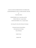
Figure 1 Effects of ceftriaxone on GLT1 expression in cerebral cortex and striatum at 9 weeks of age. Immunoblots (A, C) and
quantitative analysis (B, D) of the percentage ratio of GLT1/b-tubulin in cerebral cortex and striatum, respectively (***P < 0.001 and **P < 0.01
relative to corresponding saline group). Error bars indicate SEM. (N = 4 for each group).
Sari et al . Journal of Biomedical Science 2010, 17:62
/>Page 3 of 5
behavioral phenotype in 8-week-old R6/2 mice [7]. It is
unlikely that starting ceftriaxone treatment in 13-week-
old R 6/2s will result in behavioral improvement given
the stage of disease progre ssion in these animals, and in
fact, we found that ceftriaxone failed to reverse the
decline in body weight, which is evident in R6/2s at this
age. But our results suggest that the increase in GLT1
expression that occurs when ceftriaxone treatment is
begun earlier will continue to occur even in late-stage
HD. T hus, GLT1 expression is likely to be an effective
therapeutic target over a relatively long time course.
Glutamate dysregulation, including a possible decline
in GLT1 activity, may play a role in several neurodegen-
erative diseases [5,24]. In fact, a phase III cl inical trial of
ceftriaxone for treatment of amyotrophic lateral sclerosis
(ALS) is already underway (for review see [25]). The
dose required to increase GLT1 in mice produces com-
parable levels of ceftriaxone in the central nervous
system of patients undergoing treatment for meningitis
(0.3-6 μmol/L) [26], indicating that our treatment proto-
col is within normal limits for this drug. Nevertheless, it
is interesting that ceftriaxone increased cortical and
striatal GLT1 expression in both R6/2 and WT mice.
WT mice, however, show no discernable behavioral con-
sequences [7], suggesting that mechanisms are in place
to compensate fo r inc reased gl utamate removal.
Whether HD mice lack these mechanisms or simply
benefit from an increased rate of glutamate uptake
remains to be determined. It appears that w ithin limits
increased GLT1 expression is not a problem, but
decreased expression, which occurs in HD, is.
Conclusions
Ceftriaxone treatment enhances GLT1 expression in
cerebral cortex and striatum of R6/2 mice at 13 weeks
of age when endogenous GLT1 levels decline. These
Figure 2 Effects of ceftriaxone on GLT1 expression in cerebral cortex and striatum at 13 weeks of age.Immunoblots(A, C) and
quantitative analysis (B, D) of the percentage ratio of GLT1/b-tubulin in cerebral cortex and striatum, respectively (**P < 0.01 and ***P < 0.001
relative to corresponding saline group). Note the reduction in the percentage ratio of GLT1/b-tubulin expression in cerebral cortex (**P < 0.01)
and striatum (*P < 0.05) of saline-treated R6/2 mice relative to saline-treated WT mice, and the elimination of this effect after ceftriaxone. Error
bars indicate SEM. (N = 5 for each group).
Sari et al . Journal of Biomedical Science 2010, 17:62
/>Page 4 of 5
data suggest that the mechanism for increasing GLT1
expression is still functional even in late stage HD.
Acknowledgements
This research was supported by NINDS (R01 NS35663; F31 NS064791) and
the METACyt Initiative of Indiana University, which is funded, in part,
through a major grant from the Lilly Endowment, Inc. The authors would
like to thank Makiko Sakai for technical contributions and Faye Caylor for
administrative assistance.
Author details
1
Program in Neuroscience, Indiana University, 1101 East 10th Street,
Bloomington, IN, USA.
2
Department of Psychological and Brain Sciences,
Indiana University, 1101 East 10th Street, Bloomington, IN, USA.
3
University of
Toledo, College of Pharmacy, Department of Pharmacology, Health Science
Campus, 3000 Arlington Avenue, Toledo, OH 43606, USA.
4
University of
Texas Southwestern Medical School, Department of Physiology. 5323 Harry
Hines Boulevard, Dallas, TX 75390, USA.
Authors’ contributions
YS participated in study design and conceptualization, collected and
analyzed data, helped with data interpretation, and drafted the manus cript.
ALP helped with data collection, analysis, and interpretation. SJB performed
statistical analyses and genotyping, and helped with data interpretation.
BRM participated in study design, and helped with data collection and
analysis. GVR conceptualized and designed the study, and revised the
manuscript for intellectual content. All authors read and approved the final
manuscript.
Competing interests
The authors declare that they have no competing interests.
Received: 24 May 2010 Accepted: 27 July 2010 Published: 27 July 2010
References
1. DiFiglia M: Excitotoxic injury of the neostriatum: a model for
Huntington’s disease. Trends Neurosci 1990, 13(7):286-289.
2. Fonnum F, Storm-Mathisen J, Divac I: Biochemical evidence for glutamate
as neurotransmitter in corticostriatal and corticothalamic fibres in rat
brain. Neuroscience 1981, 6(5):863-873.
3. Harper PS: Huntington’s Disease. W.B. Saunders London, 2 1996.
4. Ross CA: Polyglutamine pathogenesis: emergence of unifying
mechanisms for Huntington’s disease and related disorders. Neuron
2002, 35(5):819-822.
5. Behrens PF, Franz P, Woodman B, Lindenberg KS, Landwehrmeyer GB:
Impaired glutamate transport and glutamate-glutamine cycling:
downstream effects of the Huntington mutation. Brain 2002, 125(Pt
8):1908-1922.
6. Lievens JC, Woodman B, Mahal A, Spasic-Boscovic O, Samuel D, Kerkerian-
Le Goff L, Bates GP: Impaired glutamate uptake in the R6 Huntington’s
disease transgenic mice. Neurobiol Dis 2001, 8(5):807-821.
7. Miller BR, Dorner JL, Shou M, Sari Y, Barton SJ, Sengelaub DR, Kennedy RT,
Rebec GV: Up-regulation of GLT1 expression increases glutamate uptake
and attenuates the Huntington’s disease phenotype in the R6/2 mouse.
Neuroscience 2008, 153(1):329-337.
8. Hassel B, Tessler S, Faull RL, Emson PC: Glutamate uptake is reduced in
prefrontal cortex in Huntington’s disease. Neurochem Res 2008,
33(2):232-237.
9. Danbolt NC: Glutamate uptake. Prog Neurobiol 2001, 65(1):1-105.
10. Robinson MB: The family of sodium-dependent glutamate transporters: a
focus on the GLT-1/EAAT2 subtype. Neurochem Int 1998, 33(6):479-491.
11. Estrada-Sanchez AM, Montiel T, Segovia J, Massieu L: Glutamate toxicity in
the striatum of the R6/2 Huntington’s disease transgenic mice is age-
dependent and correlates with decreased levels of glutamate
transporters. Neurobiol Dis 2009, 34(1):78-86.
12. Rothstein JD, Patel S, Regan MR, Haenggeli C, Huang YH, Bergles DE, Jin L,
Dykes Hoberg M, Vidensky S, Chung DS, Toan SV, Bruijn LI, Su ZZ, Gupta P,
Fisher PB: Beta-lactam antibiotics offer neuroprotection by increasing
glutamate transporter expression. Nature 2005, 433(7021):73-77.
13. Cepeda C, Wu N, Andre VM, Cummings DM, Levine MS: The corticostriatal
pathway in Huntington’s disease. Prog Neurobiol 2007, 81(5-6):253-271.
14. Walker AG, Miller BR, Fritsch JN, Barton SJ, Rebec GV: Altered information
processing in the prefrontal cortex of Huntington’s disease mouse
models. J Neurosci 2008, 28(36):8973-8982.
15. Mangiarini L, Sathasivam K, Seller M, Cozens B, Harper A, Hetherington C,
Lawton M, Trottier Y, Lehrach H, Davies SW, Bates GP: Exon 1 of the HD
gene with an expanded CAG repeat is sufficient to cause a progressive
neurological phenotype in transgenic mice. Cell 1996, 87(3):493-506.
16. Sari Y, Smith KD, Ali PK, Rebec GV: Upregulation of GLT1 attenuates cue-
induced reinstatement of cocaine-seeking behavior in rats. J Neurosci
2009, 29(29):9239-9243.
17. Carter RJ, Lione LA, Humby T, Mangiarini L, Mahal A, Bates GP, Dunnett SB,
Morton AJ: Characterization of progressive motor deficits in mice
transgenic for the human Huntington’s disease mutation. J Neurosci
1999, 19(8):3248-3257.
18. Karin M: Nuclear factor-kappaB in cancer development and progression.
Nature 2006, 441(7092):431-436.
19. Lee SG, Su ZZ, Emdad L, Gupta P, Sarkar D, Borjabad A, Volsky DJ, Fisher PB:
Mechanism of ceftriaxone induction of excitatory amino Acid
transporter-2 expression and glutamate uptake in primary human
astrocytes. J Biol Chem 2008, 283(19):13116-13123.
20. Huang K, El-Husseini A: Modulation of neuronal protein trafficking and
function by palmitoylation. Curr Opin Neurobiol 2005, 15(5):527-535.
21. Huang K, Kang MH, Askew C, Kang R, Sanders SS, Wan J, Davis NG,
Hayden MR: Palmitoylation and function of Glial Glutamate Transporter-1
is reduced in the YAC128 mouse model of Huntington disease.
Neurobiology of Disease .
22. Arzberger T, Krampfl K, Leimgruber S, Weindl A: Changes of NMDA
receptor subunit (NR1, NR2B) and glutamate transporter (GLT1) mRNA
expression in Huntington’s disease–an in situ hybridization study. J
Neuropathol Exp Neurol 1997, 56(4):440-454.
23. Ganel R, Ho T, Maragakis NJ, Jackson M, Steiner JP, Rothstein JD: Selective
up-regulation of the glial Na+-dependent glutamate transporter GLT1
by a neuroimmunophilin ligand results in neuroprotection. Neurobiol Dis
2006, 21(3):556-567.
24. Maragakis NJ, Rothstein JD: Glutamate transporters in neurologic disease.
Arch Neurol 2001, 58(3):365-370.
25. Traynor BJ, Bruijn L, Conwit R, Beal F, O’Neill G, Fagan SC, Cudkowicz ME:
Neuroprotective agents for clinical trials in ALS: a systematic
assessment. Neurology 2006, 67(1):20-27.
26. Nau R, Prange HW, Muth P, Mahr G, Menck S, Kolenda H, Sorgel F:
Passage
of cefotaxime and ceftriaxone into cerebrospinal fluid of patients with
uninflamed meninges. Antimicrob Agents Chemother 1993, 37(7):1518-1524.
doi:10.1186/1423-0127-17-62
Cite this article as: Sari et al.: Ceftriaxone-induced up-regulation of
cortical and striatal GLT1 in the R6/2 model of Huntington’s disease.
Journal of Biomedical Science 2010 17:62.
Submit your next manuscript to BioMed Central
and take full advantage of:
• Convenient online submission
• Thorough peer review
• No space constraints or color figure charges
• Immediate publication on acceptance
• Inclusion in PubMed, CAS, Scopus and Google Scholar
• Research which is freely available for redistribution
Submit your manuscript at
www.biomedcentral.com/submit
Sari et al . Journal of Biomedical Science 2010, 17:62
/>Page 5 of 5