Overexpression of hTERT increases stem-like properties and decreases spontaneous differentiation in human mesenchymal stem cell lines ppt
Bạn đang xem bản rút gọn của tài liệu. Xem và tải ngay bản đầy đủ của tài liệu tại đây (7.49 MB, 13 trang )
RESEA R C H Open Access
Overexpression of hTERT increases stem-like
properties and decreases spontaneous
differentiation in human mesenchymal stem
cell lines
Chih-Chien Tsai
1†
, Chun-Li Chen
2†
, Hwa-Chung Liu
3
, Yi-Ting Lee
1,4
, Hsei-Wei Wang
5
, Lein-Tuan Hou
2*
,
Shih-Chieh Hung
1,4*
Abstract
To overcome loss of stem-like properties and spontaneous differentiation those hinder the expansion and applica-
tion of human mesenchymal stem cells (hMSCs), we have clonally isolated permanent and stable human MSC lines
by ectopic overexpression of primary cell cultures of hMSCs with HPV 16 E6E7 and human telomerase reverse tran-
scriptase (hTERT) genes. These cel ls were found to have a differentiation potential far beyond the ordinary hMSCs.
They expressed trophoectoderm and germline specific markers upon differentiation with BMP4 and retinoic acid,
respectively. Furthermore, they displayed higher osteogenic and neural differentiation efficiency than primary
hMSCs or hMSCs expressed HPV16 E6E7 alone with a decrease in methylation level as proven by a global CpG
island methylation profile analysis. Notably, the demethylated CpG islands were highly associated with develop-
ment and differentiation associated genes. Principal component analysis further pointed out the expression profile
of the cells converged toward embryonic stem cells. These data demonstrate these cells not only are a useful tool
for the studies of cell differentiation both for the mesenchymal and neurogenic lineages, but also provide a valu-
able source of cells for cell therapy studies in animal models of skeletal and neurological disorders.
Introduction
Bone marrow derived human mesenchymal stem cells
(hMSCs) are considered one of the most promising and
prospective resources for cell and gene therapy in
mesenchymal and non-mesenchymal applications
because of their great self-renewal and versatile plasticity
in vitro and in vivo [1]. However, there are still two
major hindrances, loss of stem-like properties, namely
self-renewal and multipotency, and spontaneous differ-
entiation, encountered during in vitro expansion of
MSCs [2]. Loss of stem-like properties could be defined
as diminished replication, altered functionality [3],
and deteriorated potential for differentiation [4].
Spontaneous differentiation, known as the emergence of
lineage-specific markers without any directed differentia-
tion, would diminish the proportion of undifferentiated
stem cells, and therefore compromised the benefit of
hMSCs for clinical application. Thus, identifying meth-
ods for inhibiting loss of stem-like properties and spon-
taneous differe ntiation, and reversing hMSCs to a more
primitive state has attracted great research interest.
In a previous attempt to immortalize hMSCs with
increased life span, we have established a cell line-KP
by transferring HPV16 E6E7 genes into hMSCs [5].
Though KP successfully overco mes the drawback of
cellular senescence and could be passaged over 100
population doublings (PDs), the phenomenon of spon-
taneous differentiation could not be avoided [6]. Telo-
merase, known to maintain the telomere length, has
been indicated to play a role inself-renewalandpluri-
potency of embryonic s tem cells (ESCs) [7]. However,
hMSCs express no telomerase activity with telomere
* Correspondence: ;
† Contributed equally
1
Stem Cell Laboratory, Department of Medical Research & Education and
Orthopaedics & Traumatology, Veterans General Hospital, Taipei, Taiwan
2
Graduate Institute of Dental Sciences and Department of Periodontology,
National Taiwan University, Taipei, Taiwan
Full list of author information is available at the end of the article
Tsai et al. Journal of Biomedical Science 2010, 17:64
/>© 2010 Tsai et al; licensee BioMed Central Ltd. This is an Open Access article distributed under the terms of the Creative Commons
Attribu tion License ( s/by/2.0), which perm its unrestricted use, distribution, and reproduction in
any medium, provided the original work is properly cited .
shortening in a rate similar t o non-stem cells (30-120
bp/population doubling), a nd cease to divide when the
telomere length is less than 10 kb [8]. Besides, ectopic
expression of hum an telomerase reverse transcrip tase
(hTERT), the catalytic component of telomerase, has
been proven not only to bypass cellular senescence
and extend life span [9], but also to influence differen-
tiation potential [10]. Notably, a recent report has
unraveled a fascinating fact that TERT might play a
crucial role in gene regulation directly or indirectly,
which finally caused profound changes in gene expres-
sions of mouse skin [11]. What’s most important, the
authors further demonstrated that the effect of TERT
on gene regulation is irrelevant to its catalytic enzyme
action at telomere ends [11].
In mammals, DNA methylat ion of cytosines in cyto-
sine guanine dinucleotide (CpG) islands, known to med-
iate epigenetic gene silencing [12,13], plays pivotal roles
in embryonic develo pme nt [14-16] and ESC diffe rentia -
tion [17]. For example, treating ESCs or somatic cells
with demethylation agent such as 5-azacytidine
(5-AzaC) resulted in dedifferentiation, thereby pointing
out the associatio n of DNA methylation with the differ-
entiation state [18-20]. These results also imply methods
that reverse the d ifferenti ation state of stem or progeni-
tor cells will induce changes in DNA methylation pat-
terns [17].
In this stud y, we hy pothesized, after ectopic expres-
sion of HPV16 E 6E7 and hTERT, hMSCs would bypass
loss of stem-like properties and block spontaneous dif-
ferentiation with changes i n DNA methylation pat-
terns. Meanwhil e, we also tried to demonstrate the
heightened differentiation potential of HPV16 E6E7
and hTERT-transfected hMSCs by directing germline
and trophoectoderm differentiation. Finally, the roles
of DNA methylation-modification factors, such as
DNA methyltransferases (DNMTs) in the reversion of
hMSCs to a more primitive st ate would be explo red.
Materials and methods
Cell Cultures
Primary hMSCs were obtained from the Tulane Center
for Preparation and Distribution of Adult Stem Cells
( The cells
were grown in alpha minimal essential medium
(aMEM; GIBCO/BRL, Carlsbad, CA; itro-
gen.com) su pplemented with 16.6% fetal bovine serum
(FBS), 100 U/ml penicillin, 100 μg/m l streptomycin, and
2 mM L-glutamine (GIBCO/BRL) at 37°C under 5%
CO2 a tmosphere. The medium was changed twice per
week and a subculture was performed after they reached
about 80% confluency.
The hMSC strain (KP) w as developed by transfection
with the type 16 human papilloma virus proteins E6E7
as described previously [6]. This strain is grown in
DMEM-LG (GIB CO/BRL) supplemented with 10% F BS,
100 U/ml penicillin, 100 μg/ml streptomycin, and 2 mM
L-glutamine. The medium was changed twice per we ek
and a subculture was performed at 1:3 to 1:5 split every
week. Using flow cytometry, cells express CD29, CD44,
CD90, CD105, SH2, and SH3.
DNA Delivery Methods
KP cells were transfected w ith phTERT-IRES2-EGFP,
which was generated by inserting a 3.45-kb EcoRI-EcoRI
fragment containing the hTERT cDNA into pIRSE2-
EGFP (Clontech, Palo Alto, CA, ntech.
com) using Nucleofector technology as recommended
by the manufacturer (Amaxa Biosystems, Cologn e, Ger-
many, ). The e fficiency of trans-
fection as evaluated by the expression of EGFP was
around 70%. The cells were then suspended in an
appropriate volume of 20% FBS-supplemented DMEM-
LG medium, seeded in 96 well plate for selecting single
cell clone by neomycin (400 μg/ml).
Reverse Transcription-Polymerase Chain Reaction
(RT-PCR)
Total RNA was extracted using the Tri Reagent (Sig ma,
St. Louis, MO. ) according
to the manufacturer’s specifications. First strand cDNA
synthesis was performed using Superscript III reverse
transcriptase (Invitrogen, Carlsbad, CA, i-
trogen.com), Random primer (Invitrogen), 10 mM
dNTPs (Invitrogen), 5× First Strand synthesis buffer, 0.1
M DTT, and RNaseOUT ribonuclease RNase inhibitor
(Invitrogen). PCR was performed using cDNA as the
template in a 50 μl reaction mixture containing a speci-
fic primer pair of each cDNA according to the published
sequences. The reaction products were resolved by elec-
trophoresis on a 1.5% agarose gel and visualized with
ethidium bromide. Sequences of PCR primers and NCBI
reference sequence numbers were listed in Additional
file 1.
Real-Time PCR
Real-Time PCR was performed using an ABI PRISM
7700 sequence detection system (Applied Biosy stem,
Foster City, CA, ) and
the TaqMan Universal Master Mix (Applied Biosys-
tems). Analysis of the results was carried out using the
software supplied with the mac hine. The software calcu-
lates each gene expression relative to the b-actin house-
keeper gene (delta CT) and then relative to controls
(delta delta CT) using the fluorescence threshold of the
amplification reaction and the comparative CT method.
Sequences of PCR primers, probe and PCR conditions
can be provided on request.
Tsai et al. Journal of Biomedical Science 2010, 17:64
/>Page 2 of 13
Differentiation Protocols
Trophoectoder m differentiation protocol was modified
from a previous method [21]. Cells at 50% of confluence
were treated with 100 ng/mL BMP4 (R&D Systems,
Minneapolis, MN, ) in
DMEM-LG supplemented with 10% FBS. Medium was
changed twice per week. Germline differentiation proto-
col was performed w ith a protocol modified from pre-
vious report [22]. In brief, cells were plated at a density
of 1~2 × 10
4
cells/cm
2
in DMEM-LG supplemented
with 10% FBS and 2 μM retinoic acid (RA, Sigma) with
medium change twice per week. For osteogenic differen-
tiation, cells were seeded at a density of 10
4
cells/cm
2
and induced in DMEM-LG s upplemented with 10%
FBS, 50 μg/ml ascorbate-2 phosphate (Nacalai, Kyoto,
Japan, al ai.co.jp), 10
-8
M dexamethasone
(Sigma) and 10 mM b-glycero phosphate (Sigma) wit h
medium change twice per week. For neurogenic differ-
entiation [23], 100 ng/ml recombinant human Noggin
(R&D Systems) was added into the serum-free DMEM-
LG culture medium.
Histochemical Studies
Cells were fixed in 2% paraformaldehyde for 10 min and
stained for alkaline phosphatase activity and in vitro
mineralization by Alizarin red-S [ 5] to reveal osteogenic
differentiation. After washing 5 times with PBS, stained
cultures were photographed.
DNA Methylation Array
DNA preparation
Genomic DNA was extracted from samples using
QIAamp® DNA mini kit (Qiagen GmbH, Hilden, Ger-
many, ) according to the manu-
facturer’s protocol.
aPRIMES
1 μg genomic DNA was restricted to completion with
10 U MseI at 37°C in a final volume of 10 μl in the buf-
fer p repared with the 10 × One-Phor-All Buffer PLUS
(GE Healthcare Bio-science Corp., Piscataway, NJ,
). Heat inactivation was
carried out at 65°C for 20 min. MseIfragmentswere
then subjected to ligation with PCR lin kers, MseI linker-
S(5’-TAA CTA GCA TGC-3’)andMseIlinker-L(5’-
AGT GGG ATT CCG CAT GCT AGT-3’)overnight.
Half of the resulting ligated MseI fragments were
digested with the restriction enzyme McrBC (New Eng-
land Biol abs , Beverly , MA, http://www.n eb.com) for 3 h
following the conditions recommended by the supplier.
The other half of the MseI fragments were digested with
the three methylation-sensitive endonucleases HpaII
(New England Biolabs; recognition si te CCGG, 3 h, 37°
C), HhaI (New England Biolabs; recognition site CGCG,
3 h, 37°C) and BstUI (New England Biolabs; recognition
site CGCG, 3 h, 60°C) according to t he recommenda-
tions of the supplier. Digested DNA fragments were
then treated with 1 μl Prot einase K (Invitrogen) for 1 h
at 37°C with subsequent heat inactivation at 80°C for
10 min. For the LM-PCR steps, 2× PCR Master Mix
(Promega, Madison, WI, ht tp://www.promega.com) was
added to a final volume of 50 μl. A MJ thermocycler
was programmed to 68°C for 10 min, followed by 27
cycle loops at 94°C (40 s), 57°C (30 s) and 68°C (75 s).
Final elongation was carried out at 72°C for 10 min.
PCR products were purified by ethanol precipitation.
DNA was eluted in 50 μl nuclease free H
2
O.
Labeling and hybridization to microarrays
Both the HpaII/HhaI/BstuI-digested and the McrBC-
digested samples were diff erentially labeled with Cy5- or
Cy3-conjugated dUTP by use of an Agilent Genomic
DNA Labeling Kit PLUS (Agilent Technologies, Palo
Alto, CA, len t.com). Labeled targets were
subsequently cleanup by the use of a Centricon YM-30
column (Millipore, Billerica, MA, lipore.
com), pooled and mixed in a 500-μl hybridization mix-
tures with 50 μgofhumanCot-1DNA(Invitrogen)in
1× hybridization buffer (Agilent Technologies). Before
hybridization to the array, the hybridization mixtures
were denatured at 95°C for 3 min and incubated at 37°C
for 30 min. To remove a ny precipitate, the mixture was
centrifuged at ≥ 14,0 00 × g for 5 min and the superna-
tant was transferred to a new tube. The labeled and
denatured DNA target was then hybridized to human
CpG island microarray (G4492A, Agilent Technologies,
USA) at 65°C fo r 40 h. The arrays were washed with 0.5
× SSC/0.005% Triton X-102 (wash 1) at room tempera-
ture f or 5 min, and then wit h 0.1 × SS C/0.0 05% Triton
X-102 (wash 2) at 37°C for 5 min.
Image and microarray data analysis
After drying by nitrogen gun blowing, microarrays were
scanned with an Agilent microarray scanner (Agilent
Technologies) at 535 nm and 625 nm for Cy3 and Cy5,
respectively. Scanne d images were analyzed by Feature
extraction 9.1 software (Agilent Technologies) to quan-
tify signal and background intensity for each feature.
Microarray data wer e firstly normalized with print-tip
loess, followed by background-correction, normal ization
and analysis by the limma package within the R environ-
ment (version 2.1.0). The methylation level was deter-
mined as the ratio o f Cy5/Cy3 in each spot. The raw
data from the array experiments is available from the
Gene Expression Omnibus (GEO; .
nih.gov/geo) under the series accession number GSE
(pending number). For Gene Ontology (GO) analysis of
the genes decreased in CpG island methylation, we
determined the statistically significant GO terms us ing
the hypergeometric probability distribution. For each
GO term, a p-value was calculated representing the
Tsai et al. Journal of Biomedical Science 2010, 17:64
/>Page 3 of 13
probability that the number of genes that are annotated
at the term could have been found by chance.
Microarray expression data sets and principal component
analyses (PCA)
The expression profile of hTERT-transfected hMSCs
was implemented by using the Af fymetrix™ HG U133
Plus 2.0. The microarray data sets of various normal tis-
sues and ESCs were r etrieved from public databa ses.
The ESCs used for microarray analysis were H9 clones
and all microarray data are available at GEO under the
accession no. of GSM249282, GSM124302 and
GSM124362. To determine the similarity of the expres-
sion profiles between hTERT-transfected hMSCs and
various n ormal human tissues, MSCs, and ESCs, PCA
was performed in 31 Affymetrix™ U133 Plus 2.0 array
data. using the Partek® Genomics Suite™ software (Partek
Incorporated, St. Louis, MO, ).
All microarray datasets in this paper are available at
GEO under the accession no. of GSE7234 and GSE9520.
Results
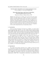
Downregulation of Oct4 and Nanog and upregulation
of developmental markers and lineage-specific genes
during expansion of primary hMSCs
Embryonic transcription factors, such as Oct4 and
Nanog, normally expressed in early embryos and ES Cs,
inhibit tissue-specific genes and enhance self-renewal
and pluripotency [24]. To evaluate whether loss of
stem-like propert ies occurred during normal passage of
hMSCs, we examined the expression of Oct4 and Nanog
in primary hMSCs isolated from three individuals. Semi-
quantative RT-PCR and real-time RT-PCR analysis
revealed higher mRNA levels of Oct4 and Nanog at pas-
sage3(P3)thanatpassage10(P10)(Figure1A),sug-
gesting loss of stem-like properties during expansion of
primary hMSCs.
ESCs, a powerfu l tool to study mammalian develop-
ment, for m e mbryoid bodies (EBs) and express a panel
of developmental markers upon removal of feeder layer
or leukemia inhibitory factor. To evaluate whether spon-
taneous differentiation with the expression of develop-
mental markers occurred during normal passage of
primary hMSCs, we examined the expression levels of
ectoderm (Pax6) [25], primitive endoderm (Gata4 and
Gata6) [26] and definitive endoderm (Sox17 and FoxA2)
[27] markers by RT-PCR. The expression levels of Pax6,
Gata4 and Fo xA2 were higher at P10 than at P3 (Figure
1Ba). We next looked at the expression of germline
markers [28], and found the expression levels of Stella,
Dazl, Vasa and Scp3 were higher at P10 (Figure 1Bb).
Finally, we examined two lineage-specific markers
expressed in EBs, t he neural (Nestin) and cardiac speci-
fic genes (Nkx 2.5 and cTn1) and found P10 had higher
expression of Nestin and cTn-1 (Figure 1Bc). These
results point to upregulation of developmental markers
and lineage-specific genes in late-passage primary
hMSCs.
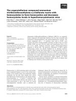
Transient upregulation of Oct4 and Nanog during early
differentiation in immortalized hMSCs
To overcome loss of stem-like properties and sponta-
neous differentiation those hinder the expansion and
application of hMSCs, we first overexpressed primary
cell cultures of hMSCs with HPV 16 E6E7 and devel-
oped the KP cells [6], which were then overexpressed
with hTERT. Several single-cell derived clones were iso-
lated and 3A6, 1C5 and 3G11 were used for further ana-
lysis. All of these clones grown in monolayer in DMEM-
LG supplemented with 10% FBS had a remarkably
shorter population doubling time (1.9 days) compared
with the parental KP cells (3.0 days). RT-PCR revealed
the expression of hTERT in all these three clones. Flow
cytometry also demonstrated these cells have a normal
surface protein profile like the no rmal hMSCs (Addi-
tional file 2).
To examine if these cells increases in stem-like prop-
erties, we chose 3A6 for further evaluation. We first
compared the expression levels of Oct4 and Nanog
between KP and 3A6. Unexpectedly, RT-PCR and real-
time RT-PCR unraveled the downregulation of both
Oct4 and Nanog in 3A6 compared with KP (Figure 2A).
Downregulation of the embryonic transcription f actors
such as Oct4 and Nanog is associated with differentia-
tion of neural stem cells, hematopoietic stem cells and
MSCs. However, an increase in Oct4 expression in ESCs
causes differentiation into primitive endoderm [29],
mesoderm [29] and early cardiac lineage [30]. Overex-
pression of Nanog also drives the expression of ecto-
derm markers [30]. The expression pattern of Oct4 and
Nanog during differentiation is completely different
between E SCs and adult stem cells such as MSCs, and
should serve as an indicator to discriminate ESCs from
MSCs [29-31]. W e therefore induced 3A6 to undergo
osteogenic and neural d ifferentiation and examined the
expression of Oct4 and Nanog. During osteogenic differ-
entiation, we noticed a continuous upregulation of Oct4
and Nanog until day 7 followed by downregulation of
both genes at day 14 (Figure 2Ba). Similarly, during
neural differentiation, the upregulation of Oct4 and
Nanog was observed during early differentiation (Figure
2Bb). These results indicated 3A6 has a differential gene
expression of embryonic markers similar to the early
differentiation of ESCs.
Downregulation of developmental markers and
lineage-specific genes in immortalized hMSCs
To clarify the blocking of spontaneous differentiation in
3A6, we compared the expression of developmental
Tsai et al. Journal of Biomedical Science 2010, 17:64
/>Page 4 of 13
markers and lineage-specific genes between 3A6 and KP
by performing RT- PCR for trophoectoderm (CDX2 and
CGb), germline (Dazl, Vasa and Scp3), osteogenic (BSP,
Bone Sialoprotein and OCN, Osteocalcin) and neural
(Pax6 and Nestin) specific markers. We noted a general
downregulation of express ion for all these gene s at 3A6
compared with KP (Figure 2C), indicating 3A6 main-
tained in an undifferentiated state.
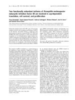
Improvement of differentiation potential in
immortalized hMSCs
After characterization of 3A6 and unraveling its relative
quiescent state, it is of great interest if the differentiation
potential of 3A6 would be sustained, enhanced and
reversed to a considerably primitive state. We first exam-
ined if 3A6 sustained the normal capabilities of hMSCs,
such as mesenchymal (osteogenic, adipogenic and chon-
drogenic) and non-mesenchymal (neural) differentiation
and hematopoietic supporting potential (cobblestone
forming). 3A6 had normal or elevated osteogenic and
chondrogenic differentiation potential compared with
one KP-derived single cell clone, whereas 3A6 had
decreased adipogenic differentiation potential (Figure
3A). These data are consistent with previous studies that
overexpression of hTERT increased osteogenic potential
and the inverse relationship between osteogenic and
Figure 1 Differential gene expression between primary cultured passage 3 (P3) and passage 10 (P10). (A) RT-PCR (left panel) and Real-
time RT-PCR (right panel) analysis of pluripotency related genes in MSCs from three individual donors (hMSC-1, -2, -3). (B) Differential expression
of (a) developmental (b) germline specific (c) lineage specific genes by RT-PCR analysis.
Tsai et al. Journal of Biomedical Science 2010, 17:64
/>Page 5 of 13
Figure 2 Differential gene expression between 3A6 and KP, and alteration of pluripotency related markers during 3A6 differentiation.
(A) RT-PCR (left panel) and Real-time RT-PCR (right panel) analysis of pluripotency related genes in 3A6 and KP. (B) Differential expression of
Oct4 and Nanog during (a) osteogenic and (b) neural differentiation in 3A6. c. RT-PCR analysis of (a) trophoectoderm (b) germline (c)
osteoblastic and (d) neural lineage specific genes.
Tsai et al. Journal of Biomedical Science 2010, 17:64
/>Page 6 of 13
adipogenic differentiation. For neural differentiation, 3A6
adopted the typical morphology of neural progenitor
cells, including bipolar elongated cell processes a nd
retracted cell bodies, and expressed neural lineage speci-
fic markers, such as Nestin and Pax6 on stimulation with
noggin in serum free conditions for 14 days (Figure 3B).
For co-cultured CD34+ hematopoietic stem cells with
3A6 cells, we noted the formation of cobblestone areas
from hematopoietic cells that transmigrated beneath the
layer of 3A6 cells (Figure 3C).
Previously, only ESCs has proven to be able to suc-
cessfully dif ferentiate toward trophoectoderm [21] and
Figure 3 Versati le differ ent iation potential of 3 A6. (A) Morphology without induction or with osteogenic (21 da ys, demonstrated by von
Kossa staining), chondrogenic (21 days, demonstrated by Alcian Blue staining) or adipogenic (14 days, demonstrated by Oil Red O staining)
differentiation. (B) Neural differentiation confirmed by the alteration of cell morphology to the round cell body with bipolar elongated cell
processes, and by RT-PCR after induction with noggin for 14 days. (C) Cobble stone formation by co-culture with hematopoietic stem cells. (D)
Trophoectoderm- and (E) germline-differentiation analyzed by RT-PCR after induction in three individual clones with BMP4 and RA, respectively.
Tsai et al. Journal of Biomedical Science 2010, 17:64
/>Page 7 of 13
germline [28] in vitro, but Johnson and others [32]
detected the e xpression of germline markers in bone
marrow and peripheral blood, and Nayernia and others
[22] further implied the germline differentiation poten-
tial of mouse MSCs. Few, if any, literature so far, how-
ever, has revealed the differentiation pote ntial of MSCs
toward tr ophoectoder m. To test the most versatile dif-
ferentiation potential of hMSCs after ectopic expression
of hTERT, we directed 3A6 and two other clones, 1C5
and 3G11 towards trophoectoderm and germline differ-
entiation upon stimulation with BMP4 [21] and retinoic
acid (RA) [33], respectively. This has been used to initi-
ate trophoblast and germline differentiation in human
ESCs. As demonstrated by RT-PCR, these cells clones
started to e xpress the trophoectoderm specific markers,
such as CDX2 and CGb (Figure 3D), and germline spe-
cific markers [28], such as Stella, Dazl, Vasa, and Scp3
(Figure 3E) after differentiation. These results together
suggest these cells not only sustained normal potential
as hMSCs, but also adopted the potential that was pre-
viously not belonged to hMSCs.
Enhanced differentiation efficiency in
immortalized hMSCs
Besides the differentiation potential, another significant
issue would be the differentiation efficiency of 3A6.
Spontaneous differentiation, noted during expansion of
primary hMSCs and KP, might hamper differentiation
efficiency because less uncommitted cells could be
directed toward specific lineage. Thus, we expected 3A6
to have better differentiation efficiency because of its
less committed state. To clarify this hypothesis, we
directed KP and 3A 6 toward osteogenic or neural line-
age and compared their differentiation efficiency by his-
tochemical staining and lineage-specific gene expression.
We observed 3A6 had higher alkaline phosphatase and
Alizarin Red S staining compared with KP at day 3 to
day 14 of osteogenic differentiation (Figure 4A). The
expression levels of osteogenic markers-BSP and OCN
were also elevated in 3A6 compared with KP during
osteogenic differentiation. The expression levels of
neural markers-Nestin and Pax6 were also e levated in
3A6 during neural differentiation (Figure 4B).
Global hypomethylation of development and
differentiation associated genes in immortalized hMSCs
To prove the recovery of stem-like properties after
immortalization might be attributed to epigenetic remo-
deling, we conducted a genome-wide analysis of DNA
methylation between 3A6 and KP cells, which contained
about 2 40000 probes for 24000 CpG isla nds. The aver-
age methylation level of 3A6 (1.630 ± 9.456) was signifi-
cantly lower than KP (1.762 ± 17.187) (Additional
file 3). The numbers (percentages) of annotated genes
detected as hypermethylated by the probes were 6 703
(16.2%) an d 7 239 (17.6%) for 3A6 and KP, respectively.
These results are consistent with the finding CpG
islands are more frequently associated with housekeep-
ing genes in an active state with hypomethylated DNA
[34] and reveal KP has greater DNA methylation level
than 3A6. Since global DNA demethylation occurs
immediately following fertilization and E SCs are near ly
Figure 4 Comparison of differentiation efficiency between 3A6 and KP. (A) Histochemical staining of alkaline phosphatase (ALP) and
Alizarin Red S (AZ-RED) after osteogenic induction for 3 to 14 days. (B) RT-PCR analysis for bone (left panel) and neuron (right panel) specific
gene expression after osteogenic and neural induction for 14 days, respectively.
Tsai et al. Journal of Biomedical Science 2010, 17:64
/>Page 8 of 13
devoid of methylation markers [17,35], the decrease in
global CpG island methylation level in 3A6 further
demonstrates its primitive state.
Due to the decrease in numbers of hypermethylated
genes in 3A6, we then analyzed genes demethylated
aft er hTERT overexpression according to different gene
categories using Gene Ontology (Figure 5). No tably, the
demethylated genes were highly associated with develop-
ment (p value = 1.09E-16) and cellular differentiation
(p value = 0.0208). However, we didn’t find a relatively
higher expression level of the demethy lated genes in
3A6 than in MSCs and differentiated ESCs by compar-
ing their transcriptome microarrays (data not shown),
suggesting the hypome thylated state didn’ tactually
assure the gene expression, but rather, kept these genes
in a state poised for activation.
Decrease in expression of DNMT genes in immortalized
hMSCs
Attempting to discover factors that might induce DNA
demethylation in 3A6, we used real-time RT-PCR to
quantify the expression level of three major DNMTs
between 3A6 and KP. Surprisingly, the levels of
DNMT1, DNMT3A and DNMT3B w ere markedly sup-
pressed in 3A6 compared with KP (Additional file 4A).
Because DNA methylation could also be controlled by
the polycomb group prote in, EZH2 [36], we checked the
expression of EZH2 by real-time RT-PCR. The expres-
sion lev els of EZH2 wer e not different between 3A6 and
KP (Additional file 4B). In addition, ChIP-on-chip stu-
dies using anti-EZH2 antibodies revealed no correlation
between demethylated genes and EZH2 binding genes in
3A6 ( data not sho wn). From these results, the decrease
in CpG island methylation in 3A6 i s associated with the
decrease in DNMT gen e expression, but n ot EZH2
associated.
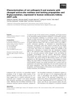
The gene expression profile of immortalized hMSCs is
similar with that of ESCs
To gain insi ght into the convergence of 3A6 toward
ESCs, we compared the expression profile of 3A6 with
various normal human tissues, MSCs and ESCs. This
data set therefore contained different tissues from
embryo, endoderm, epithelial, or mesenchymal origins.
The expression profiles of each chip were compared
using principal component analysis (PCA) to discover the
similarity of the expression profiles within and across the
cells or tissues. PCA using all probe sets showed ESC and
MSC each formed a distinct group and were quite differ-
ent from all the normal human tissues. Interestingly, the
3A6 expression profile located very close to ESCs rather
than near MSCs, signaling the expression profile of 3A6
converged toward ESCs (Figure 6).
Discussion
To c ircumvent the probl ems associated with expanded
hMSCs, we found that ectopic expression of HPV 16
E6E7 and hTERT enhanced proliferation and stem-like
properties, a nd blocked spontaneous differentiation in
primary culture of hMSCs. Surp risingly, all of the three
examined cell clones had differentiation potential far
beyond the normal hMSCs. They expressed trophoecto-
derm and germline specific markers at day 7 of induced
differentiation with BMP4 and RA, respectively. Besides
unlimited differentiation potential, we further showed
these cells displayed higher osteogenic and neural differ-
entiation efficiency than their parental cells. The
increased differentiatio n efficiency was a ttributable to
the decrease in committed cells that have spontaneously
undergone differentiation and might be limited in di rec-
ted differentiation potential.
DNA methylation and chromatin structure are major
epigenetic factors that regulate gene expression [37].
Increase in CpG island methylation was notice d during
ESC differentiation [38, 39] and de leting the three major
DNMTs would c ause hypomethylation and thorough
blockage of differentiation of ESCs [40,41]. These find-
ings plus the fact global methylation marks are erased
after fertilization and formation of embryo, and increase
during in vitro expansion [42] suggest the CpG island
methylatio n prof ile may serve as an indicator of “primi-
tiveness” of stem cells. Therefore, the decrease in CpG
island methylation in 3A6 suggests its increase in primi-
tiveness. More import antly, DNA demethylation
occurred mainly in the CpG islands of development and
differentiation associated genes, and ensured these genes
the accessibility for activation upon cues of s timulation
and further explained the unlimited differentiation
potential. To elucidate if the enhancement of stem-like
properties and blockage of spontaneous differentiation
by hTERT overexpression is restricted merely to the
immortalized cell line, we also inspected the effects of
ectopic expression of hTERT in primary hMSCs.
Although overexpression of hTERT inhibited the
expressions of DNMTs (Addi tional file 5), it did not
induce a significant change in pluripotency and lineage
gene expression. These results suggest hTERT alone or
downregulation of DNMTs is not enough to trigger
reversion of stem-like properties in hMSCs, which needs
a combinational activation of many factors or molecul es
as demonstrated previously [43].
In the curren t study, CpG island hypomethylation did
not i nduce an increase in the average gene expression
level in 3A6. Weber [15] clarified most of the unmethy-
lated promoters with high CpG frequency (HCPs)
remain inactive. Mikkelsen and others [44] further
explored the chromatin st ate of HCPs in ESCs and
Tsai et al. Journal of Biomedical Science 2010, 17:64
/>Page 9 of 13
Figure 5 Gene Ontology classification of genes decreased in CpG island methylation in hTERT-transfected hMSCs (A), and sub-
classification of development (B).
Tsai et al. Journal of Biomedical Science 2010, 17:64
/>Page 10 of 13
revealed monovalent promoters (H3K4me3) generally
regulate genes with “ housekeeping” func tions, and
otherwise, bivalent promoters (H3K4me3 and H3K27
me3) are associated with g enes related to key develop-
mental transcription factors. Most importantly, they
found low activity of bivalent HCPs, compatible with
the findings that most of the development associated
genes are quiescent in pluripotent cells. Therefore, the
low activity of demethylated development-associated
genes in 3A6 might be due to transient repression by
chromatin modifications, and indeed the hypomethy-
lated state of these genes enable them to recapitulate
expression upon later development or cellular
differentiation.
Despite further investigations needed to elucidate
exact demethylation mechanism, the effect of global
DNA demethylation on stem-like properties and beha-
vior of stem cells is still of great significance. Taylor
and others first described that treatment with 5-AzaC
increased the differentiation potential of C3H/10T1/2
cells [19]. Similarly, 5-AzaC also induce d dedifferentia-
tion in partially differentiated ESCs [18] or trophoblast
stem cells [20]. The se findings support our findings
that 3A6 with a significant global decrease in CpG
island methylation level behaved like ESCs and such
alteration in stem-like properties might be achieved by
DNA demethylation of development and differentiation
associated genes after immortalization.
Therefore, transfection of hMSCs with HPV 16 E6E7
and h TERT might elicit change of epigenetic marks to
reverse stem-like properties, which finally contributes to
unlimited differentiation potential and increased differ-
entiation efficiency. However, there are several limita-
tions to this study. First, because the cells require
transfection to improve stem cell properties and prevent
spontaneous differentiation, these transfected cells may
not be applied for clinical uses. Second, the increase of
multipotency to trophoectoderm and germ cells was
only de monstrated by the expression of several lineage
markers, without any in vivo st udy it is still limited to
define these potential. Although the hidden ulterior con-
nection between overexpression of the se genes and epi-
genetic remodeling in stem cell biology is needed, the
current results are a great step forward in establishing
the feasibility and applicability of adult stem cells in
future clinical applications.
Figure 6 The expression profile of 3A6 immortalized hMSCs converges toward embryonic stem cells. Principal component analysis
comparing the gene expression profiles of 3A6, embryonic stem cells (ESC), mesenchymal stem cells (MSC), and various tissues using all the
transcriptome data. Each plotted data point represents a single profile.
Tsai et al. Journal of Biomedical Science 2010, 17:64
/>Page 11 of 13
Additional material
Additional file 1: Primer sets and NCBI reference sequence number.
Additional file 2: (A) Detection of hTERT mRNA expression in 1C5,
3G11 and 3A6, and (B) Characterization of CD molecules in 3A6.
Cytofluorimetric profiles of 3A6 reacted first with (solid line) or without
(broken line) mouse MAbs specific for each marker, and second with
fluorescein-labeld antimouse Ig antibody.
Additional file 3: Box plots show average methylation levels of
genes contain CpG islands. P value was calculated using a t-test.
Additional file 4: Real-time RT-PCR analysis of expression levels of
(A) DNMT1, DNMT3A and DNMT3B, and (B) EZH2 in 3A6 and KP.
Additional file 5: (A) RT-PCR analysis of expression levels of hTERT
and GAPDH, and (B) Real-time RT-PCR analysis of expression levels
of DNMT1, DNMT3A and DNMT3B in primary human mesenchymal
stem cells transfected with plasmids carrying control and hTERT
vectors. Data are presented as mean ± S.D. *p < 0.01 compared with
control as calculated using a t-test.
Acknowledgements
These studies were supported in part by grants from National Scientific
Council (NSC-NSC-95-2627-B-010-009-, NSC-96-2627-B-010-009-, 97-3111-B-
010-001) and grants from National Yang-Ming University, Ministry of
Education, Aim for the Top University Plan, and Veterans General Hospital-
Taipei (V95E1-002 & V95E1-003). This work was assisted in part by the
Division of Experimental Surgery of The Department of Surgery, Taipei
Veterans General Hospital.
Author details
1
Stem Cell Laboratory, D epartment of Medical Research & Education and
Orthopaedics & Traumatology, Veterans General Hospital, Taipei, Taiwan.
2
Graduate Institute of Dental Sciences and Department of Periodontology,
National Taiwan University, Taipei, Taiwan.
3
Graduate Institute of Medical
Engineering & Department of Orthopedics, College of Medicine, National
Taiwan University, Taipei, Taiwan.
4
Institute of Clinical Medicine &
Department and Institute of Pharmacology, School of Medicine, National
Yang-Ming University, Taipei, Taiwan.
5
Institute of Microbiology and
Immunology, National Yang-Ming University, Taipei, Taiwan.
Authors’ contributions
CCT performed research and analyzed data, CLC designed research,
performed research and wrote the paper, HCL contributed vital new
reagents or analytical tools, YTL performed research, HWW contributed vital
new reagents or analytical tools and analyzed data, LTH contributed vital
new reagents or analytical tools and wrote the paper, SCH designed
research, contributed vital new reagents or analytical tools and wrote the
paper. All authors read and approved the final manuscript.
Competing interests
The authors declare that they have no competing interests.
Received: 24 March 2010 Accepted: 29 July 2010
Published: 29 July 2010
References
1. Pittenger MF, Mackay AM, Beck SC, Jaiswal RK, Douglas R, Mosca JD,
Moorman MA, Simonetti DW, Craig S, Marshak DR: Multilineage potential
of adult human mesenchymal stem cells. Science (New York, NY) 1999,
284:143-147.
2. Woodbury D, Reynolds K, Black IB: Adult bone marrow stromal stem cells
express germline, ectodermal, endodermal, and mesodermal genes prior
to neurogenesis. Journal of neuroscience research 2002, 69:908-917.
3. Beausejour C: Bone marrow-derived cells: the influence of aging and
cellular senescence. Handbook of experimental pharmacology 2007, 67-88.
4. Bonab MM, Alimoghaddam K, Talebian F, Ghaffari SH, Ghavamzadeh A,
Nikbin B: Aging of mesenchymal stem cell in vitro. BMC cell biology 2006,
7:14.
5. Hung SC, Chen NJ, Hsieh SL, Li H, Ma HL, Lo WH: Isolation and
characterization of size-sieved stem cells from human bone marrow.
Stem cells (Dayton, Ohio) 2002, 20:249-258.
6. Hung SC, Yang DM, Chang CF, Lin RJ, Wang JS, Low-Tone Ho L, Yang WK:
Immortalization without neoplastic transformation of human
mesenchymal stem cells by transduction with HPV16 E6/E7 genes. Int J
Cancer 2004, 110:313-319.
7. Amit M, Carpenter MK, Inokuma MS, Chiu CP, Harris CP, Waknitz MA,
Itskovitz-Eldor J, Thomson JA: Clonally derived human embryonic stem
cell lines maintain pluripotency and proliferative potential for prolonged
periods of culture. Developmental biology 2000, 227:271-278.
8. Baxter MA, Wynn RF, Jowitt SN, Wraith JE, Fairbairn LJ, Bellantuono I: Study
of telomere length reveals rapid aging of human marrow stromal cells
following in vitro expansion. Stem cells (Dayton, Ohio) 2004, 22:675-682.
9. Bodnar AG, Ouellette M, Frolkis M, Holt SE, Chiu CP, Morin GB, Harley CB,
Shay JW, Lichtsteiner S, Wright WE: Extension of life-span by introduction
of telomerase into normal human cells. Science (New York, NY) 1998,
279:349-352.
10. Shi S, Gronthos S, Chen S, Reddi A, Counter CM, Robey PG, Wang CY: Bone
formation by human postnatal bone marrow stromal stem cells is
enhanced by telomerase expression. Nature biotechnology 2002,
20:587-591.
11. Choi J, Southworth LK, Sarin KY, Venteicher AS, Ma W, Chang W, Cheung P,
Jun S, Artandi MK, Shah N, Kim SK, Artandi SE: TERT promotes epithelial
proliferation through transcriptional control of a Myc- and Wnt-related
developmental program. PLoS genetics 2008, 4:e10.
12. Watt F, Molloy PL: Cytosine methylation prevents binding to DNA of a
HeLa cell transcription factor required for optimal expression of the
adenovirus major late promoter. Genes & development 1988, 2:1136-1143.
13. Boyes J, Bird A: DNA methylation inhibits transcription indirectly via a
methyl-CpG binding protein. Cell 1991, 64:1123-1134.
14. Hashimshony T, Zhang J, Keshet I, Bustin M, Cedar H: The role of DNA
methylation in setting up chromatin structure during development.
Nature genetics 2003, 34:187-192.
15. Weber M, Hellmann I, Stadler MB, Ramos L, Paabo S, Rebhan M,
Schubeler D: Distribution, silencing potential and evolutionary impact of
promoter DNA methylation in the human genome. Nature genetics 2007,
39:457-466.
16. Siegmund KD, Connor CM, Campan M, Long TI, Weisenberger DJ,
Biniszkiewicz D, Jaenisch R, Laird PW, Akbarian S: DNA Methylation in the
Human Cerebral Cortex Is Dynamically Regulated throughout the Life
Span and Involves Differentiated Neurons. PLoS ONE 2007, 2:e895.
17. Meshorer E, Misteli T: Chromatin in pluripotent embryonic stem cells and
differentiation. Nature reviews 2006, 7:540-546.
18. Tsuji-Takayama K, Inoue T, Ijiri Y, Otani T, Motoda R, Nakamura S, Orita K:
Demethylating agent, 5-azacytidine, reverses differentiation of
embryonic stem cells. Biochemical and biophysical research communications
2004, 323:86-90.
19. Taylor SM, Jones PA: Multiple new phenotypes induced in 10T1/2 and
3T3 cells treated with 5-azacytidine. Cell 1979, 17:771-779.
20. Hattori N, Nishino K, Ko YG, Hattori N, Ohgane J, Tanaka S, Shiota K:
Epigenetic control of mouse Oct-4 gene expression in embryonic stem
cells and trophoblast stem cells. The Journal of biological chemistry 2004,
279:17063-17069.
21. Xu RH, Chen X, Li DS, Li R, Addicks GC, Glennon C, Zwaka TP, Thomson JA:
BMP4 initiates human embryonic stem cell differentiation to
trophoblast. Nature biotechnology 2002, 20:1261-1264.
22. Nayernia K, Lee JH, Drusenheimer N, Nolte J, Wulf G, Dressel R, Gromoll J,
Engel W: Derivation of male germ cells from bone marrow stem cells.
Laboratory investigation; a journal of technical methods and pathology 2006,
86:654-663.
23. Pera MF, Andrade J, Houssami S, Reubinoff B, Trounson A, Stanley EG,
Ward-van Oostwaard D, Mummery C: Regulation of human embryonic
stem cell differentiation by BMP-2 and its antagonist noggin. Journal of
cell science 2004, 117:1269-1280.
24. Boiani M, Scholer HR: Regulatory networks in embryo-derived pluripotent
stem cells. Nature reviews 2005, 6:872-884.
Tsai et al. Journal of Biomedical Science 2010, 17:64
/>Page 12 of 13
25. Hill RE, Favor J, Hogan BL, Ton CC, Saunders GF, Hanson IM, Prosser J,
Jordan T, Hastie ND, van Heyningen V: Mouse small eye results from
mutations in a paired-like homeobox-containing gene. Nature 1991,
354:522-525.
26. Fujikura J, Yamato E, Yonemura S, Hosoda K, Masui S, Nakao K, Miyazaki Ji J,
Niwa H: Differentiation of embryonic stem cells is induced by GATA
factors. Genes & development 2002, 16:784-789.
27. Kubo A, Shinozaki K, Shannon JM, Kouskoff V, Kennedy M, Woo S,
Fehling HJ, Keller G: Development of definitive endoderm from
embryonic stem cells in culture. Development (Cambridge, England) 2004,
131:1651-1662.
28. Clark AT, Bodnar MS, Fox M, Rodriquez RT, Abeyta MJ, Firpo MT, Pera RA:
Spontaneous differentiation of germ cells from human embryonic stem
cells in vitro. Human molecular genetics 2004, 13:727-739.
29. Niwa H, Miyazaki J, Smith AG: Quantitative expression of Oct-3/4 defines
differentiation, dedifferentiation or self-renewal of ES cells. Nature
genetics 2000, 24:372-376.
30. Zeineddine D, Papadimou E, Chebli K, Gineste M, Liu J, Grey C, Thurig S,
Behfar A, Wallace VA, Skerjanc IS, Pucéat M: Oct-3/4 dose dependently
regulates specification of embryonic stem cells toward a cardiac lineage
and early heart development. Developmental cell 2006, 11:535-546.
31. Darr H, Mayshar Y, Benvenisty N: Overexpression of NANOG in human ES
cells enables feeder-free growth while inducing primitive ectoderm
features. Development (Cambridge, England) 2006, 133:1193-1201.
32. Johnson J, Bagley J, Skaznik-Wikiel M, Lee HJ, Adams GB, Niikura Y,
Tschudy KS, Tilly JC, Cortes ML, Forkert R, Spitzer T, Iacomini J, Scadden DT,
Tilly JL: Oocyte generation in adult mammalian ovaries by putative germ
cells in bone marrow and peripheral blood. Cell 2005, 122:303-315.
33. Geijsen N, Horoschak M, Kim K, Gribnau J, Eggan K, Daley GQ: Derivation of
embryonic germ cells and male gametes from embryonic stem cells.
Nature 2004, 427:148-154.
34. Saxonov S, Berg P, Brutlag DL: A genome-wide analysis of CpG
dinucleotides in the human genome distinguishes two distinct classes
of promoters. Proceedings of the National Academy of Sciences of the United
States of America 2006, 103:1412-1417.
35. Li E: Chromatin modification and epigenetic reprogramming in
mammalian development. Nat Rev Genet 2002, 3:662-673.
36. Viré E, Brenner C, Deplus R, Blanchon L, Fraga M, Didelot C, Morey L, Van
Eynde A, Bernard D, Vanderwinden JM, Bollen M, Esteller M, Di Croce L, de
Launoit Y, Fuks F: The Polycomb group protein EZH2 directly controls
DNA methylation. Nature 2006, 439:871-874.
37. Meshorer E: Chromatin in embryonic stem cell neuronal differentiation.
Histology and histopathology 2007, 22:311-319.
38. Kremenskoy M, Kremenska Y, Ohgane J, Hattori N, Tanaka S, Hashizume K,
Shiota K: Genome-wide analysis of DNA methylation status of CpG
islands in embryoid bodies, teratomas, and fetuses.
Biochemical and
biophysical research communications 2003, 311:884-890.
39. Shen Y, Chow J, Wang Z, Fan G: Abnormal CpG island methylation occurs
during in vitro differentiation of human embryonic stem cells. Human
molecular genetics 2006, 15:2623-2635.
40. Carlone DL, Lee JH, Young SR, Dobrota E, Butler JS, Ruiz J, Skalnik DG:
Reduced genomic cytosine methylation and defective cellular
differentiation in embryonic stem cells lacking CpG binding protein.
Molecular and cellular biology 2005, 25:4881-4891.
41. Jackson M, Krassowska A, Gilbert N, Chevassut T, Forrester L, Ansell J,
Ramsahoye B: Severe global DNA hypomethylation blocks differentiation
and induces histone hyperacetylation in embryonic stem cells. Molecular
and cellular biology 2004, 24:8862-8871.
42. Maitra A, Arking DE, Shivapurkar N, Ikeda M, Stastny V, Kassauei K, Sui G,
Cutler DJ, Liu Y, Brimble SN, Noaksson K, Hyllner J, Schulz TC, Zeng X,
Freed WJ, Crook J, Abraham S, Colman A, Sartipy P, Matsui S, Carpenter M,
Gazdar AF, Rao M, Chakravarti A: Genomic alterations in cultured human
embryonic stem cells. Nature genetics 2005, 37:1099-1103.
43. Takahashi K, Yamanaka S: Induction of pluripotent stem cells from mouse
embryonic and adult fibroblast cultures by defined factors. Cell 2006,
126:663-676.
44. Mikkelsen TS, Ku M, Jaffe DB, Issac B, Lieberman E, Giannoukos G, Alvarez P,
Brockman W, Kim TK, Koche RP, Lee W, Mendenhall E, O’Donovan A,
Presser A, Russ C, Xie X, Meissner A, Wernig M, Jaenisch R, Nusbaum C,
Lander ES, Bernstein BE: Genome-wide maps of chromatin state in
pluripotent and lineage-committed cells. Nature 2007, 448:553-560.
doi:10.1186/1423-0127-17-64
Cite this article as: Tsai et al.: Overexpression of hTERT increases stem-
like properties and decreases spontaneous differentiation in human
mesenchymal stem cell lines. Journal of Biomedical Science 2010 17:64.
Submit your next manuscript to BioMed Central
and take full advantage of:
• Convenient online submission
• Thorough peer review
• No space constraints or color figure charges
• Immediate publication on acceptance
• Inclusion in PubMed, CAS, Scopus and Google Scholar
• Research which is freely available for redistribution
Submit your manuscript at
www.biomedcentral.com/submit
Tsai et al. Journal of Biomedical Science 2010, 17:64
/>Page 13 of 13