Enterovirus type 71 2A protease functions as a transcriptional activator in yeast ppsx
Bạn đang xem bản rút gọn của tài liệu. Xem và tải ngay bản đầy đủ của tài liệu tại đây (1.71 MB, 9 trang )
RESEARC H Open Access
Enterovirus type 71 2A protease functions as a
transcriptional activator in yeast
Chee-Hing Yang
1
, Hui-Chun Li
2
, Jeng-Geng Jiang
1
, Che-Fang Hsu
3
, Yi-Jen Wang
3
, Meng-Jiun Lai
1,3
, Yue-Li Juang
4
,
Shih-Yen Lo
1,3,5*
Abstract
Enterovirus type 71 (EV71) 2A protease exhibited strong transcriptional activity in yeast cells. The transcriptional
activity of 2A protease was independent of its protease activity. EV71 2A protease retained its transcriptional activ-
ity after truncation of 40 amino acids at the N-terminus but lost this activity after truncation of 60 amino acids at
the N-terminus or deletion of 20 amino acids at the C-terminus. Thus, the acidic domai n at the C-terminus of this
protein is essential for its transcriptional activity. Indeed, deletion of amino acids from 146 to 149 (EAME) in this
acidic domain lost the transcriptional activity of EV71 2A pro tein though still retained its protease activity. EV71 2A
protease was detected both in the cytoplasm and nucleus using confocal microscopy analysis. Coxsackie virus B3
2A protease also exhibited transcriptional activity in yeast cells. As expected, an acidic domain in the C-terminus of
Coxsackie virus B3 2A protease was also identified. Truncation of this acidic domain resulted in the loss of tran-
scriptional activity. Interestingly, this acidic region of poliovirus 2A protease is critical for viral RNA replication. The
transcriptional activity of the EV71 or Coxsackie virus B3 2A protea se should play a role in viral replication and/or
pathogenesis.
Background
Enterovirus type 71 (EV71) is the causative agent of sev-
eral human diseases, including hand-foot-and-mouth
disease, encephalitis, and meningitis. EV71 i s a single-
stranded, positive-sense RNA virus, which belongs to
the Picornaviridae family [1]. Genomic R NA of picorna-
viruses (e.g. polioviruses) encodes a polyprotein precur-
sor, which is processed by three proteases (the
maturation protease, 2A protease, and the 3C protease)
into at least 11 different proteins, which are arranged in
the order of NH2-VP4-VP2-VP3-VP1-2A-2B-2C-3A-
VPg-3C-3D-COOH [1]. The 2A protease of poliovirus, a
representative member of the Picornaviridae,isa
cysteine protease with multiple functions [2]. Similar to
poliovi rus 2A protease, expression of EV71 2A protease
led to cleavage of the eukaryotic init iation factor 4GI, a
key factor for host protein synt hesis [3,4]. Moreover,
transient expression of EV71 2A protease alone also
resulted in the induction of apoptotic change [5,6].
However,thefunctionofEV712Aproteaseisnotwell
characterized. The biologic function of EV71 2A pro-
tease was investigated by fusing it with the DNA-bind-
ing domain of Gal4 and examining i ts possible
interaction with cellular factors [7].
Materials and Methods
Plasmid construction
Procedures used in our previ ous studies were followed
to construct the plasmids [ 8,9]. The PCR primers used
in this study are listed in Table 1. To clone the DNA
fragment encoding the full-length EV71 2A protease
(nucleotides from 3332 to 3781 of strain pinf7-54A) for
yeast two-hybrid screening, oligonucleotide primers
(2AY-S and 2AY-AS) were used to perform PCR. After
the PCR, the DNA fragment was treated with T4 poly-
nucleotide kinase, digested by the restriction enzyme
EcoRI, and cloned into the pBDGal4 Cam (Stratagen e,
USA) expres sion vector, which had been linearized with
EcoRI and SmaI. Using the same approach, PCR was
performed with primer pairs (2AY-21 S and 2AY-AS,
2AY-41 S and 2AY-AS, 2AY-61 S and 2AY-AS) to clone
the DNA fragments encoding EV71 2A protease with
the N-terminal truncation of 20, 40, 60 amino acids
respectively, while another PCR was performed with
* Correspondence:
1
Department of Laboratory Medicine and Biotechnology, Tzu Chi University,
Hualien, Taiwan
Full list of author information is available at the end of the article
Yang et al. Journal of Biomedical Science 2010, 17:65
/>© 2010 Yang et al; licensee BioMed Cent ral Ltd. This is an Open Access article distributed under the terms of the Creativ e Co mmons
Attribution License ( g/licenses/by/2.0), which permits unrestricted use, distribution, and reproduction in
any medium, provided the original work is properly cited.
primer pairs (2AY-S and 2AY-130AS, 2AY-S and 2 AY-
110AS, 2AY-S and 2AY-90AS) to clone the DNA frag-
ments encoding EV71 2A protease with the C-terminal
deletion of 20, 40, 60 amino acids respectively. Primers
(2AY-S and 2AY-AS101) were used to perform PCR to
clone the DNA fragment encoding EV71 2A protease
withoutaminoacidsfrom146to149usingthesame
approach.
To clone the DNA fragment encoding the full-length
Coxsackie virus B3 2A protease for yeast two-hybrid
screening, mRNA extra cted from a patient infected with
Coxsackie virus B3 was converted into cDNA and
oligonucleotide primers (CoxB2AY-S and CoxB2AY-AS)
were used to perform PCR ( the sequence is t he same as
nucleotides from 3304 to 3744 of GI:323419). PCR was
performed using primer pairs (CoxB2AY-61 S and Cox-
B2AY-AS) to clone the DNA fragments encoding Cox-
sackie virus 2A protease with the N- terminal truncation
of 60 amino acids, while another P CR was performed
with primer pairs (CoxB2AY-S and CoxB2AY-127AS) to
clone the DNA fragments encoding Coxsackie virus 2A
protease with the C-terminal deletion of 20 amino acids.
Again, after the PCR, the DNA fragments were treated
with T4 polynucleotide kinase, digested by the restric-
tion enzyme EcoRI, and cloned into the pBDGal4 Cam
(Stratagene, USA) expression vector which had been lin-
earized with EcoRI and SmaI.
To clone the DNA fragment encoding the C-terminus
of EV71 VP1 and the full-length 2A protease (nucleo-
tides from 3124 to 3781 of strain pinf7-54A) for transi-
ent expression in mammalian cells, PCR was performed
using oligonucleotide primers (VP1/2A-S and 2AY-
AS2). After the PCR, the DNA fragment was digested
by restriction enzymes (ClaI/XbaI), together with the
EMCV IRES sequence (digested with EcoRI/ClaI), and
cloned into the expression vector pcDNA3 (Invitrogen,
USA) which had been linearized with EcoRI/XbaI. To
mutate amino aci d 110 of EV71 2A protease from Cys
to Ala, primers (VP1/2A-S and C110A-AS) were used to
amplify the 5’ -end of the gene fragment while primers
(C110A-S and 2AY-AS2) were used to amplify the
3’-end fragment. These two DNA fragments were linked
together by PCR using primers (VP1/2A-S and 2AY-
AS2). After the PCR, the DNA fragment was digested
by restriction enzymes (ClaI/XbaI), together with the
EMCV IRES sequence (digested with EcoRI/ClaI), and
cloned into the expression vector pcDNA3 (Invitrogen,
USA) which had been linearized with EcoRI/XbaI.
To clone the DNA fragment encoding the C-terminus
of EV71 VP1 and full-length 2A protease with the V5
tag in the C-terminus for confocal microscopy analysis
in mammalian cells, PCR was performed using oligonu-
cleotide primers (VP1/2A-S and 2AY-AS3). After the
PCR, the DNA fragment was digested by restriction
enzymes (ClaI/XbaI), together with the EMCV IRES
DNA sequence (digested with EcoRI/ClaI), and cloned
into the expression vector pcDNA3.1-V5-His A (Invitro-
gen, USA) whi ch had been lineariz ed with E coRI/ XbaI.
To clone the EV71 2A protease with mutation of amino
acid 110 from Cys to Ala for confocal microscopy analy-
sis, the DNA templ ate containing this mutation and pri-
mers (2A-S10 and 2A-AS3) was used to amplify the
DNA fragment of full-length EV71 2A protein with
mutation of amino acid 110 from Cys to Ala. After the
PCR, the DNA fragment was digested by the restriction
enzymes (EcoRI/XbaI), and cloned into the expression
Table 1 PCR primers used in this study
Name Sequence
2AY-S (5’-GGAATTCGGGAAATTTGGACAG-3’)
2AY-AS (5’-CCGCTCGAGTTACTGCTCCATGGCTTC-3’)
2AY-21S (5’-GGAATTCCATCTTGCTACTCATAA-3’)
2AY-41S (5’-GGAATTCCTCGTATCATCTACCAC-3’)
2AY-61S (5’-GGAATTCGGAGTGTATTATTGTAA-3’)
2AY-90AS (5’-TTATTAATAATACTCGCTGGCCTC-3’)
2AY-110AS (5’-TTATTAGCAATCCCCTGGTTCCGA-3’)
2AY-130AS (5’-TTATTAGCAATCCCCTGGTTCCGA-3’)
VP1/2A-S (5’-CCATCGATATGATGGGTACGTTC-3’)
2A-S10 (5’-GGAATTCATGGGGAAATTTGGACAGCAG-3’)
2A-AS2 (5’-GCTCTAGACTACTGCTCCATGGCTTCATCATC-3’)
2A-AS3 (5’-GCTCTAGACTGCTCCATGGCTTCATCATC-3’)
C110A-S (5’-CCAGGGGATGCCGGTGGCATTCTTAGATGC-3’)
C110A-AS (5’-AATGCCACCGGCATCCCCTGGTTCCGAATG-3
’)
L30/43-S (5’-CATAATGACTGGGCAAACTCATCTACCACTGCTCAA-3’)
L30/43-AS (5’-TTGAGCAGTGGTAGATGAGTTTGCCCAGTCATTATG-3’)
2AY-AS101 (5’-CCGCTCGAGTTACTGATCATCCAACCACAGAAG-3’)
2A-AS301 (5’-GCTCTAGACTGATCATCCAACCACAGAAG-3’)
CoxB2AY-S (5’-GGAATTCATGGGACAACAATCAGGGGC-3’)
CoxB2AY-AS (5’-TTATTACTGTTCCATTGCATCATC-3’)
CoxB2AY-61S (5’-GGAATTCTTTTGTGCGTCCAAAAAC-3’)
CoxB2AY-127AS (5’-TTATTAGCCTTCACCCCCCATGGT-3’)
PCBP2-S (5’-CTCTCACCATCCGGCTACTTAT-3’)
PCBP2-AS (5’-GCTGCTTATGTCCTCTTCCAGT-3’)
PTBP1-S (5’-CTACATCCAGTTCTCCAACCAC-3’)
PTBP1-AS (5’-GCTGCTTATGTCCTCTTCCAGT-3’)
RTN3-S (5’-ACTCTGTCCTCAGAAGCTTTCC-3’)
RTN3-AS (5’-CTCATAGACAATCGGGACACTG-3’)
GBF1-S (5’-CCCACTATTGCTGCTCTCTCTT-3’)
GBF1-AS (5’-CTGGGCAGGTTCTCAATAGACT-3’)
CD55-S (5’-CCGTCTTCTATCTGGTTCTCGT-3’)
CD55-AS (5
’-GTTACTAGCGTCCCAAGCAAAC-3’)
SAM68-S (5’-CGAAGGCTATTACAGCCAGAGT-3’)
SAM68-AS (5’-CATATGGGTGCTCTCTGTATGC-3’)
Note: Nucleotides for restriction enzyme cutting sites are italicized.
Nucleotides for point mutations are bold and italicized. Nucleotides for start
and stop codons are marked with bold letters. Primers for the detection of
cellular genes were used in real-time RT-PCR.
Yang et al. Journal of Biomedical Science 2010, 17:65
/>Page 2 of 9
vector pcDNA3.1-V5-His A (Invitrogen, USA) which
had been linearized with EcoRI/XbaI. To clone the
EV71 2A protease without potential NES (amino acid 31
to 42) for confocal microscopy analysis, the DNA frag-
ment containing the mutation of amino acid 110 from
Cys to Ala was used as t he PCR template. Primers (2A-
S10andL30/43-AS)wereusedtoamplifythe5’ -end o f
the gene fragment while primers (L30/43-S and 2A-
AS3) were used to amplify the 3’-end fragment. These
two DNA fragments were linked together by PCR usi ng
primers (2A-S10 and 2A-AS3). After the PCR, the DNA
fragment was digested by restriction enzymes (EcoRI/
XbaI), and cloned in to the expression vector pcDNA3 .1-
V5-His A (Invitrogen, USA) which had been linearized
with EcoRI/XbaI. The same approach was used to clo ne
the DNA fragment encoding the C-terminus of EV71
VP1 and 2A protease d eleting the amino acids 146-149
with the V5 tag in t he C-terminus using primers (VP1/
2A-S and 2A-AS301) to perform PCR.
All of the expression plasmids were verified by
sequencing.
Yeast two-hybrid screening
The yeast two-hybrid system used for screening was
purchased from Clontech Laboratories (USA). The
experimental procedures were conducted according to
the manufacturer’s instructions.
Protein expression and Western blot analysis
HeLa cells were maintained in RPMI (Chemicom, USA)
medium containing 10% fetal bovine serum, 1% gluta-
mine (200 mM, Gibco, USA), and 100 ug/ml penicillin/
streptomycin (Gibco BRL, USA). Cultured cells were
maintained at 37°C with 5% CO
2
. Cells were seeded at a
density of approximately 4 × 10
5
cells per 60-mm cul-
ture dish. After overnight inc ubation, cells were trans-
fected with plasmids (1-4 ug) u sing the ExGen 500 in
vitro transfection reagent (Fermentas, USA) or Arrest-
In™transfection reagent (Open Biosystems, USA). At 48
hours after transfection, recombinant proteins expressed
in cells were analyzed by Western blot.
Our previous procedures were followed for Western
blot analysis [7,10]. Rabbit polyclonal antibodies against
ERK-2 and eIF4G were purchased from Santa Cruz Bio-
technology (USA). Monoclonal antibodies agains t PARP
were purchased from SEROTEC (UK). Monoclonal anti-
bodies against V5 tag were purchased from Invitrogen
(USA). Rabbit antibodies against EV71 2A protease were
generated in the lab.
Confocal microscopy analysis
HeLa cells were seeded at a density of about 2.5 × 10
5
cells per 35 mm culture dish. After overnight incuba-
tion, cells were transfected with plasmids (0.5 - 2 ug)
using the ExGen 500 in vitro transfection reagent (Fer-
mentas, USA) or Arrest-In™ transfection reagent (Open
Biosystems, USA). At 48 hours after transfection,
recombinant proteins expressed in cells were analyzed
by confocal microscopy.
Cells with recombinant proteins were fixed with 1%
methanol/acetone at 0°C for 10 minutes, washed with
incubati on buffer (0.05% NaN
3
, 0.02% saponin, 1% sk im
milk in PBS) twice for 2 minutes each, and then incu-
bated with the anti-V5 antibody (1:200 dilution) at 37°C
for 30 minutes. Cells were washed with PBS at room
temperature for five minutes three times, and then incu-
bated with Cy3-conjugated goat a nti-mouse IgG anti-
body (1:20 dilution) at 37°C for 30 minutes. Cells were
washed three more times with PBS. DAPI (Merck, Ger-
many) was used to stain the nucleus.
Real-time reverse transcriptase-polymerase chain
reaction (RT-PCR)
HeLa cells were transfected with plasmids of vector
alone or pcDNA3.1-IRES-2A using Arrest-In™transfec-
tion reagent (Open Biosystems, USA). At 24 hours after
transfection, G418 was used to select the cells with
transfected plasmid. After 72 hours, cellular mRNAs
were extracted and our previous procedures were fol-
lowed for real-time RT-PCR [11].
Results
EV71 2A protease exhibited strong transcriptional
activity in yeast cells
EV71 2A protease, when fused with the DNA-binding
domain of Gal4, activates the reporter genes in yeast
cells (Figure 1). This reaction is quite specific since
none of the other proteins we studied at the same time
exhibited this activity, including EV 71 3C protein,
hepatitis C virus NS5A protein, NS3 protein(data not
shown), or ARFP [7]. Truncation of 40 but not 60
amino acids at the N-terminus of EV71 2A protease did
not affect its transcriptional activation activity (Figure 1).
On the other hand, deletion of 20 amino acids at the C-
terminus of EV71 2A protease resulted in the loss of
transcriptional activity (Figure 1).
Transcriptional activity of EV71 2A protease is
independent of its protease activity
Amino acid residues His 20, Asp 38, and Cys 109 com-
prise the catalytic core of poliovirus 2A protease [ 12].
The corresponding residue of EV71 2A protease essen-
tial for its protease activity is Cys in amino acid 110
(Figure 2A). The expression plasmids encoding the
C-terminus of VP1 protein, full- length 2A protease
wild-type or with mutati on in amino acid 110 from Cys
to Ala were constructed and transfected into HeLa cells.
Mutation of amino acid 110 from Cys to Ala of EV71
Yang et al. Journal of Biomedical Science 2010, 17:65
/>Page 3 of 9
2A protein blocked the auto-protease activity of this
protein (Figure 3A), suppressed the cleavage of cellular
eIF4G protein (Figure 3B), and reduced the induction of
apoptosis in HeLa cells (Figure 3C). However, EV71 2A
protease with this mutation still possessed transcrip-
tional activity in yeast cells (Figure 1).
Sub-cellular localization of EV71 2A protease
No potential nuclear localization signal (NLS) was
found within the EV71 2A protease asy.
org/index.html. However, it is known that ions, smaller
metabolites, and globular proteins up to 20-40 kDa
can passively diffuse through the central aqueous
region of the nuclear pore complex [13]. Thus, EV71
2A protease with 150 amino acids could passively dis-
use into the nucleus. Confocal microscopy analysis was
used to examine the sub-cellular localization of EV71
2A protein. The expression plasmid encoding the C-
terminus of VP1 protein, full-length 2A protease and
V5 tag was constructed and transfected into HeLa cells
before confocal microscopy analysis. The same
approach was used to construct and transfect the DNA
fragment encoding full-length 2A protease with muta-
tion of amino acid 110 from Cys to Ala. Protein
expression of these constructs was demonstrated using
Western blot analysis (Figure 4A). Both the wild-type
and mutant EV71 2A proteins l ocalized in both cyto-
plasm and nucleus ( Figure 4B). Amino acids 31 to 42
of EV71 2A protein (Figure 2A) were i dentified as a
potential nuclear export signal (NES) asy.
org/index.html. However, similar to full-length EV71
2A protease, this protein without amino acids 31 to 42
Figure 1 Growth of yeasts either mock-transfected or transfected with plasmids encoding EV71 2A protease of different sizes in YEPD
medium (A), YEPD without tryptophan (B), or YEPD without tryptophan and histidine (C). (D) X-gal staining of yeasts in (C).
Yang et al. Journal of Biomedical Science 2010, 17:65
/>Page 4 of 9
Figure 2 Analysis of EV71 2A protease protein. (A) Amino acid sequence of EV71 2A protein. The predicted 9aa TAD (a.a. 27-35) is indicated
with red letters. Potential NES (a.a. 31-42) is underlined. The acidic domain (the last fifteen amino acids) is also underlined. (B) Charge
distribution of EV71 2A protease: the C-terminus of this protein is highly acidic.
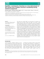
Figure 3 Western bl otting analysis of wild-t ype EV71 2A protease or with amino acid 110 mutation from Cys to Ala in HeLa cells.
HeLa cells were transfected with vector only (lane 1), or with the plasmid encoding the C-terminus of VP1 and wild-type 2A (lane 2), or with the
plasmid encoding the C-terminus of VP1 and 2A with amino acid 110 mutation from Cys to Ala (lane 3). After transfection, cell lysates were
analyzed and detected using rabbit anti-EV71 2A protein polyclonal antibody (A), mouse anti-eIF4G monoclonal antibody (B), or mouse anti-
PARP monoclonal antibody (C). The thin arrows indicate the uncleaved proteins (VP1-2A, intact eIF4G, or intact PARP) while the thick arrows
indicate the cleaved products (2A, cleaved eIF4G, or cleaved PARP).
Yang et al. Journal of Biomedical Science 2010, 17:65
/>Page 5 of 9
localized in both the cytoplasm and the nucleus but
not in the nucleu s only (Figure 4B) .
Deletion of amino acids from 146 to 149 of EV71 2A
protease lost its transcriptional activity but retained its
protease activity
A previous report demonstrated that the C-terminal
acidic region of poliovirus 2Apro is critical for viral
RNA replication but not f or cis- or trans- proteolytic
cleavage [14]. To determine whether mutation of the
amino acids in the C-terminal acidic region affect its
transcriptional activity, EV71 2A protease without
amino acids 146-149 (EAME) was constructed. Indeed,
EV71 2A protease without amino acids 146-149 still
retained its protease activity (Figure 5A) b ut lost its
transcriptional activity (Figure 5B).
EV71 2A protease did not transactivate cellular genes
reported to enhance the replication of poliovirus or EV 71
Some cellular genes were reported previously to
enhance t he replication of poliovirus or EV71: poly(rC)
binding proteins [15-17], cellular COPII proteins [18],
the polypyrimidine tract binding proteins [19], Reticulon
3 [20], and GBF1 [21]. Real-time RT-PCR was per-
formed to determine whether EV71 2A protease could
transactivate PCBP2, PTBP1, RTN3, GBF1, CD55, or
SAM68 gene. However, EV71 2A protease repressed
rather than transactivated all of these cellular genes
(data not shown).
Coxsackie virus B3 2A protease exhibited transcriptional
activity in yeast cells
To investigate whether otherpicornaviral2Aproteases
possess transcriptional activity, the DNA fragment
encoding the full-length Coxsackie virus B3 2A
protease was amplified by PCR and fused with the
DNA-binding domain of Gal4. This fusion protein also
activates reporter genes in yeast (Figure 6). Again,
Coxsackie virus B3 2A protease lost its transcriptional
activity after truncation of 60 amino acids at the N-
terminus or deletion of 20 amino acids at the C-termi-
nus (Figure 6).
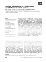
Figure 4 Analysis of various EV71 2A protein mutants in HeLa cells. (A) Protein expression of various EV71 2A protein mutants with V5 tag
in the C-terminus. HeLa cells were transfected with vector only (lane 1) or with the plasmid encoding the C-terminus of VP1 and wild-type 2A
(lane 2), or with the plasmid encoding 2A with amino acid 110 mutation from Cys to Ala (lane 3), or with the plasmid encoding 2A protein
deleting amino acids from 32 to 41 (lane 4). After transfection, cell lysates were analyzed by Western blot using mouse anti-V5 tag monoclonal
antibody. The thin arrow indicates the uncleaved protein (VP1-2A in lane 2) while the thick arrow indicates the 2A protein (lanes 2 and 3). The
thick line indicates the location of 2A protein deleting amino acids from 32 to 41 (lane 4). Erk2 protein served as a loading control. (B) Confocal
microscopy analysis of various EV71 2A protein mutants. After HeLa cells were transfected with the indicated plasmids, cells were fixed and
stained with mouse anti-V5 tag monoclonal antibody, followed by Cy3-conjugated anti-mouse IgG. DAPI (Merck, Germany) was used to stain
DNA for localization of the nucleus.
Yang et al. Journal of Biomedical Science 2010, 17:65
/>Page 6 of 9
Discussion
EV71 2A protease is expected to enter the nucleus by
passive diffusion since it i s a small protein with no
potential NLS. This protein would not be actively
exported from the nucleus since no functional NES was
detected (Figure 4). These findings explain why only
small portion of EV71 2A protease localized in the
nucleus and the majority of this protein was retained in
the cytoplasm (Figure 4). Interestingly, 2A proteins of
poliovirus and EMCV were reported to localize in the
nucleus [22,23].
As a transcriptional activator, EV71 2A protease did
not contain a glutamine-rich domain, a leucine zipper
domain, or a proline-rich domain as are found in some
other eukaryotic trans criptional activators such as CTF/
NF-1 or the amino terminal deletion mutants of HCV
NS5A protein [24-27]. The P XXXP motif necessary for
full transactivation of HIV Tat protein was also not
found in EV71 2A protease (Figure 2A) [28]. However,
one acidic domain (rich in Glu (E) or Asp (D), Figure
2B), functioning universally in eukaryotic tr anscriptional
activators from yeast to human [29,30], was found in
the C-terminus of EV71 2A protease (6 amino acids
within the last 15 amino acids are acidic, Figure 2A).
Moreover, 9aa TAD possessing an autonomous transac-
tivation activity in yeast and mammalian cells was also
found at the N-terminus of EV71 2A protease (from aa
27 to 35) (Figure 2A) [31]. Deletion analysis revealed
the acidic domain in the C-terminus but not 9 aa TAD
in the N-terminus of EV71 2A protease is essential for
the transcriptional activation activity of this protein
(Figure 1).
In addition to EV71 2A protease (Figure 1) , Coxsackie
virus B3 2A protease is also a transcription activator
(Figure 6). Interestingly, there is an acidic domain in the
C-terminus of this protein (6 amino acids within the
last 15 amino acids are acidic, Table 2). The 2A pro-
teases of other members of the Enterovirus genus, such
as Coxsackie viruses and polioviruses, also contain an
acidic domain in the C-terminus (Table 2). On the
other hand, there is no such an acidic domain in the
C-terminus of 2A proteases of rhinoviruses (2 or 3
amino acids within the last 15 amino acids are acidic,
Table 2) or cardiovirus (3 amino acids within the last 15
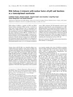
Figure 5 EV71 2A protease without amino acids 146-149 still retained its protease activity but lost its transcriptional activity. (A) HeLa
cells were transfected with vector only (lane 1) or with the plasmid encoding the C-terminus of VP1 and wild-type 2A (lane 2), or with the
plasmid encoding the C-terminus of VP1 and 2A deleting amino acids 146-149 (lanes 3 and 4). After transfection, cell lysates were analyzed by
Western blot using mouse anti-V5 tag monoclonal antibody. The thin arrow indicates the uncleaved protein (VP1-2A in lanes 2-4) while the thick
arrow indicates the 2A protein (lanes 2-4). Erk2 protein served as a loading control. (B) Growth of yeasts either mock-transfected or transfected
with plasmids encoding EV71 2A or 2A protein without amino acids 146-149 in YEPD medium, YEPD without tryptophan, or YEPD without
tryptophan and histidine.
Yang et al. Journal of Biomedical Science 2010, 17:65
/>Page 7 of 9
amino acids are acidic, Table 2). These observations
suggest that 2A proteases of enteroviruse s but not other
distinctly related picornaviruses (e.g. rhinoviruses, cardi-
oviruses) possess transcriptional activity. Interesti ngly, a
previous report demonstrated that this acidic region of
poliovirus 2Apro is critical for viral RNA replication but
not for cis- or trans- proteolytic cleavage [14]. Our
results also demonstrated that EV71 2A protease
without amino acids 146-149 s till retained its protease
activity (Figure 5A) but lost its transcriptional activity
(Figure 5B). Thus, enteroviral 2A proteases may transac-
tivate some cellular genes to b enefit virus replication.
Somecellulargenes,e.g.PCBP2,PTBP1,RTN3,GBF1,
CD55, and SAM68 gene, were reported to enhance the
replication of poliovirus or EV71. However, EV71 2A
protease suppressed rather than increased the tra nscrip-
tion of these cellular genes (data not shown). These
results were consistent with several reports regarding
the shut-off of host cell mRNA synthesis caused b y
EV71 3C protein [32,33]. If enteroviral 2A proteases
could in deed transactivate some cellular genes to bene-
fit virus repl ication, further investigati ons are needed to
determine its cellular target(s) and DNA-bindin g activ-
ity. Alternatively, EV71 2A protease m ay only help its
own viral RNA synthesis in cytoplasm, whose mechan-
ism is similar to the cellular transcription, rather than
transactivate cellular genes to benefit virus replication.
Further studies are needed to elucidate the function of
this protein.
Conclusions
In summary, 2Apro of enterovirus type 71 and Cox-
sackie virus B3 possesses transcriptional activity. The
transcriptional activity of 2A protease was independent
of its protease activity. Furthermore, the acidic domain
Figure 6 Growth of yeasts either mock-transfected or transfected with plasmids encoding CoxB3 2A protease of different sizes in
YEPD medium, YEPD without tryptophan, or YEPD without tryptophan and histidine. X-gal staining of yeasts in YEPD without tryptophan
and histidine.
Table 2 The C-terminal 15 amino acid residues of
picornaviral 2A protease sequences
Virus Name GI Sequence
Enterovirus type 71 66967945 DVRDLLWLDDEAMEQ
Coxsackie virus B3 323419 DIRDLLWLEDDAMEQ
Coxsackie virus B5 59045 DVRDLLWLEDDAMEQ
Coxsackie virus A17 238015862 SDIRDLYAYEEEAME
Poliovirus 1 193245090 DIRDLYAYEEEAMEQ
Poliovirus 1 193245074 DIRDLYAYEEEAMEQ
Poliovirus 2 332890 DIRDLYAYEEEAMEQ
Poliovirus 332895 DIRDLYAYEEEAMEQ
Poliovirus 3 61112 DIRDLYAYEEEAMEQ
Human rhinovirus 24 217316510 VAFIDLRHFHCADEQ
Human rhinovirus 52 217316506 CFADIRQLDFIAETQ
Human rhinovirus 94 217316500 VAFIDLRHFHCAEEQ
Human rhinovirus C 255115692 AFIDLRNYSSLSEHQ
Encephalomyocarditis virus 9626692 YFADLLIHDIETNPG
Yang et al. Journal of Biomedical Science 2010, 17:65
/>Page 8 of 9
at the C-te rminus of 2Apro is essential for its transcr ip-
tional activity. Enteroviral 2A proteases may transacti-
vate some cellular genes to benefit virus replication.
Acknowledgements
We would like to thank Ms. Chingyn Chang and Dr. Shin-Ru Shih for
providing viral DNA fragments of EV71 and Coxsackie virus B3. This work
was supported by grants from the National Science Council of Taiwan (NSC
97-3112-B-320-001) and from the Tzu Chi University (TCIRP96004-05) to Dr.
Shih-Yen Lo.
Author details
1
Department of Laboratory Medicine and Biotechnology, Tzu Chi University,
Hualien, Taiwan.
2
Department of Biochemistry, School of Medicine, Tzu Chi
University, Hualien, Taiwan.
3
Graduate Institute of Medical Biotechnology, Tzu
Chi University, Hualien, Taiwan.
4
Department of Microbiology, School of
Medicine, Tzu Chi University, Hualien, Taiwan.
5
Department of Laboratory
Medicine, Buddhist Tzu Chi General Hospital, Hualien, Taiwan.
Authors’ contributions
CHY conducted majority of the experiments, HCL analyzed the data and
wrote the manuscript, JGJ constructed the plasmids for Figures 1 and 4, CFH
conducted the experiment of Figure 1, YJW conducted the experiment of
Figure 3, MJL conducted the work of Figure 2, YLJ helped with the yeast
two-hybrid experiment, and SYL designed the experiments and wrote the
manuscript. All authors read and approved the final manuscript.
Competing interests
The authors declare that they have no competing interests.
Received: 7 April 2010 Accepted: 4 August 2010
Published: 4 August 2010
References
1. Knipe DM, Howley PM: Fields Virology Philadelphia: LIPPINCOTT WILLIAMS &
WILKINS 2007.
2. Jurgens CK, Barton DJ, Sharma N, Morasco BJ, Ogram SA, Flanegan JB:
2Apro is a multifunctional protein that regulates the stability, translation
and replication of poliovirus RNA. Virology 2006, 345:346-357.
3. Calandria C, Irurzun A, Barco A, Carrasco L: Individual expression of
poliovirus 2Apro and 3Cpro induces activation of caspase-3 and PARP
cleavage in HeLa cells. Virus Res 2004, 104:39-49.
4. Kempf BJ, Barton DJ: Poliovirus 2A(Pro) increases viral mRNA and
polysome stability coordinately in time with cleavage of eIF4G. J Virol
2008, 82:5847-5859.
5. Goldstaub D, Gradi A, Bercovitch Z, Grosmann Z, Nophar Y, Luria S,
Sonenberg N, Kahana C: Poliovirus 2A protease induces apoptotic cell
death. Mol Cell Biol 2000, 20:1271-1277.
6. Kuo RL, Kung SH, Hsu YY, Liu WT: Infection with enterovirus 71 or
expression of its 2A protease induces apoptotic cell death. J Gen Virol
2002, 83:1367-1376.
7. Ma HC, Lin TW, Li H, Iguchi-Ariga SM, Ariga H, Chuang YL, Ou JH, Lo SY:
Hepatitis C virus ARFP/F protein interacts with cellular MM-1 protein
and enhances the gene trans-activation activity of c-Myc. J Biomed Sci
2008, 15:417-425.
8. Lee YN, Chen LK, Ma HC, Yang HH, Li HP, Lo SY: Thermal aggregation of
SARS-CoV membrane protein. J Virol Methods 2005, 129:152-161.
9. Hsieh YC, Li HC, Chen SC, Lo SY: Interactions between M protein and
other structural proteins of severe, acute respiratory syndrome-
associated coronavirus. J Biomed Sci 2008, 15:707-717.
10. Ma HC, Ku YY, Hsieh YC, Lo SY: Characterization of the cleavage of signal
peptide at the C-terminus of hepatitis C virus core protein by signal
peptide peptidase. J Biomed Sci 2007, 14:31-41.
11. Chang CW, Li HC, Hsu CF, Chang CY, Lo SY: Increased ATP generation in
the host cell is required for efficient vaccinia virus production. J Biomed
Sci 2009, 16:80.
12. Yu SF, Lloyd RE: Characterization of the roles of conserved cysteine and
histidine residues in poliovirus 2A protease. Virology 1992, 186:725-735.
13. Lodish H, Berk A, Kaiser CA, Krieger M, Scott MP, Bretscher A, Ploegh H,
Matsudaira P: Molecular Cell Biology New York: W.H. Freeman and Company
2008.
14. Li X, Lu HH, Mueller S, Wimmer E: The C-terminal residues of poliovirus
proteinase 2A(pro) are critical for viral RNA replication but not for cis- or
trans-proteolytic cleavage. J Gen Virol 2001, 82:397-408.
15. Murray KE, Roberts AW, Barton DJ: Poly(rC) binding proteins mediate
poliovirus mRNA stability. RNA 2001, 7
:1126-1141.
16. Perera R, Daijogo S, Walter BL, Nguyen JH, Semler BL: Cellular protein
modification by poliovirus: the two faces of poly(rC)-binding protein.
J Virol 2007, 81:8919-8932.
17. Toyoda H, Franco D, Fujita K, Paul AV, Wimmer E: Replication of poliovirus
requires binding of the poly(rC) binding protein to the cloverleaf as well
as to the adjacent C-rich spacer sequence between the cloverleaf and
the internal ribosomal entry site. J Virol 2007, 81:10017-10028.
18. Rust RC, Landmann L, Gosert R, Tang BL, Hong W, Hauri HP, Egger D,
Bienz K: Cellular COPII proteins are involved in production of the vesicles
that form the poliovirus replication complex. J Virol 2001, 75:9808-9818.
19. Florez PM, Sessions OM, Wagner EJ, Gromeier M, Garcia-Blanco MA: The
polypyrimidine tract binding protein is required for efficient picornavirus
gene expression and propagation. J Virol 2005, 79:6172-6179.
20. Tang WF, Yang SY, Wu BW, Jheng JR, Chen YL, Shih CH, Lin KH, Lai HC,
Tang P, Horng JT: Reticulon 3 binds the 2C protein of enterovirus 71 and
is required for viral replication. J Biol Chem 2007, 282:5888-5898.
21. Lanke KH, van der Schaar HM, Belov GA, Feng Q, Duijsings D, Jackson CL,
Ehrenfeld E, van Kuppeveld FJ: GBF1, a guanine nucleotide exchange
factor for Arf, is crucial for coxsackievirus B3 RNA replication. J Virol 2009,
83:11940-11949.
22. Aminev AG, Amineva SP, Palmenberg AC: Encephalomyocarditis viral
protein 2A localizes to nucleoli and inhibits cap-dependent mRNA
translation. Virus Res 2003, 95:45-57.
23. Bienz K, Egger D, Rasser Y, Bossart W: Accumulation of poliovirus proteins
in the host cell nucleus. Intervirology 1982, 18:189-196.
24. Kato N, Lan KH, Ono-Nita SK, Shiratori Y, Omata M: Hepatitis C virus
nonstructural region 5A protein is a potent transcriptional activator. J
Virol 1997, 71:8856-8859.
25. Mermod N, O’Neill EA, Kelly TJ, Tjian R: The proline-rich transcriptional
activator of CTF/NF-I is distinct from the replication and DNA binding
domain. Cell 1989, 58:741-753.
26. Chung KM, Song OK, Jang SK: Hepatitis C virus nonstructural protein 5A
contains potential transcriptional activator domains. Mol Cells 1997,
7:661-667.
27. Tanimoto A, Ide Y, Arima N, Sasaguri Y, Padmanabhan R: The amino
terminal deletion mutants of hepatitis C virus nonstructural protein
NS5A function as transcriptional activators in yeast. Biochem Biophys Res
Commun 1997, 236:360-364.
28. Reddy MV, Desai M, Jeyapaul J, Prasad DD, Seshamma T, Palmeri D,
Khan SA: Functional analysis of the N-terminal domain of Tat protein of
the human immunodeficiency virus type 1. Oncogene 1992, 7
:1743-1748.
29. Ma J, Ptashne M: A new class of yeast transcriptional activators. Cell 1987,
51:113-119.
30. Hope IA, Mahadevan S, Struhl K: Structural and functional characterization
of the short acidic transcriptional activation region of yeast GCN4
protein. Nature 1988, 333:635-640.
31. Piskacek S, Gregor M, Nemethova M, Grabner M, Kovarik P, Piskacek M:
Nine-amino-acid transactivation domain: establishment and prediction
utilities. Genomics 2007, 89:756-768.
32. Sharma R, Raychaudhuri S, Dasgupta A: Nuclear entry of poliovirus
protease-polymerase precursor 3CD: implications for host cell
transcription shut-off. Virology 2004, 320:195-205.
33. Weng KF, Li ML, Hung CT, Shih SR: Enterovirus 71 3C protease cleaves a
novel target CstF-64 and inhibits cellular polyadenylation. PLoS Pathog
2009, 5:e1000593.
doi:10.1186/1423-0127-17-65
Cite this article as: Yang et al.: Enterovirus type 71 2A protease
functions as a transcriptional activator in yeast. Journal of Biomedical
Science 2010 17:65.
Yang et al. Journal of Biomedical Science 2010, 17:65
/>Page 9 of 9