Báo cáo y học: " High cell density and latent membrane protein 1 expression induce cleavage of the mixed lineage leukemia gene at 11q23 in nasopharyngeal carcinoma cell line" ppt
Bạn đang xem bản rút gọn của tài liệu. Xem và tải ngay bản đầy đủ của tài liệu tại đây (877.95 KB, 8 trang )
RESEA R C H Open Access
High cell density and latent membrane protein 1
expression induce cleavage of the mixed lineage
leukemia gene at 11q23 in nasopharyngeal
carcinoma cell line
Peter Han-Chung Yee, Sai-Peng Sim
*
Abstract
Background: Nasopharyngeal carcinoma (NPC) is commonly found in Southern China and South East Asia.
Epstein-Barr virus (EBV) infection is well associated with NPC and has been implicated in its pathogenesis.
Moreover, various chromosome rearrangements were reported in NPC. However, the underlying mechanism of
chromosome rearrangement remains unclear. Furth ermore, the relationship between EBV and chromosome
rearrangement with respect to the pa thogenesis of NPC has not been established. We hypothesize that during
virus- or stress-induced apoptosis, chromosomes are initially cleaved at the base of the chromatin loop domain
structure. Upon DNA repair, cell may surviv e with rearranged chromosomes.
Methods: In this study, cells were seeded at various densities to induce apoptosis. Genomic DNA extracted was
processed for Southern hybridization. In order to investigate the role of EBV, especially the latent membrane
protein 1 (LMP1), LMP1 gene was overexpressed in NPC cells and chromosome breaks were analyzed by inverse
polymerase chain (IPCR) reaction.
Results: Southern analysis revealed that high cell density resulted in cleavage of the mixed lineage leukemia (MLL)
gene within the breakpoint cluster region (bcr). This high cell density-induced cleavage was significantly reduced
by caspase inhibitor, Z-DEVD-FMK. Similarly, IPCR analysis showed that LMP1 expression enhanced cleavage of the
MLL bcr. Breakpoint analysis revealed that these breaks occurred within the matrix attachment region/scaffold
attachment region (MAR/SAR).
Conclusions: Since MLL locates at 11q23, a common deletion site in NPC, our results suggest a possibility of
stress- or virus-induced apoptosis in the initiation of chromosome rearrangements at 11q23. Th e breakpoint
analysis results also support the role of chromatin structure in defining the site of chromosome rearrangement.
Background
Nasopharyngeal carcinoma (NPC) is a common cancer in
Asia, especially in Southern China and South East Asia
[1]. NPC is well associated with chromosome rearrange-
ments. Among them, chromosome gains are commonly
found in 12p11.2-p12, 12q14-q21, 2q24-q31, 1q31-qter,
3q13, 1q13.3, 5q21, 6q14-q22, 7q21, 8q11.2-q23
and 18q12-qter. On the other hand, chromosome
deletions are commonly found in 3p14-p21, 11q23-qter,
16q21-qter and 14q24-qter [2]. Much effort has been
made to identify the candidate tumor suppressor genes
and oncoge nes, but studies investigating the mech-
anism(s) leading to the chromosome anomalies are
rather lacking.
Epstein-Barr virus (EBV) is strongly associated with
NPC [3] although the EBV genome is not required for
epithelial to mesenchymal transition of NPC cells [4].
Nevertheless, various EBV antigens had been used in
the diagnosis of NPC [5]. The actual mechanism of EBV
infection co ntributing to carcinogenesis in NPC remains
* Correspondence:
Faculty of Medicine and Health Sciences, Universiti Malaysia Sarawak, Lot 77,
Seksyen 22 KTLD, Jalan Tun Ahmad Zaidi Adruce, 93150 Kuching, Sarawak,
Malaysia
Yee and Sim Journal of Biomedical Science 2010, 17:77
/>© 2010 Yee and Sim ; licensee BioMed Central Ltd. This is an Open Access article distributed under t he terms of the Creative Commons
Attribution Lic ens e ( which permits unre stricted use, distribution, and reproduction in
any medium, pro vided the original work is properly cited.
unclear. Ne vertheless, EBV infe ction was found to
induce apoptosis in neutrophills [6], and, overexpression
of the EBV latent membrane pro tein 1 (LMP1)induced
apoptosis in epithelial cells [7]. EBV infection also
results in high molecular weight (HMW) DNA fra gmen-
tation [8] that is recognized as the initial chromosome
breaks during early apoptosis [9]. HMW DNA fragmen-
tation results from excision of chromosomal loops at
their attachment sites to the nuclear scaffold via the
matrix attachment region/scaffold attachment region
(MAR/SAR) sequence [10]. Various enzymes including
DNA t opoisomera se II, caspase-activated DNase (CAD)
and endonuclease G are involved in this chromosomal
loop excision [10,11].
Apoptosis is a naturally occurring programmed cell
death process, w hich can also be induce d by a wide
range of stimuli, including oxidative stress [12] and high
cell density [13]. Initially apoptosis was thought to be an
irreversible cell death process, however, there are emer-
ging reports suggested that cells can survive apoptosis.
These c ells were shown to possess rearranged chromo-
somes [14,15] where the role of CAD was implicated
[16]. Taken together the findi ngs that EBV infectio n (as
well as LMP1 expression) and stress induce or enhance
apoptosis, while the apoptotic process may contribute to
chromosome anomalies, it is possible that EBV infec-
tion-induced apoptosis may serve as a mechanism that
leads to chromosome anomalies in NPC. Furthermore
other virus has also been shown to induc e chromosome
aberrations in infected cells [17]. Therefore we hyp othe-
size that during apoptosis induced by EBV infection or
other apoptotic stimuli , chromoso me breaks and rejoin-
ing oc cur at non-random sites. As a result, the surviving
cells may harbor chromosome anomalies that are widely
observed in NPC.
Any of the chromosome ano malies in NPC would
first require the chromosome to break. To date, EBV
or LMP1-induced apoptosis has not been reported to
induce chromosome breaks within any s pecific gene.
Therefore, in order to test our hypothesis, we induced
NPC cells to undergo apoptosis followed by analysis
of chromosome breaks within the mixed lineage leu-
kemia (MLL) breakpoint cluster region (bcr). The
MLL gene was chosen because: (1) MLL gene locates
at 11q23 [18], which is a site commonly deleted in
NPC [2], (2) MLL gene is commonly translocated in
leukemia [19] and (3) MLL bcr contains MAR/SAR
sequence [20].
In this report, we showed that both high cell density
and LMP1 expression induced apoptosis in NPC cells
and resulted in cleavage of the MLL bcr at the MAR/
SAR region. This cleavage is mediated predominantly by
CAD and partially by other nucleases.
Methods
Cell lines
NPC cell lines SUNE1 and HONE1 , as well as the EBV
genome-positive marmoset cell line, B95-8 (gifts from
Prof. Dr. Choon-Kook Sam, National University of Sin-
gapore, Singapore) were cultured in RPMI 1640 medium
supplemented with 10% heat-ina ctivated fetal bovine
serum, L-glutamine ( 2 mM), penicillin (100 units/ml)
and streptomycin (100 μg/ml), at 37°C with 5% CO
2
.
The Epstein-Barr virus LMP1 recombinant plasmid was
a generous gift from Dr. Eng-Lai Tan (Internation al
Medical University, Malaysia) and Prof. Dr. Choon-Kook
Sam.
Polymerase chain reaction (PCR) for digoxigenin (DIG)-
labeled DNA probes synthesis
DIG-labeled DNA probe was synthesized using PCR
Digoxigenin (DIG) Probe Synthesis Kit (Roche, Penz-
berg, Germany). The primers were MLL8005 5′-CC
CTGAGTGCCTGGGACCAAACTAC-3′ and MLL8342
5′ -GGATCCACAGCTCTTACAGCGAACACAC-3′ .
pKS-MLLp (from Prof. Lero y Liu, USA), harboring a
section of the MLL bcr was used as DNA template.
Briefly, PCR reaction was carried out with 10 pg of
DNA template, 50 pmol each of the primers, 200 μM
each of dNTP, 1× of PCR buffer with 1.5 mM of
MgCl
2
and 2.6 U of ready to use enzyme mix in a
total reaction volume of 50 μl. The initial denaturation
step was carried out at 95°C for 5 min. This was fol-
lowed by 30 cycles of denaturation at 94°C for 1 min,
annealing at 60°C for 1 min and elongation at 72°C for
40 sec. A final elongation step of 72°C for 5 min was
performed. The DNA probe synthesized detects the
3′-most 338 nucleotides of the MLL bcr, corresponding
to nucleotides 8005-8342 of the MLL bcr [GenBank:
U04737].
Cell density-induced apoptosis and Southern analysis
Three 60 mm dishes were each seeded with 0.4 × 10
5
,
2×10
5
and 4 × 10
5
cells. In experiments where caspase
inhibitor was used, cells were either treated with 50 μM
of caspase-3 inhibitor II, Z- DEVD-FMK (Calbiochem,
San Diego, CA) or the solvent DMSO. Cells were then
allowed to grow for 4 days. Genomic DNA was
extracted using Blood and Cell Culture DNA Mini Kit
(QIAGEN, Hilden, Germany) following the manufac-
turer’s protocol. Extracted genomic DNA was digested
with 100 U of BamH I (NEB, USA ), followed by ethanol
precipitation and analysis on 1% agarose gel together
with the DIG-labeled DNA Molecular Weight Marker
VII (Roche, Penzberg, Germany). Southern blotting
was performed as described [21] except that 20× SSC
was used. DIG-labeled DNA probe for Southern
Yee and Sim Journal of Biomedical Science 2010, 17:77
/>Page 2 of 8
hybridization was synthesized as described above. South-
ern hybridization was performed using t he DIG system
and detection by DIG Luminescent Detection Kit
(Roche, Penzberg, Germany) according to the manufac-
turer’s protocol.
Subcloning of LMP1 gene
The recombinant plasmid for LMP1 gene, pcDNA3.1/
V5-His-TOPO-B95 (in short pcDNA-LMP1), was a gen-
erous gift from Prof. Choo n-Kook Sam and Dr. Eng-Lai
Tan. The LMP1 gene fragment was excised by Kpn I-
Xba I (NEB, USA) digestion and subsequently subcloned
into expression vector pTracer™-EF/V5-His B (in short
pTracer) (Invitrogen, Carlsbad, USA). The resulting
LMP1 recombinant plasmid is thus named pT racer-
LMP1.
Transfection of NPC cells with LMP1 expression plasmids
SUNE1 cells were seeded in RPMI medium without
antibiotics and allowed to grow overnight to approxi-
mately 70% confluency in 60 mm culture dish. Trans-
fection was carried out using LipofectAMINE™reagent
and PLUS reagent (Invitrogen, Carlsbad, USA) follow-
ing the manufacturer’ s protocol. Briefly, 2 μg each of
the control vectors, pcDNA and pTracer; as well as the
LMP1 expression plasmids, pcDNA-LMP1 and pTra-
cer-LMP1 was individually diluted with serum free cul-
ture medium. PLUS reagent was then added to the
plasmid DNA and the mixture was incubated at room
temperature for 15 min to form the pre-complexed
DNA. Separately, LipofectAMINE™ reagent was also
diluted with serum free culture medium and then
combined with the pre-complexed DNA, followed by
15 min incubation at room temperature to form the
DNA-PLUS-LipofectAMINE complex. Growth medium
of the S UNE1 cells was then replaced with serum free
culture medium, followed by addition of the DNA-
PLUS-LipofectAMINE complex. The cells were then
incubated at 37°C for 3 hrs in the presence of 5% CO
2
,
followed by replacing the transfe ction medium with
complete medium.
SDS-PAGE and immunoblotting for detection of LMP1
expression
Transfected SUNE1 ce lls were harvested and washed
with ice-cold phosphate buffered saline (PBS) followed by
lysis in 2× S DS sample loading buffer [21]. Samples were
boiled for 10 min, centrifuged, and equal volumes of the
supernatant were an alyzed on 10% SDS-polya crylamide
gel, followed by transfer onto Immobilon-P membrane
(Millipore, Burlington, MA). Immunoblotting was per-
formed with anti-V5 antibody (Invitrogen, Carlsbad,
USA) at 1:1,000 dilution and S12 anti-LMP1 antibody
(BD PharMinge n, San Diego, CA) at 1:3,000 dilution.
The blot was then exposed to SuperSignal® West Pico
Chemiluminescent Substrate (Pierce, Erembodegem,
Belgium) followed by autoradiography. Lysate from the
B95-8 marmoset cell was used as positive control.
Nested inverse polymerase chain reaction (IPCR)
IPCR was carried out as described with modification
[22]. Briefly, genomic DNA was extracted and digested
with BamH I (NEB, USA) at 37°C overnight. Klenow
fill-in with DNA Polymerase I Large Fragment (NEB,
USA) was performed, followed by cyclization by T4
DNA Ligase (NEB, USA) and subsequently linearization
by Msc I (NEB, USA). Nested IPCR was performed with
200 ng of Msc I-digested template DNA, 10 pmol each
of the primers, 200 μM each of the dNTP and 0.4 unit
of Phusion polymerase (Finnzyme, Finland). PCR cycle
condition was: 1 cycle at 98°C for 30 sec, followed by 30
cycles at 98°C f or 10 sec, 61°C for 30 sec, 72°C for 15
sec and a final cycle at 72°C for 10 min. Second round
PCR was performed with 2 μl of 5 time-diluted first
round PCR products with similar cycle condition, except
that the annealing and extension steps were carried out
at 54°C for 30 sec and 72°C fo r 11 sec respectively. PCR
products were analyzed on 1% agarose gel in 0.5× TBE
buffer. The primers used were 5′-GCCAGTGGACTA
CTAAAACC-3′ and 5′-CTTGTGGGTCAGCAATT
CCTTC-3′ in the f irst round, 5′ -CTTCTATCTTCC-
CATGTTC-3′ and 5′-TCCTCACTCACCTGATTC-3′ in
the second round.
Results
High cell density induces apoptosis and subsequent
cleavage of the MLL breakpoint cluster region (bcr)
To investigate the role of high cell density-induced
apoptosis in causing chromosome cleavage, SUNE1 and
HONE1 NPC cells were seeded at different densities
and allowed to grow for 4 days. Genomic DNA was
extracted, dige sted and analyzed on agarose gel. As
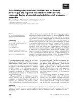
shown in Fig. 1A, accumulation of small fragments of
DNA was observed in cells seeded at higher density
(lanes 2, 3, 5 and 6) as compared to those seeded a t
lower density (lanes 1 and 4). Subsequently, Southern
hybridization was perf ormed using a DNA probe hybri-
dizing to the telo meric end of t he MLL b cr as shown in
Fig. 1B. This probe detects the 8.3 kb intact MLL bcr
encompassed by the BamH I restriction sites. Any clea-
vage within the MLL bcr, centromeric t o the probe will
be detected as fragments smaller than 8.3 kb. For both
SUNE1 and HONE1 cell lines, a 1.5 kb fragment was
detected at high cell density in addition to the 8.3 kb
intact MLL bcr, (Fig. 1C, lanes 2, 3, 5 and 6). This indi-
cates that high cell density-induced apoptosis results in
chromosome break within the MLL bcr 1.5 kb from the
telomeric end (Fig. 1B).
Yee and Sim Journal of Biomedical Science 2010, 17:77
/>Page 3 of 8
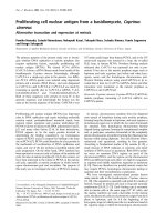
Caspase inhibitor reduces high cell density-induced MLL
bcr cleavage
The apoptotic nuclease , casp ase-activated DNase (CAD)
exists as a complex with the inhibitor of CAD (ICAD)
[23]. During apoptosis induction, caspase cascade is acti-
vated, where casp ase-3 cleaves ICAD, thus releasing the
activated CAD [24]. Therefore, to investigate if the
apoptotic nuclease is involved in cleavage of the MLL
bcr during high cell density-induced apoptosis in NPC
cells, caspase inhibitor was used to inhibit CAD. Consis-
tent with the observation shown in Fig. 1C, high cell
density resulted in cleavage o f the MLL bcr, evidenced
bythepresenceofthe1.5kbfragment(Fig.2,lanes2
and 3). This cleavage was significantly reduced in cells
treated with caspase inhibitor(Fig.2,lanes5and6).
This finding suggests that CAD is involved in cleavage
of the MLL bcr resulted from high cell density-induced
apoptosis.
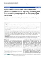
Expression of LMP1 gene induces apoptosis in SUNE1
cells
In order to investigate the relationship between EBV,
apoptosis and chromosomal rearrangements in NPC,
SUNE1 cells were transfected with either the vector or
the LMP1 expression plasmid to asse ss apoptosis induc-
tion and MLL bcr cleavage. Two expression plasmids
were used, namely pcDNA and pTracer. Since the green
fluorescent protein (GFP)geneandtheLMP1 gene are
on the same plasmid vector (pTracer), the expression of
GFP is indicative of plasmid uptake by the cells and
most likely the expression of LMP1 as well. As shown in
Fig. 3A, pTracer-transfected cells showed normal mor-
phology. Cells expressing GFP retained the normal mor-
phology as well (Fig. 3B). By contrast, cells transfected
with LMP1 expression plasmid showed the typical apop-
totic morphology (Fig. 3C). Moreover, most of the GFP-
expressing cells were found to be f loating and blebbing,
with a smaller population of GFP-expressing cells
remained attaching to the flask (Fig. 3D). These results
suggest that expression of LMP1 induces apoptosis in
SUNE1 cells. Cells transfected with the pcDNA set of
plasmids showed similar results under bright field
Figure 1 High cell density induces apoptosis and subsequent
cleavage of the MLL bcr. (A) Ethidium bromide-stained agarose
gel. SUNE1 (lanes 1-3) and HONE1 (lanes 4-6) seeded at cell number
of 0.4 × 10
5
(lanes 1 and 4), 2 × 10
5
(lanes 2 and 5) and 4 × 10
5
(lanes 3 and 6) were harvested for genomic DNA extraction after
4 days of growth. DNA was digested with BamH I and analyzed on
1% agarose gel. M represents the 1 kb DNA marker. (B) A schematic
diagram illustrating the 8.3 kb MLL breakpoint cluster region (bcr).
B represents the BamH I restriction site. Black box indicates the
position of the DNA probe and down arrow shows the anticipated
site of DNA cleavage. (C) Southern hybridization analysis. Southern
hybridization was performed using the DNA probe shown in (B).
Arrows labeled 8.3 kb and 1.5 kb show the positions of the intact
and the cleaved MLL bcr respectively. M
Dig
represents the
DIG-labeled DNA marker (Roche, Penzberg, Germany).
Figure 2 Caspase inhibitor reduces high cell density-induced
MLL bcr cleavage. SUNE1 cell seeded at cell number of 0.4 × 10
5
(lanes 1 and 4), 2 × 10
5
(lanes 2 and 5) and 4 × 10
5
(lanes 3 and 6)
were allowed to grow for 4 days in the absence (lanes 1-3) or
presence (lanes 4-6) of 50 μM caspase-3 inhibitor II (Z-DEVD-FMK).
Extracted genomic DNA was processed for Southern hybridization
as described in methods. Arrows labeled 8.3 kb and 1.5 kb show
the positions of the intact and the cleaved MLL bcr respectively.
M
Dig
represents the DIG-labeled DNA marker.
Yee and Sim Journal of Biomedical Science 2010, 17:77
/>Page 4 of 8
microscopy (data not shown). Dark-field microscopy
result is not available for the pcDNA set as it does not
carry the GFP gene.
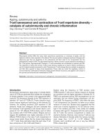
Expression of LMP1 gene induces DNA breaks within the
MLL bcr
Expression of LMP1 gene was c onfirmed by Western
blotting using anti-V5 (Fig. 4A) and S12 anti-LMP1
antibody (Fig. 4B). Expression was demonstrated in
LMP1 transfectants (Fig. 4A, lanes 2 and 4; Fig 4B, lanes
3 and 5) as compar ed to the controls (Fig. 4A, lanes 1
and 3; Fig 4B, lanes 2 and 4). EBV-positive B95 cell was
included as a positive control and the reported 63 kDa
LMP1 protein was detected (Fig. 4B, lane 1). The discre-
pancy in the protein size observed (72 kDa in trans-
fected cells and 6 3 kDa in B95-8 cell) is due to the
reason that LMP1 was expressed in fusion with V5 epi-
tope and His-tag in the transfected cells. In addition,
multiple bands of possibly degraded LMP1 were also
detected in these cells (Fig. 4B, lanes 3 and 5).
Subsequent to the observation of apoptotic morphol-
ogy in LMP1-transfected cells, we intended to test
whether expression of LMP1 results in cleavage of the
MLL bcr by nested IPCR. As shown in Fig. 4C, both
the vector-transfected and LMP1-transfected cells
demonstrated the presence of a 2 kb band, which was
derived from the intact MLL gene (Fig. 4C, lanes 1-4).
Interestingly, cells transfected with the vectors
(Fig.4C,lanes1and3)showedfaintbandsofsizesof
less than 2 kb. From our experience, these bands
might be contributed by those c ells that were dying
naturally while in culture as well as during the trans-
fection process. On the other hand, cells tran sfected
with LMP1 expression plasmids (Fig. 4C, lanes 2 and
4) showed very distinct and intense bands of sizes
smaller than 2 kb. DNA sequencing of these bands
(600bpand300bpIPCRproductsrecoveredfromFig.
4C lanes 2 and 4 respectively) confirmed that they
were the result of DNA cleavage within the MLL bcr.
The precise breakpoints of the 600 bp and 300 bp were
mapped to coordinates 7215 and 6782 respectively
Figure 3 Transfection of SUNE1 cell with LMP1 induces
apoptotic cell death. SUNE1 cells were transiently transfected with
pTracer vector (A and B) or LMP1 expression plasmid, pTracer-LMP1
(C and D). Cell morphology was monitored under bright-field
microscopy (A and C) as well as dark field microscopy (B and D).
Expression of the green fluorescence protein, GFP, is observed as
green colored cells.
Figure 4 LMP1 expression induces cleavage of the MLL bcr.(A)
Detection with anti-V5 antibody. SUNE1 cells were either
transfected with vectors, pcDNA or pTracer (lanes 1 and 3); or LMP1
expression plasmids, pcDNA-LMP1 or pTracer-LMP1 (lanes 2 and 4).
Cell lysate was analyzed on 10% SDS PAGE, and LMP1 expression
was detected by anti-V5 antibody. (B) Detection with S12 anti-LMP1
antibody. SUNE1 cells were either transfected with vectors, pcDNA
or pTracer (lanes 2 and 4); or LMP1 expression plasmids, pcDNA-
LMP1 or pTracer-LMP1 (lanes 3 and 5). Cell lysate was analyzed on
10% SDS PAGE, and LMP1 expression was detected by S12 anti-
LMP1 antibody. Lysate from the EBV-positive B95 cell line was
included as positive control (lane 1). Arrows labeled 72 kDa and 63
kDa indicate the size of the expressed LMP1 (with V5 epitope and
His-tag) and the endogenous LMP1 of B95-8 respectively. (C)
Detection of MLL bcr cleavage by IPCR. SUNE1 cells transfected with
vectors, pcDNA or pTracer (lanes 1 and 3); or LMP1 expression
plasmids, pcDNA-LMP1 or pTracer-LMP1 (lanes 2 and 4) were
collected for genomic DNA extraction. DNA was processed for
nested IPCR as described in methods. Arrow labeled 2 kb indicates
the position of the IPCR product of the intact MLL bcr. Arrows
labeled 600 bp and 300 bp indicate the positions of the IPCR
products of the cleaved MLL bcr. M
1
and M
2
represent the 1 kb and
100 bp DNA marker respectively.
Yee and Sim Journal of Biomedical Science 2010, 17:77
/>Page 5 of 8
[GenBank:U04737]. These results suggest that expres-
sion of LMP1 induces apoptosis in NPC cells, and sub-
sequently results in cleavage of the MLL bcr.
Discussion
The association of EBV with NPC is well documented
[3], and various chromosome anomalies are well
reported in NPC [2]. However, the actual role of EBV in
the pathogenesis of NPC is unclear and EBV’sinvolve-
ment in chromosome rearrangements remains to be elu-
cidated. Other virus has been shown to induce
chromosome aberrations in infected cells [17]. Similarly,
LMP1 expression was found to induce aneuploidy in
human epithelial cells [25]. Knowing that EBV infectio n
and LMP1 expression induce apoptosis in mammalian
cells [6,7], we wanted to answer a further question: is
EBV-induced apoptosis a mechanism of chromosome
rearrangement in NPC? H ere, our results for the first
time show that LMP1 expression and high cell density
induce apoptosis in NPC cells and subsequently result
in enhanced DNA cleavage within the MLL bcr at
11q23, a common chromosome deletion site in NPC.
It is important to note that, the breakpoints identified
in this study fall within the bcr of the MLL gene. Clea-
vage of the MLL bcr has been extensively studied in leu-
kemic cells, relating to chromosome translocation
mech anism invol ving topoisomerase II [26] and apopto-
tic nuclease [14,21]. However, this is the first demon-
stration of apoptosis-induced cleavage of the MLL bcr
in NPC cells. Since the MLL gene locates at 11q23 [18],
a common chromosome deletion site in NPC [2], our
findings support the possibility that chromosome dele-
tion at 11q23 in NPC could begin at the MLL gene.
In our study, treatm ent with caspase inhibitor signifi-
cantly reduced the MLL bcr cleavage. This parallels t he
observations in leukemic cells, suggesting the involve-
ment of a caspase-d ependent apoptotic nuclease [21],
possibly the caspase-activated DNase (CAD) [23]. CAD
associates with the nuclear matrix of apoptotic cells
[27], facilitating its role in cleaving the base of the chro-
matin loops at the nuclear matrix or scaffold, generating
high molecular weight (HMW) DNA during early stage
apoptosis [28]. CAD was also shown to cause DNA frag-
mentation producing the characteristic nucleosomal
DNA ladder [23]. However, CAD is not the sole enzyme
for DNA cleavage at nuclear matrix, as it was found to
be dispensable for HMW DNA fragmentation during
early stage apoptosis in chicken DT40 cells [29]. This
observ ati on tallies with our resul t that caspase inhibitor
did not abolish the MLL cleavage completely, suggesting
the possible involvement of other nucleases. O ne pro-
mising candidate is endonuclease G (Endo G) [11],
which is one of the effectors of caspase-independent cell
death pathway [30]. Interestingly, both CAD and Endo
G preferentially cleave DNA at the internucleosomal
linker DNA. They also cleave at t he borders of chroma-
tin loops, releasing chromatin domains of sizes ≥ 50 kb
[11]. This chromatin loop domain structure is main-
tained by the interaction of specific sequences known as
the matrix attachment region/scaffold attachment region
(MAR/SAR), with the nuclear matrix proteins [31]. Dur-
ing early apoptosis, genomic DNA is cleaved at the base
of the chromatin loop, results in the formation of
HMW DNA of 50 - 300 kb [32].
In this study, the MLL cleavage sites observed in the
NPC cells localized within the MAR/SAR sequence of
the MLL bcr [20], suggesting t hat both CAD and Endo
G could be involved i n introducing the b reaks during
early apoptosis. This is a very crucial observation as we
hypothesize that during apo ptosis, the genomic DNA is
being cleaved at the base of the loop, and rejoined erro-
neously upon the cell’ sattemptedrepair.Asaresult,
cells that survive the apopt otic process may harbor var-
ious kinds of chromosome anomalies. Logically, only
those cells that are at the early stage of apoptosis can be
rescued and survive apoptosis.
In addition to CAD and E ndo G, DNA topoisomerase
II is another important player in the excision of the
chromatin loops during early apoptosis [33]. Poisoning
of topoisomerase II by etoposide and oxidative stress
resulted in chromatin loop excision [10,33]. This is
entirely logical as topoisomerase II is one of the two
major proteins found in the nuclear scaffold [34]. Inter-
estingly, CAD interacts with topoisomerase II and
enhances topoisomerase II’ s decatenation activity in
vitro [35]. Since EBV infection introduces oxidative
stress to the cell [36], thus our results of MLL bcr clea-
vagecouldbepartlymediatedbytopoisomeraseIIand
Endo G in addition to CAD.
Conventionally, apoptosis is known to be an irreversi-
ble programmed cell death process [37]. However, some
of the cells can surv ive apoptosis. These cells may har-
bor rearranged chromosomes that contribute to leuke-
mogenesis [15]. This is supported by the observation
that apoptotic triggers resulted in the formation o f
MLL-AF9 fusion gene in leukemic cells that are capable
of division [14]. A lthough various mechanisms h ave
been proposed, chromatin structures at the breakpoint
cluster regions were recently suggested to contribute to
chromosome translocations in chronic and acute leuke-
mia [ 38]. Our results of chromosome breaks within th e
MAR/SARsequencesupportedtheroleofchromatin
structure in chromosome rearrangements.
Since EB V infection and LMP1 expression both
resulted in apoptosis and DNA fragmentation [7,8,39], it
is possible that during EBV infection, apoptosis is
induced and resulted in chromosome breaks that lead to
chromosome rearrangements in cells that survive
Yee and Sim Journal of Biomedical Science 2010, 17:77
/>Page 6 of 8
apoptosis. A single event of infection may not be
sufficient to initiate cancer, however, multiple cycles of
infection or reactivation and latency would increase the
possibility of tumorigenesis by increasing the number of
chromosome anomalies. This notion is supported by a
study reporting that recurrent chemical reactivations of
EBV promotes genome instability as well as enhances
tumor progression of nasopharyngeal carcinoma
cells [40].
Conclusions
High cell density and LMP1 expression induced apop-
tosis in NPC cells and subsequ ently resulted in MLL
bcr cleavage at the MAR/SAR region. This cleavage is
most likely mediated predominantly by CAD and par-
tially by other nucleases. Since MLL locates at 11q23, a
common deletion site in NPC, our results suggest a
possibility of stress- or virus-induced apoptosis in the
initiation of chromosome rearrangements at 11q23,
where the chromatin structure plays a role in defining
the site of chromosome rearrangement . These results
tally with f indings in leukemia, su ggesting a possibl e
common mechanism of chromosome rearrangement in
different cancer types.
Acknowledgements
We would like to thank Prof. Dr. Choon-Kook Sam for the NPC cell lines and
the EBV genome-positive marmoset cell line, B95-8; Dr. Eng-Lai Tan and Prof.
Dr. Choon-Kook Sam for the EBV LMP1 recombinant plasmid; Prof. Dr. Leroy
Fong Liu for the cloning plasmid and the clone for DNA probe. This project
was supported by the Ministry of Science, Technology and Innovation,
Malaysia (grant number: 06-02-09-1020-PR0054/05-02).
Authors’ contributions
SPS contributes to the main idea of the project, the design of the study,
interpretation of data and writing of manuscript. PHCY have been involved
in the detailed experimental design, acquisition of data, interpretation of
data and analysis. All authors read and approved the final manuscript.
Competing interests
The authors declare that they have no competing interests.
Received: 19 July 2010 Accepted: 22 September 2010
Published: 22 September 2010
References
1. Fandi A, Altun M, Azli N, Armand JP, Cvitkovic E: Nasopharyngeal cancer:
epidemiology, staging, and treatment. Semin Oncol 1994, 21:382-397.
2. Chien G, Yuen PW, Kwong D, Kwong YL: Comparative genomic
hybridization analysis of nasopharygeal carcinoma: consistent patterns
of genetic aberrations and clinicopathological correlations. Cancer Genet
Cytogenet 2001, 126:63-67.
3. Raab-Traub N: Epstein-Barr virus and nasopharyngeal carcinoma. Semin
Cancer Biol 1992, 3:297-307.
4. Lin JC, Liao SK, Lee EH, Hung MS, Sayion Y, Chen HC, Kang CC, Huang LS,
Cherng JM: Molecular events associated with epithelial to mesenchymal
transition of nasopharyngeal carcinoma cells in the absence of Epstein-
Barr virus genome. J Biomed Sci 2009, 16:105.
5. Gan YY, Fones-Tan A, Chan SH, Gan LH: Epstein-Barr Viral Antigens Used
in the Diagnosis of Nasopharyngeal Carcinoma. J Biomed Sci 1996,
3:159-169.
6. Larochelle B, Flamand L, Gourde P, Beauchamp D, Gosselin J: Epstein-Barr
virus infects and induces apoptosis in human neutrophils. Blood 1998,
92:291-299.
7. Lu JJ, Chen JY, Hsu TY, Yu WC, Su IJ, Yang CS: Induction of apoptosis in
epithelial cells by Epstein-Barr virus latent membrane protein 1. JGen
Virol 1996, 77(Pt 8):1883-1892.
8. Kawanishi M: Epstein-Barr virus induces fragmentation of chromosomal
DNA during lytic infection. J Virol 1993, 67:7654-7658.
9. Bortner CD, Oldenburg NB, Cidlowski JA: The role of DNA fragmentation
in apoptosis. Trends Cell Biol 1995, 5:21-26.
10. Li TK, Chen AY, Yu C, Mao Y, Wang H, Liu LF: Activation of topoisomerase
II-mediated excision of chromosomal DNA loops during oxidative stress.
Genes Dev 1999, 13:1553-1560.
11. Widlak P, Garrard WT: Discovery, regulation, and action of the major
apoptotic nucleases DFF40/CAD and endonuclease G. J Cell Biochem
2005, 94:1078-1087.
12. Kim GS, Choi YK, Song SS, Kim WK, Han BH: MKP-1 contributes to
oxidative stress-induced apoptosis via inactivation of ERK1/2 in SH-SY5Y
cells. Biochem Biophys Res Commun 2005, 338:1732-1738.
13. Kluck RM, Chapman DE, Egan M, McDougall CA, Harmon BV, Moss DJ,
Kerr JF, Halliday JW: Spontaneous apoptosis in NS-1 myeloma cultures:
effects of cell density, conditioned medium and acid pH. Immunobiology
1993, 188:124-133.
14. Betti CJ, Villalobos MJ, Diaz MO, Vaughan AT: Apoptotic stimuli initiate
MLL-AF9 translocations that are transcribed in cells capable of division.
Cancer Res 2003,
63:1377-1381.
15. Vaughan AT, Betti CJ, Villalobos MJ: Surviving apoptosis. Apoptosis 2002,
7:173-177.
16. Hars ES, Lyu YL, Lin CP, Liu LF: Role of apoptotic nuclease caspase-
activated DNase in etoposide-induced treatment-related acute
myelogenous leukemia. Cancer Res 2006, 66:8975-8979.
17. Siew VK, Duh CY, Wang SK: Human cytomegalovirus UL76 induces
chromosome aberrations. J Biomed Sci 2009, 16:107.
18. Ziemin-van der Poel S, McCabe NR, Gill HJ, Espinosa R, Patel Y, Harden A,
Rubinelli P, Smith SD, LeBeau MM, Rowley JD, Diaz MO: Identification of a
gene, MLL, that spans the breakpoint in 11q23 translocations associated
with human leukemias. Proc Natl Acad Sci USA 1991, 88:10735-10739.
19. Rowley JD: Rearrangements involving chromosome band 11Q23 in acute
leukaemia. Semin Cancer Biol 1993, 4:377-385.
20. Broeker PL, Super HG, Thirman MJ, Pomykala H, Yonebayashi Y, Tanabe S,
Zeleznik-Le N, Rowley JD: Distribution of 11q23 breakpoints within the
MLL breakpoint cluster region in de novo acute leukemia and in
treatment-related acute myeloid leukemia: correlation with scaffold
attachment regions and topoisomerase II consensus binding sites. Blood
1996, 87:1912-1922.
21. Sim SP, Liu LF: Nucleolytic cleavage of the mixed lineage leukemia
breakpoint cluster region during apoptosis. J Biol Chem 2001,
276:31590-31595.
22. Betti CJ, Villalobos MJ, Diaz MO, Vaughan AT: Apoptotic triggers initiate
translocations within the MLL gene involving the nonhomologous end
joining repair system. Cancer Res 2001, 61:4550-4555.
23. Enari M, Sakahira H, Yokoyama H, Okawa K, Iwamatsu A, Nagata S: A
caspase-activated DNase that degrades DNA during apoptosis, and its
inhibitor ICAD. Nature 1998, 391:43-50.
24. Sakahira H, Enari M, Nagata S: Cleavage of CAD inhibitor in CAD activation
and DNA degradation during apoptosis. Nature 1998, 391:96-99.
25. Man C, Rosa J, Lee LT, Lee VH, Chow BK, Lo KW, Doxsey S, Wu ZG,
Kwong YL, Jin DY, Cheung AL, Tsao SW: Latent membrane protein 1
suppresses RASSF1A expression, disrupts microtubule structures and
induces chromosomal aberrations in human epithelial cells. Oncogene
2007, 26:3069-3080.
26. Strissel PL, Strick R, Rowley JD, Zeleznik L: An in vivo topoisomerase II
cleavage site and a DNase I hypersensitive site colocalize near exon 9 in
the MLL breakpoint cluster region. Blood 1998, 92:3793-3803.
27. Lechardeur D, Xu M, Lukacs GL: Contrasting nuclear dynamics of the
caspase-activated DNase (CAD) in dividing and apoptotic cells. J Cell Biol
2004, 167:851-862.
28. Sakahira H, Enari M, Ohsawa Y, Uchiyama Y, Nagata S: Apoptotic nuclear
morphological change without DNA fragmentation. Curr Biol 1999,
9:543-546.
Yee and Sim Journal of Biomedical Science 2010, 17:77
/>Page 7 of 8
29. Samejima K, Tone S, Earnshaw WC: CAD/DFF40 nuclease is dispensable
for high molecular weight DNA cleavage and stage I chromatin
condensation in apoptosis. J Biol Chem 2001, 276:45427-45432.
30. van Loo G, Schotte P, van Gurp M, Demol H, Hoorelbeke B, Gevaert K,
Rodriguez I, Ruiz-Carrillo A, Vandekerckhove J, Declercq W, Beyaert R,
Vandenabeele P: Endonuclease G: a mitochondrial protein released in
apoptosis and involved in caspase-independent DNA degradation. Cell
Death Differ 2001, 8:1136-1142.
31. Laemmli UK, Kas E, Poljak L, Adachi Y: Scaffold-associated regions: cis-
acting determinants of chromatin structural loops and functional
domains. Curr Opin Genet Dev 1992, 2:275-285.
32. Lagarkova MA, Iarovaia OV, Razin SV: Large-scale fragmentation of
mammalian DNA in the course of apoptosis proceeds via excision of
chromosomal DNA loops and their oligomers. J Biol Chem 1995,
270:20239-20241.
33. Solovyan VT, Bezvenyuk ZA, Salminen A, Austin CA, Courtney MJ: The role
of topoisomerase II in the excision of DNA loop domains during
apoptosis. J Biol Chem 2002, 277:21458-21467.
34. Earnshaw WC, Halligan B, Cooke CA, Heck MM, Liu LF: Topoisomerase II is
a structural component of mitotic chromosome scaffolds. J Cell Biol 1985,
100:1706-1715.
35. Durrieu F, Samejima K, Fortune JM, Kandels-Lewis S, Osheroff N,
Earnshaw WC: DNA topoisomerase IIalpha interacts with CAD nuclease
and is involved in chromatin condensation during apoptotic execution.
Curr Biol 2000, 10:923-926.
36. Gruhne B, Sompallae R, Marescotti D, Kamranvar SA, Gastaldello S,
Masucci MG: The Epstein-Barr virus nuclear antigen-1 promotes genomic
instability via induction of reactive oxygen species. Proc Natl Acad Sci
USA 2009, 106:2313-2318.
37. Cohen JJ, Duke RC, Fadok VA, Sellins KS: Apoptosis and programmed cell
death in immunity. Annu Rev Immunol 1992, 10:267-293.
38. Strick R, Zhang Y, Emmanuel N, Strissel PL: Common chromatin structures
at breakpoint cluster regions may lead to chromosomal translocations
found in chronic and acute leukemias. Hum Genet 2006, 119:479-495.
39. Sbih-Lammali F, Clausse B, Ardila-Osorio H, Guerry R, Talbot M, Havouis S,
Ferradini L, Bosq J, Tursz T, Busson P: Control of apoptosis in Epstein Barr
virus-positive nasopharyngeal carcinoma cells: opposite effects of CD95
and CD40 stimulation. Cancer Res 1999, 59:924-930.
40. Fang CY, Lee CH, Wu CC, Chang YT, Yu SL, Chou SP, Huang PT, Chen CL,
Hou JW, Chang Y, Tsai CH, Takada K, Chen JY: Recurrent chemical
reactivations of EBV promotes genome instability and enhances tumor
progression of nasopharyngeal carcinoma cells. Int J Cancer 2009,
124:2016-2025.
doi:10.1186/1423-0127-17-77
Cite this article as: Yee and Sim: High cell density and latent membrane
protein 1 expression induce cleavage of the mixed lineage leukemia
gene at 11q23 in nasopharyngeal carcinoma cell line. Journal of
Biomedical Science 2010 17:77.
Submit your next manuscript to BioMed Central
and take full advantage of:
• Convenient online submission
• Thorough peer review
• No space constraints or color figure charges
• Immediate publication on acceptance
• Inclusion in PubMed, CAS, Scopus and Google Scholar
• Research which is freely available for redistribution
Submit your manuscript at
www.biomedcentral.com/submit
Yee and Sim Journal of Biomedical Science 2010, 17:77
/>Page 8 of 8