Báo cáo y học: "Structural and functional characterization of human apolipoprotein E 72-166 peptides in both aqueous and lipid environments" pot
Bạn đang xem bản rút gọn của tài liệu. Xem và tải ngay bản đầy đủ của tài liệu tại đây (1.29 MB, 9 trang )
RESEARC H Open Access
Structural and functional characterization of
human apolipoprotein E 72-166 peptides in
both aqueous and lipid environments
Yi-Hui Hsieh, Chi-Yuan Chou
*
Abstract
Backgrounds: There are three apolipoprotein E (apoE) isoforms involved in human lipid homeostasis. In the
present study, truncated apoE2-, apoE3- and apoE4-(72-166) peptides that are tailored to lack domain interaction s
are expressed and elucidated the structural and functional consequences.
Methods & Results: Circular dichroism analyses indicated that their secondary structure is still well organized.
Analytical ultracentrifugation analyses demonstrated that apoE-(72-166) produces more complicated species in PBS.
All three isoforms were significantly dissociated in the presence of dihexanoylphosphatidylcholine.
Dimyristoylphosphatidylcholine turbidity clearance assay showed that apoE4-(72-166) maintains the highest lipid-
binding capacity. Finally, only apoE4-(72-166) still maintained significant LDL receptor binding ability.
Conclusions: Overall, apoE4-(72-166) peptides displayed a higher lipid-binding and comparable receptor-binding
ability as to full-length apoE. These findings provide the explanation of diverged functionality of truncated apoE
isoforms.
Introduction
Human apolipoprotein E (apoE)
1
comprises 299 amino
acids and there are three isoforms, apoE2, apoE3, and
apoE4, encoded by the ε2, ε3, and ε4 genes, respectively.
These isoforms differ from each other only at residues
112 and 158 i.e. Cys112 and Arg158 in apoE3, a cysteine
at both positions in apoE2, and an arginine at both posi-
tions in apoE4 [1]. The amino-terminal (NT) d omain of
apoE contains four amphipathic a-helices and has
pronounced kinks in the helices near the end of the
four-helix bundle that correlates with the lipid binding
ability (Figure 1) [2,3]. The residues between 140-150 in
the fourth a-helix, comprising many basic amino acids,
has been identified as the low-density lipoprotein recep-
tor (LDLR) binding region [4], with the lipid binding
regionshowntobeinthecarboxyl-terminal(CT)
domain [5,6]. The lipid association is required for high
affinity binding of apoE to the LDLR because of the
increased exposure of basic region on the fourth a-helix
after interacting with lipids [7].
ApoE is involv ed in facilitating the transportation of
plasma chylomicron remnant to the liver through either
the remnant receptor or LDLR [8,9]. Owing to distinct
domain interactions, apoE2 and apoE3 bind preferen-
tially to small lipoproteins such as high-density lipopro-
tein (HDL), whereas apoE4 has a higher affinity to
very-low-density lipoprot ein (VLDL) [6,10]. Different to
apoE3, apoE4 is prone to raise the plasma LDL to high
levels and cause high oxidative s tress that can facilitate
atherosclerosis progression [11,12], whilst apoE2 is asso-
ciated with type III hyperlipoproteinemia [13]. The ε4
allele is also associated with familial late-onset and
sporadic Alzheimer’ s disease (AD) [14,15]. ApoE4 has
been found to interact with beta-amyloid peptides (Ab)
and induce neurofibrillary tangle (NFT) formation
[16,17]. It preferentially undergoes proteolysis to yield
NT- and CT-truncated that interact with cytoske letal
components to form NFT-like inclusions in neuronal
cells [16]. To understand the pathogenesis of different
isofomic apoE, most studies are f ocused on the delinea-
tion of the structure and function characterization of
the full-length apoE, varied length CT, or a “ four
a-helix bundle” NT domain [18-21].
* Correspondence:
Department of Life Sciences and Institute of Genome Sciences, National
Yang-Ming University, Taipei 112, Taiwan
Hsieh and Chou Journal of Biomedical Science 2011, 18:4
/>© 2011 Hsieh and Chou; licensee BioMed Central Ltd. This is an Open Access article distr ibuted under the terms of the Creative
Commons Attribution License (http://creat ivecommons.org/licenses/by/2.0), which perm its unrestricted use, distribution, and
reproduction in any medium, provided the original work is properly cited.
In the present stu dies, we attempted to clarify th e
structural and functional consequences of NT- and
CT-truncated apoE peptides, i.e. apoE-(72-166). This
truncation still maintains the LDLR binding region, and
removes the first two a-helices and the complete CT
domain. The aim is to create a shorter but still functional
apoE for potential therapeutic approach. Analytical ultra-
centrifugation was used to elucidate the quaternary struc-
tural properties of the three apoE-(72-166) isoforms. In
the presence of lipid, the degree of apoE-(72-166) disso-
ciation and extended conformation was significantly
elevated. The functional assays conclude that apoE-(72-
166) peptides still maintain comparable LDLR and higher
lipid binding ability as to full-length apoE, particularly
apoE4-(72-166). T hese findings suggest a crucial role of
shorter NT-domain in the biological function of apoE
and provide the basis for the explanation of diverged
functionality of truncated apoE isoforms.
Materials and methods
Plasmids
The construction of pET-apoE2, apoE3, apoE4, apoE3-
(72-166), and apoE4-(72-166) vectors were described
previously [22]. The ap oE2-(72-166) DNA fragment was
amplified by PCR, and t he forward primer was 5’-AAA-
CATATGAAGGCCTACAAATCGGA, whereas the
reverse primer was 5’-AACTCGAGGGCCCCGGCCT.
The NdeI-XhoI digested apoE2-(72-166) cDNA was then
ligated to the 5.2-kb NdeI-XhoI pET-29a(+) fragment.
Expression and Purification of ApoE Proteins
Protein induction and purification procedures have bee n
described previously [22,23]. Typical yields of the apoE-
(72-166) proteins were 5-10 mg after purification from 1
liter of E. coli culture medium. The purity of all recombi-
nant proteins was estimated by SDS-PAGE to be > 95%
and the molecular mass of the apoE-(72-166) proteins
was 12 kDa. The purified proteins were buffer-changed
to phosphate buffered saline (PBS) (pH7.3) using Amicon
Ultra-4 10-kDa centrifugal filter (Millipore).
Preparation of Micelle Solution
Dihexanoylphosphatidylcholine (DHPC) has a critical
micelle concentrat ion of 16 mM, at whic h micelle mono-
mers are formed containing 19 to 40 molecules based on
ultracentrifugation, NMR, and small angle neutron scat-
tering, respectively [24-26]. We used several concentra-
tions of DHPC (5, 50, and 100 mM) to establish an
appropriate lipid environment containing submicelles or
micelles. In current studies, all experiments related to
DHPC were executed at 20°C for the same lipid state.
Circular Dichroism Spectroscopy
Circular dichroism (CD) spectra of the apoE-(72-166)
peptides using a JASCO J-810 spectropolarimeter
(Tokyo, Japan) showed measurements from 250 nm to
190 nm at 20°C in PBS (pH 7.3) with or without 50 mM
DHPC. The protein concentration was 0.5 m g/ml. In
wavelength scanning, the width of the cuvette was 0.1
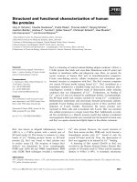
Figure 1 Structure of human apoE proteins. The model structure illustrating the full-length apoE with NT and CT domains. The structure was
modified from apoE299_20K (S. Y. Sheu, unpublished data). The polymorphic sites (residues 112 and 158) that distinguished the three isoforms
are highlighted. The picture was produced with PyMOL [46].
Hsieh and Chou Journal of Biomedical Science 2011, 18:4
/>Page 2 of 9
mm. The far-UV CD spectrum data were analyzed with
the CDSSTR program [27,28]. In this analysis, the
a-helix, b-sheet, and random coil were split. To estimate
the goodness-of-fit, the normalized root mean square
deviation (NRMSD) was calculated.
Unfolding of the ApoE-(72-166) Proteins in Guanidinium
Chloride
ApoE-(72-166) proteins (0.1 mg/ml) with or without 50
mM DHPC were unfolded with different concentrations
ofGdnClinPBS(pH7.3)at4°Covernighttoreach
equilibrium. The unfolding of the proteins was moni-
tored by measuring the CD signal of 222 nm at 20°C
and t he width of the cuvette was 1 mm. The unfolding
data were analyzed using thermodynamic models by
global fitting of the measurements to the two-state
unfolding model [29] as follows:
y
yye
e
obs
NU
GmGdnCl
RT
G
HO
NU
NU
HO
N
=
+•
+
−
−
[]
⎛
⎝
⎜
⎜
⎞
⎠
⎟
⎟
−
→
→
→
Δ
Δ
()
()
2
2
1
UU
NU
mGdnCl
RT
−
[]
⎛
⎝
⎜
⎜
⎞
⎠
⎟
⎟
→
(1)
where y
obs
is the observe d biophysical signal; y
N
and
y
U
are the calculated signals of the native and unfolded
states, respectively. GdnCl is the GdnCl concentration,
and
ΔG
HON U()
2
→
isthefreeenergychangeforthe
N®U process. m
N®U
is the sensitivity of the unfolding
process to a denaturant concentration.
Sedimentation Velocity
Sedimentation velocity (SV) experiments were per-
formed with an XL-A analytical ultracentrifuge (Beck-
man, Fullerton, CA) as described previously [23]. All
studies were performed at 20°C with a rotor speed of
42,000 rpm in PBS (pH 7.3) with or without DHPC.
The protein concentration was 0.5 mg/ml. Multiple
scans at different time periods were then fitted to a con-
tinuous c(s) distribution model using the SEDFIT
program as described previously [30,31]. All continuous
size distributions were calculated using a confidence
level of p = 0.95, a best fitted average anhydrous friction
ratio (f
r
), a resolution value N of 200, and sedimentation
coefficients between 0 and 20 S. For the da ta fitting of
apoE-(72-166) in PBS and 5 mM DHPC, the partial spe-
cific volume was set to 0.73 for proteins species. Differ-
ently, for those in 50 and 100 mM DHPC, the value was
set to 0.86 because the influence of DHPC micelle.
Previous studies have suggested that DHPC’s partial spe-
cific volume is 0.99 ml/g [32]. According to our calcula-
tion, higher partial specific volume will lower the best
fitted average f
r
, while the c(s) distribution will not have
any difference.
Sedimentation Equilibrium
Sedimentation equilibrium (SE) experiments were per-
formed with six-channel epon charcoal-filled center-
pieces as described previously [22]. The cells were then
mounted into an An-60 Ti rotor and centrifuged at
10,000 rpm, 15,000 rpm, and 20,000 rpm, respective ly,
each for 18 h at 20°C. Ten A
280 nm
measurements with
a time interval of 8-10 min were performed for each dif-
ferent rotor speed to check the equilibrium state. The
SV and SE spectrum of each apoE-(72-166) protein
under the same environments were combined and then
fitted to a global discrete species model using
SEDPHAT program as described previously [22,33].
DMPC Turbidity Clearance Assay
The preparation of DMPC (Sigma, St Louis, MO) multi-
lamellar vesicles (mLV) has been described previously
[22,34-36]. ApoE (250 μg) was added to DMPC mLV
solution (0.5 mg/ml) in a quartz cuvette which had been
preincubated at 24°C in a Perkin-Elmer Lamb da 35
spectrophotometer with water circulated temperature
control. Vesicle solubilization was monitored as a
decrease in the absorbance at 325 nm. The time course
of the clearance measurements were fitted by nonlinear
regression to the biexponential decay equation,
YAe Be C
kt kt
=⋅ +⋅ +
−⋅ −⋅
12
(2)
where Y is the absorbance at 325 nm and k, k
1
or k
2
are the rate constants for different kinetic phases of the
solution clearance. A and B are the changes in turbidity
for different phases (pool sizes), t is the time, and C is
the remaining turb idity at the completion o f the
reaction.
In vitro VLDL Binding Assay
ApoE proteins were incubated with apoE(-) mice serum
at 37°C. The molar ratio of apoE and VLDL was 1:1 for
the apoE and 5:1 for the apoE-(72-166) proteins. After a
4 h incubation, the apoE-VLDL particles and free apoE
were separated by NaBr density ultracentrifugation
(Optima L-90K ultracentrifuge, Beckman). At first, the
density of serum was corrected to 1.211 g/ml by adding
NaBr. The serum solution was then loaded into 10-ml
ultracentrifuge bottles (polycarbonate, Beckman, Fuller-
ton, CA) and centrifugation was performed for 24 h
with a rotor (Beckman 70.1 Ti) speed of 44,000 rpm at
4°C. After centrif ugation, the lipoproteins ( HDL, LDL,
and VLDL) float on the solution surfac e and can be
recovered by pipetting. The binding of apoE-VLDL was
then confirmed by lipoprotein electrophoresis (hydragel
lipo + Lp(a) K20, Sebia) at 50 V, a current of 25 mA,
and a power setting of 5 W for 3 h. The LDL, VLDL,
and HDL molecules were separated by their charge and
Hsieh and Chou Journal of Biomedical Science 2011, 18:4
/>Page 3 of 9
the VLDL band was shifted with the binding of apoE
proteins.
LDLR Binding Assay
The detailed procedures for the LDLR binding assay
have been described previously [22,37,38]. Briefly,
human hepatoblastoma cells (HepG2) were incubated in
DMEM with 10% fetal bovine serum at 37°C followed
by incubatio n with DMEM containing
3
H-LDL and
different receptor binding competitors (apoE proteins)
at 4°C for 2 h. After washing, cells were released, lysed,
and the radioactivity was determined using a liquid
scintillation counter (Beckman, Fullerton, CA).
Results and Discussions
Secondary Structures of the apoE-(72-166) peptides is
well organized and a-helical dominant
Based on the far-UV CD measurements we made,
apoE2-, apoE3-, and apoE4-(72-166) peptides main-
tained 49, 48, and 53% a-helical structure in PBS; and
47, 49, an d 45% in DHPC micellar solution, respectively
(Additional file 1: F igure S1A, B, and Table S1). The
structure of apoE-(72-166) peptides was estimated to be
a-h elix dominant in both aqueous and DHPC micellar
solution, although the content of a-helix was lower than
the value from the solved crystal structure of NT
domain (residues 23-166, pdb code: 1LPE), which is 74%
[39]. The shorter length of our peptides and lower pro-
tein concentration used in CD may be the reason. Over-
all, the content of a-helix in all three isoforms did not
change too much in the two environments, while the
content of b-strand increased by 8-10% in DHPC micel-
lar solution. Consequently, their random coil decreased
by 1-11%. These data indicated that in the aqueous or
DHPC micellar solution, the secondary structure of
apoE-(72-166)waswellorganizedanddidnotshow
very significant isoformic difference.
The secondary structure of apoE-(72-166) was more
stable in the solution containing DHPC micelles
To delineate the structural stability of the apoE-(72-166)
peptides with or without DHPC, the GdnCl denaturation
experiments were executed. The denaturation of the
three apoE-(72-166) proteins follow ed a two-state transi-
tion (Additional file 1: Figure S1C, D). Our experimental
data was then fitted using equation 1 to calculate the
change of free energy, m value, and [GdnCl]
0.5
(Table 1).
InthepresenceofDHPCmicelle,themvalueofthe
three isoforms showed a significant decrease, while
ΔG
HON U()
2
→
didnot.Itresultedinthe[GdnCl]
0.5
of the
three isoform increased by 0.8-0.86 M, respectively, com-
paring to those in PBS. These differences suggeste d that
the secondary structure of apoE-(72-166) was more
stable in the solution containing DHPC micelles. Recent
studies for apolipoprotein C-II amyloid fibrils have
shown similar phenomenon that phospholipid interac-
tions can stabilize regular secondary structure formations
and molecular-level polymorphisms [40].
Similar to full-length apoE proteins in a lipid-free
solution [20], the differences between the apoE-72-166
protein isoforms in terms of structural stability was in
the order of apoE2 > apoE3 > apoE4. Previous structural
studies indicated that Cys112 of apoE3 is partially
buried between helices 2 and 3, while Arg112 of apoE4
could be easily accommodated by filling the solvent
region surrounding the helix pair [39]. This variation
may cause apoE4 more unstable. By the way, it further
suggests that the structure of apoE4-(72-166) is more
easily opened and exposed more hydrophobic residues.
Indeed, by 1-anilino-8-naphtha lenesulfonic acid titration
analysis (our unpublished data), the apoE4-(72-166)
shows the highest hydrophobic exposure, which can
further explain the highest ability of DMPC turbidity
clearance of apoE 4-(72-166) (see belo w). Differently but
not surprisingly, apoE-(72-166) displayed a two-state
transition, whereas full-length apoE showed a three-state
unfolding process. We also found that the [GdnCl]
0.5
values for apoE2-, and apoE3-(72-166) were about
1.1-1.4 M, very close to the [GdnCl]
0.5,N-I
of full-length
apoE2 and apoE3. How ever, the [GdnCl]
0.5
of apoE4-
(72-166) was only 0.6 M, which w as lower than the
[GdnCl]
0.5,N-I
measurement of full-length apoE4 (0.9 M).
Remarkably, the relatively unstable apoE4-(72-166) frag-
ment still possessed a 53 % a-helical structure. More
Table 1 Guanidine hydrochloride denaturation of apoE-(72-166) proteins with and without DHPC
Buffer Protein
ΔG
HON U()
2
→
a
(kcal mol
-1
) m (kcal mol
-1
M
-1
) [GdnCl]
0.5
(M)
PBS apoE2-(72-166) 1.93 ± 0.14 1.37 ± 0.09 1.40 ± 0.14
apoE3-(72-166) 1.71 ± 0.18 1.51 ± 0.13 1.13 ± 0.15
apoE4-(72-166) 1.52 ± 0.20 2.45 ± 0.27 0.62 ± 0.11
PBS + 50 mM DHPC apoE2-(72-166) 1.89 ± 0.24 0.84 ± 0.11 2.25 ± 0.41
apoE3-(72-166) 2.18 ± 0.23 1.13 ± 0.11 1.93 ± 0.28
apoE4-(72-166) 1.30 ± 0.26 0.88 ± 0.15 1.48 ± 0.39
a
The denaturation data were analyzed by the two-state unfolding model (eq. 1). The R
sqr
of each result was from 0.975 to 0.997.
Hsieh and Chou Journal of Biomedical Science 2011, 18:4
/>Page 4 of 9
detailed structural analysis may be required to explain
the reciprocal low structural stability and high a-h elical
content of apoE4-(72-166) in aqueous environment.
Our SV experiments and c(s) distribution analysis
demonstrate a different species distribution of
apoE-(72-166) in aqueous and lipid environments
In PBS, apoE-(72-166) proteins showed a distribution
pattern of two major species (Figure 2A). The first of
these showed a sedimentatio n coefficient distribution of
20 % for apoE2-(72-166) and 23 % for apoE3-(72-166) at
s = 2.0, bu t only 6 % for the same species of apoE4-(72-
166). The second major species was a broad peak at
s=3.5to6.5,withatotaloccupancyof46%for
apoE2-(72-166), 55 % for a poE3-(72-166), and 59 % for
apoE4-(72-166). This region may be the result of a con-
tribution by multi-oligomers. Besides, there were 22-35
% distribution belonged to large aggregated forms. In
the 5 mM DHPC submicellar solution, the smal l species
(s = 2) of the three apoE-(72-166) increased by 1.3 to
4 % (Figure 2B), whereas the major species at s = 3.5-
6.5 decreased by 2 to 8 %. It suggested that submicellar
DHPC can induce the dissociation of apoE-(72-166)
peptide s but not very significantly. In 50 mM DHPC, 76
to 82 % of the apoE-(72-166) proteins dissociated to a
species at s = 1.2-1.5 (Figure 2C). Finally, whilst apoE2-
(72-166) maintained a two species distribution (s = 1.1
and 2.0) in 100 mM DHPC, its apoE3 and apoE4 coun-
terparts maintained a single major species at s = 1 .1
(Figure 2D). Fur thermore, by c(s) distrib ution analysis
we found t hat the average f
r
of apoE-(72-166) in PBS
was around 1.3-1.5, but in 5-50 mM DHPC was around
1.7-1.8, which increased to 1.7-2.1 in 100 mM DHPC
(partial specific volume at 0.86). These differences indi-
cated that when the DHPC conc entration increases,
apoE- (72-166) not only displays a dissociation tendency,
but also adopts a more elongated conformation.
The mass variation of the apoE-(72-166) in PBS and in
DHPC was analyzed by global discrete species model
To further clarify the mass variation of the three apoE-
(72-166) peptides in PBS and also in the presence of
DHPC, SE experiments we re performed. The SE and SV
data were combined and globally fitted to a multiple
discrete species model using SEDPHAT. Figure 3
showed the best-fit results of apoE3-(72-166) in PBS.
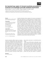
Figure 2 c(s) distributi on of apoE-(72-166) proteins in PBS with or without DHPC. The sedimentation velocity data was fitted with the
SEDFIT program using the continuous c(s) distribution model [30]. The fitted curves for apoE2-, apoE3-, and apoE4-(72-166) are shown as dotted,
dash, and solid lines, respectively. Panels A-D: proteins were in PBS, and with 5 mM, 50 mM, or 100 mM DHPC, respectively. Insets, grayscale of
the residual bit map showing the quality of data fitting.
Hsieh and Chou Journal of Biomedical Science 2011, 18:4
/>Page 5 of 9
According to the results of c(s) distribution (Figure 2),
the data were adequately described and fit ted by a three
(those in PBS and 5 mM DHPC) and two (those in
DHPC micelle) discrete species model, respectively. The
best-fit results are summarized in Table 2. The
calculated local concentration and sedimentation coeffi-
cient of each discrete species showed a similar content to
those in c(s). Most major s pecies detected in SV were
also detected in SE experi ments. In the aqueous PBS
solution, apoE -(72-166) peptides showed a major species
of dimer, tetramer (for apoE4-(72-166)) or hexamer (for
apoE2- an d apoE3-(72-166)), and la rge aggregates,
respectively, which indicated a significant polymerization.
In the 5 mM DH PC submicellar solution, the content of
each species did not show significant change, although
the hexamer of apoE2- and apoE3-(72-166) dissociated to
tetramer. It may suggest that apoE-(72-166) peptides
begin to dissociate, which is consistent with the observa-
tion by c(s). In the presence of 50 mM DHPC micelles,
all three apoE-(72-166) proteins maintained a major spe-
cies of 19-20 kDa, which may be a complex structure of a
monomeric apoE-(72-166) peptides (12 kDa) with a
smaller DHPC micelle (20 molecules, 9 kDa). As a ellip-
soid micelle with 20 DHPC molecules, the radius of gyra-
tion of the fatty acyl core region is 15.6 Å [41], whose
circumferen ce is about 100 Å, just identical to the lengt h
of apoE-(72-166) a-helical region. Besides, by SE experi-
ments, apoE-(72-166) showed a major species of dimer
(for apoE3- and apoE4-(72-166)) or tetramer (for apoE2-
(72-166)) with a larger DHPC micelle (40 molecules, 18
kDa). As a micelle with 40 DHPC molecule s, which has
surface area of 2 times, the circumference will be about
140 Å. It may result in that apoE-(72-166) peptides do
not form a complete belt around the micelle but are stag-
gered at a suitable angle to each other [42]. Similarly,
most apoE-(72-166) proteins in the presence of 100 mM
DHPC micelles were found to have a major complex spe-
cies of monomeric peptides w ith a m icelle. The peptide-
lipid complex with higher molar mass was also found by
SE experiments.
Nevertheless, our study demonstrates that DHPC may
provide a lipid or hydrophobic rich environment that
will facilitate the maintenance of a dissociated and
extended conformation for apoE-(72-166). This
tendency also positive ly correlates with the increasing
concentration of DHPC.
Protein-lipid interactions and Protein-LDLR binding of
ApoE-(72-166) Proteins
To identify and compare the lipid binding ability of the
three apoE-(72-166) peptides, we assessed the DMPC
turbidity clearance ability of apoE2-(72-166) (Additional
file 1: Figure S2). Compared with the other two isoforms
[22], apoE4-(72-166) had the highest DMPC turbidity
clearance ability. By fitting to biexponential decay model
(Eq. 3), it suggested that the rate constants of apoE4-
(72-166) in both phase were 4-13 times faster than
apoE2 and a poE3 counterparts and 99.9% turbidity was
removed, which indicated that all DMPC mLV have
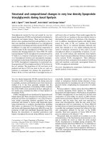
Figure 3 Global analysis of the apoE3-(72-166) proteins in PBS
(pH 7.3). The SV experiment (A) was centrifuged to 42,000 rpm
(circles) at 20°C for 4 h. The speed of centrofugation for SE
experiments (B) was 10,000 rpm (circles), 15,000 rpm (triangles), and
20,000 rpm (squares) at 20°C each for 18 h. The solid lines in A-B
are the best fit distributions from global analysis of the three
discrete species model by SEDPHAT according to eq. 4. The molar
mass and sedimentation coefficients of the species were floated
and fitted. The residuals of each fit are shown below the panels and
have a local RMSD for each channel of 0.0054 (A) and 0.0050 (B).
The discrete species distribution of apoE3-(72-166) from SV (closed
circles) and SE (open circles) are shown in C. The parameters by
best fit are shown in Table 3.
Hsieh and Chou Journal of Biomedical Science 2011, 18:4
/>Page 6 of 9
been solubilized by apoE4-( 72-166) (Additional file 1:
Table S2). Furthermore, to evaluate if apoE-(7 2-166)
peptides can bind to lipoprotein particles, the in vitro
binding experiment of apoE(-) mice VLDL with the
apoE proteins was analyzed using zone electrophoresis,
which can separate the lipoproteins by their charge [43].
In these experiments , the interaction of VLDL and apoE
proteins increased the charge of VLDL particles, result-
ing in the migration of VLDL band (lane 2 vs. lane 3-4
in Figure 4). Remarkably, the three apoE-(72-166)
proteins also showed significant VLDL shifts (lane 6 vs.
7-9 in Figure 4), which indicated that the region c on-
taining residues 72-166 was sufficient for binding VLDL.
In our previous study, we have evaluated the LDLR
binding ability of apoE3-(72-166) and apoE4-(72-166)
[22]. Here we further analyzed the LDLR binding ability
of apoE2-(72-166) peptides as a comparison with apoE3
and apoE4 counterparts (Additional file 1: Figure S3).
As previously, we employed HepG2 cells as the LDLR
carriers [22].
3
H-LDL was used as the ligand and the
apoE proteins with or without DMPC were therefore
the competitors. Overall, apoE-DMPC complex showed
better
3
H-LDL competition than apoE. Among the three
isoforms, apoE4-(72-166)-DMPC complex decreased the
3
H-LDL binding by 55%, comparing with 19% for
apoE2-(72-166)-DMPC and 26% for apoE3-(72-166)-
DMPC. At the same dose, apoE4-(72-166)-DMPC main-
tained almost identical LDLR binding ability to that of
full length apoE-DMPC, while those of apoE2- and
apoE3-(72-166) were significantly lower [22]. This indi-
cated t hat alone of the three isoforms, only apoE4-(72-
166) did not lose its LDLR binding ability. Comparing
to the apoE2 and apoE3 counterpart, apoE4-(72-166)
shows the highest lipid binding ability (Additional file 1:
Figure S2 and Table S2). The lipid association is
required for high affinity binding of apoE to the LDLR
because of the increased exposure of basic region on the
fourth a-helix after interacting with lipids [7].
Table 2 Global discrete species analysis of apoE-(72-166) with different environments
a
In
b
ApoE2-(72-166) ApoE3-(72-166) ApoE4-(72-166)
S
c
(Svedberg)
M
d
(kDa)
Local C of SV
and SE (A
280
)
e
S
c
(Svedberg)
M
d
(kDa)
Local C of SV
and SE (A
280
)
e
S
c
(Svedberg)
M
d
(kDa)
Local C of SV
and SE (A
280
)
e
PBS 2.2 19 0.07, 0.10 2.1 18 0.07, 0 2.7 24 0.02, 0
5.3 67 0.17, 0 4.8 68 0.16, 0.04 4.7 51 0.18, 0.05
7.8 186 0.05, 0.11 7.7 251 0.03, 0.06 8.6 210 0.05, 0.12
5mM
DHPC
2.3 25 0.07, 0.08 2.5 21 0.08, 0.08 2.2 22 0.01, 0
4.9 49 0.18, 0 4.9 52 0.15, 0 4.8 51 0.17, 0.07
8.2 190 0.04, 0.09 8.4 232 0.03, 0.06 8.5 217 0.04, 0.19
50 mM
DHPC
1.3 19 0.35, 0.13 1.2 20 0.34, 0 1.1 19 0.29, 0
4.3 71 0.01, 0.15 3.1 40 0.02, 0.15 3.8 45 0.03, 0.14
100 mM
DHPC
1.2 30 0.31, 0 1.0 19 0.32, 0.08 0.8 20 0.27, 0.04
3.3 49 0.05, 0.17 2.3 50 .0, 0.10 3.0 58 0.01, 0.11
a
The SV and SE experiments of apoE-(72-166) were combined and fitted to the global discrete model by SEDPHAT [33]. The best-fit local root mean square
errors of SE were from 0.0041 to 0.0118 and those of SV were from 0.0054 to 0.0088.
b
The partial specific volume was set by 0.73 in PBS and 5 mM DHPC and by 0.86 in 50 and 100 mM DHPC (see materials and methods for detail).
c, d, e
Best-fit calculated sedimentation coefficients (s), molar mass (M), and local concentrations (C) of different species were shown. The local concentration of SV
was from the discrete distribution of SV and that of SE was from the SE experiments. The concentration units were signal units (A
280
).
Figure 4 Lipoprotein electrophoresis of apoE-VLDL particles.
Various apoE proteins were incubated with apoE(-) mice serum at
37°C for 4 h, respectively. After removing the free proteins by NaBr
density ultracentrifugation, the VLDL particles were checked by zone
electrophoresis (separation by charge). Lanes 1 and 5, human serum
sample; lane 2 and 6, apoE(-) mice serum sample; lane 3-4 and 7-9,
apoE(-) mice serum incubated with full length apoE3 and apoE4,
and with apoE2-, apoE3-, and apoE4-(72-166) proteins, respectively.
The VLDL bands were shifted with the binding of apoE proteins.
Detailed procedures are described in Materials and Methods.
Hsieh and Chou Journal of Biomedical Science 2011, 18:4
/>Page 7 of 9
Conclusion
To illustrate the interaction of apoE-(72-166) peptides
with lipids, a model for apoE-(72-166) in PBS with or
without DHPC is proposed (Figure 5). ApoE-(72-166) was
found to be prone to polymerize in PBS. When apoE-(72-
166) interacts with DHPC submicelles, these DHPC mole-
cules will intercalate into its hydrophobic region causing
hydrophobic exposure. In the DHPC micellar solution,
apoE-(72-166) will dissociate and interact to a D HPC
micelle with an extended conformation. We demonstrate
herein that unlike the four a-helical bundle NT domain
which maintains a stable monomer [22], apoE-(72-166), as
a less structured peptide, may have less lateral contacts
and tend to aggreg ate in PBS, but dissociates at the exis-
tence of DHPC micelle which may stabilize back these
contacts. Besides, the truncated apoE peptides, especially
apoE4-(72-166), still displays the comparable LDLR bind-
ing and higher lipid binding abilities as to full-length apoE
[22]. Compared with a fused peptide which may have
shorter half-life [44,45], the remarkable lipid binding and
LDLR binding avidity of the apoE4-(72-166) suggests the
possible feasibility for designing a competitive peptide
against atherosclerosis or AD.
Additional material
Additional file 1: Tables S1 and S2. Figures S1-S3.
Abbreviations
1
Aβ: β-amyloid peptide; AD: Alzheimer’s disease; apoE: apolipoprotein E; CD:
circular dichroism; CT: carboxyl-terminal; DHPC: dihexanoylphosphatidylcholine;
DMPC: dimyristoylphosphatidylcholine; f
r
: frictional ratio; GdnCl: guanidinium
chloride; HDL: high-density lipoprotein; LDLR: low-density lipoprotein receptor;
Meq: equivalent molar mass; mLV: multilamellar vesicles; NFT: neurofibrillary
tangle; NRMSD: normalized root mean square deviation; NT: amino-terminal;
PBS: phosphate buffered saline; SE: sedimentation equilibrium; SV:
sedimentation velocity; VLDL: very-low-density lipoprotein
Acknowledgements
We are grateful to Prof. Sheh-Yi Sheu in the same faculty for providing the
apoE model structure. This research was supported in part by grants from
the Taiwan National Science Council (NSC 98-2320-B-010-026-MY3) and
National Health Research Institute, Taiwan (NHRI-EX99-9947SI) to CYC. We
also thank NYMU for its financial support (Aim for Top University Plan from
Ministry of Education).
Authors’ contributions
YHH carried out most experiments and helped to draft the manuscript. CYC
conceived the study, participated in experimental design, analyzed the AUC
data, and drafted and revised the manuscript. Both authors read and
approved the final manuscript.
Competing interests
The authors declare that they have no competing interests.
Received: 17 September 2010 Accepted: 10 January 2011
Published: 10 January 2011
References
1. Weisgraber KH, Rall SC Jr, Mahley RW: Human E apoprotein heterogeneity.
Cysteine-arginine interchanges in the amino acid sequence of the apo-E
isoforms. J Biol Chem 1981, 256:9077.
2. Aggerbeck LP, Wetterau JR, Weisgraber KH, Wu CS, Lindgren FT: Human
apolipoprotein E3 in aqueous solution. II. Properties of the amino- and
carboxyl-terminal domains. J Biol Chem 1988, 263:6249.
3. Wetterau JR, Aggerbeck LP, Rall SC Jr, Weisgraber KH: Human
apolipoprotein E3 in aqueous solution. I. Evidence for two structural
domains. J Biol Chem 1988, 263:6240.
4. Mahley RW: Apolipoprotein E: cholesterol transport protein with
expanding role in cell biology. Science 1988, 240:622.
5. Westerlund JA, Weisgraber KH: Discrete carboxyl-terminal segments of
apolipoprotein E mediate lipoprotein association and protein
oligomerization. J Biol Chem 1993, 268:15745.
Figure 5 Proposed model. Schematic diagram for the apoE-(72-166) peptides in PBS, 5 mM DHPC submicelles, and 50 mM DHPC micelles. The
yellow and green cylinders show the positions of residues 87-124 and 131-162, respectively.
Hsieh and Chou Journal of Biomedical Science 2011, 18:4
/>Page 8 of 9
6. Dong LM, Wilson C, Wardell MR, Simmons T, Mahley RW, Weisgraber KH,
Agard DA: Human apolipoprotein E. Role of arginine 61 in mediating the
lipoprotein preferences of the E3 and E4 isoforms. J Biol Chem 1994,
269:22358.
7. Lund-Katz S, Zaiou M, Wehrli S, Dhanasekaran P, Baldwin F, Weisgraber KH,
Phillips MC: Effects of lipid interaction on the lysine microenvironments
in apolipoprotein E. J Biol Chem 2000, 275:34459.
8. Mahley RW, Rall SC Jr: Apolipoprotein E far more than a lipid transport
protein. Annu Rev Genomics Hum Genet 2000, 1:507.
9. Strittmatter WJ, Bova Hill C: Molecular biology of apolipoprotein E. Curr
Opin Lipidol 2002, 13:119.
10. Saito H, Dhanasekaran P, Baldwin F, Weisgraber KH, Phillips MC, Lund-
Katz S: Effects of polymorphism on the lipid interaction of human
apolipoprotein E. J Biol Chem 2003, 278:40723.
11. Gregg RE, Brewer HB Jr: The role of apolipoprotein E and lipoprotein
receptors in modulating the in vivo metabolism of apolipoprotein B-
containing lipoproteins in humans. Clin Chem 1988, 34:B28.
12. Greenow K, Pearce NJ, Ramji DP: The key role of apolipoprotein E in
atherosclerosis. J Mol Med 2005, 83:329.
13. Weisgraber KH, Innerarity TL, Mahley RW: Abnormal lipoprotein receptor-
binding activity of the human E apoprotein due to cysteine-arginine
interchange at a single site. J Biol Chem 1982, 257:2518.
14. Lane RM, Farlow MR: Lipid homeostasis and apolipoprotein E in the
development and progression of Alzheimer’s disease. J Lipid Res 2005,
46:949.
15. Tanzi RE, Bertram L: Twenty years of the Alzheimer’s disease amyloid
hypothesis: a genetic perspective. Cell 2005, 120:545.
16. Huang Y, Liu XQ, Wyss-Coray T, Brecht WJ, Sanan DA, Mahley RW:
Apolipoprotein E fragments present in Alzheimer’s disease brains induce
neurofibrillary tangle-like intracellular inclusions in neurons. Proc Natl
Acad Sci USA 2001, 98:8838.
17. Strittmatter WJ, Saunders AM, Schmechel D, Pericak-Vance M, Enghild J,
Salvesen GS, Roses AD: Apolipoprotein E: high-avidity binding to beta-
amyloid and increased frequency of type 4 allele in late-onset familial
Alzheimer disease. Proc Natl Acad Sci USA 1993, 90:1977.
18. Barbier A, Clement-Collin V, Dergunov AD, Visvikis A, Siest G, Aggerbeck LP:
The structure of human apolipoprotein E2, E3 and E4 in solution 1.
Tertiary and quaternary structure. Biophys Chem 2006, 119
:158.
19. Chroni A, Pyrpassopoulos S, Thanassoulas A, Nounesis G, Zannis VI,
Stratikos E: Biophysical analysis of progressive C-terminal truncations of
human apolipoprotein E4: insights into secondary structure and
unfolding properties. Biochemistry 2008, 47:9071.
20. Clement-Collin V, Barbier A, Dergunov AD, Visvikis A, Siest G, Desmadril M,
Takahashi M, Aggerbeck LP: The structure of human apolipoprotein E2,
E3 and E4 in solution. 2. Multidomain organization correlates with the
stability of apoE structure. Biophys Chem 2006, 119:170.
21. Hatters DM, Zhong N, Rutenber E, Weisgraber KH: Amino-terminal domain
stability mediates apolipoprotein E aggregation into neurotoxic fibrils. J
Mol Biol 2006, 361:932.
22. Chou CY, Jen WP, Hsieh YH, Shiao MS, Chang GG: Structural and
functional variations in human apolipoprotein E3 and E4. J Biol Chem
2006, 281:13333.
23. Chou CY, Lin YL, Huang YC, Sheu SY, Lin TH, Tsay HJ, Chang GG, Shiao MS:
Structural variation in human apolipoprotein E3 and E4: secondary
structure, tertiary structure, and size distribution. Biophys J 2005, 88:455.
24. Chou JJ, Baber JL, Bax A: Characterization of phospholipid mixed micelles
by translational diffusion. J Biomol NMR 2004, 29:299.
25. Lin TL, Chen SH, Gabriel NE, Roberts MF: The use of small-angle neutron
scattering to determine the structure and interaction of
dihexanoylphosphatidylcholine micelles. J Am Chem Soc 1986, 108:3499.
26. Tausk RJ, Karmiggelt J, Oudshoorn C, Overbeek JT: Physical chemical
studies of short-chain lecithin homologues. I. Influence of the chain
length of the fatty acid ester and of electrolytes on the critical micelle
concentration. Biophys Chem 1974, 1:175.
27. Sreerama N, Woody RW: Estimation of protein secondary structure from
circular dichroism spectra: comparison of CONTIN, SELCON, and CDSSTR
methods with an expanded reference set. Anal Biochem 2000, 287:252.
28. Whitmore L, Wallace BA: DICHROWEB, an online server for protein
secondary structure analyses from circular dichroism spectroscopic data.
Nucleic Acids Res 2004, 32:W668.
29. Pace CN: Measuring and increasing protein stability. Trends Biotechnol
1990, 8:93.
30. Brown PH, Schuck P: Macromolecular size-and-shape distributions by
sedimentation velocity analytical ultracentrifugation. Biophys J 2006,
90:4651.
31. Schuck P: Size-distribution analysis of macromolecules by sedimentation
velocity ultracentrifugation and lamm equation modeling. Biophys J 2000,
78:1606.
32. Koynova R, Koumanov A, Tenchov B: Metastable rippled gel phase in
saturated phosphatidylcholines: calorimetric and densitometric
characterization. Biochim Biophys Acta 1996, 1285:101.
33. Schuck P: Modern Analytical Ultracentrifugation: Techniques and
Methods. In Cambridge. Edited by: Scott DJ, Harding SE, Rowe AJ. The
Royal Society of Chemistry; 2005:26-50.
34. Choy N, Raussens V, Narayanaswami V: Inter-molecular coiled-coil
formation in human apolipoprotein E C-terminal domain. J Mol Biol 2003,
334:527.
35. Pownall HJ, Massey JB, Kusserow SK, Gotto AM Jr: Kinetics of lipid–protein
interactions: interaction of apolipoprotein A-I from human plasma high
density lipoproteins with phosphatidylcholines. Biochemistry 1978,
17:1183.
36. Segall ML, Dhanasekaran P, Baldwin F, Anantharamaiah GM, Weisgraber KH,
Phillips MC, Lund-Katz S: Influence of apoE domain structure and
polymorphism on the kinetics of phospholipid vesicle solubilization. J
Lipid Res 2002, 43:1688.
37. Krempler F, Kostner GM, Friedl W, Paulweber B, Bauer H, Sandhofer F:
Lipoprotein binding to cultured human hepatoma cells. J Clin Invest
1987, 80:401.
38. Lundberg BB, Suominen LA:: Physicochemical transfer of [3H]cholesterol
from plasma lipoproteins to cultured human fibroblasts. Biochem J 1985,
228:219.
39. Wilson C, Wardell MR, Weisgraber KH, Mahley RW, Agard DA: Three-
dimensional structure of the LDL receptor-binding domain of human
apolipoprotein E. Science 1991, 252:1817.
40. Griffin MD, Mok ML, Wilson LM, Pham CL, Waddington LJ, Perugini MA,
Howlett GJ: Phospholipid interaction induces molecular-level
polymorphism in apolipoprotein C-II amyloid fibrils via alternative
assembly pathways. J Mol Biol 2008, 375:240.
41. Bockmann RA, Caflisch A: Spontaneous formation of detergent micelles
around the outer membrane protein OmpX. Biophys J 2005, 88:3191.
42. Peters-Libeu CA, Newhouse Y, Hatters DM, Weisgraber KH: Model of
biologically active apolipoprotein E bound to
dipalmitoylphosphatidylcholine. J Biol Chem 2006, 281:1073.
43. Campos E, Fievet P, Caces E, Fruchart JC, Fievet C: A screening method for
abnormally high lipoprotein(a) concentrations by agarose lipoprotein
electrophoresis. Clin Chim Acta 1994, 230:43.
44. Datta G, Garber DW, Chung BH, Chaddha M, Dashti N, Bradley WA,
Gianturco SH, Anantharamaiah GM: Cationic domain 141-150 of apoE
covalently linked to a class A amphipathic helix enhances atherogenic
lipoprotein metabolism in vitro and in vivo. J Lipid Res 2001, 42:959.
45. Gupta H, White CR, Handattu S, Garber DW, Datta G, Chaddha M, Dai L,
Gianturco SH, Bradley WA, Anantharamaiah GM: Apolipoprotein E mimetic
Peptide dramatically lowers plasma cholesterol and restores endothelial
function in watanabe heritable hyperlipidemic rabbits. Circulation 2005,
111:3112.
46. DeLano WL: The Pymol manual. San Carlos, CA: DeLano Scientific; 2002.
doi:10.1186/1423-0127-18-4
Cite this article as: Hsieh and Chou: Structural and functional
characterization of human apolipoprotein E 72-166 peptides in
both aqueous and lipid environments. Journal of Biomedical Science 2011
18:4.
Hsieh and Chou Journal of Biomedical Science 2011, 18:4
/>Page 9 of 9