Báo cáo y học: " Mitochondrial targeting of human NADH dehydrogenase (ubiquinone) flavoprotein 2 (NDUFV2) and its association with early-onset hypertrophic cardiomyopathy and encephalopathy" ppt
Bạn đang xem bản rút gọn của tài liệu. Xem và tải ngay bản đầy đủ của tài liệu tại đây (2.21 MB, 17 trang )
RESEARCH Open Access
Mitochondrial targeting of human NADH
dehydrogenase (ubiquinone) flavoprotein
2 (NDUFV2) and its association with early-onset
hypertrophic cardiomyopathy and
encephalopathy
Hsin-Yu Liu, Pin-Chao Liao , Kai-Tun Chuang and Mou-Chieh Kao
*
Abstract
Background: NADH dehydrogenase (ubiquinone) flavoprotein 2 (NDUFV2), containing one iron sulfur cluster ([2Fe-
2S] binuclear cluster N1a), is one of the core nuclear-encoded subunits existing in human mitochondrial complex I.
Defects in this subunit have been associated with Parkinson’s disease, Alzheimer’s disease, Bipolar disorder, and
Schizophrenia. The aim of this study is to examine the mitochondrial targeting of NDUFV2 and dissect the
pathogenetic mechanism of one human deletion mutation present in patients with early-onset hypertrophic
cardiomyopathy and encephalopathy.
Methods: A series of deletion and point-mutated constructs with the c-myc epitope tag were generated to
identify the location and sequence features of mitochondrial targeting sequence for NDUFV2 in human cells using
the confocal microscopy. In addition, various lengths of the NDUFV2 N-terminal and C-terminal fragments were
fused with enhanced green fluorescent protein to investig ate the minimal region required for correct
mitochondrial import. Finally, a deletion construct that mimicked the IVS2+5_+8delGTAA mutation in NDUFV2 gene
and would eventually produce a shortened NDUFV2 lacking 19-40 residues was generated to explore the
connection between human gene mutation and disease.
Results: We identified that the cleavage site of NDUFV2 was located around amino acid 32 of the precursor
protein, and the first 22 residues of NDUFV2 were enough to function as an efficient mitochondrial targeting
sequence to carry the passenger protein into mitochondria. A site-directed mutagen esis study showed that none
of the single-point mutations derived from basic, hydroxylated and hydrophobic residues in the NDUFV2
presequence had a significant effect on mitochondrial targeting, while increasing number of mutations in basic
and hydrophobic residues gradually decreased the mitochondrial import efficacy of the protein. The deletion
mutant mimicking the human early-onset hypertrophic cardiomyopathy and encephalopathy lacked 19-40 residues
in NDUFV2 and exhibited a significant reduction in its mitochondrial targeting ability.
Conclusions: The mitochondrial targeting sequ ence of NDUFV2 is located at the N-terminus of the precursor
protein. Maintaining a net positive charge and an amphiphilic structure with the overall balance and distribution of
basic and hydrophobic amino acids in the N-terminus of NDUFV2 is important for mitochondrial targeting. The
results of human disease cell model established that the impairment of mitochondrial localization of NDUFV2 as a
mechanistic basis for early-onset hypertrophic cardiomyopathy and encephalopathy.
* Correspondence:
Institute of Molecular Medicine & Department of Life Science, National Tsing
Hua University, 101, Sec. 2, Kuang-Fu Rd., Hsinchu 30013, Taiwan, R.O.C
Liu et al. Journal of Biomedical Science 2011, 18:29
/>© 2011 Liu et al; licensee BioMed Central Ltd. This is an Open Access article distributed under the terms of the Creative Commons
Attribution License ( which permits unrestricted use, distribution, and reproduction in
any medium, provided the original work is prop erly cited.
Background
Mammalian NADH:ubiquinone oxidoreductase (com-
plex I) (EC 1.6.5.3) is the first, largest and most compli-
cated respiratory complex in mitochondria [1]. It is one
of the electrons entry sites in the oxidative phosphoryla-
tion system (OXPHOS), and catalyzes NADH oxidation,
followed by transferring two electrons to ubiquinone [2].
To date, 45 different subunits have been identified in
bovine heart mitochondrial complex I [3,4]. Among
them, seven subunits of complex I, including ND1-6
and ND4L, are encoded by mitochondrial DNA, and the
others are encoded by nuclear DNA [5]. In contrast,
bacterial complex I (also called NDH-1) is much sim-
pler. It contains only 13-14 unlike subunits [6]. These
subunits of bacterial origins are conserved in mitochon-
drial complex I and considered as the “ minimal” struc-
ture required for correct function. The two recently
published crystal structures of the complete complex I
from prokaryote Thermus thermophilus and eukaryote
Yarrowia lipolytica indicated that this enzyme complex
is L-shaped and separated into two arms: a hydrophobic
arm embedded in the periplasm/the inner membrane
and a hydrophilic arm protruding into the cytoplasm/
the matrix [7,8]. The bacterial complex I possesses nine
Fe-S clusters, including two [2Fe-2S] clusters (N1a and
N1b) and seven [4Fe-4S] clusters (N3, N4, N5, N6a,
N6b, N7 and N2), to manage the passage of two elec-
trons [9]. According to the T. thermophilus model, the
main pathway for electron transfer i n complex I is
NADH- FMN- N3- N1b- N4- N5- N6a- N6b- N2- qui-
nine [10,11].
Human NADH dehydrogenase (ubiquinone) flavo-
protein 2 (NDUFV2) subunit, also called 24-kDa, is
one of the complex I core subunits which are very
conserved from bacteria to mammals [12]. The
NDUFV2 gene has been cloned and assigned to human
chromosome 18p11.31-p11.2 [13]. The entire gene
spans approximately 20 kb and contains 8 exons, a nd
the expressed protein is homologous to 24-kDa of Bos-
taurus and Neurospora crassa [14], NuoE of Escheri-
chia coli [15] and Rhodobacter capsulatus [16], NQO2
of Paracoccus denitrificans [17] and T. thermophilus
[18], and NUHM of Y. lipolytica [19]. Human
NDUFV2 contains a binuclear [2Fe-2S] cluster c alled
N1a. This iron-sulfur clust er has a binding motif, Cys-
(X)
4
-Cys-(X)
35
-Cys-(X)
3
-Cys, which is very conserved
among orthologues [20]. Based on the crystal structure
of the hydrophilic domain fr om T. thermophilus com-
plex I, cluster N1a can accept electrons from FMN,
but is unable to pass them to cluster N3, which is too
far away from N1a [10,11]. One hypothesis suggests
that cluster N1a may act as an antioxidant to accept
the excessive electrons to prevent the generation of
reactive oxygen species (ROS) [10,11].
The fungus N. crassa is an eukaryotic organism which
is frequentl y used as a model to study the structure and
function of complex I [21]. In t he N. crassa studies, it
was found that the lacking of 24-kDa subunit would
reduce the levels of 51-kDa subunit (a homologous of
human NDUFV1) and affect the NADH:ferricyanide
reductase activity, suggesting that the 24-kDa subunit is
essential for a proper assembly of 51 kDa subunit and
complex I activity [14]. This phenotype may explain
why the deficiency of NDUFV2 subunit has been asso-
ciated with some neurodegenerative diseases, including
Parkinson disease [22], Alzheimer’sdisease[23],Bipolar
disorder, and Schizophrenia [24,25].
Most nuclear DNA-encoded mitochondrial proteins,
including NDUFV2, are synthesized in the cytosol on
free ribosomes as a precursor protein which carries a
mitochondrial targeting sequence (MTS) for c orrect
import. These mitochondrial preproteins are then trans-
ported into or across mitochondrial membranes with
the help of several distinct complexes, including the
translocase of outer membrane (TOM) complex and the
translocase of inner membrane (TIM) complex [26,27].
The final location of the protein will be determined by
the combined actions of the involved translocation path-
way and the targeting message encoded within the p ro-
tein. For most proteins targeted to the mitochondrial
matrix and some of those destined for the intermem-
brane space and the inner membrane, a cleavable exten-
sion is frequently present in the N-terminus of the
precursor protein. This sequence contains about 10-80
amino acid residues that have a high content of basic,
hydrophobic and hydroxylated amino acids but a lack of
negatively charged amino acids [28]. The positive resi-
dues are considered to play an important role in mito-
chondrial t argeting, and are thought to assist the MTS
across the inner membrane driving by the membrane
potential. Having the potential to form amphiphilic a-
helices is another common feature that is proposed for
receptor recognition. The molecular structure of a gen-
eral import receptor TOM20 interacting with a mito-
chondrial presequence suggests the importance of the
amphiphilic a-helical structure and the involvement of
hydrophobic residues in binding to this mitochondrial
import receptor [29]. However, the result from an
import study based on several artificial presequences
fused with a passenger protein suggested that amphiphi-
lic ity is necessary for mitochondrial impor t but forming
a helical structure may not be essential [30]. Except
these characteristics, there is no sequence identity
shared between MTSs, even between closely related
orthologs. Most of the N-terminal MTSs are cleaved
from precursors by the mitochondrial processing pepti-
dase (MPP) in o ne step, some others are processed
sequentially by MPP and the mitochondrial intermediate
Liu et al. Journal of Biomedical Science 2011, 18:29
/>Page 2 of 17
peptidase (MIP) in a two-step reaction [28]. Infre-
quently, the MTS can be found to be present at the C-
terminus.
The mature 24-kDa of complex I has been purified
from bovine heart and the primary structure of this pro-
tein has been partially determined [31]. According to
the complementary DNA (cDNA) sequence of NDUFV2
and its close relatedness with the bovine sequence, the
possible human precursor and mature sequences of
NDUFV2 subunit were predicted [32]. In a recent
report, a 4-bp deletion in intron 2 (IVS2+5_+8delG-
TAA) in the NDUFV2 gene has been shown to associate
with patients with early-onset hypertrophic cardiomyo-
pathy and en cephalopathy [33]. This muta tion altered
the splicing donor site and caused the exon 2 missing in
the mRNA of NDUFV2. The truncated RNA transcript
is predicted to encode a shorter protein not only lacking
part of the MTS but also losing the cleavage-processing
site. Biochemical analyses indic ated that patients with
this mutation had a 70% reduction in the amount of
NDUFV2 protein and a significant complex I deficiency
[33]. S cientists have tried to simulate this exon 2 skip-
ping mutation by deleting the corresponding region of
orthologous NUHM gene in the obligate aerobic yeast
Y. lipolytica [19]. Surprisingly, the results showed that
this mutant was indistinguishable from normal cells in
activity, inhibit or sensi tivity and EPR signals of complex
I in this yeast model.
The mitochondrial targeting of N DUFV2 has not been
experimentally established. In the present study, a series
of N-terminal truncated, C-terminal truncated and
point-mutated constructs with the c-myc epitope tag
were generated to identify the location and sequence
features of MTS for NDUFV2 in human cells. In addi-
tion, various lengths of the NDUFV2 N-terminus and
C-terminus were fused with enhanced green fluorescent
protein (EGFP) to i nvestigate the minimal functional
region required for c orrect mitochondrial import.
Finally, a deletion constructthatmimicstheIVS2+5_
+8delGTAA mutation and would produce a shortened
precursor protein lacking 19-40 residues in NDUFV2
was generated to dissect the pathogenetic mechanism of
this mutation.
Methods
Cell and bacterial culture
T-REx-293 cells (Invitrogen, Carlsbad, CA, USA),
human embryonic kidney cells with the t etracycline-
regulated expression system, were cultured at 37°C and
5% CO
2
with saturating humi dity in Dulbeccos modified
Eagle media (DMEM) which contained 10% fetal bovine
serum (FBS), 100 U/ml penicillin and 100 μg/ml strep-
tomycin. Escherichia coli DH5a strain and Top10F’
strain were used for gene cloning, and the bacteria were
grown i n Luria Bertani (LB) media or on LB agar plates
containing ampicillin (100 μg/ml) at 37°C.
Plasmid construction
Construction of plasmids expressing full-length NDUFV2
proteins
The Mammalian Gene Collection (MGC) cDNA clone
encoding human NDUFV2 (accession numbers
NM_021074, clone number: MGC-15943, IMAGE:
3537815) was obtained from the I.M.A.G.E Consortium.
The derived plasmid was used as the template for ampli-
fication by polymerase chain reaction (PCR) using Pfu
DNA polymerase. The sequences of primers used are
shown in Addit ional file 1-(1). The resultant fragment
was then cloned into the pGEM-T vector (Promega,
Madison, WI, USA) and the sequence was confirmed by
sequencing. The resulting plasmid was digested with
EcoRI/XhoI, a nd the DNA fragm ent containing th e
desired cDNA was then purified and ligated with the
pcDNA4/TO/myc-His A vector (Invitrogen) using the
same restriction sites to generate th e pcDNA4-NDUFV2
expressing vector.
Construction of plasmids expressing truncated NDUFV2
proteins
The pcDNA4-NDUFV2 vector was used as the template
for generation of i ts N-terminal deletion constructs
(pcDNA4-△1-18 NDUFV2, pcDNA4-△1-32 NDUFV2
and p cDNA4-△1-50 NDUFV2) and C-terminal deletion
constructs ( pcDNA4-△183-249 NDUFV2 and pcDNA4-
△198-249 NDUFV2). The sequences of primers used are
shown in Additional file 1-(2). In addition, the construct
(named pcDNA4-△19-40 NDUFV2) which mimics the
human pathogenic IVS2+5_+8delGTAA mutation in
NDUFV2 gene in patients with hypertrophic cardiomyo-
pathy and encephalomyopathy was generated with the
primers shown in Additional file 1-(3).
Construction of plasmids expressing various lengths of
NDUFV2-EGFP
Using the pcDNA4-NDUFV2 plasmid as the template,
various DNA fragments encoding different N-terminal
proteins of NDUFV2 were designed and generated to
fuse with EGFP gene in the pEGFP-N3 expression vec-
tor (Clontech Laboratories, Mountain view, CA, USA).
The restriction enzyme sites used for this purpose were
XhoIandEcoRI. These resulting constructs included
NDUFV2 full-length (pEGFP-N3 NDUFV2
1-249
),
pEGFP-N3 NDUFV2
1-32
, pEGFP-N3 NDUFV2
1-22
,
pEGFP-N3 NDUFV2
1-21
, pEGFP-N3 NDUFV2
1-20
and
pEGFP-N3 NDUFV2
1-18
. The sequences of primers used
are shown in Additional file 1-(4).
Construction of plasmids expressing NDUFV2 missense
mutants
ThepcDNA4-NDUFV2wasusedasthetemplatefor
introduction of missense mutations on basic, hydroxylated
Liu et al. Journal of Biomedical Science 2011, 18:29
/>Page 3 of 17
and hydrophobic residues in the first 1-32 amino acids of
NDUFV2 using the site-directed mutagenesis methodol-
ogy based on the QuickChange manual (Stratagene, La
Jolla, CA, USA). All of the used primers are shown in
Additional file 1-(5, 6, 7).
Transient transfection and immunofluorescent staining
T-REx-293 cells were seeded in 24-well plates containing
cover glasses. When cell growth reached approximately
60-70% confluency, TransIT-LT1 transfection Reagent
(Mirus, Madison, WI, USA) pre-mixing with the desired
plasmid was introduced for transfection. After 24-h incu-
bation, the culture medium was removed and the fresh
medium containing tetracycline to a final concentration
of 0.5 μg/ml was added to the cell culture. Following 24 h
of tetracycline induction at 37°C, cells were incubated
with the growth medium containing 100 nM Mito
Tracker Red (CMX-Ros; Molecular probe, Eugene, USA)
for 30 min, followed by washing once in the phosphate-
buffered saline (PBS) buffer. Next, cells were permeated
and fixed with the acetone and methanol mixture (acet-
one: methanol = 3: 1 in volume proportion) for 5 min on
ice. After fixation, cells were first incubated with growth
media at room temperature for 2 h and then with diluted
monoclonal mouse anti-c-myc antibody (Calbiochem,
1:100 dilution) at room temperature for 1 h. After 5
times of washing with the PBS buffer, the cells were incu-
bated with goat anti-mouse IgG-FITC (Invitrogen, 1:100
dilution)atroomtemperatureforanother1h,and
washed again by the PBS buffer. Finally, the cover glass
was mounted with the V ECTASHIELD Mounting Med-
ium (Vector Laboratories, Burlingame, CA, USA). When
the EGFP fusion constructs were applied for analyses,
cells were fixed with 4 % paraformaldehyde in P BS for 15
min at r oom temperature and then permeabilized with
0.5 ml methanol for 5 min on ice. The f ollowing steps
were executed as the procedure described for staining
with antibodies. Immunofluorescence was visualized by
the LSM510 laser scanning confocal microscope (Carl
Zeiss, Oberkochen, Germany) using excitation and emis-
sion filters at 488 and 510 nm, respectively, for the FITC
or EGFP signal, and 543 and 565 nm, respectively, for the
Mito Tracker Red signal. The resulting images were
merged for evaluation of co-localization. For assessing
the efficiency of mitochondrial targeting, fusio n protein
(EG FP-fused or c-myc-tagged) import into mitochondria
was monitored by confocal microscopy in at least 50
fusion protein-expressing cells, and quantified as the
ratio of the number of cells in which the fusion protein
was co-localized with mitochondria (labelled with Mito
Tracker Red) relative to the total number of fusion pro-
tein-expressing cells. For each construct, the confocal
image analysis was per form ed in three separate transfec-
tion experiments.
Western blotting analyses and antibodies
For Western blotting analyses, T-REx-293 cells were
transfected with the desired plasmids as described
above. Cells with tetracycline induction were collected
by trypsinization and centrifuged with 1000 × g force
for 5 min at 4°C. The pellet was washed once and cen-
trifuged at the same conditions for another 5 min. The
collected p ellet was then suspended with the lysis buffer
(0.15 M NaCl, 5 mM EDTA pH 8, 1% T riton-X 100, 10
mM Tris -Cl, pH 7.4) for 20 min on ice and centrifuged
at 12000 × g for 10 min at 4°C. The supernatant was
transferred to a new eppendorf tube and the protein
concentration was determined by the BCA protein assay
kit (Thermo Scientific, Rockford, IL, USA). The 4× pro-
tein loading dye was then added to the supernatant and
the resulting mixture was boiled for 5 m in. Next, the
proteins were separated by 10 or 15% SDS-PAGE
(sodium dodecyl sulphate polyacrylamide gel electro-
phoresis) and transferred onto a polyvinylidene fluoride
(PVDF) membrane at 350 mA constant current for 90
min. The membrane was then blockin g with 5% skin
milk in the PBS buffer at room temperature for 90 min
and incubated with the diluted primary antibody at
room temperature for 1 h. After three times of PBS
washing for 10 min each, the membrane was then incu-
bated with a proper secondary antibody at room tem-
perature for 1 h, and followed by several washes using
the PBS buffer. Finally, the enhanced chemilumines-
cence (ECL) system (PerkinElmer, Walcham, MA, USA)
was applied for detection. The primary antibody used in
this study included monoclonal mouse anti-c-myc anti-
body (Calbiochem, San Diego, CA, USA), monoclonal
mouse anti-b-tubulin antibody (Santa Cruz Biotechnol-
ogy, Santa Cruz, CA, USA), monoclonal mouse anti-b-
actin antibody, (Novus Biologicals, Littleton, CO, USA)
and monoclonal mouse anti-ATP synthase subunit a
antibody (Invitrogen). The secondary antibody included
goat anti-mouse IgG-HRP (Invitrogen).
Subcellular fractionation
Subcellular fractio nation of cells to separate mitochon-
drial and cytos olic fractions was conducted according to
a published differential centrifugation method with
some modifications [34]. T- REx-293 cells collected fr om
three 10-cm culture dishes with trypsination were
washed once with the PBS buffer and then resuspended
in 1 ml hypotonic buffer (10 mM HEPES, 1 mM
KH
2
PO
4
,10mMNaCl,5mMNaHCO
3
, 1 mM CaCl
2
,
0.5 mM MgCl
2
and 5 mM EDTA). After incubation on
ice for 5 min to promote hypotonic swelling, cells were
homogenized by 30 up-and-down strokes with a glass
homogenizer, followed by the addit ion of 100 μl2.5M
sucrose to prevent organelles of cells from bursting. The
homogenate was centrifuged at 1000 × g for 10 minutes
Liu et al. Journal of Biomedical Science 2011, 18:29
/>Page 4 of 17
at 4°C and the collected supernatant was transferred to
a clean chilled tube for further centrifugation at 12000 ×
g for 15 min at 4°C. The supernatant representing the
cytosolic fraction was collected without any treatment
and stored at -20°C for later analyses. The pellet was
washed once with the mitochondrial isolation buffer
(250 mM suc rose, 0.1 mM EGTA and 20 mM HEPES,
pH 7.4). The resulting pellet representing the mitochon-
drial fracti on was finally resuspended in 40 μlPBScon-
taining 0.1% SDS.
Results
The MTS of NDUFV2 was located at the N-terminus of the
protein
NDUFV2 is a nuclear-encoded mitochondrial protein
which is assembled into the L-shaped complex I and is
localized in the hydrophilic arm protruding into the
matrix. Therefore, this protein is expected to be
imported into mitochondria t hrough a pathway specific
for mitochon drial matrix proteins. Analyses of this pro-
tein by MitoProt II [35] suggested a 99.6% probability of
mitochondrial targeting of NDUFV2. A very similar
result was also obtained from th e prediction from the
TargetP server [36]. Protein sequence alignment of
NDUFV2 from various species revealed that the proteins
from eukaryotic species have a non-conserved region
located at the N-terminus (Figure 1a). It has been pre-
dicted that the first 32 amino acids of NDUFV2 may be
the MTS of this protein [32]. To test this prediction,
full-length, various N-terminal and C-terminal deletion
constructs were generated to determine the location and
orientation of MTS in NDUFV2 (Figure 2a). A c-myc
epitope tag was appended to the C-terminus of these
constructs to facilitate detection and analysis by the
immunofluorescent staining method. All of the designed
constructs were successfully engineered from the
NDUFV2 cDNA and conf irmed by direct sequenc ing.
After transient transfection, mouse anti-c-myc antibody
was applied to detect the expressed proteins, and the
Mito Tracker Red dye was used to mark the mitochon-
dria in T-REx-293 cells. The results showed that the
full-length NDUFV2 construct had a punctuated cytoso-
lic staining pattern that was typically observed when
mitochondria were immunostained, indicating applicable
of th e experimental strategy. When the C-terminal dele-
tion constructs (pcDNA4-△183-249 NDUFV2 and
pcDNA4-△1 98-249 NDUFV2) were individually trans-
fected into T-REx-293 cells, both of the truncated pro-
teins were still colocalized with mitochondria. However,
N-terminal truncations of NDUFV2 including △1-18
NDUFV2, △1-32 NDUFV2 and △1-50 NDUFV2, all lost
their mitochondrial localization (Figure 2b). These
observations agree well with the suggestion from protein
sequence alignment and the protein domain prediction
programs, and indicate that the MTS of NDUFV2 is
located at the N-terminus of the precursor protein.
NDUFV2 was processed in vivo by proteolytic removal of
the N-terminal MTS at a cleavage site around amino acid
residue 32
Most of the N-terminal presequences of mitochondrial
matrix proteins are cleavable, primarily through the
actions of MPP [28]. Typically, a single cleavage by MPP
is sufficient for the maturation of most matrix protein
precursors. However, when a not very well-defined octa-
peptide-containing precursor appears, two sequential
cleavages carried out by MPP and MIP may occur (Fig-
ure 3a) (24). To determine whether NDUFV2 is pro-
cessed by matrix proteases and estimate the
approximate cleavagesiteofthisproteinin vivo ,the
full-length construct and three constructs encoding
NDUFV2 suffering from an N-terminal truncation of a
different length (△1-18 NDUFV2, △1-32 NDUFV2 and
△1-50 NDUFV2) were transiently expressed in T-REx-
293 cells and the sizes of these recombinant proteins
were determined by Western blotting with the mouse
anti-c-myc antibody (Figure 3b). The slower migration
of the △1-18 NDUFV2 mutant protein t han the wild-
type NDUFV2 indicates that that the region containing
the first 18 amino acids of NDUFV2 is essential for
mitochondrial targeting of NDUFV2 and its subsequent
proper processing. In contrast, the deletion mutant lack-
ing the first 50 amino acids (△1-50 NDUF V2) was smal-
ler than the natively processed NDUFV2, indicating that
the native cleavage site must be in a position within the
first 50 residues. Finally, the truncated NDUFV2 protein
lacking the first 32 amino acids (△1-32 NDUFV2) had a
similar migration rate with that of the natively processed
NDUFV2. This finding strongly suggests that the final
cleavage site for generation of the mature NDUFV2 pro-
tein is most likely located around residue 32 from the
N-terminus of the precursor protein. It has to be specifi-
cally noted that the amount of both △1-18 and △1-32
NDUFV2 mutant proteins appe ars less when compared
with that of the mature NDUFV2 or t he △ 1-50
NDUFV2 mutant protein, indicating these two mutant
proteins are less stable.
The cleavage site for MPP is usually indicated by an
arginine residue at position -2 relative to the cleavage
site, which is -10 relative to the amino terminus of the
mature protein. To evaluate the involvement of this resi-
due i n NDUFV2 cleavage, we substituted the -10 argi-
nine with alanine (i.e. R23A mutation) in the
presequence of NDUFV2, and investigated the status of
its processing in the mitochondrial fraction. As shown
in Figure 3c, only one band with a similar intensity and
size to that of the wild-type, mature NDUFV 2 was pre-
sent in the mitochondrial fraction. This result suggested
Liu et al. Journal of Biomedical Science 2011, 18:29
/>Page 5 of 17
that mutation of the -1 0 argini ne alone in the precursor
has little effect on the formation of mature NDUFV2.
The first 22 amino acids in the N-terminal sequence of
NDUFV2 were essential and efficient for mitochondrial
targeting
After identification of the MTS in NDUFV2 as well as
theprobablecleavagesiteof the protein, this study
attempted to define the minimal region required for
mitochondrial targeting. A series of chimeric constructs
for expression of NDUFV2 MTS-EGFP fusion protein
were generated (Figure 4a). As shown in Figure 4b,
EGFP along without any targeting sequence addition
was present throughout the c ell, with some accumula-
tion in the nucleus. On the other hand, EGFP fused
with the full-length NDUFV2
1-249
or the newly identified
MTS (NDUFV2
1-32
) colocalized very we ll with that of
the Mito Tr acker Red dye, indicating that the first 32
amino acid acids in the N-terminus of NDUFV2 had a
mitochondrial targeting ability comparable to that of th e
full-length NDUFV2. It was interesting to observe that
the protein fragment containing the first 22 amino acid
residues of NDUFV2 was sufficient to carry most (if not
all) of the EGFP into mitochondria successfully, whereas
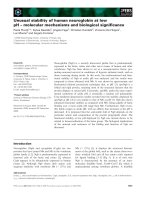
Figure 1 Sequence comparison and secondary structure analysis of the N-terminal region of NDUFV2. (a) Multiple sequence alignment
of NDUFV2 proteins from different species. Sequence alignment was generated by EMBL-EBI Clustal W2 [47] and displayed by BOXSHADE server
[48]. The abbreviations used are: H. sapiens, Homo sapiens NDUFV2 (UniProt: P19404); B. Taurus, Bos taurus 24 kDa (UniProt: P04394); N. crassa,
Neurospora crassa NUO-24 (UniProt: P40915); Y. lipolytica, Yarrowia lipolytica NUHM (UniProt: Q9UUT9); P. denitrificans, Paracoccus denitrificans
NQO2 (UniProt: P29914); T. thermophilus, Thermus thermophilus Nqo2 (UniProt: Q56221); E. coli, Escherichia coli strain K12 NuoE (UniProt: P0AFD1).
Residues identical to the consensus are highlighted in reversed-out lettering on a black background; residues not identical but similar to the
consensus are shown on a grey-shaded background. (b) The secondary structure prediction of wild-type and NDUFV2 IVS2+5_+8delGTAA
disease mutant. Secondary structure of the N-terminal region of NDUFV2 was predicted by the PSIPRED server [38]. H, a-helix; C, coil; E, strand.
Liu et al. Journal of Biomedical Science 2011, 18:29
/>Page 6 of 17
the regions containing either the first 21 (NDUFV2
1-21
-EGFP) or 20 (NDUFV2
1-20
-EGFP) amino acid residues
in the N-terminal sequence of NDUFV2 showed a much
lower efficiency. When the first 18 amino acid residues
were used as the signal peptide, the majority of mito-
chondrial targeting ability of this hybrid protein was lost
(Figure 4b). The NDUFV2
8-22
-EGFP was also incapable
of targeting to mitochondria. Together, these results
indicate that the entire 1-22 residues are necessary for
mitochondria targeting of NDUFV2.
Moreover, when the N-terminal 22 resid ues of
NDUFV2 were moved to the C-terminus of EGFP, the
mitochondrial targeting c apability of this newly identi-
fied MTS functional region was completely lost (Figure
4b). This result implies that t he MTS of NDUFV2 is
directional and needs to be located at the N-terminus of
NDUFV2 to be functional.
Effects of basic residue and hydrophobic residue
mutations in NDUFV2 MTS on mitochondrial targeting
As shown in Figure 1a, the first 1-32 amino acids which
we just demonstrated to function as the MTS have a net
positive charge (contributing by 4 arginines, 1 lysine, 3 his-
tidines, and the N-term inal methionine) but no acid ic
amino acids. Based on the Eisenberg method of hydropho-
bic moment calculation with Hmoment server [37], the
MTS of NDUFV2 had a hydrophobic region roughly in
the middle of the presequence. The secondary structure
prediction using PSIPRED server [38] indicated that the
first1-32residuesofNDUFV2containtwoa-helical
structures (one in residues 4-16, the other in residues 22-
30) with one short coil structure in bet ween (Figure 1b).
When Helical Wheel Projections program [39] was
applied to construct the a-helical wheel model for the N-
terminus of NDUFV2, it was clear that the N-terminal
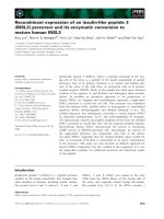
Figure 2 Effects of NDUFV2 N-terminal and C-terminal truncation on mitochondrial targeting of the protein.(a)Theconstructs
generated to express full-length and truncated NDUFV2 proteins. Full-length NDUFV2 (A), N-terminal truncation (B, △1-18 NDUFV2; C, △1-32
NDUFV2; D, △1-50 NDUFV2) and C-terminal truncation (E, △198-249 NDUFV2; F, △183-249 NDUFV2) were fused with c-myc epitope tag, and
expressed in T-REx-293 cells. The number of (+) symbols indicates that the proportion of cells exhibiting FITC fluorescence have a typical
punctuated staining pattern and mitochondrial colocalization in (b). The (++++) symbol indicates all of the FITC fluorescence signals in
transfected cells are fully colocalized with mitochondria. The (-) symbol indicates that there is no cell producing FITC fluorescence within the
mitochondrial compartment. (b) The distribution of c-myc fusion proteins was detected by anti-c-myc-FITC antibody (green color) and
mitochondria were labeled by Mito Tracker Red (red color). Only merged images are shown (colocalization of expressed protein and
mitochondria is indicated by yellow signals). Photos A-F are corresponding to constructs A-F shown in (a). Scale bars = 10 μm.
Liu et al. Journal of Biomedical Science 2011, 18:29
/>Page 7 of 17
region of NDUFV2 contains a t ypical amphiphilic struc-
ture with hydrophobic residues on one side and polar resi-
dues on the other side of the a-helix (Figure 5).
To examine the effect of basic, hydrophobic and
hydroxylated residues within the N-terminal region of
NDUFV2 on mitochondrial targeting, a site-directed
mutagenesis methodology was applied systematically on
these three groups of residues. The positively charged
arginine, l ysine and histidine residues were changed to
non-charged residues, hydrophobic residues were
replaced with hydrophilic residues and hydroxylated
residues were substituted with residues without a hydro-
xyl group. The N-terminal 1-32 amino acids of
NDUFV2 contain eight basic residues, including Arg8,
Arg10, His17, Arg20, His21, Arg23, His26 and Lys27
(Figure 6a). Surprisingly, none of the substitutions at
each individual basic amino acid residue affected the
mitochondrial targeting function of the protein (data
not shown). When three arginine residues (Arg8, Arg10
and Arg20) and one histidine (His17) w ere mutated at
the same time to generate a quadruple mutant (Figure
6b), the resulting protein still yielded a mitochondrial
localization pattern indistinguishable from that of the
wild-type NDUFV2. However, when the fifth amino acid
substitution (H21A) was introduced into the quadruple
mutant, a slight reduction in the mitochondrial targeting
was observed in the resulting protein (Figure 6b). With
the introduction of increasing number of mutations in
the basic residues, the resulting mutant gradually lost its
capability of mitochondrial import. When all of the
eight basi c residues were mutated at the same time (the
R8G+R10A+H17A+R20A+H21A+R23A+H26A+K27A
octuple mutant), the ability of mitochondrial targeting of
the protein was almost completely destroyed (Figure 6b).
To further confirm the result obtained f rom confocal
images, the strategy of subcellular fractionation, followed
with quantitative analyses by Western blots was also
applied on several mutants with a single-pointed muta-
tion or multiple-pointed mutations on the basic resi-
dues. As shown in Figure 6c, the quantitative signals for
the single-pointed mutant (R23A), quintuple mutant
(R8G+R10A+H17A+R20A+H21A) and sextuple mutant
(R8G+R10A+H17A+R20A+H21A+R23A) were 92%, 74%
and 2 2%, respectively, of those of the wild-ty pe T-REx-
293 cell. This result is corresponding very well with the
data derived from aforementioned confocal image ana-
lyses. Interestingly, when the same mutagenesis
approach was applied to investigate the role of hydro-
phobic residues in the MTS of NDUFV2, a similar phe-
nomenon was observed. Eight hydrophobic residues in
total, inc luding Phe2, Phe3, Leu7, Leu14, Trp18, Val22,
Leu25 and Ala29 (Figure 7a), were selected for mutation
to evaluate the effects of these changes on mitochon-
drial import but all of the single-point mutants showed
an import efficiency comparable to that of the wild-type
NDUFV2 (data not shown). A clear deficiency in mito-
chondrial targeting of these mutants was started to be
observed when five hydrophobic residues in NDUFV2
N-terminus were mutated (the L7Q+L14Q+V22G+
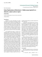
Figure 3 Cleavage of the presequ ence occurs around residue
32 in the N-terminal region of NDUFV2. (a) Two possible
mitochondrial processing sites of NDUFV2 were predicted by the
TargetP server [36]. The diagram shows a part of the N-terminal
sequence of NDUFV2 (residues 17-51), with the MPP and MIP
consensus cleavage sequence, R-10 motif (xRx↓(F/L/I)xx(S/T/G)xxxx↓),
above it. The arrows indicate the expected MPP and MIP cleavage
sites on NDUFV2. (b) The cleavage site of NDUFV2 in vivo is located
around amino acid residue 32. Lanes 1-5, the total cell lysates of T-
REx-293 transfected with the c-myc-tagged full-length NDUFV2
(lanes 1 and 5) and the c-myc -tagged NDUFV2 lacking the first 18,
32, and 50 residues respectively (lanes 2-4). Cell lysates were
resolved by 15% SDS-PAGE, transferred, and probed with a mouse
monoclonal anti-c-myc antibody. b-actin (42 kDa) was used as an
internal control for Western blotting. (c) Mutation of the -10
arginine alone (i.e. R23A mutation) in the precursor has little effect
on the formation of mature NDUFV2. Western blot analyses were
conducted using mitochondrial extracts from T-REx-293 cells
transiently transfected with the wild-type (lane 1) or NDUFV2 R23A
mutant (lane 2) construct. The expressed proteins were detected by
an anti-c-myc antibody. ATP synthase subunit a (ATP a) was used
as a mitochondrial marker.
Liu et al. Journal of Biomedical Science 2011, 18:29
/>Page 8 of 17
L25Q +A29G quintuple mutant shown in Figure 7b).
When 7 h ydrophobic residues were mutated simulta-
neously (the L7Q+L14Q+V22G+ L25Q +A29G+W18Y
+F3Y septuple mutant) the mitochondrial localization
pattern was completely abolished. Finally, the only three
hydroxylated residues, inclu ding Ser4, Thr15 and Thr28
in the NDUFV2 presequence were used for mutation,
and the result showed that all of the mutations includ-
ing single-, double- and triple-point mutations did not
have a significant effect on the mitochondrial targeting
of this protein (data not shown).
Establishing the human disease mechanism of the early-
onset hypertrophic cardiomyopathy and encephalopath
The patients of early-onset hypertrophic cardiomyopathy
and encephalopathy were shown to have a homozygous
mutation, a 4-bp deletion in intron 2 (IVS2+5_+8delG-
TAA), in NDUFV2 gene [33]. This mutated gene finally
produced a shortened NDUFV2 that lacks 19-40 resi-
dues due to a splicing donor site is affected (Figure 8a).
The affected patients had a significant complex I defi-
ciency and NDUFV2 missing. In a study using yeast Y.
lipolytica as the model, the corresponding amino acids
Figure 4 The N-terminal 22-amino acid region of NDUFV2 is essential and efficient for mitochondrial targeting. (a) The diagra mmatic
representation of EGFP fusion proteins carrying an NDUFV2 N-terminal peptide of a different length. A series of chimeric cDNA were
constructed for expression of fusion proteins containing the full-length (NDUFV2
1-249
-EGFP), N-terminal (NDUFV2
1-32
-EGFP, NDUFV2
1-22
-EGFP,
NDUFV2
1-21
-EGFP, NDUFV2
1-20
-EGFP, NDUFV2
1-18
-EGFP) or internal fragment (NDUFV2
8-22
-EGFP) in the MTS of NDUFV2 with EGFP at the C-
terminus or at the N-terminus (EGFP-NDUFV2
1-22
). The number of (+) symbols indicates the relative number of cells that exhibited EGFP
fluorescence within the mitochondrial compartment in (b). The number of (+) symbols indicates that the proportion of cells exhibiting EGFP
fluorescence have a typical punctuated staining pattern and mitochondrial colocalization in (b). The (++++) symbol indicates all of the EGFP
fluorescence signals in transfected cells are fully colocalized with mitochondria. The (-) symbol indicates there is no cell producing EGFP
fluorescence within the mitochondrial compartment. (b) The distribution of EGFP fusion proteins in transfected T-REx-293 cells was detected by
EGFP fluorescence and mitochondria were labeled by Mito Tracker Red (red color). Only merged images are shown (colocalization of expressed
protein and mitochondria is indicated by yellow signals). Photos A-I are corresponding to constructs A-I shown in (a). Scale bars = 10 μm.
Liu et al. Journal of Biomedical Science 2011, 18:29
/>Page 9 of 17
17-32 from the orthologous NUHM protein have been
deleted to mimic the disease condition. However, i t was
found that the resulting mutant produced a normal
amount of NUHM, and this protein was fully assembled
into complex I with a normal function [19]. This finding
contradicted the situation described for the patients
with early-onset hypertrophic cardiomyopathy and ence-
phalopathy and thus prompted us to test the same
mutation using the human cell model. The DNA frag-
ment encoded residues 19-40 of NDUFV2 was removed
from th e wild-type NDUFV2 constru ct and the resulting
plasmid was introduced into T-REx-293 cells for analy-
sis. When confocal microscopy was used for tracking
the expressed human disease associated NDUFV2
mutant protein (△19-40 NDUFV2), diffuse fluorescence
was present throughout the cytoplasm and only a very
limited mitochondrial localization was observed (Figure
8b). To confirm the immunofluorescent results, subcel-
lular fractions prepared from T-Rex-293 cells transiently
transfected with the wild-type and human disease
mutant NDUFV2 constructs were applied for Western
blotting analyses. As controls for pr oper cytosolic and
mitochondrial separation, tubulin and ATP synthase a-
subunit was used as a marker for the cytosol and mito-
chondria, respectively. In accor dance with the immuno-
fluorescent results, the wild-type NDUFV2 was found to
be localized only in mitochondria whereas the △ 19-40
NDUFV2 mutant protein was detected mainly in the
cytosol (Note: Equal amounts of total protein were
loaded in each lane of gel and the △19-40 N DUFV2
mutant was expected to be less concentrated in the
cytosol than in mitochondria) (Figure 8c). In additio n,
the size of △19-40 NDUFV2 (227 amino acids) observed
in the Western blotting was slightly larger than that o f
the mature wild-type NDUFV2 (217 amino acids),
implying that the △19-40 NDUFV2 mutant protein was
not processed.
According to the original finding, fibroblasts from
patients suffering from early-onset hypertr ophic cardio-
myopathy and encephalopathy had a significant reduc-
tion in the quality of NDUFV2 protein in mitochondria
[33]. This observation agreed with the result of our
aforementioned Western blotting analyses on the sub-
cellular fractionation samples. However, it couldn’tbe
completely ruled out that the reduced level of the
mutant protein might also contribute to the pathophy-
siology of the disease. To evaluate this possibility, we
conducted an experiment to investigate the expression
levels of wild-type and mutant proteins in the whole cell
lysates. As shown in F igure 8d, in spite of having a
slightly larger size, the expression level of the △19-40
NDUFV2 mutant protei n observed in the Western blot-
ting was similar to that of the wild-type protein. This
finding confirmed that the loss of mitochondrial import
of the △19-40 NDUFV2 mutant protein is the major
cause for early-onset hypertrophic cardiomyopathy and
encephalopathy.
Discussion
There are several lines of evidences indicating that
applying an in vitro import system for mitochondrial
targeting studies can lead to artif icial results [40]. For
this reason, in vivo analyses were used instead to investi-
gate NDUFV2 import in this study. As the conventional
subcellular fractionation requires large quantities of
starting material which is very difficult to acquire using
the transient transfection approach, confocal microscopy
was applied as a convenient alternative to track the loca-
tion of the transiently expressed protein. To confirm the
immunofluorescence result, biochemical fractionation
techniques was also adopted in the human pathogenic
NDUFV2 deletion part of the study. The results derived
from these two approaches were consistent with each
other, indicating the confocal microscopy approac h
could be a reliable met hod to study the mitochondrial
targeting of NDUFV2.
The N-terminal 1-32 amino acids of bovine 24-kDa
and human NDUFV2 presequences have been suggested
to contain the mitochondrial targeting sequence [31,32].
In this report, we experimentally characterized the
human NDUFV2-MTS by deletion mapping and ident i-
fied that the minimal sequence required for efficient
mitochondrial targeting was located at the N-terminal
amino acids 1-22. The location of this minimal MTS
was directional: Addition of this sequence in the N-ter-
minus of passenger protein EGFP promoted m itochon-
drial targeting of the fusion protein, but the
phenomenon of mitochondrial localization was
Figure 5 The a-helical wheel diagram of the first 32 amino
acids of NDUFV2. The a-helical wheel model for the first 32
residues of NDUFV2 was constructed using Helical wheel
projections [39]. The output presents the hydroxylated residues as
yellow circles, hydrophobic residues as green diamonds, potentially
basic (or positively charged) residues as blue pentagons, and the
remaining residues as grey circles.
Liu et al. Journal of Biomedical Science 2011, 18:29
/>Page 10 of 17
Figure 6 Effects of basic residue mutation in NDUFV2 MTS on mitochondrial targeting. (a) The sites of basic residue in NDUFV2 N-terminal
1-32 amino acids were underlined and marked. (b) The effect of basic residue mutation within the N-terminal region of NDUFV2 on
mitochondrial targeting was evaluated by confocal image analyses. A series of point mutations targeting at arginine, lysine and histidine residues
were introduced into NDUFV2 with the c-myc epitope tag and expressed in T-REx-293 cells. The expressed proteins with basic residue mutations
were detected by an anti-c-myc-FITC antibody in transfected cells (green color), mitochondria were labeled by Mito Tracker Red (red color), and
colocalization of expressed protein and mitochondria is shown as a merged image and indicated by yellow signals. The number of (+) symbols
indicates that the proportion of cells exhibiting FITC fluorescence have a typical punctuated staining pattern and mitochondrial colocalization.
The (++++) symbol indicates all of the FITC fluorescence signals in transfected cells are fully colocalized with mitochondria. The (-) symbol
indicates that there is no cell producing FITC fluorescence within the mitochondrial compartment. Scale bars = 10 μm. (c) The effect of basic
residue mutation on mitochondrial targeting was investigated using subcellular fractionation and Western blotting analyses. Western blotting
analyses were conducted using mitochondrial extracts from T-REx-293 cells transiently transfected with the wild-type (lane 1), quintuple mutant
(R8G+R10A+H17A+R20A+H21A, lane 2), sextuple mutant (R8G+R10A+H17A+R20A+H21A+R23A, lane 3) or NDUFV2 R23A single-pointed mutant
(R23A, lane 4) construct. The expressed proteins were detected by an anti-c-myc antibody. ATP synthase subunit a (ATP a) was used as a
mitochondrial marker.
Liu et al. Journal of Biomedical Science 2011, 18:29
/>Page 11 of 17
completely lost when it was appended to the C-terminus
of EGFP. Accord ing to t he result of secondary structure
prediction, two a-helical structures (one in residues 4-
16, the other in resid ues 22-30) connecte d by one short
coil structure were evident in the signal peptide of the
NDUFV2 (Figure 1b). The essentiality of the N-terminal
amphiphilic a-helix in the MTS for mitochondrial tar-
geting-recognition has been greatly discussed, and the
importance of the a-helical structure in the C-terminal
domain of MTS for mito chondrial processing-
Figure 7 Effects of hydrophobic residue mutation in NDUFV2 MTS on mitochondrial targe ting. (a) The sites of hydrophobic residue in
NDUFV2 N-terminal 1-32 amino acids were underlined and marked. (b) The effect of hydrophobic residue mutation within the N-terminal region
of NDUFV2 on mitochondrial targeting. A series of point mutations targeting at hydrophobic residues were introduced into NDUFV2 with the c-
myc epitope tag and expressed in T-REx-293 cells. The expressed proteins with hydrophobic residue mutations were labeled by an anti-c-myc-
FITC antibody in transfected cells (green color), mitochondria were labeled by Mito Tracker Red (red color), and colocalization of expressed
protein and mitochondria is shown as a merged image and indicated by yellow signals. The number of (+) symbols indicates that the
proportion of cells exhibiting FITC fluorescence have a typical punctuated staining pattern and mitochondrial colocalization. The (++++) symbol
indicates all of the FITC fluorescence signals in transfected cells are fully colocalized with mitochondria. The (-) symbol indicates that there is no
cell producing FITC fluorescence within the mitochondrial compartment. Scale bars = 10 μm.
Liu et al. Journal of Biomedical Science 2011, 18:29
/>Page 12 of 17
Figure 8 The human pathogenic NDUFV2 deletion mutant lost most of its mitochondrial targeting ability. (a) Schematic representation
of the genomic structure of NDUFV2 in the first three exons. The 4-bp deletion of human pathogenic IVS2+5_+8delGTAA mutation is indicated
by underlined red letters (GTAA). Dotted line represents the wild-type splicing form, and continuous line indicates the abnormal splicing form.
Schematic structures of c-myc fusion proteins corresponding to the wild-type splicing form and the abnormal splicing form are also shown here.
E1, E2 and E3 represent exon 1, exon 2 and exon 3, respectively. (b) The protein distribution patterns of wild-type and NDUFV2 IVS2+5_
+8delGTAA mutant. The expressed proteins were labeled by an anti-c-myc-FITC antibody in transfected cells (green color), mitochondria were
labeled by Mito Tracker Red (red color), and colocalization of expressed protein and mitochondria is shown as a merged image and indicated by
yellow signals. The number of (+) symbols indicates that the proportion of cells exhibiting FITC fluorescence have a typical punctuated staining
pattern and mitochondrial colocalization. The (++++) symbol indicates all of the FITC fluorescence signals in transfected cells are fully colocalized
with mitochondria. Scale bars = 10 μm. (c) Subcellular localization of the wild-type and NDUFV2 pathogenic mutant. Western blot analyses were
conducted using cytosolic (Cy) and mitochondrial (Mi) extracts from T-REx-293 cells transiently transfected with the wild-type or NDUFV2
pathogenic mutant construct. (c) The expression levels of the wild-type and the △19-40 NDUFV2 mutant proteins in the whole cell lysates.
Western blot analyses were conducted using the whole cell lysates from T-REx-293 cells transiently transfected with the wild-type or the △19-40
NDUFV2 mutant construct. The expressed proteins were detected by an anti-c-myc antibody. b-tubulin was used as a cytosolic marker and ATP
synthase subunit a (ATP a) was used as a mitochondrial marker.
Liu et al. Journal of Biomedical Science 2011, 18:29
/>Page 13 of 17
recognition has also been pointed out [41]. Based on the
prediction, the N-terminal amino acids 1-22 contained a
complete amphiphilic a-helix and our experimental
results demonstrated that this N-terminal sequence was
not only essential but also efficient for mitochondrial
targeting of NDUFV2 and the passenger protein EGFP.
Most of the N-terminal presequences of mitochondrial
matrix proteins are cleavable and removed to become
mature proteins. When NDUFV2 w as applied in Mito-
Prot II [35] for MTS processing analyses, a cleavage site
between residues 43 and 44 was predicted, whereas Tar-
getP server [36] suggested a cleavage site between resi-
dues 32 and 33. The MitoProt II prediction fitted the R-
2 motif rule, xRx ↓ x(S/x), with the presence of an argi-
nine residue at the -2 position from the cleavage site
(↓ ). As for the prediction from TargetP server, an R-10
motif, xRx↓(F/L/I)xx(S/T/G)xxxx↓[28], appeared in the
presequence and implied that NDUFV2 could be cleaved
first by MPP, followed by MIP with the arginine residue
located at position -2 from the MPP cleavage site a nd
-10 from the MIP cleavage site (Figure 3a). In this work
we tested three deletions, △1-18, △1-32 and △1-50, in
the N-terminal part of NDUFV2, and showed that
NDUFV2 was processed in vivo probably by proteolytic
removal of the N-terminal MTS at a cleavage site
around amino acid residue 32 from the N-terminus o f
NDUFV2 precursor protein. Because the two cutting
sites pre dicted for MPP and MIP are separated only by
8 amino acids and the Western blotting result of the
nat ively processed NDUFV2 only showed a single band,
it is very difficult for us to conclude whether this pro-
tein is processed through a single step or two-step clea-
vage. However, the result from our experiments fitted
very well with the R-10 motif rule and implied that the
precursor NDUFV2 might be sequentially processed by
MPP and MIP in the mitochondrial matrix. Further-
more, our result also showed that mutation of the -10
arginine alone in the precursor has little effect on the
formation of mature NDUFV2, indicating that NDUFV2
precursor without arginin e at position -10 can still be
the substrate for cleavage. This result is not surprising
becauseitiswelldocumentedthatsite-directedmuta-
genesis of the -2, -3 or -10 arginine in different precur-
sor molecules has displayed variable results, ranging
from complete or partial inhibition o f processing, gen-
eration of novel cleavage sites, to lack of any obvious
effect [28]. In addition, it has been suggested that the
structural elements in the presequence, or even in the
mature portion of the protein may hide the most impor-
tant determinants for mitochondrial processing o f any
given precursor [28].
It is well recognized that the positively charged resi-
dues in the MTS are imp ortant for mito chondrial tar-
geting. However, in the present report, single-point
mutations derived from eight basic residues in the
NDUFV2 MTS had n o marked effect on mitochondrial
import, while their gra dual mutation decreased the
mitochondrial import efficacy of the protein. From this
point of view, the t argeting function of NDUFV2 MTS
does not depend on a specific basic amino acid but may
instead depend on the net positive charge and its overall
presence. The NDUFV2 MTS, similar to most mito-
chondrial presequence, is predicted to maintain an N-
terminal amphiphi lic a-helical structure. The essentiality
of the amphiphilicity of the N-terminal part of MTS is
well recognized but the importance of the a-helix is
controversial because some presequences do not have
this structural property [30,42]. In addition, an experi-
mental strategy which introduces point mutations to
interrupt the predicted a-helical structure has a very
high possibilit y to modify residues which are associated
with the amphip hilic features of the MTS and thus
makes the interpretation difficult. For t his reason our
present study was focused on the role of the hydrophilic
and hydrophobic residues in NTUFV2 MTS. As shown
in the prediction derived from the Helical Wheel Projec-
tions program [39], the N-terminus of NDUFV2 holds a
typical amphiphilic structure with hydrophobic residues
on one face and hydrophilic residues on the other face
of the a-helix (Figure 5). The majority of these hydro-
philic residues are actually those basic residues we just
discussed that may contribute to the net po sitive charge
of the NDUFV2 MTS. As for those hydrophobic resi-
dues, none of the single-point mutations had a s ignifi-
cant effe ct on the import efficiency but the influence of
these residues was gradually observed when the number
of mutations was increased. All of these mutation results
point t o a conclusion that none of a single amino acid
in t he MTS of NDUFV2 is absolutely required for mito-
chondrial targeting of this protein, but maintaining a
net positive charge and an amphiphilic structure with
the overall balance and distribution of basic and hydro-
phobic amino acids is important for correct localization
of NDUFV2.
Previous data from clinical researches indicated t hat
the patients suffering from early-onset hypertrophic car-
diomyopathy and encephalopathy disease frequently
contain a 4-bp deletion in the NDUFV2 gene and p ro-
duce a shortened NDUFV2 protein that lacks 19-40 resi-
dues [33]. When yeast Y. lipolytica wasusedasthe
model to simulate the deletion in the orthologous
NUHM gene, it was found unexpectedly that the trun-
cated protein lacking residues 17-32 re sidues was still
fully assembled into complex I and carried out the nor-
mal function [19]. In contrast, our current results
derived from the human cell model indicated that the
majority of expressed human pathogenic NDUFV2
mutant with the disease corresponding deletion was
Liu et al. Journal of Biomedical Science 2011, 18:29
/>Page 14 of 17
unable to target to mitochondria. This deletion caused
the NDUFV2 mutant to just have its first 18 residues of
MTS remained in the N-terminus and thus significantly
reduced its mitochondrial targeting ability. According to
the prediction, this modification changed the secondary
structure of NDUFV2 MTS greatly (Figure 1c). One half
of the original first a-helix was remained but the second
a-hel ix was completely lost in the MTS region of
NDUFV2. Although a small amount of the NDUFV2
mutant could still be translocated into mitochondria, it
could not be processed in the matrix due to the missing
of its mitochondrial processing sites. This conclusion is
also agreed with the mitochondrial targeting data from
NDUFV2
1-18
- EGFP constructs (Figure 4b). When func-
tional NDUFV2 could not be imported to mitochondria
by the defected MTS, complex I would lose its function
in the energy transduction pathway. These data eluci-
date the deletion in the NDUFV2-MTS as a cause for
early-onset hypertrophic cardiomyopathy and
encephalopathy.
Nevertheless, why results derived from these two
eukaryotic model systems have such big contradiction?
According to the result of sequence identity and similar-
ity analyzed by EMBOSS Pairwise Alignment Algorithms
[43], though there is h igh conservation between human
NDUFV2 and Y. lipolytica NUHM with 51.8% identity
and 66.5% similarity, the identity and similarity for their
presequences are only 18.8% and 28.1%, respectively.
This comparison agrees with the general recognition
that the MTS could be very diverse even between clo-
sely related orthologs. I n addition, the functional MTS
of NUHM has not been experimentally identified. We
hypothesize that losing 17-32 amino acid residues in
NUHM does not disrupt the functional MTS region of
this protein so that this mutant protein could still be
transported to mitochondria and retain its normal
assembly and activities in complex I. In order to support
this hypothesis, we used the MitoProt server [35] to pre-
dict the possible MTS in NUHM protein and the result
suggested that it is located at the first 1-16 amino acids
of NUHM and carries a R-3 cleavage motif (xRx(Y/
x)↓(S/A/x)x). In addition, we also applied t he Predotar
server [44] to predict the location of MTS in the mutant
NUHM, and found that the △ 17-32 NUHM mutant still
has a high degree o f mitochondrial targeting score simi-
lar to that of the intact NUHM (Table 1). Also, it was
reported that human NDUFV2 cDNA could not com-
plementaNUHMsubunitdeletionintheY. lipolytica
model study. Moreover, the N-terminal region o f △17-
32 NUHM mutant did not lose its amphiphilic a-helical
pattern p redicted by Helical Wheel Projections program
(data not shown) [39]. The real situation of mitochon-
drial targeting in Y. lipolytica requires further experi-
ments to confirm.
Pathogenic mutations found to affect protein localiza-
tion are called mislocalization mutations. Modification
in mitochondrial targetin g signals can cause a protein
not arriving at its final destination and eventually lead
to hu man diseases. An arginine-to-proline substitution
in the MTS of E1a subunit of the mitochondri al matrix
protein complex pyruvate dehydrogenase (PDH) was the
fir st reported case related to the malfunctio n of a mito-
chondrial targeting signal [45]. Infants and children car-
rying this mutation showed a significant X-linked PDH
deficiency and developed primary lactic acidosis which
led to severe microcephaly and cerebral atrophy. Bio-
chemical analyses indicated the PDH activity and the
level of PDH E1a protein were dramatically reduced in
cultured skin fibroblasts. A similar but in the opposite
twist of disorders associated with mitochondrial target-
ing is that some diseases are caused by the mislocaliza-
tion of a protein which is normally not present in
mitochondria. One example is the association of mist ar-
geting of the peroxisoma l alanine:glyoxylate aminotrans-
ferase (AGT) to mitochondria with patients having
primary hyperoxaluria type 1. A single mutation (pro-
line-to-leucine) in the AGT activates a cryptic MTS,
that accompanying with the second mutation (glycine-
to-arginine) in the other part of the protein, changes the
AGT t arget ing from peroxisome s to mito chondri a [46].
This mislocalization of AGT disrupts peroxisomal func-
tion and finally leads to diseases. These examples sup-
port our argument that the mislocalization of NDUFV2
caused by the IVS2+5_+8delGTAA mutation in
NDUFV2 gene is associated with early-onset hyper-
trophic cardiomyopathy and encephalopathy.
Conclusions
In conclusion, the MTS of NDUFV2 is located at the N-
terminus of the precursor protein and is proteolytically
removed at a cleava ge site around amino acid residue
32.Thefirst22residuesofNDUFV2areessentialand
efficient to carry the passenger protein into mitochon-
dria and the location of this minimal MTS is directional.
None of a single amino acid in the MTS of NDUFV2 is
absolutely required for mitochondrial targeting of this
protein, but maintaining a net positive charge and an
Table 1 Prediction of subcellular location of NDUFV2-
related proteins by the Predotar v.1.03 server [44]
Mitochondria Endoplasmic reticulum Elsewhere
Protein Possibility
NUHM 0.95 0.01 0.05
△17-32 NUHM 0.94 0.01 0.06
NDUFV2 0.85 0.01 0.15
△19-40 NDUFV2 0.46 0.01 0.54
△1-22 NDUFV2 0.04 0.01 0.95
Liu et al. Journal of Biomedical Science 2011, 18:29
/>Page 15 of 17
amphiphilic structure with the overall balance and dis-
tribution of basic and hy drophobic amino ac ids are
important. The results of human disease cell model
establish that the impairment of mitochondrial localiza-
tion of NDUFV2 as a mechanistic basis for early-onset
hypertrophic cardiomyopathy and encephalopathy.
Additional material
Additional file 1: Sequences of the primers used in this study.
Abbreviations
DAPI: diamidino-2-phenylindole; DMEM: Dulbecco’s modified Eagle’s
medium; EGFP: enhanced green fluorescence protein; EPR: electron
paramagnetic resonance; ETC: electron transport chain; FBS: fetal bovine
serum; FITC: fluorescein isothiocyanate; FMN: flavin mononucleotide; MIP:
mitochondrial intermediate peptidase; MMP: mitochondrial membrane
potential; MPP: mitochondrial processing peptidase; MTS: mitochondrial
targeting sequence; NDUFV2: NADH dehydrogenase (ubiquinone)
flavoprotein 2; OXPHOS: oxidative phosphorylation system; TIM: translocase
of inner membrane; TOM: translocase of outer membrane.
Acknowledgements
We thank Dr. Hwan-You Chang for critical reading of the manuscript and Dr.
Yen-Chung Chang for helpful advice and discussion.
The study was supported by grants NSC98-2311-B-007-011-MY3 and NSC95-
2311-B-007-023-MY3 from the National Science Council, Taiwan, R.O.C.
Authors’ contributions
HYL contributed to the study design, did most of the experiments, and
wrote the first draft of the manuscript. PCL and KTC participated in the
design and conducted subcellular fractionation experiments and Western
blotting analyses. MCK, the correspondence author, organized the whole
study design, team discussion, and final revision of this paper. All authors
read and approved the final manuscript.
Competing interests
The authors declare that they have no competing interests.
Received: 1 December 2010 Accepted: 6 May 2011
Published: 6 May 2011
References
1. Yagi T, Matsuno-Yagi A: The proton-translocating NADH-quinone
oxidoreductase in the respiratory chain: the secret unlocked. Biochemistry
2003, 42:2266-2274.
2. Schultz BE, Chan SI: Structures and proton-pumping strategies of
mitochondrial respiratory enzymes. Annu Rev Biophys Biomol Struct 2001,
30:23-65.
3. Carroll J, Fearnley IM, Shannon RJ, Hirst J, Walker JE: Analysis of the
subunit composition of complex I from bovine heart mitochondria. Mol
Cell Proteomics 2003, 2:117-126.
4. Carroll J, Fearnley IM, Skehel JM, Shannon RJ, Hirst J, Walker JE: Bovine
complex I is a complex of 45 different subunits. J Biol Chem 2006,
281:32724-32727.
5. Smeitink JA, Loeffen JL, Triepels RH, Smeets RJ, Trijbels JM, van den
Heuvel LP: Nuclear genes of human complex I of the mitochondrial
electron transport chain: state of the art. Hum Mol Genet 1998,
7:1573-1579.
6. Yagi T, Yano T, Di Bernardo S, Matsuno-Yagi A: Procaryotic complex I
(NDH-1), an overview. Biochim Biophys Acta 1998, 1364:125-133.
7. Efremov RG, Baradaran R, Sazanov LA: The architecture of respiratory
complex I. Nature 2010, 465:441-445.
8. Hunte C, Zickermann V, Brandt U: Functional modules and structural basis
of conformational coupling in mitochondrial complex I. Science 2010,
329:448-451.
9. Ohnishi T: Iron-sulfur clusters/semiquinones in complex I. Biochim Biophys
Acta 1998, 1364:186-206.
10. Hinchliffe P, Sazanov LA: Organization of iron-sulfur clusters in respiratory
complex I. Science 2005, 309:771-774.
11. Sazanov LA, Hinchliffe P: Structure of the hydrophilic domain of
respiratory complex I from Thermus thermophilus. Science 2006,
311:1430-1436.
12. Brandt U: Energy converting NADH:quinone oxidoreductase (complex I).
Annu Rev Biochem 2006, 75:69-92.
13. de Coo R, Buddiger P, Smeets H, Geurts van Kessel A, Morgan-Hughes J,
Weghuis DO, Overhauser J, van Oost B: Molecular cloning and
characterization of the active human mitochondrial NADH:ubiquinone
oxidoreductase 24-kDa gene (NDUFV2) and its pseudogene. Genomics
1995, 26:461-466.
14. Almeida T, Duarte M, Melo AM, Videira A: The 24-kDa iron-sulphur subunit
of complex I is required for enzyme activity. Eur J Biochem 1999,
265:86-93.
15. Weidner U, Geier S, Ptock A, Friedrich T, Leif H, Weiss H:
The gene locus of
the
proton-translocating NADH: ubiquinone oxidoreductase in
Escherichia coli. Organization of the 14 genes and relationship between
the derived proteins and subunits of mitochondrial complex I. J Mol Biol
1993, 233:109-122.
16. Duborjal H, Dupuis A, Chapel A, Kieffer S, Lunardi J, Issartel JP: Immuno-
purification of a dimeric subcomplex of the respiratory NADH-CoQ
reductase of Rhodobacter capsulatus equivalent to the FP fraction of
the mitochondrial complex I. FEBS Lett 1997, 405:345-350.
17. Yano T, Sled VD, Ohnishi T, Yagi T: Expression of the 25-kilodalton iron-
sulfur subunit of the energy-transducing NADH-ubiquinone
oxidoreductase of Paracoccus denitrificans. Biochemistry 1994, 33:494-499.
18. Yano T, Chu SS, Sled VD, Ohnishi T, Yagi T: The proton-translocating
NADH-quinone oxidoreductase (NDH-1) of thermophilic bacterium
Thermus thermophilus HB-8. Complete DNA sequence of the gene
cluster and thermostable properties of the expressed NQO2 subunit. J
Biol Chem 1997, 272:4201-4211.
19. Kerscher S, Benit P, Abdrakhmanova A, Zwicker K, Rais I, Karas M, Rustin P,
Brandt U: Processing of the 24 kDa subunit mitochondrial import signal
is not required for assembly of functional complex I in Yarrowia
lipolytica. Eur J Biochem 2004, 271:3588-3595.
20. Zu Y, Di Bernardo S, Yagi T, Hirst J: Redox Properties of the [2Fe-2S]
Center in the 24 kDa (NQO2) Subunit of NADH:Ubiquinone
Oxidoreductase (Complex I)†. Biochemistry 2002, 41:10056-10069.
21. Videira A: Complex I from the fungus Neurospora crassa. Biochim Biophys
Acta 1998, 1364:89-100.
22. Hattori N, Yoshino H, Tanaka M, Suzuki H, Mizuno Y: Genotype in the 24-
kDa subunit gene (NDUFV2) of mitochondrial complex I and
susceptibility to Parkinson disease. Genomics 1998, 49:52-58.
23. Kim SH, Vlkolinsky R, Cairns N, Fountoulakis M, Lubec G: The reduction of
NADH ubiquinone oxidoreductase 24- and 75-kDa subunits in brains of
patients with Down syndrome and Alzheimer’s disease. Life Sci 2001,
68:2741-2750.
24. Nakatani N, Hattori E, Ohnishi T, Dean B, Iwayama Y, Matsumoto I, Kato T,
Osumi N, Higuchi T, Niwa S, Yoshikawa T: Genome-wide expression
analysis detects eight genes with robust alterations specific to bipolar I
disorder: relevance to neuronal network perturbation. Hum Mol Genet
2006, 15:1949-1962.
25. Karry R: Mitochondrial complex i subunits expression is altered in
schizophrenia: a postmortem study. Biological Psychiatry 2004, 55:676-684.
26. Neupert W, Herrmann JM: Translocation of proteins into mitochondria.
Annu Rev Biochem 2007, 76:723-749.
27. Kutik S, Guiard B, Meyer HE, Wiedemann N, Pfanner N: Cooperation of
translocase complexes in mitochondrial protein import. J Cell Biol 2007,
179:585-591.
28. Gakh O, Cavadini P, Isaya G:
Mitochondrial processing peptidases. Biochim
Biophys
Acta 2002, 1592:63-77.
29. Abe Y, Shodai T, Muto T, Mihara K, Torii H, Nishikawa S, Endo T, Kohda D:
Structural basis of presequence recognition by the mitochondrial
protein import receptor Tom20. Cell 2000, 100:551-560.
Liu et al. Journal of Biomedical Science 2011, 18:29
/>Page 16 of 17
30. Roise D, Theiler F, Horvath SJ, Tomich JM, Richards JH, Allison DS, Schatz G:
Amphiphilicity is essential for mitochondrial presequence function.
EMBO J 1988, 7:649-653.
31. von Bahr-Lindstrom H, Galante YM, Persson M, Jornvall H: The primary
structure of subunit II of NADH dehydrogenase from bovine-heart
mitochondria. Eur J Biochem 1983, 134:145-150.
32. Pilkington SJ, Walker JE: Mitochondrial NADH-ubiquinone reductase:
complementary DNA sequences of import precursors of the bovine and
human 24-kDa subunit. Biochemistry 1989, 28:3257-3264.
33. Benit P, Beugnot R, Chretien D, Giurgea I, De Lonlay-Debeney P, Issartel JP,
Corral-Debrinski M, Kerscher S, Rustin P, Rotig A, Munnich A: Mutant
NDUFV2 subunit of mitochondrial complex I causes early onset
hypertrophic cardiomyopathy and encephalopathy. Hum Mutat 2003,
21:582-586.
34. Guillemin I, Becker M, Ociepka K, Friauf E, Nothwang HG: A subcellular
prefractionation protocol for minute amounts of mammalian cell
cultures and tissue. Proteomics 2005, 5:35-45.
35. MitoProt II server. [ />36. TargetP server. [ />37. EMBOSS explorer hmoment server. [ />bin/emboss/hmoment/].
38. PSIPRED server. [ />39. Helical Wheel Projections program. [ />wheel.cgi].
40. Silva-Filho MD, Wieers MC, Flugge UI, Chaumont F, Boutry M: Different in
vitro and in vivo targeting properties of the transit peptide of a
chloroplast envelope inner membrane protein. J Biol Chem 1997,
272:15264-15269.
41. Sjoling S, Eriksson AC, Glaser E: A helical element in the C-terminal
domain of the N. plumbaginifolia F1 beta presequence is important for
recognition by the mitochondrial processing peptidase. J Biol Chem 1994,
269:32059-32062.
42. Allison DS, Schatz G: Artificial mitochondrial presequences. Proc Natl Acad
Sci USA 1986, 83:9011-9015.
43. EMBOSS Pairwise Alignment Algorithms. [ />emboss/align/].
44. Predotar server. [ />45. Takakubo F, Cartwright P, Hoogenraad N, Thorburn DR, Collins F, Lithgow T,
Dahl HH: An amino acid substitution in the pyruvate dehydrogenase E1
alpha gene, affecting mitochondrial import of the precursor protein. Am
J Hum Genet 1995, 57:772-780.
46. Purdue PE, Allsop J, Isaya G, Rosenberg LE, Danpure CJ: Mistargeting of
peroxisomal L-alanine:glyoxylate aminotransferase to mitochondria in
primary hyperoxaluria patients depends upon activation of a cryptic
mitochondrial targeting sequence by a point mutation. Proc Natl Acad
Sci USA 1991, 88:10900-10904.
47. EMBL-EBI ClustalW2. [ />48. BOXSHADE server. [ />doi:10.1186/1423-0127-18-29
Cite this article as: Liu et al.: Mitochondrial targeting of human NADH
dehydrogenase (ubiquinone) flavoprotein 2 (NDUFV2) and its
association with early-onset hypertrophic cardiomyopathy and
encephalopathy. Journal of Biomedical Science 2011 18:29.
Submit your next manuscript to BioMed Central
and take full advantage of:
• Convenient online submission
• Thorough peer review
• No space constraints or color figure charges
• Immediate publication on acceptance
• Inclusion in PubMed, CAS, Scopus and Google Scholar
• Research which is freely available for redistribution
Submit your manuscript at
www.biomedcentral.com/submit
Liu et al. Journal of Biomedical Science 2011, 18:29
/>Page 17 of 17

![Tài liệu Báo cáo khoa học: Specific targeting of a DNA-alkylating reagent to mitochondria Synthesis and characterization of [4-((11aS)-7-methoxy-1,2,3,11a-tetrahydro-5H-pyrrolo[2,1-c][1,4]benzodiazepin-5-on-8-oxy)butyl]-triphenylphosphonium iodide doc](https://media.store123doc.com/images/document/14/br/vp/medium_vpv1392870032.jpg)