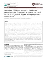
Báo cáo y học: "Decreased GABAB receptor function in the cerebellum and brain stem of hypoxic neonatal rats: Role of glucose, oxygen and epinephrine resuscitation" pot
Bạn đang xem bản rút gọn của tài liệu. Xem và tải ngay bản đầy đủ của tài liệu tại đây (593.29 KB, 11 trang )
RESEARCH Open Access
Decreased GABA
B
receptor function in the
cerebellum and brain stem of hypoxic neonatal
rats: Role of glucose, oxygen and epinephrine
resuscitation
Thoppil R Anju, Sadanandan Jayanarayanan and Cheramadatikudiyil S Paulose
*
Abstract
Background-: Hypoxia during the first week of life can induce neuronal death in vulnerable brain regions usually
associated with an impairment of cognitive function that can be detected later in life. The neurobiological changes
mediated through neurotransmitters and other signaling molecules associated with neonatal hypoxia are an
important aspect in establishing a proper neonatal care.
Methods-: The present stud y evaluated total GABA, GABA
B
receptor alterations, gene expression changes in GABA
B
receptor and glutamate decarboxylase in the cerebellum and brain stem of hypoxic neonatal rats and the
resuscitation groups with glucose, oxygen and epinephrine. Radiolabelled GABA and baclofen were used for
receptor studies of GABA and GABA
B
receptors respectively and Real Time PCR analysis using specific probes for
GABA
B
receptor and GAD mRNA was done for gene expression studies.
Results-: The adaptive response of the body to hypoxic stress resulted in a reduction in total GABA and GABA
B
receptors along with decreased GABA
B
receptor and GAD gene expression in the cerebellum and brain stem.
Hypoxic rats supplemented with glucose alone and with oxygen showed a reversal of the receptor alterations and
changes in GAD. Resuscitation with oxygen alone and epinephrine was less effective in reversing the receptor
alterations.
Conclusions-: Being a source of immediate energy, glucose can reduce the ATP-depletion-induced changes in
GABA and oxygenation, which helps in encountering hypoxia. The present study suggests that reduction in the
GABA
B
receptors functional regulation during hypoxia plays an important role in central nervous system damage.
Resuscitation with glucose alone and glucose and oxygen to hypoxic neonatal rats helps in protecting the brain
from severe hypoxic damage.
Keywords: GABA
B
neonatal hypoxia, cerebellum and brain stem
Background
Hypoxia is one of th e most common reasons for neona-
tal morbidity and mortality, causing reduced oxygen
supply to the vital organs [1] and inju ry to the develop-
ing brain [2-5]. The response of central nervous system
to hypoxia is vital in revealing mechanisms that
participate in coordinated behavior of respiratory and
vasomotor activities [6,7].
The ventilatory response to acute hypoxia (hypoxic
ventilatory response; HVR) in humans and some other
mammalian species is biphasic [8,9]. The initial rise in
ventilation(earlyphaseoftheHVR)isfollowedbya
marked decline after several minutes to values above the
prehypoxic level. This decline in ventilation has been
termed “ventilatory roll-off” or “ hypoxic ventilatory
decline” (HVD). Several neurotransmitters and neuro-
modulators, such as g-aminobutyric acid (GABA),
* Correspondence:
Molecular Neurobiology and Cell Biology Unit, Centre for Neuroscience,
Department of Biotechnology, Cochin University of Science and Technology,
Cochin-682022 Kerala, India
Anju et al. Journal of Biomedical Science 2011, 18:31
/>© 2011 Anju et al; licensee BioMed Central Ltd. This is an Open Access article distributed under the terms of the Creative Commons
Attribution License (http://creativecommo ns.org/license s/by/2.0), which permits unrestricted use, distribution, and reproduction in
any medium, pro vided the original work is properly cited.
[10-13] serotonin [14], adenosine, [15,16] and platelet-
derived growth factor [17,18] play important roles in
HVD. The alterations in neurotransmitter signaling in
the respiratory control centers in brain stem and
stressed breathing facilitating regions in cerebellar deep
nuclei highly influence the ventilatory response of the
body.
At synaptic transmission level, experimental hypoxia
or hypoxia/ischemia increases the release of aminoacid
neurotransmitters [19-23], causing an imbalance in nor-
mal activity of glutamatergic and GABAergic neurones,
resulting in acute cell excitotoxicity. Endogenous GABA
acting on GABA
A
or GABA
B
receptors modulates venti-
lation during room air breathing as well that the ventila-
tory response to acute and sustained hypoxia [24].
Rhythm generation in mature respiratory networks is
influenced strongly by synaptic inhibition. Zhang et al,
2002 [24] reported th at GABA
B
-receptor-mediated post-
synaptic modulation plays an important role in the
respiratory network from P0 on. GABA
B
-receptor-
mediated presynaptic modulation develops with a longer
postnatal latency, and becomes predominant within the
first postnatal week [25].
GABA
B
receptors may contribute essentially t o the
modulation of respiratory rhythm in adult mammals
and may be involved in the control of respiratory neuro-
nal discharge [26]. GABA, which is met abolized in
GABA shunts, is produced through a-decarboxylation
of glutamic acid catalyzed by glutamate decarboxylase
(GAD; EC 4.1.1.15) under the presence of cofactor pyri-
doxal 5’-phoshate. GAD, the rate limiting enzyme of
GABA synthesis and a key protein in the GABA path-
way, is used as a marker for GABAergic activity.
Thus, understanding the diagnosis, pathogenesis,
resuscitation and treatment of those infants suffering
hypoxic brain injury is paramount to reducing disability,
improving s urvival and enhancing quality of life. Upon
delivery, 5–10% of all newborns r equire some degree of
resuscitation and assistance to begin breathing [27-29].
The aim of resuscitation is to prevent neonatal death
and adverse long-term neurodevelopment sequelae asso-
ciated with neonatal hypoxic event [30] and rapidly
reverse fetal hypoxemia, and acidosis [31]. Debate
regarding the optimal concentration of oxygen at initia-
tion of resuscitation continues in the international com-
munity. The present study focused on understanding
the alterations in GABA content, total GABA and
GABA
B
receptors and GAD expression in the cerebel-
lum and brain stem of hypoxic neonatal rats and the
effects of various resuscitations on these alterations. The
effectiveness of various resuscitation methods like
administration of 100% oxygen and intravenous fluids
like 10% glucose and 0.10 g/Kg body wt epinephrine
alone and in combinations in the management of
hypoxia was analyzed to understand the neuroprotective
role of glucose supplementation. Understanding the
molecular mechanisms involved in the regulation of
neurotransmitter receptors will lead to better therapies
for neonatal hypoxia-ischemia.
Materials and methods
Animals
Neonatal Wistar rats were purchased from Amrita Insti-
tute of Medical Sciences, Kochi. Neona tal rats of four
days old were weighed and used for experiments. All
groups of neonat al rat were maintained with t heir
mothers under optimal conditions - 12 hour light and
12 hour dark periods and were fed standard food and
water ad libitum. All animal care and procedures were
taken in accordance with the institutional, National
Institute of Health guidelines and CPCSEA guidelines.
Induction of Acute Hypoxia in Neonatal Rats
Wistar ne onatal rats of 4-days old (body weight, 6.06 ±
0.45 g) were used for the experiments and were grouped
into seven as follows: (i) Control neonatal rats were
given atmospheric air (20.9% oxygen) for 30 minutes
(C); (ii) Hypo xia was induced by placing the neonatal
rats in a hypoxic chamber provided with 2.6% oxygen
for 30 minutes (Hx); (iii) Hypoxic neonatal rats were
injected 10% dextrose (500 mg/Kg body wt) intra-perito-
neally (i.p.) (Hx+G). ( iv) Hypoxic neonatal rats were
supplied with 100% oxygen for 30 minutes (Hx+O); (v)
Hypoxic neonatal rats were injected 10% dextrose (500
mg/Kg body wt. i.p.) and treated with 100% oxygen for
30 minutes (Hx+G+O) . (vi) Hypoxic neonatal rats were
injected 10% dextrose (500 mg/Kg body wt), epinephrine
(0.1 μg/Kg body wt. i.p.) and treated with 100% oxygen
for 30 minutes (Hx+G+E+O) (vii) Hypoxic neonatal ra ts
were injected with epinephrine (0.10 g/Kg body wt) i.p.
(Hx + E). The experimental animals were maint ained in
the room temperature for one week.
Tissue preparation
Control and experimental neonatal rats were sacrificed
by decapitation. The cerebellum and brain stem were
dissected out quic kly over ice according to the proce-
dure of Glowinski and Iversen, 1966 [32] and was stored
at -80°C for various experiments.
Quantification of GABA content Using [
3
H]Radioligands
GABA content in the cerebellum and brain stem of con-
trol and experimental rat groups was quantified by dis-
placement method of Kurioka et al, 1981 [33] where the
incubation mixture contained 30 nM [
3
H]GABA with
and without GABA at a concentration range of 10
-8
M
to 10
-4
M. The unknown concentrations were deter-
mined from the stan dard displacement curve using
Anju et al. Journal of Biomedical Science 2011, 18:31
/>Page 2 of 11
appropriate dilutions and calcu late d for μ moles/gm wt.
of the tissue
GABA Receptor Binding Assay
[
3
H] GABA binding t o the GABA receptor was assayed
in Triton X-100 treated synaptic membranes [ 33].
Crude synaptic membranes were prepared using
sodium-free 10 mM tris buffer, pH 7.4. Each assay tube
contained a protein concentration of 0.1 - 0.2 mg. In
saturation binding experiments, 5 nM to 40 nM concen-
trations of [
3
H]GABA was incubated with and without
excess of unlabelled GABA (100 μM) and in compe ti-
tion binding experiments the incubation mixture con-
tained 30 nM of [
3
H] GABA with and without GABA at
a concentration range of 10
-8
Mto10
-4
M were used.
GABA
B
Receptor Binding Assay
[
3
H] baclofen binding to the GABA
B
receptor was
assayed in Triton X-100 treated synaptic membranes
[33]. Crude synaptic membranes were prepared using
sodium-free 10 mM tris buffer, pH 7.4. Each assay tube
contained a protein concentration of 0.1 - 0.2 mg. In
saturation binding experiments, 5 nM to 40 nM concen-
trations of [
3
H]baclofen was incubated with and without
excess of unlabelled baclofen (100 μM) were used.
Protein was measured by the method of Lowry et al,
1951 [34] using bovine serum albumin as standard.
Linear regression analysis of the receptor binding data
for Scatchard plots
The data was analysed according t o Scatchard, 1949
[35]. The specific binding was determined by subtracting
non-specific binding from the total. The binding para-
meters, maximal bind ing (B
max
)andequilibriumdisso-
ciation constant (K
d
), were derived by linear regression
analysis by plotting the specific binding of the radioli-
gand on X-axis and bound/free on Y-axis. The maximal
binding is a measure of the total number of rece ptors
present in the tissue and the equilibrium dissociation
constant is the measure of the affinity of the receptors
for the radioligand. The K
d
is inversely related to recep-
tor affinity.
Nonlinear regression analysis for displacement curve
Competitive binding data was analyzed using non-linear
regression curve-fitting procedure (GraphPad PRISM™,
San Diego, USA). The data of the competitive binding
assa ys were represented graphically with the log of con-
centration of the competing drug on x-axis and percen-
tage of the radioligand bound on the y-axis. The
steepness of the binding curve can be quantified with a
slope factor, often called a Hill slope. A one-site compe-
titive binding curve that follows the law of mass action
has a slope of 1.0 and a two site competitive binding
curve has a slope less than 1.0. The concentration of
competitor that competes for half the specific binding
was defined as EC
50
, which is same as IC
50
. The affinity
of the receptor for the competing drug is designated as
K
i
and is defined as the concentration of the competing
ligand that binds to half the binding sites at equilibrium
in the absence of radioligand or other competitors.
Gene expression studies in cerebellum and brain stem
RNA was isolated from the cerebellum and brain stem
using Tri reagent. Total cDNA synthesis was performed
using ABI PRISM cDNA Archive kit. Real-Time PCR
ass ays were performed in 96-well plates in an ABI 7300
Real-Time PCR instrument (Applied Biosystems, Foster
City, CA, USA). PCR analyses were conducted with
gene-specific primers and fluorescently labeled Taq
probe for GABA B (Rn 00578911) and GAD1 (Rn
00690304_g1) designed by Applied Biosystems. Endo-
genous control (b-actin) labeled with a reporter dye was
used as internal control. All reagents were purchased
from Applied Biosystems. The real-time data were ana-
lyzed with Seq uence Detection Systems software version
1.7. All reactions were performed in duplicate.
The ΔΔCT method of r elative quantification was used
to determine the fold change in expression. This was
done by first normalizing the resulting threshold cycle
(CT) values of the target mRNAs to the CT values of
the internal control b-actin in the same samples (ΔCT =
CT
Target
-CT
b-actin
). It was further normalized with
the contro l (ΔΔCT = ΔCT-CT
Control
). The fold
change in expression was then obtained (2
-ΔΔCT
).
Statistical analysis
The equality of all the groups was tested by the analysis
of variance (ANOVA) technique for different values of
p. Further the pair wise comparisons of all the experi-
mental groups were studied using Students-Newman-
Keuls test at different significance levels. The testing
was performed using GraphPad Instat (Ver. 2.04a, San
Diego, USA) computer program.
Results
GABA Content in the cerebellum and brain stem of
control and experimental neonatal rats
The GABA content was decreased significantly (p <
0.001) in the cerebellum and brain stem of hypoxic neo-
natal rats compa red to control. The decreased content
was reversed to near normal in glucose supplemented
groups - Hx + G and Hx + G + O (Table 1).
Total GABA receptors in the cerebellum and brain stem
of control and experimental neonatal rats
Receptor studies for total GABA showed a significant
decrease in receptor number compared to control in the
Anju et al. Journal of Biomedical Science 2011, 18:31
/>Page 3 of 11
cerebellum and brain stem (p < 0.01, p < 0.001 respec-
tively) of hypoxic neonatal rats. In glucose supplemented
groups, H x + G and Hx + G + O , the receptor number
was reversed to near control (p < 0.001) in both the
brain regions. Epinephrine supplemented groups, Hx +
EandHx+G+E+O,showednosignificantreversal
in the altered receptor number to control level. In Hx +
O, the Bmax was significantly decreased (p < 0.001)
compared to control (Table 2).
Non linear regression analysis of total GABA receptors in
the cerebellum and brain stem
The binding d ata were confirme d by compe tition bind-
ing assay with [
3
H] GABA against different concentra-
tions of GABA. GABA affinity in the cerebellum and
brain stem of control and hypoxic neonatal rats fitted to
a t wo site model with Hill slope value away fro m unity.
GABAaffinityofHx+O,Hx+G,Hx+G+O,Hx+
EandHx+G+E+Oalsofittedtoatwositemodel
with Hill slope value away from unity. The Ki(H)
increased in hypoxic neonatal rats along with an
increase in the log (EC
50
)-1 indicating a shift in high
affinity towards low affinity. Ki(L) also showed an
increase in hypoxic neonatal rats with an increase in log
(EC
50
)-2 denoting a shift in the low affinity site towards
much lower affinity (Figure 1 & 2).
GABA
B
receptors in the cerebellum and brain stem of
control and experimental neonatal rats
GABA
B
receptors was significantly decreased (p < 0.001)
with a significant increase in its affini ty (p < 0.001, p <
0.05) in the cerebellum and brain stem of hypoxic neo-
natal rats compared to control. Hx + G and Hx + G +
O showed a significant reversal of B
max
(p < 0.001) and
K
d
(p < 0.01) to near control in the cerebellum and a
significant reversal of B
max
(p < 0.01, p < 0.001 respec-
tively) to near control in the brain stem. In epinephrine
and 100% oxygen supplemented groups, no reversal was
observed (Table 3).
Gene expression of GABA
B
receptor mRNA in the
cerebellum and brain stem
GABA
B
receptor mRNA was significantly down regu-
lated (p < 0.001) in the cerebellum and brain stem of
hypoxic neonatal rats compared to control. In the cere-
bellum, Hx + G, Hx + G + O and Hx + O showed a sig-
nificant reversal of GABA
B
receptor expression (p <
0.001, p < 0.001 and p < 0.05 respectively) to near con-
trol where as epinephrine supplement ed groups, Hx + E
and Hx + G + E + O, showed no significant reversal of
altered expression. In the brain stem, glucose supple-
mentedgroups,Hx+G,Hx+G+O,showeda
Table 1 GABA Content (μmoles/g wet wt.) in cerebellum
and brain stem of Control and Experimental Groups of
Neonatal Rats
Experimental groups GABA Content (μmoles/g wet wt.)
Cerebellum Brain stem
Control 6.45 ± 1.2 8.45 ± 1.8
Hx 2.02 ± 1.0
a
4.06 ± 1.4
a
Hx + G 6.25 ± 1.4
b
9.85 ± 2.2
b
Hx + G + O 6.60 ± 1.4
b
8.66 ± 1.4
b
Hx + O 3.55 ± 1.8
b
6.01 ± 1.5
b
Hx + E 3.05 ± 1.2
a
4.55 ± 1.6
a
Hx + G + E + O 3.12 ± 1.1
a
5.02 ± 1.4
a
Values are Mean ± S.E.M of 4-6 separate experiments. Each group consist 6-8
rats.
a
p < 0.001 when compared to Control
b
p < 0.001,
c
p < 0.01 when compared to hypoxic group
Hypoxic rats- Hx, Hypoxic rats glucose treated - Hx+G, Hypoxic rats oxygen
treated - Hx+O, Hypoxic rats glucose and oxygen treated - Hx+ G+O, Hypoxic
rats epinephrine treated - Hx + E, Hypoxic rats glucose, epinephrine and
oxygen treated - Hx+G+E+O
Table 2 Total GABA receptor binding parameters in the cerebellum and brain stem of control and experimental
neonatal rats.
Experimental groups Cerebellum Brain stem
B
max
(fmoles/mg protein) K
d
(nM) B
max
(fmoles/mg protein) K
d
(nM)
Control 71.50 ± 2.41 11.11 ± 0.95 153.36 ± 3.7 4.77 ± 0.44
Hx 50.01 ± 1.80
a
14.82 ± 0.82
a
116.68 ± 2.8
a
3.77 ± 0.22
a
Hx + G 62.18 ± 1.50
b
9.85 ± 0.36
b
173.36 ± 2.5
b
6.78 ± 0.35
a, b
Hx + G + O 66.33 ± 2.00
b
12.54 ± 0.42 160.84 ± 3.4
b
5.01 ± 0.26
a, b
Hx + O 55.34 ± 2.50
a
15.72 ± 0.54
a
136.68 ± 2.3
a, b
4.73 ± 0.29
b
Hx + E 44.02 ± 3.20
a
10.46 ± 0.10
b
122.08 ± 2.6
a
3.30 ± 0.14
a
Hx + G + E + O 45.50 ± 2.50
a
7.46 ± 0.11
a, b
125.84 ± 4.5
a
4.10 ± 0.22
b
Values are Mean ± S.E.M of 4-6 separate experiments. Each group consist 6-8 neonatal rats.
a
p < 0.001 when compared with control
b
p < 0.001 when compared with hypoxic group.
Hypoxic rats- Hx, Hypoxic rats glucose treated - Hx+G, Hypoxic rats oxygen treated - Hx+O, Hypoxic rats glucose and oxygen treated - Hx+G+O, Hypoxic rats
epinephrine treated - Hx + E, Hypoxic rats glucose , epinephrine and oxygen treated - Hx+G+E+O
Anju et al. Journal of Biomedical Science 2011, 18:31
/>Page 4 of 11
significant reversal of the gene expression (p < 0.001) to
near contr ol, whereas Hx + O, Hx + E and Hx + G + E
+ O showed a down regulated GABA
B
receptor expres-
sion (p < 0.01, p < 0. 001, p < 0.00 1 respectively) with
out a significant reversal to near control (Figure 3).
Gene expression of GAD mRNA in the cerebellum and
brain stem
The expression of glutamate decarboxylase in cerebel-
lum and brain stem also showed a significant down reg-
ulation (p < 0.001) in the hypoxic group compared to
control. The cerebellar and brain stem GAD expression
was significantly reversed to near control in Hx + G, Hx
+ G + O and Hx + O whereas in Hx + E and Hx + G +
E + O, there was no significant reversal to near control
(Figure 4).
Discussion
Hypoxia–ischemia (HI) occurring before or shortly after
birth is a major cause of life-threatening injury and life-
long disability [36]. HI results in multi-organ failure and
structural/functional damage especially devastating to
the cardiovascular, renal, gastrointestinal and central
nervous systems [37,38]. HI brain inj ury is very complex
and has different neuropathological manifestations
depending on the maturity of the newborn. Many of the
structural changes that occur during the initial postnatal
period in rodents are consistent with those seen during
the late prenatal period in human brain development.
Thus, exposure of rat to hypoxia on postnatal day 4
includes many of the neurodevelo pmental events that
may be affected by hypoxia in preterm human infants.
In the pre sent study, we investigated the functional reg-
ulation of GABA
B
receptors and GAD in hypoxic neo-
natal rats and the role of glucose, oxygen and
epinephrine in altering the receptor status.
Numerous studies by different groups have confirmed
that both inhibitory and excitatory amino acids are
involved in the acute hypoxic ventilatory response
[39-42]. Increases in GABA as a consequence of brain
hypoxia can lead to depression of ventilation, which
becomes more apparent in the absence of peripheral
chemoreceptors. Blockade of GABA by biccuculine can
significantly reduce this depressive effect of GABA on
ventilation during hypoxia in chemodenervated animal
or the newborn [43-45].
Figure 1 Displacement of [
3
H] GABA again st GABA in cerebellum of control and experimental neonatal rats. Competiti on studies were
carried out with 30 nM [
3
H] GABA in each tube with the unlabelled GABA concentrations varying from 10
-8
to10
-4
M. Values are representation
of 4-6 separate experiments. Data from the curves as determined from nonlinear regression analysis using computer program PRISM fitted to a
two-site model. The affinity for the first and second site for the competing drug is designated as Ki-1 (for high affinity) and Ki-2 (for low affinity).
EC
50
is the concentration of competitor that competes for half the specific binding. The equation built-in to the program is defined in terms of
the log (EC
50
). If the concentrations of unlabelled compound are equally spaced on a log scale, the uncertainty of the log (EC
50
) will be
symmetrical, but uncertainty of the EC50 will not be symmetrical
Anju et al. Journal of Biomedical Science 2011, 18:31
/>Page 5 of 11
The present study reports a significant decrease in total
GABA and GABA
B
receptor number with a down regu-
lated receptor expression and glutamate decarboxylase
expression in the cerebellum and brain stem regions of
hypoxic neonatal rat s. The decreased expression of GAD
in turn results in the inhibition of GABA synthesizing
pathway, which can be correlated to the decreased GABA
receptors. The decreased GABA receptor is a response of
the body to encounter hypoxic ventilatory decline. The
reduction in GABA
B
receptor may help in overcoming
Figure 2 Displacement of [
3
H] GABA against GABA in brain stem of control and experimental neonatal rats. Competit ion studies were
carried out with 30 nM [
3
H] baclofen in each tube with the unlabelled baclofen concentrations varying from 10
-12
to10
-4
M. Values are
representation of 4-6 separate experiments. Data from the curves as determined from nonlinear regression analysis using computer program
PRISM fitted to a two-site model. The affinity for the first and second site for the competing drug is designated as Ki-1 (for high affinity) and Ki-2
(for low affinity). EC
50
is the concentration of competitor that competes for half the specific binding. The equation built-in to the program is
defined in terms of the log (EC
50
). If the concentrations of unlabelled compound are equally spaced on a log scale, the uncertainty of the log
(EC
50
) will be symmetrical, but uncertainty of the EC50 will not be symmetrical.
Table 3 GABA
B
receptor binding parameters in the cerebellum and brain stem of control and experimental neonatal
rats.
Experimental groups Cerebellum Brain stem
B
max
(fmoles/mg protein) K
d
(nM) B
max
(fmoles/mg protein) K
d
(nM)
Control 71.50 ± 2.41 11.11 ± 0.95 74.27 ± 1.20 13.31 ± 1.00
Hx 50.01 ± 1.80
a
14.82 ± 0.82
a
51.84 ± 1.50
a
14.44 ± 0.99
b
Hx + G 62.18 ± 1.50
b
9.85 ± 0.36
b
69.41 ± 1.40
b
20.47 ± 0.99
a
Hx + G + O 66.33 ± 2.00
b
12.54 ± 0.42 70.47 ± 1.10
c
26.10 ± 1.20
a
Hx + O 55.34 ± 2.50
a
15.72 ± 0.54
a
49.10 ± 1.10
a
16.36 ± 1.50
a
Hx + E 44.02 ± 3.20
a
10.46 ± 0.10
b
43.59 ± 1.5
a
14.53 ± 0.99
b
Hx + G + E + O 45.50 ± 2.50
a
7.46 ± 0.11
a, b
53.95 ± 1.5
a
13.90 ± 0.99
b
Values are Mean ± S.E.M of 4-6 separate experiments. Each group consist 6-8 neonatal rats.
a
p < 0.001,
b
p < 0.05 when compared with control
c
p < 0.001 when compared with hypoxic group.
Hypoxic rats- Hx, Hypoxic rats glucose treated - Hx+G, Hypoxic rats oxygen treated - Hx+O, Hypoxic rats glucose and oxygen treated - Hx+G+O, Hypoxic rats
epinephrine treated - Hx + E, Hypoxic rats glucose , epinephrine and oxygen treated - Hx+G+E+O
Anju et al. Journal of Biomedical Science 2011, 18:31
/>Page 6 of 11
the ventilatory decline during hypoxia but at the cost of
severe central nervous system dysfunction. Louzoun-
Kaplan et al, 2008 [46] reported that prenatal hypoxia at
gestation day 17 in mice caused an imme diate decrease
in fetal cerebral cortex levels of glutamate decarboxylase.
Decreased levels of key proteins in the GABA pathway in
the cerebral cortex may lead to high susceptibility to sei-
zures and epilepsy in newborns after prenatal or perinatal
hypoxia. In the elevated plus maze, the agonist of GABA-
B receptor was reported to i mprove consolidation of pas-
sive avoidance in rats undergoing hypoxia [47]. GABA
B
receptor-mediated activation of TASK-1 or a related
channel provides a presynaptic autoregulatory f eedback
mechanism that modulates fast synaptic transmission in
the rat carotid body [48]. The signaling cascade that trig-
gers the altered transcription of GABA-B receptor and
GAD under hypoxic stress can be related to the activa-
tion of apopto tic pathways by triggering Bax expression
and deactivati ng CREB expressi on coupled with the acti-
vation of HIF. The accumulation of HIF-1a in ischemic
or hypoxi c tissues promote adaptive mechanisms for cell
survival [49] and was found to be an important mediator
of hypoxia-induced tolerance to ischemia [50]. Although
HIF-1a is essential for adaptation to low oxygen levels, it
has also been shown in vitro to mediate hypoxia-induced
growth arrest and apoptosis [51]. The increased Hif 1
mRNA expression under hypoxia facilitates angiogenesis,
vasodialation and erythropoiesis. But in severe hypoxic
cases, HIF-1a is accumulated and leads to cell death by
activating different target genes [52]. The role of HIF-1 a
in mediating pro death and pro survival responses, is
dependent on the duration [53] and types of pathological
stimuli [54] as well as the cell type that it induces [55].
We observed that glucose su pplementation to hyp oxic
neonates alone and along with 100% oxygen showed a
reversal in the altered GABA
B
receptor parameters and
Figure 3 Real time PCR amplification of GABA
B
receptor subunit in mRNA form the cerebellum (A) and brain stem (B) of control and
experimental neonatal rats. The ΔΔCT method of relative quantification was used to determine the fold change in expression. The relative
ratios of mRNA levels were calculated using the ΔΔCT method normalized with b-actin. CT value as the internal control and Control CT value as
the caliberator. PCR analyses were conducted in the cerebellum (A) and brain stem (B) with gene-specific primers and fluorescently labeled Taq
probe GABA
B
(Rn 00578911)
Anju et al. Journal of Biomedical Science 2011, 18:31
/>Page 7 of 11
GAD expression in the cerebellum and brain stem. Glu-
cose supplementation provides an instant source of
energy to the brain cells thereby preventing ATP deple-
tion mediated cell death. Hattori and Wasterlain, 2004
[56] observed a reduction in the blood glucose levels
and substantially increased cerebral glucose utilization
[57] as a result of hypoxic stress in experimental rats.
Mónica Lemus et al, 2008 [58] reported that GABA
B
receptor agonist (baclofen) or antagonists (phaclofen
and CGP55845A) locally injected into nucleus tractus
solitarius modified arterial glucose levels and brain glu-
cose retention.
The standard approach to resuscitation neonatal
hypoxia is to use 100% O2. Further, resuscitation with
100% is recommended as a beneficial short-term therapy
that is generally thought to be non-to xic [31,59].
Although th e use of 100% O 2 appears intuitive to maxi-
mize the gradient required to drive O2 into hypoxic
cells [30], a building body of evidence derived from
animal models, has demons trated that although resusci-
tation with 100% O2 improves restoration of cerebral
and cortical perfusion, it may occur at the price of
greater biochemical oxidative stress [31]. Resuscitation
with 100% O
2
significantly increased glutamate and gly-
cine in the dorsal cortex contralateral to the ligated
common carotid artery, compared to piglets resuscitated
with 21% O
2
. These data suggest that persistent changes
in neurochemistry occur 4 days after hypoxic ischemia
and further studies are warranted to elucidate the conse-
quences of this on neonatal brain development [60]. We
observed that 100% oxygen resuscitation for neonatal
hypoxia is not as effective as the combination of glucose
and oxygen or administration of glucose alone. In cere-
bellum and brain stem of 100% oxygen resuscitated
groups, GABA
B
receptors showed a significant decrease
compared to control. One hundred percentage of oxy-
gen generated abnormally high levels of reactive oxygen
species (ROS) which causes dysfunction of defensive
Figure 4 Real time PCR amplification of GAD mRNA form the cerebellum (A) and brain stem (B) of control and experimental neonatal
rats. The ΔΔCT method of relative quantification was used to determine the fold change in expression. The relative ratios of mRNA levels were
calculated using the ΔΔCT method normalized with b-actin. CT value as the internal control and Control CT value as the caliberator. PCR
analyses were conducted in the cerebellum (A) and brain stem (B) with gene-specific primers and fluorescently labeled Taq probe GAD1 (Rn
00690304_g1).
Anju et al. Journal of Biomedical Science 2011, 18:31
/>Page 8 of 11
antioxidant system of cells by altering enzyme activity
[61,62] and act as a factor for neurodegeneration [63].
Hypoxemic piglets resuscitated with 100% O
2
also
showed increased cerebral injury, cortical damage and
early neurologic disorders [64-66]. Previous studies on
acetylcholinesterase [67], GABA
A
and serotonin recep-
tors [68] reported the neuroprotective role of glucose
and combination of glucose and oxygen resuscitation
and the damaging effects of oxygen supplementation
alone. The reduction in GABA
B
receptor number in the
cerebellar and brain stem region s during oxygen supple-
mentation is suggested to be due to tissue damage
caused by the formation of free radicals or reactive oxy-
gen species and the changes in amino acids resulting in
neuronal cell death. During oxygen resuscitation, the
accumulation of ROS activates the over stimulation of
HIF 1 alpha w hich can in turn results in the activation
of apoptotic pathways by altering the expression of tran-
scription factors like CREB and NF-Kappa-B.
Epinephrine is routinely used in the resuscitation for
persistent severe neonatal hypoxia. The present study
points out the adverse effects of epinephrine supplemen-
tation, alone and even in combination with glucose a nd
oxygen, by studying the changes in GABA
B
receptor,
expression of GABA
B
receptor and GAD in the brain
stem and cerebellum. The GABA
B
receptor was signifi-
cant ly decreased in epinephr ine treated groups. A ref lex
action of epinephrine firing occurs during hypoxia. Sup-
plementation of epinephrine to already excited system
results in its hyper activity and it affects the balance of
various neurotransmitters like dopamine [69] and gluta-
mate. Epinephrine induces a hypoxia-neovascularizati on
cascade and plays a primary role in vascular prolifera-
tion within soft tissues [70]. It is reported that repetitive
hypoxic stress induced by labour is a powerful stimulus
for catecholamine release in fetus and is accompanied
by typical alterations of fetal heart rate. The high influx
of this excitatory neurotransmitter affects the balance of
other n eurotransmitters thereby disrupting the cascade
of signal transduction.
There has been much interest in the acute neurologi-
cal changes associated with neonatal hypoxia, along with
the mechanisms of subsequent central nervous system
dysfunction in the adult [71-74]. Hypoxia during the
first week of life can induce neuronal death in vulner-
able brain regions usually associated with an impairment
of cognitive function that can be detected later in life
[75]. Postnatal hypoxia resulting from lung immaturity
and respiratory disturbances in infants is an important
pathophysiological mechanism underlying the devastat-
ing neurological complications. This points th e impor-
tance of a proper resuscitation program to overcome
neonatal hypoxia for a better intellect in the later stages
of life.
Conclusions
Our studies point out the neuropr otective role of glu-
cose in the management of neonatal hypoxic stress. The
down regulated GABA
B
receptor in cerebellum and
brain stem led to hypoxia induced ventilatory decline
and activation of a poptotic pathways. These receptor
alterations are reversed back to near control by the
timely resuscitation with glucose, alone and in combina-
tion with oxygen. The deleterious effect of oxygen alone
and epinephrine resuscita tion in neuronal response
through alterations in neurotransmitters was also
observed. Thus it is suggest ed that glucose administra-
tion immediately after hypoxia with oxygenated air as a
resuscitation programme will be of tremendous advan-
tage especially in neonatal care. Deeper understanding
of mechanisms, through which hypoxia regulates the
neurotra nsmitters, could poin t towards the development
of new therapeutic approaches to reduce or suppress
the pathological effects of hypoxia.
Acknowledgements
This work was supported by the research grants from DBT, DST, ICMR, Govt.
of India and KSCSTE, Govt. of Kerala to Dr. C. S. Paulose. Anju T R thanks
Council of Scientific and Industrial Research for Senior Research Fellowship.
Authors’ contributions
TRA carried out the receptor assays, gene expression and drafted the
manuscript. SJ participated participated in the design of the study and
performed the statistical analysis. CSP conceived of the study and
participated in its design and coordination. All authors read and approved
the final manuscript.
Competing interests
The authors declare that they have no competing interests.
Received: 5 January 2011 Accepted: 12 May 2011
Published: 12 May 2011
References
1. Low JA, Froese AB, Galbraith RS, Smith JT, Sauerbrei EE, Derrick EJ: The
association between preterm newborn hypotension and hypoxemia and
outcome during the first year. Acta Paediatrica 1993, 82:433-437.
2. Delivoria-Papadopoulos M, Mishra POM: Mechanisms of perinatal cerebral
injury in fetus and newborn. Annals of the New York Academy of Sciences
2000, 900:159-168.
3. Li C, Jackson RM: Reactive species mechanisms of cellular
hypoxicreoxygenation injury. American Journal of Physiology 2002,
282:227-241.
4. Rodrigo J, Fernandez AP, Serrano J, Peinado MA, Martinez A: The role of
free radicals in cerebral hypoxia and ischemia. Free Radical Biology and
Medicine 2005, 39:26-50.
5. Xu W, Chi L, Row BW, et al: Increased oxidative stress is associated with
chronic intermittent hypoxia-mediated brain cortical neuronal cell
apoptosis in a mouse model of sleep apnea. Neuroscience 2004,
126:313-323.
6. Acker T, Acker H: oxygen sensing need in CNS function: Physiological
and pathological implications. Journal of Experimental Biology 2004,
207:3171-3188.
7. Solomon IC: Excitation of phrenic and sympathetic output during acute
hypoxia: Contribution of medullary oxygen detectors. Respiration
Physiology 2000, 121:101-117.
8. Neubauer JA, Melton JE, Edelman NH: Modulation of respiration during
brain hypoxia. J Appl Physiol 1990, 68:441-451.
Anju et al. Journal of Biomedical Science 2011, 18:31
/>Page 9 of 11
9. Weil JV, Zwillich CW: Assessment of ventilatory response to hypoxia.
Chest 1976, 70(1):124-128.
10. Kneussl MP, Pappagianopoulos P, Hoop B, Kazemi H: Reversible
depressiozn of ventilation and cardiovascular function by
ventriculocisternal perfusion with γ-aminobutyric acid in dogs. Am Rev
Respir Dis 1986, 133:1024-1028.
11. Taveira da Silva AM, Hartley B, Hamosh P, Quest JA, Gillis RA: Respiratory
depressant effects of GABA α- and β-receptor agonists in the cat. J Appl
Physiol 1987, 62:2264-2272.
12. Kazemi H, Hoop B: Glutamic acid and gamma-aminobutyric acid
neurotransmitters in central control of breathing. J Appl Physiol 1991,
70:1-7.
13. Richter DW, Schmidt-Garcon P, Pierrefiche O, Bischoff AM, Lalley PM:
Neurotransmitters and neuromodulators controlling the hypoxic
respiratory response in anaesthetized cats. J Physiol 1999, 514:567-578.
14. Di Pasquale E, Morin D, Monteau R, Hilaire G: Serotonergic modulation of
the respiratory rhythm generator at birth: an in vitro study in the rat.
Neurosci Lett 1992, 143:91-95.
15. Neylon M, Marshall JM: The role of adenosine in the respiratory and
cardiovascular response to systemic hypoxia in the rat. J Physiol 1991,
440:529-545.
16. Elnazir B, Marshall JM, Kumar P: Postnatal development of the pattern of
respiratory and cardiovascular response to systemic hypoxia in the
piglet: the roles of adenosine. J Physiol 1996, 492:573-585.
17. Gozal D, Simakajornboon N, Czapla MA, Xue YD, Gozal E, Vlasic V, Lasky JA,
Liu JY: Brainstem activation of platelet-derived growth factor-β receptor
modulates the late phase of the hypoxic ventilatory response.
J Neurochem 2000, 74:310-319.
18. Simakajornboon N, Kuptanon T: Maturational changes in
neuromodulation of central pathways underlying hypoxic ventilatory
response. Respir Physiol Neurobiol 2005, 149:273-286.
19. Cataltepe O, Towfighi J, Vannucci RC: Cerebrospinal fluid concentrations
of glutamate and GABA during perinatal cerebral hypoxia-ischemia and
seizures. Brain Res 1996, 709:326-330.
20. Hagberg H, Andersson P, Kjellmer I, Thiringer K, Thordstein M: Extracellular
overflow of glutamate, aspartate, GABA and taurine in the cortex and
basal ganglia of fetal lambs during hypoxia-ischemia. Neurosci Lett 1987,
78:311-317.
21. Rego AC, Santos MS, Oliveira CR: Oxidative stress, hypoxia, and ischemia-
like conditions increase the release of endogenous amino acids by
distinct mechanisms in cultured retinal cells. J Neurochem 1996,
66:2506-2516.
22. Saransaari P, Oja SS:
Enhanced GABA release in cell-damaging conditions
in
the adult and developing mouse hippocampus. Int J Devl Neurosci
1997, 15(2):163-174.
23. Saransaari P, Oja SS: Release of endogenous glutamate, aspartate, GABA,
and taurine from hippocampal slices from adult and developing mice
under cell-damaging conditions. Neuochem Res 1998, 23:563-570.
24. Zhang W, Barnbrock A, Gajic S, Pfeiffer A, Ritter B: Differential ontogeny of
GABA
B
-receptor-mediated pre- and postsynaptic modulation of GABA
and glycine transmission in respiratory rhythm-generating network in
mouse. The Journal of Physiology 2002, 540:435-446.
25. Suzuki M, Tetsuka M, Endo M: GABA(B) receptors in the nucleus tractus
solitarii modulate the carotid chemoreceptor reflex in rats. Neurosci Lett
1999, 260:21-24.
26. Yang AL, Lo MJ, Ting H, Chen JS, Huang CY, Lee SD: GABAA and GABAB
receptors differentially modulate volume and frequency in ventilatory
compensation in obese Zucker rats. J Appl Physiol 2007, 102:350-357.
27. Schubert S, Brandl U, Brodhun M, Ulrich C, Spaltmann J, Fiedler N, Bauer R:
Neuroprotective effects hypoxia–ischemia in newborn piglets. Brain Res
2005, 1058:129-136.
28. Davis PG, Tan A, O’Donnell CPF, Schulze A: Resuscitation of newborn
infants with 100% oxygen or air: a systematic review and meta-analysis.
Lancet 2004, 364:1329-33.
29. Tan A, Schulze A, O’Donnell CPF, Davis PG: Air versus oxygen for
resuscitation of infants at birth. The cochrane database of systemic reviews
2006 2005, 2.
30. Corff KE, McCann DL: Room air resuscitation versus oxygen resuscitation
in the delivery room. J Perinat Neonat Nurs 2005, 19:379-90.
31. Martin RJ, Walsh MC, Carlo WA: Reevaluating neonatal resuscitation with
100% oxygen. Am J Respir Crit Care Med 2005, 172:1360.
32. Glowinski J, Iversen LL: Regional studies of catecholamines in the rat
brain: The disposition of [3H] Norepinephrine, [3H] DOPA in various
regions of the brain. J Neurochem 1966, 13:655-669.
33. Kurioka S, Toshiaki K, Makoto M: Effects of sodium and bicarbonate ions
on gamma amino butyric acid receptor binding in synaptic membranes
of rat brain. J Neurochem 1981, 37:418-421.
34. Lowry OH, Rosebrough NJ, Farr AL, Randall J: Protein measurement with
folin phenol reagent. J Biol Chem 1951, 193:265-275.
35. Scatchard G: The attractions of proteins for small molecules and ions.
Ann NY Acad Sci 1949, 51:660-672.
36. du Plessis AJ, Volpe JJ: Perinatal brain injury in the preterm and term
newborn. Curr Opin Neurol 2002, 15:151-7.
37. Shah P, Riphagen S, Beyene J, Perlman JM: Multiorgan dysfunction in
infants with post-asphyxial hypoxic–
ischemic encephalopathy. Arch
Dis
Child Fetal Neonatal Ed 2004, 89:F152-5.
38. Vento M, Sastre J, Asensi MA, Vina J: Room-air resusciatation causes less
damage to heart and kidney than 100% oxygen. Am J Respir Crit Care
Med 2005, 172:1393-8.
39. Mitra J, Prabhakar NR, Overholt JL, Cherniack NS: Respiratory effects of N-
methyl-D-aspartate on the ventrolateral medullary surface. J Appl Physiol
1989, 67:1814-1819.
40. Lin J, Suguihara C, Huang J, Hehre D, Devia C, Bancalari E: Effect of N-
methyl-D-aspartate-receptor blockage on hypoxic ventilatory response
in unanesthetized piglets. J Appl Physiol 1996, 80:1759-1763.
41. Gozal D, Gozal E, Torres JE, Gozal YM, Nuckton TJ, Hornby PJ: Nitric oxide
modulates ventilatory responses to hypoxia in the developing rat. Am J
Respir Crit Care Med 1997, 155:1755-1762.
42. Gozal D, Graff GR, Torres JE, Khicha SG, Nayak GS, Simakajornboon N, Gozal E:
Cardiorespiratory responses to systemic administration of a protein
kinase C inhibitor in conscious rats. J Appl Physiol 1998, 84:641-648.
43. Hayashi F, Lipski J: The role of inhibitory amino acids in control of
respiratory motor output in an arterially perfused rat. Respir Physiol 1992,
89:47-63.
44. Huang J, Suguihara C, Hehre D, Lin J, Bancalari E: Effects of GABA receptor
blockage on the respiratory response to hypoxia in sedated newborn
piglets. J Appl Physiol 1994, 77:1006-1010.
45. Soto-Arape I, Burton MD, Kazemi H: Central amino acid neurotransmitters
and the hypoxic ventilatory response. Am J Respir Crit Care Med 1995,
151:1113-1120.
46. Louzoun-Kaplan V, Zuckerman M, Regino Perez-Polo J, Golan HM: Prenatal
hypoxia down regulates the GABA pathway in newborn mice cerebral
cortex; partial protection by MgSO
4.
. International Journal of
Developmental Neuroscience 2008, 26:77-85.
47. Car H, Oksztel R, Nadlewska A, Wi K: Baclofen prevents hypoxia-induced
consolidation impairment for passive avoidance in rats. Pharmacological
Research 2001, 44:329-335.
48. Fearon IM, Zhang M, Vollmer C, Nurse CA: GABA mediates autoreceptor
feedback inhibition in the rat carotid body via presynaptic GABA
B
receptors and TASK-1. The Journal of Physiology 2003, 553:83-94.
49. Bergeron M, Gidday JM, Yu AY, Semenza GL, Ferriero DM, Sharp FR: Role of
hypoxia-inducible factor-1 in hypoxia-induced ischemic tolerance in
neonatal rat brain. Ann Neurol 2000, 48:285-296.
50. Bernaudin M, Nedelec AS, Divoux D, MacKenzie ET, Petit E, Schumann-
Bard P: Normobaric hypoxia induces tolerance to focal permanent
cerebral ischemia in association with an increased expression of
hypoxia-inducible factor-1 and its target genes, erythropoietin and
VEGF, in the adult mouse brain. J Cereb Blood Flow Metab 2002,
22:393-403.
51. Goda N, Ryan HE, Khadivi B, McNulty W, Rickert RC, Johnson RS: Hypoxia-
inducible factor 1alpha is essential for cell cycle arrest during hypoxia.
Mol Cell Biol 2003, 23:359-369.
52. Semenza GL, Agani F, Feldser D, Iyer N, Kotch L, Laughner E, Yu A: Hypoxia,
HIF-1, and the pathophysiology of common human diseases. Adv Exp
Med Biol 2000, 475:123-130.
53. Halterman MW, Federoff HJ: HIF-1alpha and p53 promote hypoxia-
induced delayed neuronal death in models of CNS ischemia. Exp Neurol
1999, 159:65-72.
54. Aminova LR, Chavez JC, Lee J, Ryu H, Kung A, Lamanna JC, Ratan RR:
Prosurvival and prodeath effects of hypoxia-inducible factor-1alpha
stabilization in a murine hippocampal cell line. J Biol Chem 2005,
280:3996-4003.
Anju et al. Journal of Biomedical Science 2011, 18:31
/>Page 10 of 11
55. Vangeison G, Carr D, Federoff HJ, Rempe DA: The good, the bad, and the
cell type-specific roles of hypoxia inducible factor-1 alpha in neurons
and astrocytes. J Neurosci 2008, 28:1988-1993.
56. Hattori H, Wasterlain CG: Posthypoxic glucose supplement reduces
hypoxicischemic brain damage in the neonatal rat. Ann Neurol 2004,
28:122-128.
57. Vannucci SJ, Hagberg H: Hypoxia-ischemia in the immature brain. J Exp
Biol 2004, 207:3749-3154.
58. Lemus Mónica, Montero S, Cadenas JL, Lara JJ, Tejeda-Chávez HR, Álvarez-
Buylla R, de Álvarez-Buylla Elena Roces: Gaba
B
receptors activation in the
NTS blocks the glycemic responses induced by carotid body receptor
stimulation. Autonomic Neuroscience 2008, 141:73-82.
59. Kuisma M, Boyd J, Voipio V, Alaspaa A, Roine RO, Rosenberg P: Comparison
of 30 and the 100% inspired oxygen concentrations during early post-
resuscitation period: a randomised controlled pilot study. Resuscitation
2006, 69:199-206.
60. Lauren LJ, Po-Yin C, Obaid L, Emara M, Johnson ST, Bigam DL, Todd KG:
Persistent neurochemical changes in neonatal piglets after hypoxia-
ischemia and resuscitation with 100%, 21% or 18% oxygen. Resuscitation
2008, 77:111-120.
61. Bandyopadhyay U, Das D, Ranajit K, Banerjee V: Reactive oxygen species:
Oxidative damage and pathogenesis. Current Science 1999, 77:658-666.
62. Anju TR, Athira B, Paulose CS: Superoxide dismutase functional regulation
in neonatal hypoxia: Effect of glucose, oxygen and epinephrine. Indian J
Biochem Biophys 2009, 46:166-171.
63. Matharan TS, Laemmel E, Duranteau J, Vicaut E: After hypoxia and glucose
depletion causes reactive oxygen species production by mitochondria in
HUVEC. American Journal of Physiology: Regulatory Integrative and
Comparative Physiology 2004, 287:R1037-R1043.
64. Temesvari P, Karg E, Bódi I, Németh I, Pintér S, Lazics K: Impaired early
neurologic outcome in newborn piglets reoxygenated with 100%
oxygen compared with room air after pneumothorax-induced asphyxia.
Pediatric Research 2001, 49:812-819.
65. Munkeby BH, Borke WB, Bjornland K, Sikkeland LL, Borge GL, Halvorsen B,
Saugstad OD: Resuscitation with 100% O
2
increases cerebral injury in
hypoxemic piglets. Pediatric Research 2004, 56(5):783-790.
66. Shimabuku R, Ota A, Pereyra S, Veliz B, Paz E, Nakachi G: Hyperoxia with
100% oxygen following hypoxia-ischemia increases brain damage in
newborn rats. Biology of Neonate 2005, 88:168-171.
67. Chathu F, Krishnakumar A, Paulose CS: Acetylcholine esterase activity and
behavioral response in hypoxia induced neonatal rats: Effect of glucose,
oxygen and epinephrine supplementation. Brain and cognition 2008,
68:59-66.
68. Anju TR, Jobin M, Jayanarayanan S, Paulose CS: Cerebellar 5-HT
2A
receptor
function under hypoxia in neonatal rats: Role of glucose, oxygen, and
epinephrine resuscitation. Respir Physiol Neurobiol 2010, 172(3):147-153.
69. Binoy J, Nandhu MS, Paulose CS: Dopamine D(1) and D(2) receptor
functional down regulation in the cerebellum of hypoxic neonatal rats:
Neuroprotective role of glucose and oxygen, epinephrine resuscitation.
Pharmacological Research 2009.
70. Karacaoglu E, Bayram I, Celiköz B, Zienowicz RJ: Does sustained
epinephrine release trigger a hypoxia-neovascularization cascade? Plast
Reconstr Surg 2007, 119:858-64.
71. Soulier V, Peyronnet J, Pequignot JM, Cottet-Emard JM, Lagercrantz H,
Dalmaz Y: Long-term impairment in the neurochemical activity of the
sympathoadrenal system after neonatal hypoxia in the rat. Pediatr Res
1997, 42:30-38.
72. Peterson BS: Brain imaging studies of the anatomical and functional
consequences of preterm birth for human brain development. Ann NY
Acad Sci 2003, 1008:219-237.
73. Lindahl E, Michelsson K, Helenius M, Parre M: Neonatal risk factors and
later neurodevelopmental disturbances. Dev Med Child Neurol 1988,
30:571-589.
74. Berg AT: Childhood neurological morbidity and its association with
gestational age, intrauterine growth retardation and perinatal stress.
Paediatr Perinat Epidemiol 1988, 2:229-238.
75. Casolini P, Zuena AR, Cinque C: Sub-neurotoxic neonatal anoxia induces
subtle behavioural changes and specific abnormalities in brain group-I
metabotropic glutamate receptors in rats. J Neurochem 2005, 95:137-145.
doi:10.1186/1423-0127-18-31
Cite this article as: Anju et al .: Decreased GABA
B
receptor function in
the cerebellum and brain stem of hypoxic neonatal rats: Role of
glucose, oxygen and epinephrine resuscitation. Journal of Biomedical
Science 2011 18:31.
Submit your next manuscript to BioMed Central
and take full advantage of:
• Convenient online submission
• Thorough peer review
• No space constraints or color figure charges
• Immediate publication on acceptance
• Inclusion in PubMed, CAS, Scopus and Google Scholar
• Research which is freely available for redistribution
Submit your manuscript at
www.biomedcentral.com/submit
Anju et al. Journal of Biomedical Science 2011, 18:31
/>Page 11 of 11