Báo cáo y học: "Minocycline fails to modulate cerebrospinal fluid HIV infection or immune activation in chronic untreated HIV-1 infection: results of a pilot stud" doc
Bạn đang xem bản rút gọn của tài liệu. Xem và tải ngay bản đầy đủ của tài liệu tại đây (618.33 KB, 8 trang )
RESEARC H Open Access
Minocycline fails to modulate cerebrospinal fluid
HIV infection or immune activation in chronic
untreated HIV-1 infection: results of a pilot study
Emily L Ho
1,4
, Serena S Spudich
1,5
, Evelyn Lee
1
, Dietmar Fuchs
2
, Elizabeth Sinclair
3
and Richard W Price
1*
Abstract
Background: Minocycline is a tetracycline antibiotic that has been shown to attenuate central nervous system
(CNS) lentivirus infection, immune activation, and brain injury in model systems. To initiate assessment of
minocycline as an adjuvant therapy in human CNS HIV infection, we conducted an open-labelled pilot study of its
effects on cerebrospinal fluid (CSF) and blood biomarkers of infection and immune responses in 7 viremic subjects
not taking antiretroviral therapy.
Results: There were no discernable effects of minocycline on CSF or blood HIV-1 RNA, or biomarkers of immune
activation and inflammation including: CSF and blood neopterin, CSF CCL2, CSF white blood cell count, and
expression of cell-surface activation markers on CSF and blood T lymphocytes and monocytes.
Conclusions: This pilot study of biological responses to minocycline suggests little potential for its use as
adjunctive antiviral or immunomodulating therapy in chronic untreated HIV infection.
Background
Human immunodeficiency virus type one (HIV) infec-
tion of the central nervous system (CNS) is a nearly ubi-
quitous facet of systemic infection that begins early after
exposure [1-6]. This CNS infection is accompanied by
local immune responses that are reflected in elevations
of CSF biomarkers of immune activation and inflamma-
tion [7-11]. Though clinically inapparent in most
patients, CNS HIV infection evolves in some to a more
‘invasive’ HIV encephalitis (HIVE)thatmanifestswith
the cognitive and motor dysfunction characteristic of
the AIDS dementia complex (ADC) [12], now com-
monly referred t o as HIV-associated dementia (HAD)
[13]. While the pathogenesis of brain injury related to
HIVE is not precisely understood, it likely involves
‘indirect’ pathways of injury in whi ch host inflammatory
mediators serve as important neuropathogenic signals
and toxins and, hence, in a broad sense can be consid-
ered immunopathological [14,15]. Chronic subclinical
CNS infection may also be accompanied by more
indolent brain injury that manifests later as cognitive
impairment [13,16,17] and possibly continues despite
antiretroviral treatment [18]. Although the pathogenesis
of this type of chronic injury is less well understood
than that of HIVE, continued immune activation may be
an important factor [8,19,20].
These indirect mechanisms of injury have led to a
search for adjuvant mode s of treatment to mitigate
brain i njury by attenuating immunopathology or inter-
fering with downstream neurotoxic pathways. While a
number of adjunctive therapies have been advocated or
tested [21], none of these has yet proved effective or
entered clinical practice. Recently, the antibiotic, mino-
cycline, has been proposed as a candidate therapy in
this broad class. Minocycline has been shown to reduce
lentivirus infection and imm une responses in model sys-
tems [22-27] and also to exert neuroprotective effects in
diverse models of neurodegeneration [28-35]. This has
led to the suggestion that it might be useful in human
HIV infection, either as an adjunct to [25] or low-cost
replacement for antiretroviral treatment, with particular
relevance to attenuation of CNS infection and disease.
Tobegintotestthisinthehumandiseasesetting,we
initiated a pilot study to evaluate minocycline in chronic
* Correspondence:
1
Department of Neurology
1
University of California San Francisco, San
Francisco, CA, USA
Full list of author information is available at the end of the article
Ho et al. AIDS Research and Therapy 2011, 8:17
/>© 2011 Ho et al; licensee BioMed Central Ltd. This is an Open Access article distributed under the terms of the Creative Commons
Attribution License ( which permits unrestricted use, distribution, and reproduction in
any medium, provided the original work is prope rly cited.
humanHIVinfectionintheabsenceofantiretroviral
therapy, using CSF and blood biomarkers as principal
indices of drug effects, with CSF infection thus serving
as a ‘ model’ of and window into CNS infection and
immunoactivation [36,37]. For this open-labelled pilot
study we hypothesized that minocycline would reduce
CSF HIV-1 RNA concentrations, both absolutely and in
relation to blood HIV-1 RNA, and diminish evidence of
CSF and blood immune activation, including CSF and
blood concentrations of neopterin [11,38], CSF concen-
trations of CCL2 (monocyte chemotactic protein-1,
MCP-1) [39,40] and T ce ll and monocyte expression of
cell-surface activation markers [10].
Results
Of 17 subjects scree ned ove r a period of 3 years (2006-
2009), 6 w ere exc luded bec ause of low CS F H IV-1 RNA
(N = 3) or unsuccessful lumbar punctures (N = 3). Three
other subjects withdrew from the study without starting
minocycl ine treatment. One subject enrolled in the study
but stopped after 4 days due to a reaction to minocy cline
(nausea and vomiting) that resolved after stopping the
drug. T he remaining 7 subjects entered the study and
were prescribed minocycline. Their baseline characteristics
are shown in Table 1. Six of these completed the study
without adverse events. One subject discontinued minocy-
cline after week 4 of the study due to elevations in serum
transaminases, but continued study participation through
the washout period and the last visit at week 14; the trans-
aminases s ubsequently returned to normal . For repeated
measures ANOVA analysis, this subject’s 4-week results
were carried forward and included in the 8-week da ta.
The six remaining subjects tolerated the treatment without
clinical or laboratory evidence of toxicity.
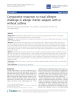
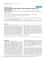
Figure 1 sho ws the changes fr om baseline in the pri-
mary and secondary outcome measures. There were no
significant changes in the virological measures. Both the
CSF (A) and plasma (B) HIV-1 RNA remained stable, as
did the CSF:plasma HIV-1 RNA ratio (not shown). Like-
wise, neither the CSF (C) nor plasma (D) neopterin chan-
ged. Similarly, none of the CSF or blood T cell (E - H) or
mono cyte (I and J) activation levels changed. There was
no reduction in the CSF WBC count (K), which is com-
posed principally of blood-derived T cells [10,41,42]. CSF
CCL2 (L), CSF:blood albumin ratio (M), and the brief
measure of neurological performance, the QNPZ-4 score
(N), also did not change significantly. Curiously, there
was a reduction of absolute CD8+ (O) and CD4+ (P)
T cell numbers in the blood, although only the latter was
statistically significant by repeated measures analysis.
Discussion
This pilot study was undertaken to exp lore the use of
minocycline as an adj uvant treatment for chronic HIV
infection, particularly for attenuating the CNS compo-
nents of immunoactivation and infection. It aimed to
provide a preliminary view of the biological effects of
minocycline on CNS HIV immune reactions and infec-
tion, and to obtain effect-size estimates for power calcula-
tions prior to planning a larger controlled trial. Our
underlying mechanist ic hypotheses centered on the pro-
posed capacity of minocycline to attenuate CNS immune
and systemic perturbations and their effects on CNS
infection as revealed by changes in CSF and blood bio-
markers. We hypothesized that attenuating these immu-
nological effects would be reflected in reductions in CSF
(and perhaps plasma) neopterin and CSF CCL2 concen-
trations, and in the expression of surface activation
Table 1 Baseline subject characteristics
Median Range
Age (years) 49.9 32.0 - 55.2
Gender (M:F) 6:1
Time since HIV diagnosis (years) 17.0 1.7 - 20.3
HIV-1 RNA (log
10
copies/mL)
Plasma 4.49 4.26 - 5.56
CSF 3.87 3.11 - 4.47
Plasma:CSF difference 1.06 0.12 - 1.58
Blood T cells (cells/μL)
CD4+ 453 267 - 806
CD8+ 1,009 575 - 2185
CSF WBCs (cells/μL) 9 2 - 18
Neopterin (nmol/L)
CSF 13.1 5.9 - 41.2
Plasma 13.4 9.2 - 53.5
CSF CCL2 (pg/mL) 479.2 397.9 - 1322.2
T Cell Activation (percent CD38+/HLA-DR+)
CSF CD4+ 14.7 3.4 - 60.2
Blood CD4+ 13.3 7.6 - 24.7
CSF CD8+ 83.4 41.8 - 97.6
Blood CD8+ 58.5 34.5 - 78.0
Monocyte Activation (percent CD16+)
CSF monocytes 93.6 80.1 - 100
Blood monocytes 10.8 4.7 - 17.0
CSF:blood albumin ratio 5.05 3.91 - 12.26
QNPZ-4 -0.32 -3.44 - 0.54
Ho et al. AIDS Research and Therapy 2011, 8:17
/>Page 2 of 8
markers on T cells and monocytes. Additionally, we
hypothesized that minocycline might also indirectly
reduce CNS infection through its effects on various
immune system-related mechanisms that contribute to
the magnitude of CNS (and CSF) infection, including:
CD4+ T cell traffic that brings both infected cells and
uninfected targets into the CNS, and CD4+ T cell and
macrophage activation that enhance viral replic ation in
these cell types. Unfortunately, in this study, none of
these effects were seen. Similarly, there were no changes
in the other secondary endpoints, including CSF WBC
counts, CSF:blood albumin ratios or the brief neurologi-
cal performance battery, the QNPZ-4.
Minocycline, a licensed tetracycline antibiotic, has
been reported to have a number of properties that make
it an attractive adjuvant therapy candidate. In various
model systems, it has been shown to have anti-inflam-
matory effects [43,44], including modulation of T cell
activation and attenuation of macrophage and microglial
activation [27,34,45,46]. It also has neuroprotective
properties in vitro and in in vivo animal models [47-52].
These and other properti es have led to trials of minocy-
cline in several conditions, including rheumatoid arthri-
tis [53], and neurodegenerative and neuroinflammatory
dise ases [33,52,54,55 ]. Minocycline also has been shown
to inhibit HIV replication in microglia in vitro [22].
Figure 1 Changes in outcome variables in the CSF and blood with minocycline treatment. The horizontal bar in panel A indicates the
period of minocycline treatment. Panels show the mean changes from baseline and 95% confidence intervals for CSF (A) and plasma (B) HIV-1
RNA concentrations; CSF (C) and plasma (D) neopterin concentrations; percent of CSF (E) and blood (F) CD8+ T cell activation, as assessed by co-
expression of CD38 and HLA-DR on CD3+CD8+ lymphocytes; percent of CSF (G) and blood (H) CD4+ T cell activation, as assessed by co-
expression of CD38 and HLA-DR on CD3+CD4+ lymphocytes; percent of CSF monocyte activation (I) as assessed by CD16 expression on CD14
+CD4loCD3lo cells; percent of blood monocyte activation (J) as assessed by CD16 expression on CD14+CD4loCD3- cells; CSF WBC counts (K);
CSF CCL2 concentration (L); QNPZ-4 performance score (N); and blood CD8+ (O) and CD4+ (P) T cell counts. Analysis of individual changes from
baseline by Kruskal-Wallis and Dunn’s post hoc testing from baseline to 8 weeks or 14 weeks and by repeated measures from baseline to 8 or
14 weeks with Dunnet’s post hoc testing of each interval found no significant changes for any of the 12 variables shown except for changes in
the blood CD4+ T cell counts (P), which was statistically significant for weeks 0 - 8 (P = 0.035) and weeks 0 - 14 (P = 0.013). Abbreviation: Act =
activation.
Ho et al. AIDS Research and Therapy 2011, 8:17
/>Page 3 of 8
Importantly, in an SIV model of accelerated CNS infec-
tion, minocycline-treated SIV-infected macaques were
noted to have less severe encephalitis, reduced expres-
sion of CNS inflammatory markers, reduced axonal
degeneration and lower levels of CNS virus replication
[23]. Recent in vitro studies on human peripheral blood
CD4+ T cells demonstrate that minocycline has anti-
viral effects in CD4+ T cells and reduces cellular CD4+
T cell activation [27]. Since all of these properties made
it an intriguing candidate for adjuvant use in CNS HIV
infection, our study results thus beg the issue of why we
did not see similar effects in the studied patients.
While it is possible that the CSF m easurements were
insensitive to salutary effects on the brain parenchyma,
including the important perivascular environment, this
does not seem likely. CSF neopterin is a marker of CNS
macrophage activation (presumably including both brain
and meningeal populations) that increases with disease
severity and is especially elevated in HIVE/HAD [11,38].
This pteridine biomarker responds well to antiretroviral
therapy [11], although it does not always return to nor-
mal levels [8,19,56]. Its blood concentration is also a
prognostic marker of disease progression [57]. Bo th CSF
and blood levels were unaffected by minocycline in our
study, suggesting that there was little effect on CNS or
systemic macrophage activation. Similarly, CSF CCL2, a
biomarker of macrophage chemotaxis that is a lso char-
acteristically elevated in HAD/HIVE [58], showed no
changes. This is especially disappointing since CSF
CCL2 has been used as a biomarker in SIV en cephal itis,
and was shown to be reduced by minocycline treatment
in the SIV model [23,40]. Increased levels of CD4+ T
cell, CD8+ T cell and monocyte activation observed
in the CSF compared to the blood is characteristic of
HIV infection [10,42,59] and is likely an important com-
ponent of both systemic [60-62] and CNS disease patho-
genesis [10,20]. These measures also were stable through
the course of minocyline treatment.
CSF HIV-1 RNA levels reflect more than one cellular
source, with the relative contributions differing depending
on the stage of systemic and CNS infection and disease
evolution [63-66]. Short-lived cells, presumably CD4+ T
cells, contribute a CSF viral population that is genetically
similar to the blood population [63]. This component has
been termed transitory infection [5,37] and is presumably
sustained by infected and susceptible CD4+ T cells traf-
ficking into the meninges and brain. In early HIV infec-
tion, this type of infection predominates and may even be
the only type detected [67]. A second viral population
turnsovermoreslowly[66].Thispopulationislikely
derived from macrophages, and is genetically distinct
from the blood population. This component, termed
autonomous or compartmentalized infection, is character-
istically detected as a minor contributor to CSF HIV
levels in neuroasymptomatic chronic infection, but predo-
minates in more advanced infection, particularly HIV
encephalitis (HIVE) [64].
Minocycline might atten uate both types of infection
by its effects on T cell a nd monocyte-macrophage acti-
vation. I n the case of transitory infection, T cell activa-
tion is critical to support HIV replication and also
promotes T cell traffic that carries infecte d and unin-
fected target CD4+ T cells into the meninges and pe ri-
vascular spaces. Hence, if minocycline alters these T cell
properties it might reduce this type of CSF infection.
Similarly, activat ion is likely important for macrophages
in sustaining infection and also, perhaps, in their entry
into the CNS, including into the perivascular spaces,
meninges and parenchyma. Mi nocycline might, there-
fore, reduce this type of autonomous infection. How-
ever, we detected no evidence of reduced CSF infection,
although in the subjects studied with relatively preserved
blood CD4+ T cell counts, the major CSF viral popula-
tion likely originated from transitory type infection,
although this was not directly examined in these
subjects.
Our methods of examining t he hypothesized actions
of minocycline should have been adequate to detect a
substantial immunological or virological effect of minocy-
cline. Possible reasons as to why there were no discern-
able effects similar to those in the SIV-infected pigtailed
macaques may have included species differences. Perhaps
more likely were differences in the disease targets. The
SIV model differs from our subjects in the relatively
shortdiseasedurationandthepresenceoffranklenti-
virus encephalitis [23]. Our study patients had a chronic
‘stable’ infection for a number of years and thus, perhaps,
presented a level of immune activation and viral replica-
tion that the drug effect was too weak to modify. In addi-
tion, the absence of en cephalitis meant that there might
have been little CNS disease to target. Our study , of
course, did not address these possibilities.
The study also did not assess the more direct neuro-
protective properties of minocycline. With one excep-
tion, o ur subjects were largely neuroasymptomatic, and
we performed only brief quantitative neurological per-
formance testing (QNPZ-4) on four measures. The
small improvement noted in this measure, which was
not statistically significant, might have related to prac-
tice effect. However, if the observed improvement was
indeed real, then a study with 20-25 subjects in each of
two treatment arms (minocycline and placebo) would be
need ed for an 80% power to detect the differ ence found
here at 8 weeks. An AIDS Clinical Trials Group study is
studying whether minocycline might improve perfor-
mance in cognitively impaired HIV-infected subjects
( and
these issues should be addressed by that study.
Ho et al. AIDS Research and Therapy 2011, 8:17
/>Page 4 of 8
The observed decline in blood CD4+ and CD8+ T cell
counts was unexpected and unexplained. Curiously, it
did not impact the CSF WBC count. This mild T-cell
lymphopenia needs to be verified in a larger study, and
if so, subject to further investigation.
Overall, this pilot study was subject to several inher-
ent design limitatio ns, including i ts small size, relatively
short duration, and absence of an untreated control
group for comparison, raising concern for Type II
error. Thus, we cannot fully di smiss the possibili ty that
the study was underpowered to detect a mild effect of
the drug or that CSF HIV and CNS immune activation
might decline further with longer exposure. However,
given the minimal changes noted in the major out-
comes, it would take a large study to test the effective-
ness of minocycline on these measures in this typ e o f
patient population. For example, if the small reduction
(-0.070 log10 copies/mL) in CSF HIV-1 RNA at 8
weeks was in deed a ‘real’ finding, then it would require
more than 100 subjects in each of the two arms (mino-
cycline and placebo) to have an 80% power to detect
this difference between the groups, a differe nce with
likely little clinical meaning. In the case of CSF neop-
terin, there was no sta tistically significant reduction,
but if the slight increase at 8 weeks (0.033 nmol/L) was
inverted and a ctually a reduction, it would take 500
subject in each group to detect this difference. Thus,
the effects of minocycline on infection and immune
activation appeared too weak to justify a study of the
requisite size, particularly when viewed in comparison
to the potent effects of combination antiretroviral on
these variables [6].
Conclusions
In conclusion, this small pilot study suggests that any
effects of minocycline on CNS HIV infection and
immune activation were not suf ficient to impact chroni c
HIV in the absence of antiretroviral treatment. There-
fore, there seems little justification or in deed ethical
basis for treati ng chronic HIV infection with minocy-
cline instead of combination antiretroviral drugs. How-
ever, given the reported in vitro and S IV effects o f this
tetracycline [23], there still may be reason for further
study, fo r example in well-treated patients in which the
level of immunoactivation is partially attenuated or in
patients with cognitive impairment in which its neuro-
protective properties may yet prove useful in concert
with combination antiretroviral treatment.
Methods
Thi s stud y was approved by the University of California
San Francisco Committee on Human Research and con-
ducted according to the principles expressed in the
Declaration of Helsinki. In formed written consent was
obtained from all subjects. The study was registered
with ClinicalTrials.gov (number: NCT01064752).
Study design
This was an open-labelled, uncontrolled, pilot study
examining the effects of 100 mg of minocycline taken
orally twice daily for 8 weeks. Subject entry criteria
included: ≥18 y ears of age; chronic HIV inf ection w ith
plasma and CSF HIV-1 RNA concentrations >1,000
copies/mL; not taking antiretroviral therapy (either naïve
to therapy or >6 weeks off treatment with no plans to
start during the period of study); predicted medication
adherence; blood CD4+ T cell counts >100 cells/ μl; no
previous adverse reaction to tetracyclines; no tetracycline
treatment for the past 6 months; no c ontraindications to
lumbar puncture (LP); no active opportunistic infection
or neurological disease confounding evaluations; ADC
stage <1 [68]; no concomitant medications altering the
metab olism or risk of minocycline; hemoglobi n >10 g/dL
and liver transaminas es <2.5 times upper limit of normal;
and not taking any other immunomodulating drugs.
After consent, subjects underwent a screening evaluation
that included lumbar puncture (LP) and CSF characteri-
zation, concurrent blood sampling, and standardized
neurological assessments as previously describ ed
[6,10,69]. For those meeting entry criteria, this also
served as the baseline visit, and they starting minocycline
100 mg twi ce daily orally for t he next 8 weeks. At four
and eight weeks, and after a 6-week washout period off
minocycline, subjects underwent repeated evaluation
similar to the baseline, including LP and CSF analysis
[6,10,69]. Treatment adherence was assessed at each on-
study visit by direct questioning and pill count.
The primary outcome measures were the change from
baseline during treatment in CSF HIV-1 RNA and CSF
neopterin concentrations as indices of CNS infection
and immunoactivation [38]. Change from baseline was
calculated at weeks four and eight after initiation of
minocycl ine treatment and after a 6-week wash-out per-
iod. Additional secondary measured outcomes included
changes in: CSF white blood cell (WBC) count; blood
CD4+ and CD8+ counts; ratio of CSF to blood albumin
as a measure of blood-brain barrier permeability [70,71];
CSF CCL2 as a measure of monocyte-macrophage che-
motaxis [58]; CSF and blood CD4+ and CD8+ T cell
and monocyte activation as measured by multiparameter
flow cytometry [10]. Four quantitative tests (timed gait,
grooved pegboard, finger tapping and digit symbol) were
used to obtain a simple quantitative neurological perfor-
mance aggregate score (QNPZ-4) [72].
CSF and blood assays
HIV-1 RNA was measured in cell-free CSF a nd plasma
by the Roche Amplicor HIV-1 Monitor assay (versions
Ho et al. AIDS Research and Therapy 2011, 8:17
/>Page 5 of 8
1.0 and 1.5, Roche Diagnostic Systems, Inc., Branchburg,
N.J). Neopterin concentrations in cell-free CSF and
plasma were measured in batch by ELISA according to
the manufacturer’s instructions (BRAHMS Aktienge-
sellschaft, Hennigsdorf, German y). Blood CD4+ and
CD8+ T cell counts were performed in the San Fran-
cisco General Hospital (SFGH) Clinical Laboratories
using standard flow cytometric methods. CCL2 was
measured in cell-free CSF by ELISA (R&D Systems,
Minneapolis, MN). Other measurements performed in
the SFGH Clinical Laboratories u sing routine clinical
methods included CSF and blood albumin (used to
compute the CSF: blood albumin ratio [70,71]), CSF
WBC count s and different ial, CSF total protein and
blood metabolic profile.
CSF and blood CD4+ and CD8+ T cell activation were
assessed by the percent of these cells in fresh specimens
co-expressing surface CD38 and HLA-DR by multipara-
meter flow cytometry as previously described [10].
Blood monocytes were defined as CD14+CD4loCD3-
cells from the mononuclear gate. CSF monocytes had
low level staining for CD3 and were defined as CD14
+CD4loCD3lo cells. Monocyte activation was defined by
the percent of these cells expressing CD16 [10]. Flow
cytometry data was compensated and analysed with
FlowJo (Tree Star, Ashland, OR).
Statistics
Changes from baseline to follow-up test intervals were
analysed by Kruskal-Wallis test with Dunn’ sposthoc
compa rison of individual intervals and additionally from
baseline through week 8 using repeated measures
ANOVA with Dunnet’ s post hoc comparison. All P
values were two-sided with values <0.05 considered sig-
nificant. Statistical analyses used Prism 5 (GraphPad
Software Inc, San Diego, CA) while power calculations
used StatMate 2.00 (GraphPad Software Inc).
Acknowledgements
This work was supported by National Institutes of Health R01 MH62701, K23
MH074466, and the National Center for Research Resources support of the
University of California San Francisco-Clinical and Translational Sciences
Institute, UL1 RR024131. Its contents are solely the responsibility of the
authors and do not represent the official views of the NIH. E.L.H. was a
recipient of a Clinical Research Training Fellowship from the American
Academy of Neurology.
These study results were presented in preliminary fashion at the Conference
on Retroviruses and Opportunistic Infections (CROI) 2010 (Poster #426) in
San Francisco, February 2010.
Author details
1
Department of Neurology
1
University of California San Francisco, San
Francisco, CA, USA.
2
Division of Biological Chemistry, Biocentre, Innsbruck
Medical University, Innsbruck, Austria.
3
Division of Experimental Medicine,
Department of Medicine, University of California San Francisco, San
Francisco, CA, USA.
4
Department of Neurology, University of Washington,
Seattle, WA, USA.
5
Department of Neurology, Yale University, New Haven, CT,
USA.
Authors’ contributions
ELH examined study participants, performed lumbar punctures, and assisted
with the analysis of the data and preparation of the manuscript. SSS
examined study participants, performed lumbar punctures, and assisted in
design of the study and reviewed the manuscript. EL served as the patient
study coordinator, aided in the design of the study, performed the
quantitative neurological performance testing and managed the data. DF
performed assays of CSF and plasma neopterin. ES designed the flow
cytometry assays, directed the SFGH Clinical Immunology Laboratory that
performed the flow cytometry assays and CSF CCL2 ELISA assays, and
analysed and interpreted flow cytometry data. RWP designed and oversaw
the study, examined study participants, performed lumbar punctures,
analysed and interpreted the data, and participated in preparation of the
manuscript. All authors read and approved of the final manuscript.
Competing interests
Dr. Price has received funding from Merck to support an investigator-
initiated research study and an honorarium from Abbott for a conference
presentation. The other authors have no competing interests.
Received: 14 January 2011 Accepted: 12 May 2011
Published: 12 May 2011
References
1. Davis LE, Hjelle BL, Miller VE, Palmer DL, Llewellyn AL, Merlin TL, Young SA,
Mills RG, Wachsman W, Wiley CA: Early viral brain invasion in iatrogenic
human immunodeficiency virus infection. Neurology 1992,
42(9):1736-1739.
2. Pilcher CD, Shugars DC, Fiscus SA, Miller WC, Menezes P, Giner J, Dean B,
Robertson K, Hart CE, Lennox JL, et al: HIV in body fluids during primary
HIV infection: implications for pathogenesis, treatment and public
health. AIDS 2001, 15(7):837-845.
3. Ellis RJ, Hsia K, Spector SA, Nelson JA, Heaton RK, Wallace MR, Abramson I,
Atkinson JH, Grant I, McCutchan JA: Cerebrospinal fluid human
immunodeficiency virus type 1 RNA levels are elevated in neurocognitively
impaired individuals with acquired immunodeficiency syndrome. HIV
Neurobehavioral Research Center Group. Ann Neurol 1997, 42(5):679-688.
4. McArthur JC, McClernon DR, Cronin MF, Nance-Sproson TE, Saah AJ, St
Clair M, Lanier ER: Relationship between human immunodeficiency virus-
associated dementia and viral load in cerebrospinal fluid and brain. Ann
Neurol 1997, 42(5):689-698.
5. Spudich SS, Nilsson AC, Lollo ND, Liegler TJ, Petropoulos CJ, Deeks SG,
Paxinos EE, Price RW: Cerebrospinal fluid HIV infection and pleocytosis:
relation to systemic infection and antiretroviral treatment. BMC Infect Dis
2005, 5 :98.
6. Spudich S, Lollo N, Liegler T, Deeks SG, Price RW: Treatment benefit on
cerebrospinal fluid HIV-1 levels in the setting of systemic virological
suppression and failure. J Infect Dis 2006, 194(12):1686-1696.
7. Gisslen M, Fuchs D, Svennerholm B, Hagberg L: Cerebrospinal fluid viral
load, intrathecal immunoactivation, and cerebrospinal fluid monocytic
cell count in HIV-1 infection. J Acquir Immune Defic Syndr 1999,
21(4):271-276.
8. Abdulle S, Hagberg L, Svennerholm B, Fuchs D, Gisslen M: Continuing
intrathecal immunoactivation despite two years of effective
antiretroviral therapy against HIV-1 infection. AIDS 2002,
16(16):2145-2149.
9. Cinque P, Brew BJ, Gisslen M, Hagberg L, Price RW: Cerebrospinal fluid
markers in central nervous system HIV infection and AIDS dementia
complex. Handb Clin Neurol 2007, 85:261-300.
10. Sinclair E, Ronquillo R, Lollo N, Deeks SG, Hunt P, Yiannoutsos CT,
Spudich S, Price RW: Antiretroviral treatment effect on immune activation
reduces cerebrospinal fluid HIV-1 infection. J Acquir Immune Defic Syndr
2008, 47(5) :544-552.
11. Hagberg L, Cinque P, Gisslen M, Brew BJ, Spudich S, Bestetti A, Price RW,
Fuchs D: Cerebrospinal fluid neopterin: an informative biomarker of
central nervous system immune activation in HIV-1 infection. AIDS Res
Ther 2010, 7:15.
12. Navia BA, Jordan BD, Price RW: The AIDS dementia complex: I. Clinical
features. Ann Neurol 1986, 19(6):517-524.
13. Antinori A, Arendt G, Becker JT, Brew BJ, Byrd DA, Cherner M, Clifford DB,
Cinque P, Epstein LG, Goodkin K, et al
: Updated
research nosology for
Ho et al. AIDS Research and Therapy 2011, 8:17
/>Page 6 of 8
HIV-associated neurocognitive disorders. Neurology 2007,
69(18):1789-1799.
14. Kaul M, Zheng J, Okamoto S, Gendelman HE, Lipton SA: HIV-1 infection
and AIDS: consequences for the central nervous system. Cell Death Differ
2005, 12(Suppl 1):878-892.
15. Gonzalez-Scarano F, Martin-Garcia J: The neuropathogenesis of AIDS. Nat
Rev Immunol 2005, 5(1):69-81.
16. Simioni S, Cavassini M, Annoni JM, Rimbault Abraham A, Bourquin I,
Schiffer V, Calmy A, Chave JP, Giacobini E, Hirschel B, et al: Cognitive
dysfunction in HIV patients despite long-standing suppression of
viremia. AIDS 2010, 24(9):1243-1250.
17. Heaton RK, Clifford DB, Franklin DR Jr, Woods SP, Ake C, Vaida F, Ellis RJ,
Letendre SL, Marcotte TD, Atkinson JH, et al: HIV-associated
neurocognitive disorders persist in the era of potent antiretroviral
therapy: CHARTER Study. Neurology 2010, 75(23):2087-2096.
18. Tozzi V, Balestra P, Bellagamba R, Corpolongo A, Salvatori MF, Visco-
Comandini U, Vlassi C, Giulianelli M, Galgani S, Antinori A, et al: Persistence
of neuropsychologic deficits despite long-term highly active
antiretroviral therapy in patients with HIV-related neurocognitive
impairment: prevalence and risk factors. J Acquir Immune Defic Syndr
2007, 45(2) :174-182.
19. Eden A, Price RW, Spudich S, Fuchs D, Hagberg L, Gisslen M: Immune
activation of the central nervous system is still present after >4 years of
effective highly active antiretroviral therapy. J Infect Dis 2007,
196(12):1779-1783.
20. Ancuta P, Kamat A, Kunstman KJ, Kim EY, Autissier P, Wurcel A, Zaman T,
Stone D, Mefford M, Morgello S, et al: Microbial translocation is associated
with increased monocyte activation and dementia in AIDS patients. PLoS
One 2008, 3(6):e2516.
21. Uthman OA, Abdulmalik JO: Adjunctive therapies for AIDS dementia
complex. Cochrane Database Syst Rev 2008, , 3: CD006496.
22. Si Q, Cosenza M, Kim MO, Zhao ML, Brownlee M, Goldstein H, Lee S: A
novel action of minocycline: inhibition of human immunodeficiency
virus type 1 infection in microglia. J Neurovirol 2004, 10(5):284-292.
23. Zink MC, Uhrlaub J, DeWitt J, Voelker T, Bullock B, Mankowski J, Tarwater P,
Clements J, Barber S: Neuroprotective and anti-human immunodeficiency
virus activity of minocycline. JAMA 2005, 293(16):2003-2011.
24. Follstaedt SC, Barber SA, Zink MC: Mechanisms of minocycline-induced
suppression of simian immunodeficiency virus encephalitis: inhibition of
apoptosis signal-regulating kinase 1. J Neurovirol 2008, 14(5):376-388.
25. Clements JE, Mankowski JL, Gama L, Zink MC: The accelerated simian
immunodeficiency virus macaque model of human immunodeficiency
virus-associated neurological disease: from mechanism to treatment.
J Neurovirol
2008, 14(4):309-317.
26.
Ratai
EM, Bombardier JP, Joo CG, Annamalai L, Burdo TH, Campbell J, Fell R,
Hakimelahi R, He J, Autissier P, et al: Proton magnetic resonance
spectroscopy reveals neuroprotection by oral minocycline in a
nonhuman primate model of accelerated NeuroAIDS. PLoS One 2010,
5(5):e10523.
27. Szeto GL, Brice AK, Yang HC, Barber SA, Siliciano RF, Clements JE:
Minocycline attenuates HIV infection and reactivation by suppressing
cellular activation in human CD4+ T cells. J Infect Dis 2010,
201(8):1132-1140.
28. Chen M, Ona VO, Li M, Ferrante RJ, Fink KB, Zhu S, Bian J, Guo L, Farrell LA,
Hersch SM, et al: Minocycline inhibits caspase-1 and caspase-3
expression and delays mortality in a transgenic mouse model of
Huntington disease. Nat Med 2000, 6(7):797-801.
29. Lin S, Zhang Y, Dodel R, Farlow MR, Paul SM, Du Y: Minocycline blocks
nitric oxide-induced neurotoxicity by inhibition p38 MAP kinase in rat
cerebellar granule neurons. Neurosci Lett 2001, 315(1-2):61-64.
30. Du Y, Ma Z, Lin S, Dodel RC, Gao F, Bales KR, Triarhou LC, Chernet E,
Perry KW, Nelson DL, et al: Minocycline prevents nigrostriatal
dopaminergic neurodegeneration in the MPTP model of Parkinson’s
disease. Proc Natl Acad Sci USA 2001, 98(25):14669-14674.
31. Van Den Bosch L, Tilkin P, Lemmens G, Robberecht W: Minocycline delays
disease onset and mortality in a transgenic model of ALS. Neuroreport
2002, 13(8) :1067-1070.
32. Wu DC, Jackson-Lewis V, Vila M, Tieu K, Teismann P, Vadseth C, Choi DK,
Ischiropoulos H, Przedborski S: Blockade of microglial activation is
neuroprotective in the 1-methyl-4-phenyl-1,2,3,6-tetrahydropyridine
mouse model of Parkinson disease. J Neurosci 2002, 22(5):1763-1771.
33. Metz LM, Zhang Y, Yeung M, Patry DG, Bell RB, Stoian CA, Yong VW,
Patten SB, Duquette P, Antel JP, et al: Minocycline reduces gadolinium-
enhancing magnetic resonance imaging lesions in multiple sclerosis.
Ann Neurol 2004, 55(5):756.
34. Michel-Monigadon D, Nerriere-Daguin V, Leveque X, Plat M, Venturi E,
Brachet P, Naveilhan P, Neveu I: Minocycline promotes long-term survival
of neuronal transplant in the brain by inhibiting late microglial
activation and T-cell recruitment. Transplantation 2010, 89(7):816-823.
35. Guimaraes JS, Freire MA, Lima RR, Picanco-Diniz CW, Pereira A, Gomes-
Leal W: Minocycline treatment reduces white matter damage after
excitotoxic striatal injury. Brain Res 2010, 1329:182-193.
36. Price RW, Staprans S: Measuring the “viral load” in cerebrospinal fluid in
human immunodeficiency virus infection: window into brain infection?
Ann Neurol 1997, 42(5)
:675-678.
37.
Price
RW: The two faces of HIV infection of cerebrospinal fluid. Trends
Microbiol 2000, 8(9):387-391.
38. Wirleitner B, Schroecksnadel K, Winkler C, Fuchs D: Neopterin in HIV-1
infection. Mol Immunol 2005, 42(2):183-194.
39. Zink MC, Clements JE: A novel simian immunodeficiency virus model that
provides insight into mechanisms of human immunodeficiency virus
central nervous system disease. J Neurovirol 2002, 8(Suppl 2):42-48.
40. Mankowski JL, Queen SE, Clements JE, Zink MC: Cerebrospinal fluid
markers that predict SIV CNS disease. J Neuroimmunol 2004, 157(1-
2):66-70.
41. Shacklett BL, Cox CA, Wilkens DT, Karl Karlsson R, Nilsson A, Nixon DF,
Price RW: Increased adhesion molecule and chemokine receptor
expression on CD8+ T cells trafficking to cerebrospinal fluid in HIV-1
infection. J Infect Dis 2004, 189(12):2202-2212.
42. Neuenburg JK, Cho TA, Nilsson A, Bredt BM, Hebert SJ, Grant RM, Price RW:
T-cell activation and memory phenotypes in cerebrospinal fluid during
HIV infection. J Acquir Immune Defic Syndr 2005, 39(1):16-22.
43. Tikka T, Fiebich BL, Goldsteins G, Keinanen R, Koistinaho J: Minocycline, a
tetracycline derivative, is neuroprotective against excitotoxicity by
inhibiting activation and proliferation of microglia. J Neurosci 2001,
21(8):2580-2588.
44. Tikka TM, Koistinaho JE: Minocycline provides neuroprotection against N-
methyl-D-aspartate neurotoxicity by inhibiting microglia. J Immunol 2001,
166(12):7527-7533.
45. Kloppenburg M, Verweij CL, Miltenburg AM, Verhoeven AJ, Daha MR,
Dijkmans BA, Breedveld FC: The influence of tetracyclines on T cell
activation. Clin Exp Immunol 1995, 102(3):635-641.
46. Maier K, Merkler D, Gerber J, Taheri N, Kuhnert AV, Williams SK, Neusch C,
Bahr M, Diem R: Multiple neuroprotective mechanisms of minocycline in
autoimmune CNS inflammation. Neurobiol Dis 2007, 25(3):514-525.
47. Yrjanheikki J, Tikka T, Keinanen R, Goldsteins G, Chan PH, Koistinaho J: A
tetracycline derivative, minocycline, reduces inflammation and protects
against focal cerebral ischemia with a wide therapeutic window. Proc
Natl Acad Sci USA 1999, 96(23):13496-13500.
48. Hersch S, Fink K, Vonsattel JP, Friedlander RM: Minocycline is protective in
a mouse model of Huntington’s disease. Ann Neurol 2003, 54(6):841,
author reply 842-843.
49. Hunter CL, Bachman D, Granholm AC: Minocycline prevents cholinergic
loss in a mouse model of Down’s syndrome. Ann Neurol 2004,
56(5)
:675-688.
50.
Stirling
DP, Khodarahmi K, Liu J, McPhail LT, McBride CB, Steeves JD,
Ramer MS, Tetzlaff W: Minocycline treatment reduces delayed
oligodendrocyte death, attenuates axonal dieback, and improves
functional outcome after spinal cord injury. J Neurosci 2004,
24(9):2182-2190.
51. Ryu JK, Franciosi S, Sattayaprasert P, Kim SU, McLarnon JG: Minocycline
inhibits neuronal death and glial activation induced by beta-amyloid
peptide in rat hippocampus. Glia 2004, 48(1):85-90.
52. Kim HS, Suh YH: Minocycline and neurodegenerative diseases. Behav
Brain Res 2009, 196(2):168-179.
53. Tilley BC, Alarcon GS, Heyse SP, Trentham DE, Neuner R, Kaplan DA,
Clegg DO, Leisen JC, Buckley L, Cooper SM, et al: Minocycline in
rheumatoid arthritis. A 48-week, double-blind, placebo-controlled trial.
MIRA Trial Group. Ann Intern Med 1995, 122(2):81-89.
54. Lampl Y, Boaz M, Gilad R, Lorberboym M, Dabby R, Rapoport A, Anca-
Hershkowitz M, Sadeh M: Minocycline treatment in acute stroke: an
open-label, evaluator-blinded study. Neurology 2007, 69(14):1404-1410.
Ho et al. AIDS Research and Therapy 2011, 8:17
/>Page 7 of 8
55. Zabad RK, Metz LM, Todoruk TR, Zhang Y, Mitchell JR, Yeung M, Patry DG,
Bell RB, Yong VW: The clinical response to minocycline in multiple
sclerosis is accompanied by beneficial immune changes: a pilot study.
Mult Scler 2007, 13(4):517-526.
56. Yilmaz A, Price RW, Spudich S, Fuchs D, Hagberg L, Gisslen M: Persistent
intrathecal immune activation in HIV-1-infected individuals on
antiretroviral therapy. J Acquir Immune Defic Syndr 2008, 47(2):168-173.
57. Mildvan D, Spritzler J, Grossberg SE, Fahey JL, Johnston DM, Schock BR,
Kagan J: Serum neopterin, an immune activation marker, independently
predicts disease progression in advanced HIV-1 infection. Clin Infect Dis
2005, 40(6) :853-858.
58. Cinque P, Vago L, Mengozzi M, Torri V, Ceresa D, Vicenzi E, Transidico P,
Vagani A, Sozzani S, Mantovani A, et al: Elevated cerebrospinal fluid levels
of monocyte chemotactic protein-1 correlate with HIV-1 encephalitis
and local viral replication. AIDS 1998, 12(11):1327-1332.
59. Neuenburg JK, Furlan S, Bacchetti P, Price RW, Grant RM: Enrichment of
activated monocytes in cerebrospinal fluid during antiretroviral therapy.
AIDS 2005, 19(13):1351-1359.
60. Deeks SG, Kitchen CM, Liu L, Guo H, Gascon R, Narvaez AB, Hunt P,
Martin JN, Kahn JO, Levy J, et al: Immune activation set point during early
HIV infection predicts subsequent CD4+ T-cell changes independent of
viral load. Blood 2004, 104(4):942-947.
61. Hunt PW: Role of immune activation in HIV pathogenesis. Curr HIV/AIDS
Rep 2007, 4(1):42-47.
62. Hunt PW, Brenchley J, Sinclair E, McCune JM, Roland M, Page-Shafer K,
Hsue P, Emu B, Krone M, Lampiris H, et al: Relationship between T cell
activation and CD4+ T cell count in HIV-seropositive individuals with
undetectable plasma HIV RNA levels in the absence of therapy. J Infect
Dis 2008, 197(1):126-133.
63. Harrington PR, Haas DW, Ritola K, Swanstrom R: Compartmentalized
human immunodeficiency virus type 1 present in cerebrospinal fluid is
produced by short-lived cells. J Virol 2005, 79(13):7959-7966.
64. Ritola K, Robertson K, Fiscus SA, Hall C, Swanstrom R: Increased human
immunodeficiency virus type 1 (HIV-1) env compartmentalization in the
presence of HIV-1-associated dementia. J Virol 2005, 79(16):10830-10834.
65. Harrington PR, Schnell G, Letendre SL, Ritola K, Robertson K, Hall C,
Burch CL, Jabara CB, Moore DT, Ellis RJ, et al: Cross-sectional
characterization of HIV-1 env compartmentalization in cerebrospinal
fluid over the full disease course. AIDS 2009, 23(8):907-915.
66. Schnell G, Spudich S, Harrington P, Price RW, Swanstrom R:
Compartmentalized human immunodeficiency virus type 1 originates
from long-lived cells in some subjects with HIV-1-associated dementia.
PLoS Pathog 2009, 5(4):e1000395.
67. Schnell G, Price RW, Swanstrom R, Spudich S: Compartmentalization and
clonal amplification of HIV-1 variants in the cerebrospinal fluid during
primary infection. J Virol 2010, 84(5):2395-2407.
68. Price RW, Brew BJ: The AIDS dementia complex. J Infect Dis 1988,
158(5):1079-1083.
69. Probasco JC, Spudich SS, Critchfield J, Lee E, Lollo N, Deeks SG, Price RW:
Failure of atorvastatin to modulate CSF HIV-1 infection: results of a pilot
study. Neurology 2008, 71(7):521-524.
70. Link H, Tibbling G: Principles of albumin and IgG analyses in neurological
disorders. III. Evaluation of IgG synthesis within the central nervous
system in multiple sclerosis. Scand J Clin Lab Invest 1977, 37(5):397-401.
71. Tibbling G, Link H, Ohman S: Principles of albumin and IgG analyses in
neurological disorders. I. Establishment of reference values. Scand J Clin
Lab Invest 1977, 37(5):385-390.
72. Price RW, Yiannoutsos CT, Clifford DB, Zaborski L, Tselis A, Sidtis JJ, Cohen B,
Hall CD, Erice A, Henry K: Neurological outcomes in late HIV infection:
adverse impact of neurological impairment on survival and protective
effect of antiviral therapy. AIDS Clinical Trial Group and Neurological
AIDS Research Consortium study team. AIDS 1999, 13(13):1677-1685.
doi:10.1186/1742-6405-8-17
Cite this article as: Ho et al.: Minocycline fails to modulate
cerebrospinal fluid HIV infection or immune activation in chronic
untreated HIV-1 infection: results of a pilot study. AIDS Research and
Therapy 2011 8:17.
Submit your next manuscript to BioMed Central
and take full advantage of:
• Convenient online submission
• Thorough peer review
• No space constraints or color figure charges
• Immediate publication on acceptance
• Inclusion in PubMed, CAS, Scopus and Google Scholar
• Research which is freely available for redistribution
Submit your manuscript at
www.biomedcentral.com/submit
Ho et al. AIDS Research and Therapy 2011, 8:17
/>Page 8 of 8