ABC OF LIVER, PANCREAS AND GALL BLADDER - PART 2 ppsx
Bạn đang xem bản rút gọn của tài liệu. Xem và tải ngay bản đầy đủ của tài liệu tại đây (365.45 KB, 11 trang )
1 Investigation of liver and biliary disease
I J Beckingham, S D Ryder
Jaundice is the commonest presentation of patients with liver
and biliary disease. The cause can be established in most cases
by simple non-invasive tests, but many patients will require
referral to a specialist for management. Patients with high
concentrations of bilirubin ( > 100 mol/l) or with evidence of
sepsis or cholangitis are at high risk of developing
complications and should be referred as an emergency because
delays in treatment adversely affect prognosis.
Jaundice
Hyperbilirubinaemia is defined as a bilirubin concentration
above the normal laboratory upper limit of 19 mol/l. Jaundice
occurs when bilirubin becomes visible within the sclera, skin,
and mucous membranes, at a blood concentration of around
40 mol/l. Jaundice can be categorised as prehepatic, hepatic,
or posthepatic, and this provides a useful framework for
identifying the underlying cause.
Around 3% of the UK population have hyperbilirubinaemia
(up to 100 mol/l) caused by excess unconjugated bilirubin, a
condition known as Gilbert’s syndrome. These patients have
mild impairment of conjugation within the hepatocytes. The
condition usually becomes apparent only during a transient rise
in bilirubin concentration (precipitated by fasting or illness) that
results in frank jaundice. Investigations show an isolated
unconjugated hyperbilirubinaemia with normal liver enzyme
activities and reticulocyte concentrations. The syndrome is often
familial and does not require treatment.
Prehepatic jaundice
In prehepatic jaundice, excess unconjugated bilirubin is
produced faster than the liver is able to conjugate it for
excretion. The liver can excrete six times the normal daily load
before bilirubin concentrations in the plasma rise.
Unconjugated bilirubin is insoluble and is not excreted in the
urine. It is most commonly due to increased haemolysis
—
for
example, in spherocytosis, homozygous sickle cell disease, or
thalassaemia major
—
and patients are often anaemic with
splenomegaly. The cause can usually be determined by further
haematological tests (red cell film for reticulocytes and
abnormal red cell shapes, haemoglobin electrophoresis, red cell
antibodies, and osmotic fragility).
Hepatic and posthepatic jaundice
Most patients with jaundice have hepatic (parenchymal) or
posthepatic (obstructive) jaundice. Several clinical features may
help distinguish these two important groups but cannot be
relied on, and patients should have ultrasonography to look for
evidence of biliary obstruction.
The most common intrahepatic causes are viral hepatitis,
alcoholic cirrhosis, primary biliary cirrhosis, drug induced
jaundice, and alcoholic hepatitis. Posthepatic jaundice is most
often due to biliary obstruction by a stone in the common bile
duct or by carcinoma of the pancreas. Pancreatic pseudocyst,
chronic pancreatitis, sclerosing cholangitis, a bile duct stricture,
or parasites in the bile duct are less common causes.
In obstructive jaundice (both intrahepatic cholestasis and
extrahepatic obstruction) the serum bilirubin is principally
conjugated. Conjugated bilirubin is water soluble and is
Box 1.1 History that should be taken from patients
presenting with jaundice
x Duration of jaundice
x Previous attacks of jaundice
x Pain
x Chills, fever, systemic symptoms
x Itching
x Exposure to drugs (prescribed and illegal)
x Biliary surgery
x Anorexia, weight loss
x Colour of urine and stool
x Contact with other jaundiced patients
x History of injections or blood transfusions
x Occupation
Box1.2 Examination of patients with jaundice
x Depth of jaundice
x Scratch marks
x Signs of chronic liver disease:
Palmar erythema
Clubbing
White nails
Dupuytren’s contracture
Gynaecomastia
x Liver:
Size
Shape
Surface
x Enlargement of gall bladder
x Splenomegaly
x Abdominal mass
x Colour of urine and stools
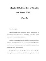
Old red blood cells
Spleen
Fe
2
+
Haem
Unconjugated
bilirubin
Conjugated
bilirubin
Bile
canaliculi
Bile
ducts
Small amount of reduced
bilirubin reabsorbed into
portal vein liver
systemic blood supply
kidneys
Bilirubin
reduced by
gut bacteria
to:
Stercobilinogen
Faeces
Terminal
ileum
Colon
Liver
Kidney
Urobilinogen
Hepatocytes
Albumin
Duodenum
Figure 1.1 Bilirubin pathway
1
This is trial version
www.adultpdf.com
excreted in the urine, giving it a dark colour (bilirubinuria). At
the same time, lack of bilirubin entering the gut results in pale,
“putty” coloured stools and an absence of urobilinogen in the
urine when measured by dipstick testing. Jaundice due to
hepatic parenchymal disease is characterised by raised
concentrations of both conjugated and unconjugated serum
bilirubin, and typically stools and urine are of normal colour.
However, although pale stools and dark urine are a feature of
biliary obstruction, they can occur transiently in many acute
hepatic illnesses and are therefore not a reliable clinical feature
to distinguish obstruction from hepatic causes of jaundice.
Liver function tests
Liver function tests routinely combine markers of function
(albumin and bilirubin) with markers of liver damage (alanine
transaminase, alkaline phosphatase, and -glutamyl transferase).
Abnormalities in liver enzyme activities give useful information
about the nature of the liver insult: a predominant rise in
alanine transaminase activity (normally contained within the
hepatocytes) suggests a hepatic process. Serum transaminase
activity is not usually raised in patients with obstructive
jaundice, although in patients with common duct stones and
cholangitis a mixed picture of raised biliary and hepatic enzyme
activity is often seen.
Epithelial cells lining the bile canaliculi produce alkaline
phosphatase, and its serum activity is raised in patients with
intrahepatic cholestasis, cholangitis, or extrahepatic obstruction;
increased activity may also occur in patients with focal hepatic
lesions in the absence of jaundice. In cholangitis with
incomplete extrahepatic obstruction, patients may have normal
or slightly raised serum bilirubin concentrations and high
serum alkaline phosphatase activity. Serum alkaline
phosphatase is also produced in bone, and bone disease may
complicate the interpretation of abnormal alkaline phosphatase
activity. If increased activity is suspected to be from bone, serum
concentrations of calcium and phosphorus should be measured
together with 5¢-nucleotidase or -glutamyl transferase activity;
these two enzymes are also produced by bile ducts, and their
activity is raised in cholestasis but remains unchanged in bone
disease.
Occasionally, the enzyme abnormalities may not give a clear
answer, showing both a biliary and hepatic component. This is
usually because of cholangitis associated with stones in the
common bile duct, where obstruction is accompanied by
hepatocyte damage as a result of infection within the biliary
tree.
Plasma proteins and coagulation
factors
A low serum albumin concentration suggests chronic liver
disease. Most patients with biliary obstruction or acute hepatitis
will have normal serum albumin concentrations as the half life
of albumin in plasma is around 20 days and it takes at least 10
days for the concentration to fall below the normal range
despite impaired liver function.
Coagulation factors II, V, VII, and IX are synthesised in the
liver. Abnormal clotting (measured as prolongation of the
international normalised ratio) occurs in both biliary
obstruction and parenchymal liver disease because of a
combination of poor absorption of fat soluble vitamin K (due to
absence of bile in the gut) and a reduced ability of damaged
hepatocytes to produce clotting factors.
Box 1.3 Drugs that may cause liver damage
Analgesics
x Paracetamol
x Aspirin
x Non-steroidal anti-inflammatory drugs
Cardiac drugs
x Methyldopa
x Amiodarone
Psychotropic drugs
x Monoamine oxidase inhibitors
x Phenothiazines (such as chlorpromazine)
Others
x Sodium valproate
x Oestrogens (oral contraceptives and hormone replacement
therapy)
The presence of a low serum albumin
concentration in a jaundiced patient
suggests a chronic disease process
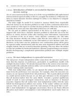
Initial consultation send results of
• Liver function tests
• Hepatitis A IgM
• Hepatitis B surface antigen
Bilirubin <100 µmol/l
Alanine transaminase = hepatitis
Hepatitis A IgM
positive
Hepatitis A IgM
negative
Treat for
hepatitis A
Refer
Alkaline phosphatase g-glutamyltransferase -
cholestasis/obstruction
Bilirubin >100 µmol/l Urgent referral
Refer
Figure 1.2 Guide to investigation and referral of patients with jaundice in
primary care
ABC of Liver, Pancreas, and Gall Bladder
2
This is trial version
www.adultpdf.com
Serum globulin titres rise in chronic hepatitis and cirrhosis,
mainly due to a rise in the IgA and IgG fractions. High titres of
IgM are characteristic of primary biliary cirrhosis, and IgG is a
hallmark of chronic active hepatitis. Ceruloplasmin activity
(ferroxidase, a copper transporting globulin) is reduced in
Wilson’s disease. Deficiency of
1
antitrypsin (an enzyme
inhibitor) is a cause of cirrhosis as well as emphysema. High
concentrations of the iron carrying protein ferritin are a marker
of haemochromatosis.
Autoantibodies are a series of antibodies directed against
subcellular fractions of various organs that are released into the
circulation when cells are damaged. High titres of
antimitochondrial antibodies are specific for primary biliary
cirrhosis, and antismooth muscle and antinuclear antibodies are
often seen in autoimmune chronic active hepatitis. Antibodies
against hepatitis are discussed in detail in a future article on
hepatitis.
Imaging in liver and biliary disease
Plain radiography has a limited role in the investigation of
hepatobiliary disease. Chest radiography may show small
amounts of subphrenic gas, abnormalities of diaphragmatic
contour, and related pulmonary disease, including metastases.
Abdominal radiographs can be useful if a patient has calcified
or gas containing lesions as these may be overlooked or
misinterpreted on ultrasonography. Such lesions include
calcified gall stones (10-15% of gall stones), chronic calcific
pancreatitis, gas containing liver abscesses, portal venous gas,
and emphysematous cholecystitis.
Ultrasonography is the first line imaging investigation in
patients with jaundice, right upper quadrant pain, or
hepatomegaly. It is non-invasive, inexpensive, and quick but
requires experience in technique and interpretation.
Ultrasonography is the best method for identifying gallbladder
stones and for confirming extrahepatic biliary obstruction as
dilated bile ducts are visible. It is good at identifying liver
abnormalities such as cysts and tumours and pancreatic masses
and fluid collections, but visualisation of the lower common bile
duct and pancreas is often hindered by overlying bowel gas.
Computed tomography is complementary to ultrasonography
and provides information on liver texture, gallbladder disease,
bile duct dilatation, and pancreatic disease. Computed
tomography is particularly valuable for detecting small lesions
in the liver and pancreas.
Cholangiography identifies the level of biliary obstruction
and often the cause. Intravenous cholangiography is rarely used
now as opacification of the bile ducts is poor, particularly in
jaundiced patients, and anaphylaxis remains a problem.
Endoscopic retrograde cholangiopancreatography is advisable
when the lower end of the duct is obstructed (by gall stones or
carcinoma of the pancreas). The cause of the obstruction (for
example, stones or parasites) can sometimes be removed by
endoscopic retrograde cholangiopancreatography to allow
cytological or histological diagnosis.
Percutaneous transhepatic cholangiography is preferred for
hilar obstructions (biliary stricture, cholangiocarcinoma of the
hepatic duct bifurcation) because better opacification of the
ducts near the obstruction provides more information for
planning subsequent management. Obstruction can be relieved
by insertion of a plastic or metal tube (a stent) at either
endoscopic retrograde cholangiopancreatography or
percutaneous transhepatic cholangiography.
Magnetic resonance cholangiopancreatography allows
non-invasive visualisation of the bile and pancreatic ducts. It is
Table 1.1 Autoantibody and immunoglobulin characteristics
in liver disease
Autoantibodies Immunoglobulins
Primary biliary
cirrhosis
High titre of
antimitochondrial antibody
in 95% of patients
Raised IgM
Autoimmune
chronic active
hepatitis
Smooth muscle antibody in
70%, antinuclear factor in
60%, Low antimitochondrial
antibody titre in 20%
Raised IgG in all
patients
Primary
sclerosing
cholangitis
Antinuclear cytoplasmic
antibody in 30%
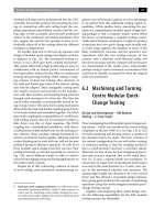
Ultrasonography is the most useful initial
investigation in patients with jaundice
Figure 1.3 Computed tomogram of ampullary carcinoma (white arrow)
causing obstruction of the bile duct (black arrow, bottom) and pancreatic
ducts (white arrowhead)
Investigation of liver and biliary disease
3
This is trial version
www.adultpdf.com
superseding most diagnostic endoscopic
cholangiopancreatography as faster magnetic resonance
imaging scanners become more widely available.
Liver biopsy
Percutaneous liver biopsy is a day case procedure performed
under local anaesthetic. Patients must have a normal clotting
time and platelet count and ultrasonography to ensure that the
bile ducts are not dilated. Complications include bile leaks and
haemorrhage, and overall mortality is around 0.1%. A
transjugular liver biopsy can be performed by passing a special
needle, under radiological guidance, through the internal
jugular vein, the right atrium, and inferior vena cava and into
the liver though the hepatic veins. This has the advantage that
clotting time does not need to be normal as bleeding from the
liver is not a problem. Liver biopsy is essential to diagnose
chronic hepatitis and establish the cause of cirrhosis.
Ultrasound guided liver biopsy can be used to diagnose liver
masses. However, it may cause bleeding (especially with liver cell
adenomas), anaphylactic shock (hydatid cysts), or tumour
seeding (hepatocellular carcinoma or metastases). Many lesions
can be confidently diagnosed by using a combination of
imaging methods (ultrasonography, spiral computed
tomography, magnetic resonance imaging, nuclear medicine,
laparoscopy, and laparoscopic ultrasonography). When
malignancy is suspected in solitary lesions or those confined to
one half of the liver, resection is the best way to avoid
compromising a potentially curative procedure.
Summary points
x An isolated raised serum bilirubin concentration is usually due to
Gilbert’s syndrome, which is confirmed by normal liver enzyme
activities and full blood count
x Jaundice with dark urine, pale stools, and raised alkaline
phosphatase and -glutamyl transferase activity suggests an
obstructive cause, which is confirmed by presence of dilated bile
ducts on ultrasonography
x Jaundice in patients with low serum albumin concentration suggests
chronic liver disease
x Patients with high concentrations of bilirubin ( > 100 mol/l) or
signs of sepsis require emergency specialist referral
x Imaging of the bile ducts for obstructive jaundice is increasingly
performed by magnetic resonance cholangiopancreatography, with
endoscopy becoming reserved for therapeutic interventions
Figure 1.4 Subcapsular haematoma: a complication of liver biopsy
ABC of Liver, Pancreas, and Gall Bladder
4
This is trial version
www.adultpdf.com
2 Gallstone disease
I J Beckingham
Gall stones are the most common abdominal reason for
admission to hospital in developed countries and account for
an important part of healthcare expenditure. Around 5.5
million people have gall stones in the United Kingdom, and
over 50 000 cholecystectomies are performed each year.
Types of gall stone and aetiology
Normal bile consists of 70% bile salts (mainly cholic and
chenodeoxycholic acids), 22% phospholipids (lecithin), 4%
cholesterol, 3% proteins, and 0.3% bilirubin. Cholesterol or
cholesterol predominant (mixed) stones account for 80% of all
gall stones in the United Kingdom and form when there is
supersaturation of bile with cholesterol. Formation of stones is
further aided by decreased gallbladder motility. Black pigment
stones consist of 70% calcium bilirubinate and are more
common in patients with haemolytic diseases (sickle cell
anaemia, hereditary spherocytosis, thalassaemia) and cirrhosis.
Brown pigment stones are uncommon in Britain
(accounting for < 5% of stones) and are formed within the
intraheptic and extrahepatic bile ducts as well as the gall
bladder. They form as a result of stasis and infection within the
biliary system, usually in the presence of Escherichia coli and
Klebsiella spp, which produce glucuronidase that converts
soluble conjugated bilirubin back to the insoluble unconjugated
state leading to the formation of soft, earthy, brown stones.
Ascaris lumbricoides and Opisthorchis senensis have both been
implicated in the formation of these stones, which are common
in South East Asia.
Clinical presentations
Biliary colic or chronic cholecystitis
The commonest presentation of gallstone disease is biliary pain.
The pain starts suddenly in the epigastrium or right upper
quadrant and may radiate round to the back in the
interscapular region. Contrary to its name, the pain often does
not fluctuate but persists from 15 minutes up to 24 hours,
subsiding spontaneously or with opioid analgesics. Nausea or
vomiting often accompanies the pain, which is visceral in origin
and occurs as a result of distension of the gallbladder due to an
obstruction or to the passage of a stone through the cystic duct.
Most episodes can be managed at home with analgesics and
antiemetics. Pain continuing for over 24 hours or accompanied
by fever suggests acute cholecystitis and usually necessitates
hospital admission. Ultrasonography is the definitive
investigation for gall stones. It has a 95% sensitivity and
specificity for stones over 4 mm in diameter.
Non-specific abdominal pain, early satiety, fat intolerance,
nausea, and bowel symptoms occur with comparable frequency
in patients with and without gall stones, and these symptoms
respond poorly to inappropriate cholecystectomy. In many of
these patients symptoms are due to upper gastrointestinal tract
problems or irritable bowel syndrome.
Acute cholecystitis
When obstruction of the cystic duct persists, an acute
inflammatory response may develop with a leucocytosis and
mild fever. Irritation of the adjacent parietal peritoneum causes
Box 1.1 Risk factors associated with formation of cholesterol
gall stones
x Age > 40 years
x Female sex (twice risk in
men)
x Genetic or ethnic variation
x High fat, low fibre diet
x Obesity
x Pregnancy (risk increases
with number of
pregnancies)
x Hyperlipidaemia
x Bile salt loss (ileal disease
or resection)
x Diabetes mellitus
x Cystic fibrosis
x Antihyperlipidaemic drugs
(clofibrate)
x Gallbladder dysmotility
x Prolonged fasting
x Total parenteral nutrition
Box 1.2 Differential diagnosis of common causes of severe
acute epigastric pain
x Biliary colic
x Peptic ulcer disease
x Oesophageal spasm
x Myocardial infarction
x Acute pancreatitis
Age (years)
% of population
30 40 50 60 70
0
10
15
20
25
30
35
40
5
Men
Women
Figure 2.1 Prevalence of gall stones in United Kingdom
according to age
Figure 2.2 Gall stones vary from pure cholesterol (white), through mixed, to
bile salt predominant (black)
5
This is trial version
www.adultpdf.com
localised tenderness in the right upper quadrant. As well as gall
stones, ultrasonography may show a tender, thick walled,
oedematous gall bladder with an abnormal amount of adjacent
fluid. Liver enzyme activities are often mildly abnormal.
Initial management is with non-steroidal anti-inflammatory
drugs (intramuscular or per rectum) or opioid analgesic.
Although acute cholecystitis is initially a chemical inflammation,
secondary bacterial infection is common, and patients should
be given a broad spectrum parenteral antibiotic (such as a
second generation cephalosporin).
Progress is monitored by resolution of tachycardia, fever,
and tenderness. Ideally cholecystectomy should be performed
during the same admission as delayed cholecystectomy has a
15% failure rate (empyema, gangrene, or perforation) and a
15% readmission rate with further pain.
Jaundice
Jaundice occurs in patients with gall stones when a stone
migrates from the gall bladder into the common bile duct or,
less commonly, when fibrosis and impaction of a large stone in
Hartmann’s pouch compresses the common hepatic duct
(Mirrizi’s syndrome). Liver function tests show a cholestatic
pattern (raised conjugated bilirubin concentration and alkaline
phosphatase activity with normal or mildly raised aspartate
transaminase activity) and ultrasonography confirms dilatation
of the common bile duct ( > 7 mm diameter) usually without
distention of the gall bladder.
Acute cholangitis
When an obstructed common bile duct becomes contaminated
with bacteria, usually from the duodenum, cholangitis may
develop. Urgent treatment is required with broad spectrum
antibiotics together with early decompression of the biliary
system by endoscopic or radiological stenting or surgical
drainage if stenting is not available. Delay may result in
septicaemia or the development of liver abscesses, which are
associated with a high mortality.
Acute pancreatitis
Acute pancreatitis develops in 5% of all patients with gall stones
and is more common in patients with multiple small stones, a
wide cystic duct, and a common channel between the common
bile duct and pancreatic duct. Small stones passing down the
common bile duct and through the papilla may temporarily
obstruct the pancreatic duct or allow reflux of duodenal fluid or
bile into the pancreatic duct resulting in acute pancreatitis.
Patients should be given intravenous fluids and analgesia and
be monitored carefully for the development of organ failure
(see later article on acute pancreatitis).
Gallstone ileus
Acute cholecystitis may cause the gall bladder to adhere to the
adjacent jejunum or duodenum. Subsequent inflammation may
result in a fistula between these structures and the passage of a
gall stone into the bowel. Large stones may become impacted
and obstruct the small bowel. Abdominal radiography shows
obstruction of the small bowel and air in the biliary tree.
Treatment is by laparotomy and “milking” the obstructing stone
into the colon or by enterotomy and extraction.
Natural course of gallstone disease
Two thirds of gall stones are asymptomatic, and the yearly risk
of developing biliary pain is 1-4%. Patients with asymptomatic
gall stones seldom develop complications. Prophylactic
cholecystectomy is therefore not recommended when stones
Box 1.3 Charcot’s triad of symptoms in severe cholangitis
x Pain in right upper quadrant
x Jaundice
x High swinging fever with rigors and chills
Figure 2.3 Ultrasonogram showing large gall stone (thin arrow)
casting acoustic shadow (thick arrow) in gall bladder
Figure 2.4 Type 1 Mirrizi’s syndrome: gallbladder stone in Hartmann’s
pouch compressing common bile duct and causing deranged liver function
Figure 2.5 Small bowel obstruction and gas in bile ducts in patient with
gallstone ileus
ABC of Liver, Pancreas, and Gall Bladder
6
This is trial version
www.adultpdf.com
are discovered incidentally by radiography or ultrasonography
during the investigation of other symptoms. Although gall
stones are associated with cancer of the gall bladder, the risk of
developing cancer in patients with asymptomatic gall stones is
< 0.01%
—
less than the mortality associated with
cholecystectomy.
Patients with symptomatic gall stones have an annual rate of
developing complications of 1-2% and a 50% chance of a
further episode of biliary colic. They should be offered
treatment.
Management of gallstone disease
Cholecystectomy
Cholecystectomy is the optimal management as it removes both
the gall stones and the gall bladder, preventing recurrent
disease. The only common consequence of removing the gall
bladder is an increase in stool frequency, which is clinically
important in less than 5% of patients and responds well to
standard antidiarrhoeal drugs when necessary.
Laparoscopic cholecystectomy has been adopted rapidly
since its introduction in 1987, and 80-90% of cholecystectomies
in the United Kingdom are now carried out in this way. The
only specific contraindications to laparoscopic cholecystectomy
are coagulopathy and the later stages of pregnancy. Acute
cholecystitis and previous gastroduodenal surgery are no
longer contraindications but are associated with a higher rate of
conversion to open cholecystectomy.
Laparoscopic cholecystectomy has a lower mortality than
the standard open procedure (0.1% v 0.5% for the open
procedure). This is mainly because of a lower incidence of
postoperative cardiac and respiratory complications. The
smaller incisions cause less pain, which reduces the requirement
for opioid analgesics. Patients usually stay in hospital for only
one night in most centres, and the procedure can be done as a
day case in selected patients. Most patients are able to return to
sedentary work after 7-10 days. This decrease in overall
morbidity and earlier recovery has led to a 25% increase in the
rate of cholecystectomy in some countries.
The main disadvantage of the laparoscopic technique has
been a higher incidence of injury to the common hepatic or
bile ducts (0.2-0.4% v 0.1% for open cholecystectomy). Higher
rates of injury are associated with inexperienced surgeons (the
“learning curve” phenomenon) and acute cholecystitis.
Furthermore, injuries to the common bile duct tend to be more
extensive with laparoscopic surgery. However, there is some
evidence suggesting that the rates of injury are now falling.
Box 1.4 Causes of pain after cholecystectomy
x Retained or recurrent stone (dilatation of common bile duct seen in
only 30% of patients)
x Iatrogenic biliary leak or stricture of common bile duct
x Papillary stenosis or dysfunctional sphincter of Oddi
x Incorrect preoperative diagnosis
—
for example, irritable bowel
syndrome, peptic ulcer, gastro-oesophageal reflux
Figure 2.6 Laparoscopic cholecystectomy reduces the risk of surgery in
morbidly obese patients
Year of audit
Annual incidence of bile duct injury (%)
1991 1992 1993 1994 1995
0
0.2
0.3
0.4
0.5
0.6
0.7
0.8
0.1
Total
Major injury
Minor injury
Figure 2.7 Annual incidence of injury to bile duct during laparoscopic
cholescystectomy, United Kingdom,1991-5. Adapted from Br J Surg
1996;83:1356-60
Figure 2.8 Injury to common bile duct incurred during laparoscopic cholecystectomy before, during, and after repair by balloon dilatation
Gallstone disease
7
This is trial version
www.adultpdf.com
Alternative treatments
Several non-surgical techniques have been used to treat gall
stones including oral dissolution therapy (chenodeoxycholic
and ursodeoxycholic acid), contact dissolution (direct instillation
of methyltetrabutyl ether or mono-octanoin), and stone
shattering with extracorporeal shockwave lithotripsy.
Less than 10% of gall stones are suitable for non-surgical
treatment, and success rates vary widely. Stones are cleared in
around half of appropriately selected patients. In addition,
patients require expensive, lifelong treatment to counteract bile
acid in order to prevent stones from reforming. These
treatments should be used only in patients who refuse surgery.
Managing common bile duct stones
Around 10% of patients with stones in the gallbladder have
stones in the common bile duct. Patients may present with
jaundice or acute pancreatitis; the results of liver function tests
are characteristic of cholestasis and a dilated common bile duct
is visible on ultrasonography.
The optimal treatment is to remove the stones in both the
common bile duct and the gall bladder. This can be performed
in two stages by endocsopic retrograde
cholangiopancreatography followed by laparoscopic
cholecystectomy or as a single stage cholecystectomy with
exploration of the common bile duct by laparoscopic or open
surgery. The morbidity and mortality (2%) of open surgery is
higher than for the laparoscopic option. Two recent
randomised controlled trials have shown laparoscopic
exploration of the bile duct to be as effective as endoscopic
retrograde cholangiopancreatography in removing stones from
the common bile duct. Laparoscopic exploration has the
advantage that the gall bladder is removed in a single stage
procedure, thus reducing hospital stay. In practice, management
often depends on local availability and skills.
In elderly or frail patients endoscopic retrograde
cholangiopancreatography with division of the sphincter of
Oddi (sphincterotomy) and stone extraction alone (without
cholecsytectomy) may be appropriate as the risk of developing
further symptoms is only 10% in this population.
When stones in the common bile duct are suspected in
patients who have had a cholecystectomy, endoscopic
retrograde cholangiopancreatography can be used to diagnose
and remove the stones. Stones are removed with the aid of a
dormia basket or balloon. For multiple stones, a pigtail stent can
be inserted to drain the bile; this often allows subsequent
passage of the stones. Large or hard stones can be crushed with
a mechanical lithotripter. When cholangiopancreatography is
not technically possible the stones have to be removed
surgically.
Box 1.5 Criteria for non-surgical treatment of gall stones
x Cholesterol stones < 20 mm in diameter
x Fewer than 4 stones
x Functioning gall bladder
x Patent cystic duct
x Mild symptoms
Summary points
x Gall stones are the commonest cause for emergency hospital
admission with abdominal pain
x Laparoscopic cholecystectomy has become the treatment of choice
for gallbladder stones
x Risk of bile duct injury with laparoscopic cholecystectomy is around
0.2%
x Asymptomatic gall stones do not require treatment
x Cholangitis requires urgent treatment with antibiotics and biliary
decompression by endoscopic retrograde
cholangiopancreatography
Further reading
x Beckingham IJ, Rowlands BJ. Post cholecystectomy problems. In
Blumgart H, ed. Surgery of the liver and biliary tract. 3rd ed. London:
WB Saunders, 2000
x National Institutes of Health consensus development conference
statement on gallstones and laparoscopic cholecystectomy Am J
Surg 1993;165:390-8
x Cuschieri A, Lezoche E, Morino M, Croce E, Lacy A, Toouli J, et al.
EAES multicenter prospective randomized trial comparing
two-stage vs single-stage management of patients with gallstone
disease and ductal calculi. Surg Endosc 1999;13:952-7
Figure 2.9 Magnetic resonance cholangiopancreatogram showing stone in
common bile duct
Figure 2.10p Large angular common bile duct stones. These are
difficult to remove endoscopically
ABC of Liver, Pancreas, and Gall Bladder
8
This is trial version
www.adultpdf.com
3 Acute hepatitis
S D Ryder, I J Beckingham
Acute hepatic injury is confirmed by a raised serum alanine
transaminase activity. The activity may be 100 times normal, and
no other biochemical test has been shown to be a better
indicator. Alkaline phosphatase and -glutamyltransferase
activities can also be raised in patients with an acute hepatic
injury, but their activites are usually proportionately lower than
that of alanine transaminase.
Acute viral hepatitis
Hepatitis can be caused by the hepatitis viruses A, B, C, D, or E.
The D and E forms are rare in the United Kingdom. A large
proportion of infections with hepatitis viruses of all types are
asymptomatic or result in anicteric illnesses that may not be
diagnosed as hepatitis. Hepatitis A virus causes a typically minor
illness in childhood, with more than 80% of cases being
asymptomatic. In adult life infection is more likely to produce
clinical symptoms, although only a third of patients with acute
hepatitis A infections are jaundiced. Infections with hepatitis B
and C viruses are also usually asymptomatic except in
intravenous drug users, in whom 30% of hepatitis B infections
are associated with jaundice.
In the preicteric phase, patients often have non-specific
systemic symptoms together with discomfort in the right upper
quadrant of the abdomen. An illness resembling serum sickness
occurs in about 10% of patients with acute hepatitis B infection
and 5-10% of patients with acute hepatitis C infection. This
presents with a maculopapular rash and arthralgia, typically
affecting the wrist, knees, elbows, and ankles. It is due to
formation of immune complexes, and patients often test
positive for rheumatoid factor. It is almost always self limiting,
and usually settles rapidly after the onset of jaundice.
Rarely, patients with acute hepatitis B infection present with
acute pancreatitis. Up to 30% of patients have raised amylase
activity, and postmortem examinations in patients with
fulminant hepatitis B show histological changes of pancreatitis
in up to 50%. Myocarditis, pericarditis, pleural effusion, aplastic
anaemia, encephalitis, and polyneuritis have all been reported
in patients with hepatitis.
Physical signs in viral hepatitis
Physical examination of patients before the development of
jaundice usually shows no abnormality, although hepatomegaly
(10% of patients), splenomegaly (5%), and lymphadenopathy
(5%) may be present. Patients with an acute illness should not
have signs of chronic liver disease. The presence of these signs
suggests that the illness is either the direct result of chronic liver
disease or that the patient has an acute event superimposed on
a background of chronic liver disease
—
for example, hepatitis D
virus superinfection in a carrier of hepatitis B virus.
A small proportion of patients with acute viral hepatitis
develop a profound cholestatic illness. This is most common
with hepatitis A and can be prolonged, with occasional patients
remaining jaundiced for up to eight months.
Table 3.1 Liver enzyme activity in liver disease
Hepatitis
Cholestasis or
obstruction “Mixed”
Alkaline phosphatase Normal Raised Raised
-glutamyltransferase Normal Raised Raised
Alanine transaminase Raised Normal Raised
Box 3.1 Common symptoms of acute viral hepatitis
x Myalgia
x Nausea and vomiting
x Fatigue and malaise
x Change in sense of smell or taste
x Right upper abdominal pain
x Coryza, photophobia, headache
x Diarrhoea (may have pale stools and dark urine)
Table 3.2 Types and modes of transmission of human
hepatitis viruses
ABCDE
Virus type Picorna-
viridae
Hepadna-
viridae
Flavi-
viridae
Delta-
viridae
Calci-
viridae
Nucleic acid RNA DNA RNA RNA RNA
Mean (range)
incubation
period (days)
30
(15-50)
80
(28-160)
50
(14-160)
Variable 40
(15-45)
Mode of transmission:
Orofaecal Yes Possible No No Yes
Sexual Yes Yes Rare Yes No
Blood Rare Yes Yes Yes No
Chronic
infection
No Yes Yes Yes No
Box 3.2 Other biochemical or haematological abnormalities
seen in acute hepatitis
x Leucopenia is common ( < 5 · 10
9
/l in 10% of patients)
x Anaemia and thrombocytopenia
x Immunoglobulin titres may be raised
Figure 3.1 Structure of hepatitis B virus
9
This is trial version
www.adultpdf.com
Acute liver failure (fulminant hepatitis)
Death from acute viral hepatitis is usually due to the
development of fulminant hepatitis. This is usually defined as
development of hepatic encephalopathy within eight weeks of
symptoms or within two weeks of onset of jaundice. The risk of
developing fulminant liver failure is generally low, but there are
groups with higher risks. Pregnant women with acute hepatitis
E infection have a risk of fulminant liver failure of around 15%
with a mortality of 5%. The risk of developing fulminant liver
failure in hepatitis A infection increases with age and with
pre-existing liver disease. Fulminant hepatitis B is seen in adult
infection and is relatively rare.
The primary clinical features of acute liver failure are
encephalopathy and jaundice. Jaundice almost always precedes
encephalopathy in acute liver failure The peak of alanine
transaminase activity does not correlate with the risk of
developing liver failure. Prolonged coagulation is the biochemical
hallmark of liver failure and is due to lack of synthesis of liver
derived factors. Prolongation of the prothrombin time in acute
hepatitis, even if the patient is clinically well without signs of
encephalopathy, should be regarded as sinister and the patient
monitored closely. Hypoglycaemia is seen only in fulminant liver
disease and can be severe.
Diagnosis of acute hepatitis
Hepatitis A
Hepatitis A infection can be reliably diagnosed by the presence
of antihepatitis A IgM. This test has high sensitivity and
specificity. Occasional false positive results occur in patients with
liver disease due to other causes if high titres of
immunoglobulin are present, but the clinical context usually
makes this obvious.
Hepatitis B
Hepatitis B infection is usually characterised by the presence of
hepatitis B surface antigen. Other markers are used to
determine if the virus is active and replicating, when it can
cause serious liver damage.
In acute hepatitis B infection the serology can be difficult to
interpret. Acute hepatitis develops because of immune
recognition of infected liver cells, which results in T cell
mediated killing of hepatocytes. Active regeneration of
hepatocytes then occurs. As well as a cell mediated immune
response, a humoral immune response develops; this is
probably important in removing viral particles from the blood
and thus preventing reinfection of hepatocytes. Because of the
immune response attempting to eradicate hepatitis B virus, viral
replication may already have ceased by the time a patient
presents with acute hepatitis B, and the patient may be positive
for hepatitis B surface antigen and negative for e antigen.
It is difficult in this situation to be certain that the patient
had acute hepatitis B and that the serology does not imply past
infection unrelated to the current episode. To enable a clear
diagnosis, most reference centres now report the titre of IgM
antibody to hepatitis B core antigen (IgM anticore). As core
antigen never appears in serum, its presence implies an
immune response against hepatitis B virus within liver cells and
is a sensitive and specific marker of acute hepatitis B infection.
Rarely, the immune response to hepatitis B infection is so
rapid that even hepatitis B surface antigen has been cleared
from the serum by the time of presentation with jaundice. This
may be more common in patients developing severe acute liver
disease and has been reported in up to 5% of patients with
fulminant hepatitis diagnosed by an appropriate pattern of
antibody response.
The onset of confusion or drowsiness in a patient with
acute viral hepatitis is always sinister
Replication of hepatitis B virus is assessed by measuring e
antigen (a truncated version of the hepatitis B core
antigen that contains the viral replication mechanism) and
hepatitis B DNA
Figure 3.2 Disconjugate gaze due to cerebral oedema in jaundiced patient
with fulminant hepatitis
Time (days)
Titre
012345678910
0
100
150
50
Viral DNA
e antigen
Anti-e antibody
Jaundice
Figure 3.3 Appearance of serological markers in acute self limiting hepatitis
B virus infection
Surface
antigen
Surface
antigen
Virion assembled
Incomplete virus
exported
Core
antigen
RNA
Proteins
Hepatitis B virus
DNA
Hepatitis B virus
DNA
Complete virion
Figure 3.4 Mechanism of assembly and excretion of hepatitis B virus from
infected hepatocytes
ABC of Liver, Pancreas, and Gall Bladder
10
This is trial version
www.adultpdf.com
Hepatitis C
Screening tests for hepatitis C virus infection use enzyme linked
immunosorbent assays (ELISA) with recombinant viral antigens
on patients’ serum. Acute hepatitis C cannot be reliably
diagnosed by antibody tests as these often do not give positive
results for up to three months.
Hepatitis C virus was the cause of more than 90% of all
post-transfusion hepatitis in Europe and the United States.
Before 1991, the risk of infection in the United Kingdom was
0.2% per unit of blood transfused, but this has fallen to 1
infection per 10 000 units transfused since the introduction of
routine serological screening of blood donors. Acute hepatitis C
infection is therefore now seen commonly only in intravenous
drug users.
Antibodies to hepatitis C appear relatively late in the course
of the infection, and if clinical suspicion is high, the patient’s
serum should be tested for hepatitis C virus RNA to establish
the diagnosis.
Non-A-E viral hepatitis
Epstein Barr virus causes rises in liver enzyme activities in
almost all cases of acute infection, but it is uncommon for the
liver injury to be sufficiently severe to cause jaundice. When
jaundice does occur in patients with Epstein Barr virus
infection, it can be prolonged with a large cholestatic element.
Diagnosis is usually relatively easy because the typical symptoms
of Epstein Barr infection are almost always present and
serological testing usually gives positive results.
Cytomegalovirus can also cause acute hepatitis. This is unusual,
rarely severe, and runs a chronic course only in
immunosuppressed patients.
The cause of about 7% of all episodes of acute presumed
viral hepatitis remains unidentified. It seems certain that other
viral agents will be identified that cause acute liver injury.
Management of acute viral hepatitis
Hepatitis A
Most patients with hepatitis A infection have a self limiting
illness that will settle totally within a few weeks. Management is
conservative, with tests being aimed at identifying the small
group of patients at risk of developing fulminant liver failure.
Hepatitis B
Acute hepatitis B is also usually self limiting, and most patients
who contract the virus will clear it completely. All cases must be
notified and sexual and close household contacts screened and
vaccinated. Patients should be monitored to ensure fulminant
liver failure does not develop and have serological testing three
months after infection to check that the virus is cleared from
the blood. About 5-10% of patients will remain positive for
hepatitis B surface antigen at three months, and a smaller
proportion will have ongoing viral replication (e antigen
positive). All such patients require expert follow up (see article
on chronic viral hepatitis).
Hepatitis C
Early identification and referral of cases of acute hepatitis C
infection is important because strong evidence exists that early
treatment with interferon alpha reduces the risk of chronic
infection. The rate of chronicity in untreated patients is about
80%; treatment with interferon reduces this to below 50%.
Box 3.3 Hepatitis D and E infection
Hepatitis D
x Incomplete RNA virus that requires hepatitis B surface antigen to
transmit its genome from cell to cell
x Occurs only in patients positive for hepatitis B surface antigen
x Usually confined to intravenous drug users in United Kingdom
Hepatitis E
x Transmitted by orofaecal route
x Produces an acute self limiting illness similar to hepatitis A
x Common in developing world
x High mortality in pregnant women
Summary points
x Symptoms of hepatitis are non-specific and often occur without the
development of jaundice
x Serum alanine transaminase is the most useful screening test for
hepatitis in general practice
x Hepatitis A rarely causes fulminant liver failure or chronic liver
disease
x In the developed world, new cases of hepatitis C are mainly seen in
intravenous drug users
x Most adults who contract hepatitis B virus clear the virus, with
< 10% developing chronic liver infection
Time (months)
Alanine transaminase (u/l)
0 1 2 3 4 5 6 7 8 9 10
0
800
1200
1000
600
200
400
Hepatitis C virus
Alanine transaminase
Antibody to hepatitis C virus
Jaundice
Figure 3.5 Appearance of hepatitis C virus RNA, antibodies to hepatitis C
virus, and raised alanine transaminase activity in acute hepatitis C infection
Hepatitis A IgM positive
Check international normalised ratio
International normalised ratio <2
Better
Review with liver function tests
in 5-7 days
No improvement
(clinical or biochemical)
International normalised ratio >2
Abnormal
Refer
Repeat liver function
tests at 6 weeks
Normal
No follow up
Figure 3.6 Management of acute hepatitis A infection in general practice
Acute hepatitis
11
This is trial version
www.adultpdf.com