Atlas of Clinical Hematology - part 4 potx
Bạn đang xem bản rút gọn của tài liệu. Xem và tải ngay bản đầy đủ của tài liệu tại đây (3.6 MB, 44 trang )
IV
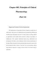
Fig. 39 e – h
e Very intense acid phosphatase reac-
tion in Gaucher cells
f Acid phosphatase. The fibrillary struc-
ture of the cytoplasm is apparent in the
slightly crushed cells
g Strong diffuse PAS reaction
h Iron stain produces marked diffuse
staining of the cytoplasm
124 Chapter IV · Blood and Bone Marrow
IV
5.5.2 Niemann-Pick Disease
Niemann-Pick disease is a sphingomyelin storage
disease (sphingolipoidosis) that is based on a de-
ficiency of sphingomyelinase. It is inherited as an
autosomal recessive trait and produces clinical
manifestations during childhood. Five different
biochemical subtypes have been identified. Char-
acteristic foam cells are found in the bone mar-
row, liver, spleen, and lymph nodes.
Another variant is type C (NPC 1-protein de-
fect) with a defect of cholesterol transport.
Here you find vacuoles of different size in the cy-
toplasm, sometimes blue granules. An infantile
and a juvenile-adult course can be distinguished.
125
5 · Bone Marrow
IV
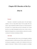
Fig. 40 a – d. Niemann-Pick disease
a, b Storage cells with very small nuclei
and fine, closely spaced, partially con-
fluent pale bluish-gray inclusions, some
of which are dislodged during staining
and appear as vacuoles (foamy cyto-
plasm)
c Relatively weak PAS reaction
d The inclusions may show marked ba-
sophilic staining like the storage cells in
sea-blue histiocytic disease, considered a
variant of Niemann-Pick disease. These
“sea-blue histiocytes” may also occur as
storage cells when cellular breakdown is
increased (as in this case)
126 Chapter IV · Blood and Bone Marrow
IV
5.5.3 Glycogen Storage Disease Type II
(Acid Maltase Deficiency, Pompe Disease)
In our examination of an adult with severe mus-
cular dystrophy, we noted severe vacuolation in
the plasma cells of the bone marrow (Fig. 41a –
d). The PAS reaction demonstrated coarse posi-
tive inclusions. Electron microscopy of semithin
sections and cytochemi cal analysis revealed the
presence of a polysaccharide- and protein-con-
taining material in the “vacuoles.”
1
1
Pralle H, Schro¨der R, Lo¨ffler H (1975) New kind of cytoplas-
mic inclusions of plasma cells in acid maltase deficiency. Acta
Haematol 53 : 109 –117
127
5 · Bone Marrow
IV
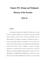
Fig. 41 a – d. Type II glycogen storage
disease (acid maltase deficiency,
Pompe disease)
a, b Plasma cells contain closely spaced
vacuoles of varying size, found on elec-
tron microscopy and cytochemical ana-
lysis to contain glycopeptide
b
c Coarse PAS-positive inclusions in the
plasma cells
d The “vacuoles” are strongly positive for
acid phosphatase
128 Chapter IV · Blood and Bone Marrow
IV
5.6 Hemophagocytic Syndromes
The phagocytosis of blood cells by macrophages
may occur in the setting of inflammatory pro-
cesses, immune responses, or malignant diseases.
An hereditary form, familial hemophagocytic
lymphohistiocytosis, predominantly affects in-
fants, with 80 % of cases occurring before the
second year of life. Marked phagocytic states
with greatly increased numbers of macrophages
were formerly described as malignant histiocy-
toses or hist iocytic medullary reticuloses. Many
of these states may be caused by viruses (e.g., cy-
tomegalovirus) and other infectious organisms.
They are most common in immunosuppressed
patients but also occur in the setting of malignant
diseases. The “malignant histiocytoses” probably
consist mainly of different forms of monocytic
leukemia, and some may represent misidentified
forms of large-cell malignant lymphoma. True
neoplasias with a macrophagic phenotype are
probably quite rare.
129
5 · Bone Marrow
IV
Fig. 42 a – h. Hemophagocytic
syndrome
a Low-power view of bone marrow
shows several macrophages that have
phagocytized platelets and erythrocytes.
The cause in this case is unknown
b Macrophages with erythrocytes,
platelets, and (at top right ) small nuclei in
the cytoplasm
c Bone marrow from the same patient
shows a phagocytized neutrophil at
upper right
d Phagocytized erythrocytes and
platelets have displaced the macrophage
nucleus to the edge of the cell
130 Chapter IV · Blood and Bone Marrow
IV
Fig. 42 e – h
e Macrophage with phagocytized
normoblasts
f Phagocytosis of two rod neutrophils
and a nuclear remnant. Macrophage
nucleus is at lower right
g Macrophages preserved in air-dried
smears for 15 months still show strong
acid phosphatase activity
h Sample from the same patient (fresh
smear) shows strong esterase activity in
the macrophages
131
5 · Bone Marrow
IV
5.7 Histiocytosis X
Histiocytosis X (Langerhans cell histiocytosis,
Fig. 43) is characterized by large cells with abun-
dant grayish-blue cytoplasm and round to oval
nuclei. CD11c, CD1, and S-100 protein serve as
markers. The Birbeck granules that are specific
for Langerhans cells can be demonstrated by elec-
tron microscopy. Multinucleated giant cells are
characteristic.
132 Chapter IV · Blood and Bone Marrow
IV
Fig. 43 a – d
a, b Bone marrow involvement by
histiocytosis X (Langerhans cell histiocy-
tosis). Note the large cells with broad,
bluish-gray cytoplasm and round to oval
nuclei
b
c Nonspecific esterase reaction (ANAE)
demonstrates fine positive granules in
the cytoplasm
d Demonstration of acid phosphatase in
the cytoplasm of malignant cells. The
reaction is weaker than in the macro-
phages
133
5 · Bone Marrow
IV
5.8 Chronic Myeloproliferative Disorders
(CMPD)
Dameshek introduced the “myeloproliferative
syndrome” as a collective term encompassing es-
sential thrombocythemia, polycythemia vera, os-
teomyelosclerosis, and chronic myeloid leuke-
mia. Since the detection of the Philadelphia chro-
mosome (Ph) by Nowell and Hungerford in 1960
and later the underlying BCR/ABL translocation
by Bartram et al., a sharp distinction must be
drawn between chronic myeloid (granulocytic)
leukemia and the other chronic myeloprolifera-
tive disorders. The concept of CMPD is justified
by certain similarities in the course of these dis-
eases. Several apparent transitions between the
different forms have been elucidated using mole-
cular genetic techniques and have been classified
as various manifestations of chron ic myeloid leu-
kemia. Many questions remain unanswered, how-
ever, and it is necessary to provide an accurate
description of individual cases.
The diagnosis of essential thrombocythemia is
based on a consistently elevated platelet count
(higher than 6 Â 10
9
/l), the exclusion of a different
cause (including chronic inflammatory disease),
and an increase of megakaryocytes in the bone
marrow, which often show only subtle abnormal-
ities (hypersegmented nuclei) and are grouped in
clusters. The peripheral blood film may show a
mild leukocytosis with slight basophilia and eosi-
nophilia in addition to thrombocytosis. These
cases require a chromosomal and/or molecular
genetic evaluation to exclude chronic myeloid
leukemia. Polycythemia vera can be diagnosed
only when findings meet the criteria defined by
the Polycythemia Vera Study Group. The cellular-
ity of the bone marrow is markedly increased, and
fat cells are completely absent in fully established
cases. There is a significant increase in megakar-
yocytes, which show an extreme diversity of sizes.
Erythropoiesis and usually granulocytopoiesis
are markedly increased, and iron stores are ab-
sent from the marrow. A slight increase of baso-
phils is observed in the blood and bone marrow.
Histologic examination is necessary for an accu-
rate quantitative evaluation of bone marrow
structures. An increase in leukocyte alkaline
phosphatase activity is detected in blood smears.
Osteomyelosclerosis or myelofibrosis is character-
ized by an increase in reticular fibers and/or can-
cellous bone ranging to the complete obliteration
of the bone marrow and by extramedullary hema-
topoiesis. The differential blood count may be
very similar to that in chronic myeloid leukemia,
but there are significant erythrocyte abnormal-
ities that include teardrop-shaped cells and the
presence of erythroblasts in the blood smear. Leu-
kocyte alkaline phosphatase is usually elevated or
normal. L. Pahl et al. (Blood 100, 2441 (2002)) de-
scribed a membrane receptor PRV-1, which is
overexpressed in polycythemia vera, partly in es-
sential thrombocythemia and myelofibrosis.
134 Chapter IV · Blood and Bone Marrow
IV
Fig. 44 a – d. Essential thrombocythe-
mia (ET)
a Blood smear reveals anisocytosis and a
greatly increased number of platelets
b Bone marrow smear in ET shows large
masses of platelets and scattered
megakaryocytes
c Three mature megakaryocytes and
large platelet aggregations
d Histologic section in ET shows a
substantial increase in moderately pleo-
morphic megakaryocytes, some ar-
ranged in clusters. There is a normal
proportion of fat cells. Giemsa stain
135
5 · Bone Marrow
IV
Fig. 45 a – e. Polycythemia vera
a Bone marrow smear shows marked
hypercellularity with a significant in-
crease in megakaryocytes, which vary
markedly in size and maturation
b High-power view shows the size
variation of the megakaryocytes
c Bone marrow area with increased
erythropoiesis and granulocytopoiesis. At
left is a basophil
d Histologic section shows residual fat
cells, a typical increase in megakaryo-
cytes of varying size, and increased ery-
thropoiesis. Giemsa stain
136 Chapter IV · Blood and Bone Marrow
IV
Fig. 45 e – h
e Blood smear shows a substantial
increase in leukocyte alkaline phospha-
tase activity (red)
f Bone marrow in myelofibrosis. Note
the clustering of the pleomorphic
megakaryocytes. Hematoxylin-eosin
stain
g Bone marrow section in myelofibro-
sis. Silver stain demonstrates heavy fiber
proliferation. At lower left is a cluster of
megakaryocytes
h Bone marrow in osteomyelosclerosis
(OMS). The marrow cavity is almost
completely obliterated by collagen and
increased cancellous trabeculae. Hema-
toxylin-eosin stain
137
5 · Bone Marrow
IV
Fig. 46 a – d
a Blood smear in osteomyelosclerosis
(OMS). Monocyte and segmented cell at
left, erythroblast at center, and promye-
locyte at right
b Blood smear in OMS shows significant
poikilocytosis with teardrop-shaped
erythrocytes. At top is a normoblast
c Blood smear in OMS shows heavy
basophilic stippling and two erythrocytes
with Cabot rings. At center is an
erythrocyte with Howell-Jolly bodies and
a nuclear remnant
d Increased leukocyte alkaline phos-
phatase (LAP) in OMS. There is one grade
1 and one grade 4 neutrophil, a normo-
blast, and a negative blast at upper left
138 Chapter IV · Blood and Bone Marrow
IV
Fig. 46 e – h
e Blood smear in chronic myeloproli-
ferative disease (CMPD) shows five
erythroblasts and, at the center of the
field, basophilic stippling. Such cases
were once termed “chronic erythremia.”
Erythroblastosis can occur in various
forms of CMPD
f Blood smear in chronic myeloid leu-
kemia (CML) during the accelerated
phase after splenectomy. At center is a
megakaryocyte nucleus, at right are two
megakaryoblasts, and at left is a myelo-
blast
g Histologic section from an iliac crest
biopsy in “pure” megakaryocytic myelosis
consists almost entirely of megakaryo-
cytes at various stages of maturity.
Giemsa stain
h Silver-stained specimen from the same
patient clearly shows the darkly stained
nuclei of the megakaryocytes and fiber
proliferation
139
5 · Bone Marrow
IV
5.8.1 Myeloid Leukemia and Transient
Abnormal Myelopoiesis (TAM)
of Down Syndrome (DS)
Acute myeloid leukemia of DS is immunologi-
cally characterized by blast cells with features
of megakaryoblasts. The blasts have a basophilic
cytoplasm which might remind of proerythro-
blasts. The disease responds quite well to the
usual treatment. There are no biological differ-
ences between MDS and AML in Down syn-
drome. AML in older children with DS (3 years
and older) behave more like AML in children
without DS and has a poorer prognosis.
Transient abnormal myelopoiesis (TAM) or
transient myeloproliferation may show a clinical
and morphological picture indistinguishable
from AML.
Spontaneous remission appears in the major-
ity within 3 months. AML develops 1– 3 years later
in about one quarter of the children.
5.8.2 Special Variants of Megakaryocyte
Proliferation
The pure malignant proliferation of megakaryo-
cytes (Fig. 46g and h) is as rare as tumorous
megakaryoblastoma (Fig. 104d and e)
In one case with an exceptional increase in
megakaryoblasts and promegakaryocytes and a
very high proportion of mitoses, we were able
to classify the disease as promegakaryocytic-
megakaryoblastic leukemia (megakaryocyte pre-
cursor cell leukemia). The cells and mitoses could
be positively identified by the immunocytochem-
ical detection of the megakaryocyte markers CD41
and CD61. This case is more characteristic of a
CMPD than an acute leukemia (Fig. 47a – h; joint
observation with D. Mu¨ller, Hof).
Figure 48a – c shows an exam ple of familial
polyglobulia with positive erythrocyte alkaline
phosphatase. Cytochemical and biochemical tests
in four family members (three generations)
showed that some of the erythrocytes and
100 % of the erythroblasts contained alkaline
phosphatase, which differs from the phosphatase
in neutrophils. It is likely that the increased
breakdown of 2,3– diphospho glycerate plays a
role in the pathogenesis of the erythro cytosis.
Fig. 46 i. Three blast cells in the periph-
eral blood in TAM. All three cells have
intensely basophilic cytoplasm, which is
hardly visible in the two cells below.
There is no morphological difference to
AML of DS
140 Chapter IV · Blood and Bone Marrow
IV
Fig. 47 a – h. Promegakaryocytic-mega-
karyoblastic leukemia (after Lo¨ffler and
Mu¨ller, unpublished)
a Six megakaryocytic mitoses and
numerous small megakaryoblasts
b Higher-power view of megakaryo-
blasts and four mitoses
c Four mitoses, blasts, and a promega-
karyocyte in the lower half of the field
d Megakaryoblasts, a mitosis, and a
promegakaryocyte
141
5 · Bone Marrow
IV
Fig. 47 e – h
e Immunocytochemical detection of
CD41. A large proportion of the mega-
karyoblasts and promegakaryocytes are
positive
f CD41: three positive mitoses are seen
at upper left and lower right. Other fea-
tures are the same as in e
g CD41: besides blasts and promega-
karyocytes, a positive mature megakar-
yocyte is visible at right
h CD61: the result is the same as with
CD41
142 Chapter IV · Blood and Bone Marrow
IV
5.8.3 Familial Erythrocytosis
(Fig. 48 a – c)
Cytochemical Detection
of Alkaline Phosphatase
a Blood smear. Erythrocytes show weak
diffuse reaction with fine positive gran-
ules
b Erythroblast cluster in bone marrow
smear with marked cytoplasmic reaction
(substrate a-naphthyl phosphate)
c Marked reaction (red) in erythroblasts
with the substrate naphthol-AS-Bi phos-
phate
143
5 · Bone Marrow
IV
5.8.4 Chronic Myeloid (Granulocytic)
Leukemia
The blood picture in chronic myeloid leukemia
(CML) often contributes more to the diagnosis
than the bone marrow. Besides the high leukocyte
count and a pathologic left shift with the appear-
ance of immature granulocytopoietic forms at all
stages (including promyelocytes and myelo-
blasts), eosinophilia and basophilia are present
in the peripheral blood and corroborate the diag-
nosis of CML. In addition, individual granulo-
cytes show qualitative changes suc h as anisocyto-
sis, nuclear-cytoplasmic asynchrony, and hypo-
segmentation (“pseudo-Pelger forms”). These
changes are largely absent during the early
chronic phase. The same changes may be found
in severe reactive leukocyto ses. The bone marrow
is very cellular. Erythropoiesis is greatly sup-
pressed in favor of granulocytopoiesis, which
dominates the cell picture. The granulocytopoie-
tic line include a great many immature forms,
producing a marked shift to the left. Marrow ba-
sophils and eosinophils are usually increased. De-
spite these findings, it can be difficult to distin-
guish the bone marrow changes from those asso-
ciated with severe reactive leukocytoses. Before
the discovery of the Philadelphia chromosome
and the BCR-ABL translocation, the demonstra-
tion of low or even negative leukocyte alkaline
phosphatase (LAP, see p. 13) w as of key impor-
tance. Today the diagnosis is established by detec-
tion of the Philadelphia chromosome (Ph). It re-
presents a reciprocal translocation between the
long arms of chromosomes 9 and 22 [i.e.,
t(9;22)], resulting in a translocation of the BCR
and ABL genes. This creates a new fusion gene
called the BCR-ABL gene. The corresponding
proteins, which have molecular weights of
210 (p210) and 190 (p190), and very rarely 230
(p230) can be detected in very low concentration
by PCR. Thus, molecular biology and its combi-
nation with cytogenetic analysis in the FISH tech-
nique provide highly sensitive detection methods
that complement morphology and cytogenetics in
the diagnosis and especially the follow-up of CML
after intensive therapy.
The differentiation of CML from chronic mye-
lomonocytic leukemia (CMML), can be difficult
to accomplish by morphology alone, since there
are “intermediate forms” that the FAB group
has classified as atypica l CML. The most reliable
differentiating method is the cytogenetic detec-
tion of the (9;22) translocation or the molecular
genetic detection of the BCR-ABL translocation.
The absence of these changes preclude s a diagno-
sis of CML (CGL).
Almost all chronic myeloid leukemias progress
to an acute phase (acute blast phase, blast crisis)
during the course of the disease. This acute phase
may arise by transformation from the chronic
phase, or an accelerated phase may precede it.
The accelerated phase can be diagnosed by its
clinical manifestations (fever, bone pain, spleno-
megaly) and an increasing left shift of the gran-
ulocytopoiesis. There may also be an increase
in basophilic granulocytes, which are already nu-
merous in this disease. An increase in leukocyte
alkaline phosphatase activity is occasionally de-
tected during the blast phase. Sometimes the
acute phase has its onset in a particular organ
such as the spleen or lymph nodes.
The blasts consist predominantly of cyto-
chemically and immunologically identifiable
myeloblasts and less commonly (20 % – 30 %)
of lymphoblasts, which may be PAS-positive
and display the immunologic features of common
ALL. Megakaryoblast or erythroblast transforma-
tion is less frequent, but mixed blast phases are
somewhat more common. Besides the t (9;22)
translocation, the accelerated phase or blast phase
is often characterized by other cytogenetic
changes that mainly consist of an extra Ph chro-
mosome, an isochromosome 17, trisomy 8, or a
combination of these.
Because the BCR-ABL translocation occurs in
early stem cells, CML affects a portion of the T
lymphocytes and may affect all hematopoietic
cells, although the involvement need not be com-
plete. It is not surprising, therefore, when a high
percentage of eosinophils or basophils are discov-
ered in variants of CML. As long as the typical
cytogenetic or molecular genetic abnormality is
present, there is no need to classify the leukemia
as “eosinophilic” or “basophilic.” True eosinophi-
lic and basophilic leukemias do exist, but they are
more aptly classified as acute leukemias and are
discussed under that heading.
144 Chapter IV · Blood and Bone Marrow
IV
Table 8. Stages of CML
a) Chronic phase
Blood smear Bone marrow
GP: Pathologic left shift GP: Very hyperplastic
Increased eosinophils Shift to the left
Increased basophils Increased eosinophils
Increased basophils
EP: Scattered normoblasts
Anisocytosis, polychromatophilia
EP: Decreased (absolute or relative)
ThP: Platelets usually increased
Anisocytosis, giant platelets
Scattered megakaryocyte nuclei
ThP: Megakaryocytes usually increased, some ab-
normal forms (microkaryocytes)
b) Accelerated phase
Blood smear Bone marrow
GP: Pathologic left shift, pseudo-Pelger forms
Increased numbers of blasts,
< 20 %
Basophils may be markedly increased,
< 30 %
GP: Pathologic left shift
Increased numbers of N.C. or “blasts,”
20 %
Basophils may be markedly increased
EP: Scattered normoblasts anisocytosis,
polychromatophilia
EP: Decreased
ThP: Platelets normal or decreased
Anisocytosis, scattered megakaryocyte
nuclei
ThP: Normal or decreased
c) Acute phase (blast crisis)
Blood smear Bone marrow
GP: Practically all cells are blasts GP: Practically all cells are blasts > 30 %
EP: Pronounced anisocytosis
Polychromatophilia, normoblasts
EP: Greatly decreased
ThP: Platelets absent or greatly decreased
Anisocytosis, megakaryocyte nuclei
ThP: Greatly decreased
GP, granulocytopoiesis;
EP, erythropoiesis;
ThP, thrombocytopoiesis, megakaryocytopoiesis
145
5 · Bone Marrow
IV
Fig. 49 a – h. Chronic myeloid leukemia
(CML)
a Blood smear shows a preponderance
of mature neutrophilic granulocytes.
Myelocyte at center, basophil at upper
right
b Promyelocyte (center) in a blood smear
c Blood smear showing a greater left
shift. At center is a myeloblast
d Two basophils in blood smear
146 Chapter IV · Blood and Bone Marrow
IV
Fig. 49 e – h
e Two blasts in blood smear – an unusual
finding during the chronic phase
f Increased granulocytopoiesis in a bone
marrow smear. Neutrophilic granulo-
cytes, two basophils, and four eosinophils
g Bone marrow smear in CML shows
greatly increased numbers of megakar-
yocytes, including many with round nu-
clei and mature cytoplasm
h Bone marrow smear. Despite the pre-
sence of a large megakaryocyte cluster,
the patient had typical CML with BCR/
ABL translocation
147
5 · Bone Marrow
IV
Fig. 50 a – d. Chronic myeloid leukemia
(CML)
a Megakaryocytes are greatly increased
and interspersed with (red) neutrophilic
granulocytes. Histologic section,
naphthol AS-D chloroacetate esterase
reaction
b Blood smear in untreated CML. All cells
are negative for leukocyte alkaline
phosphatase (index 0)
c Histologic section from an iliac crest
core biopsy (Giemsa stain) illustrates the
extremely high cellular density and
paucity of fat cells
d Histologic section during chronic
phase of CML. Naphthol AS-D chloro-
acetate esterase reaction shows an ex-
treme increase in neutrophilic granulo-
cytopoiesis
148 Chapter IV · Blood and Bone Marrow