A–Z of Haematology - part 6 pps
Bạn đang xem bản rút gọn của tài liệu. Xem và tải ngay bản đầy đủ của tài liệu tại đây (513.19 KB, 25 trang )
Griscelli syndrome partial albinism
with immunodeficiency—defective nat-
ural killer cell function, absent delayed
hypersensitivity reactions and sometimes
secondary hypogammaglobulinaemia
Grocott’s methenamine silver (GMS)
stain
a stain used for the detection of
fungi
growth factor a protein secreted by one
cell that promotes growth of cells of
another lineage
GSH2 a gene, gene map locus 4q11,
encoding a brain-specific homeobox
gene homologous to the Drosophila
gene ‘intermediate neuroblasts defective’
(ind); GSH2 is upstream of the CHIC2-
ETV6/TEL fusion gene and is dysregu-
lated in acute myeloid leukaemia
associated with t(4;12)(q11;p13)
GTP guanosine triphosphate
guanine a purine base of DNA or RNA,
pairs with cytosine
guanine nucleotide exchange factors
(GEFs)
a family of molecules that bind
to inactive GTPases, e.g. Rho, RAS and
RAC, and induce conformational changes
allowing GDP release and replacement
by GTP (see also RAS)
GVHD graft-versus-host disease
GVL graft-versus-leukaemia
GYPA a gene at 4q28.2-q31.1, also known
as GPA and MN locus, encoding gly-
cophorin A; the M and N antigens are
encoded by alleles of GYPA
GYPB a gene at 4q28.2-q31.1, also known
as GPB and Ss locus, encoding glyco-
phorin B; the S and s antigens are encoded
by alleles of GYPB
GYPC a gene at 2q14-q24 encoding
glycophorin C and glycophorin D, which
carries the Gerbich blood group antigens
granulocyte-colony stimulating fac-
tor (G-CSF)
a cytokine that promotes
granulopoiesis, leading to an increased
neutrophil count in vivo and supporting
growth of granulocyte colonies in vitro,
encoded by a gene at 17q11.2-21; recom-
binant G-CSF is available as a therapeu-
tic product
granulocyte immunofluorescence
test (GIFT)
a test for anti-neutrophil
antibodies
granulocyte/macrophage colony-
forming unit (GM-CFU)
a progenitor
cell which can give rise to a mixed colony
of granulocytes and macrophages on in
vitro culture
granulocyte/macrophage colony-
stimulating factor (GM-CSF)
a
haemopoietic growth factor, synthesized
by B cells, T cells, NK cells and macro-
phages, which stimulates production of
granulocytes and macrophages, leading
to neutrophilia and monocytosis in vivo
and sustaining growth of, mixed granulo-
cyte/macrophage colonies in vitro,
encoded by a gene at 5q31
granulocytic sarcoma chloroma, a soft
tissue tumour composed of leukaemic
myeloblasts with or without maturing
cells
granuloma (i) a cohesive cluster of
epithelioid macrophages with or without
lymphocytes and other inflammatory cells
(ii) a cohesive cluster of altered macro-
phages containing lipid vacuoles
granulomere the granular part of a
platelet
granulopoiesis the process by which
granulocytes are produced (see Fig. 25,
p. 95)
grey platelet syndrome an inherited
platelet defect in which platelets lack α
granules and thus, when stained, appear
pale blue or grey and agranular
GYPC 115
HAE-G 01/13/2005 05:11PM Page 115
H4(10S170) a gene, gene map locus
10q21, encoding a leucine zipper protein
the function of which is unknown; con-
tributes to:
• a fusion gene, H4(10S17)-PDGFRB,
in atypical chronic myeloid leukaemia
associated with t(5;10)(q33;q22);
• an H4-BCL6 fusion gene in non-
Hodgkin’s lymphoma;
The leucine zipper domain is present
in the chimaeric proteins generated in
each of these cases and permits their
oligomerization
H & E haematoxylin and eosin stain
HAART highly active antiretroviral
therapy
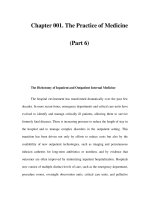
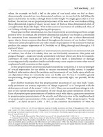
haem a porphyrin structure that con-
tains iron and that forms part of the
haemoglobin molecule; it is synthesized
partly within mitochondria and partly in
the cytosol (Fig. 34)
haematemesis the vomiting of blood
haematocrit (Hct) the proportion of a
column of centrifuged blood which is
occupied by erythrocytes or an equiva-
lent estimation produced by an auto-
mated blood counter
haematogone a primitive lymphoid cell
which morphologically resembles a lym-
phoblast but is a normal reactive cell
haematology the study of blood and its
diseases
haematopoiesis a synonym for haem-
opoiesis, this term being generally used in
the USA
haematopoietic pertaining to haem-
atopoiesis, a synonym for haemopoietic,
this term being generally used in the USA
haematoxylin a basic dye used in cytology
and to stain parasites; used in combina-
tion with eosin to stain tissue sections
H
Glycine + succinyl Coa
δ aminolaevulinic acid
Porphobilinogen
Hydroxymethylbilane
Uroporphyrinogen III
Coproporphyrinogen III
Protoporphyrinogen III
Protoporphyrin IX
Fe
2+
Haem
Mitochondrion
Cytosol
Mitochondrion
ala-synthase
+ pyridoxal-5'-
phosphate
ala-dehydrase
Porphobilinogen
deaminase
Uroporphyrinogen
III decarboxylase
Coproporphyrinogen
III oxidase
Protoporphyrinogen
III oxidase
Ferrochelatase
Uroporphyrinogen
III synthase
Figure 34 Haem synthesis.
The process by which haem is synthesized. Enzymes
are shown in italics and enzyme products in upright
script.
116
HAE-H 01/13/2005 05:12PM Page 116
haemoglobin C a variant haemoglobin
with an amino acid substitution in the
beta chain, mainly found in those of
African ancestry
haemoglobin Constant Spring a
variant haemoglobin with a structurally
abnormal alpha chain which is synthe-
sized at a reduced rate, leading to
αα
thalassaemia
haemoglobin D the designation of a
group of haemoglobin variants, some α
chain variants and some β chain variants,
that have the same mobility as haemo-
globin S on haemoglobin electrophoresis
at alkaline pH
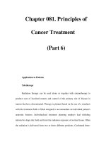
haemoglobin dissociation curve a
plot of percentage saturation of haemo-
globin against partial pressure of oxygen
(Fig. 37)
Haemoglobin Distribution Width
(HDW)
a measurement made by some
automated blood counters that indicates
the amount of variation in haemoglobin
concentration between erythrocytes; an
increased HDW correlates with aniso-
chromasia on a blood film
haemoglobin E a variant haemoglobin
with an amino acid substitution in the
beta chain, mainly found in South-east
Asia and parts of the Indian subcontinent
haemoglobin electrophoresis a me-
thod of separating normal and variant
haematoxylin and eosin (H & E) the
standard stain used for staining tissue
sections, a mixture of basic haemotoxylin
and acidic eosin
haematuria the presence of red cells in
the urine
haemiglobin cyanide an alternative
designation of cyanmethaemoglobin, the
form of haemoglobin which results from
interaction with cyanide in the cyan-
methaemoglobin method for estimation
of haemoglobin concentration
haemochromatosis see hereditary hae-
mochromatosis
haemocytometer a counting chamber
for counting blood cells
haemodialysis a method of treating
acute or chronic renal failure by passing
the patient’s blood through a dialysis
machine; blood and dialysis fluid are sep-
arated by a semipermeable membrane so
that exchange of solutes can occur
haemoflagellates flagellated blood par-
asites such as trypanosomes and leishmania
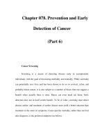
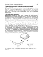
haemoglobin a complex molecule com-
posed of four globin chains, each of which
partially encloses a haem molecule
(Fig. 35), which has as its major function
the transport of oxygen from the lungs to
the tissues
haemoglobin A the major haemoglobin
component present in most adults, hav-
ing two α chains and two β chains
haemoglobin A
2
a minor haemoglobin
component present in adults and, as an
even lower proportion of total haemo-
globin, in neonates and infants; it has two
α chains and two δ chains
haemoglobin Bart’s an abnormal
haemoglobin with four γ chains and no
α chains, present as the major haemo-
globin component in haemoglobin Bart’s
hydrops fetalis and as a minor component
in neonates with haemoglobin H disease
or alpha thalassaemia trait
haemoglobin Bart’s hydrops fetalis
a fatal condition of a fetus or neonate,
resulting from homozygosity or compound
heterozygosity for
αα
0
thalassaemia
(Fig. 36); as there are no alpha genes
there can be no production of haemo-
globin A, A
2
or F
haemoglobin electrophoresis 117
β
2
α
2
β
1
α
1
Figure 35 The haemoglobin molecule.
A sketch of the haemoglobin molecule showing that
it is composed of two α globin chains and two β
globin chains, each enclosing a haem moiety.
HAE-H 01/13/2005 05:12PM Page 117
118 haemoglobin F
haemoglobins from each by applying a
haemolysate to a membrane or gel across
which there is an electrical gradient; the
pH and the nature of the membrane or gel
determines the rate at which different
haemoglobins migrate in the electrical
field (Fig. 38)
haemoglobin F fetal haemoglobin, the
major haemoglobin of the fetus and
neonate (Fig. 39), which is present as a
very minor component in most adults
and in higher amounts in a minority;
adult levels have usually been reached by
about one year of age
haemoglobin G the designation of a
group of haemoglobin variants, some of
which are alpha chain variants and some
of which are beta chain variants, that
have the same mobility as haemoglobin S
on haemoglobin electrophoresis at alka-
line pH; whether a variant haemoglobin
100
90
80
70
60
50
40
30
20
10
0
2.5 5 7.5 10 12.5
Alkalosis
decreased
2,3BPG
Acidosis
increased
2,3BPG
fever
kPa
Figure 37 A haemoglobin dissociation curve.
A haemoglobin dissociation curve showing the
sigmoid form of the normal dissociation curve and
the factors which shift the curve to the left or right.
Mother
α
0
thalassaemia
heterozygosity
Father
α
0
thalassaemia
heterozygosity
Hb Bart's
hydrops fetalis
(homozygosity
for α
0
thalassaemia)
α thalassaemia
trait
(heterozygosity
for α
0
thalassaemia)
α thalassaemia
trait
(heterozygosity
for α
0
thalassaemia)
Normal
Figure 36 haemoglobin Bart’s hydrops fetalis.
A diagrammatic representation of the possible outcomes in a family at risk of
producing a fetus with haemoglobin Bart’s hydrops fetalis.
HAE-H 01/13/2005 05:12PM Page 118
haemoglobin Lepore 119
is designated haemoglobin D or haemo-
globin G is completely arbitrary
haemoglobin Gower an embryonic
haemoglobin; haemoglobin Gower 1 is
ζ
2
ε
2
and haemoglobin Gower 2 is α
2
ε
2
haemoglobin H a variant haemoglobin
with four β chains and no α chains,
present in haemoglobin H disease and, in
small quantities, in
αα
thalassaemia trait
haemoglobin H disease a thalas-
saemic condition caused by marked
underproduction of α chains, often but
not always resulting from compound
heterozygosity for
αα
+
thalassaemia and
αα
0
thalassaemia with consequent lack of
three of the four alpha genes (Fig. 40)
haemoglobin H inclusions small
round evenly dispersed erythrocyte
inclusions composed of haemoglobin H;
they can be stained with vital dyes
haemoglobin Lepore a variant
haemoglobin resulting from the fusion
of part of a δ globin gene with part of
Figure 38 Haemoglobin electrophoresis.
The results of haemoglobin electrophoresis on a
cellulose acetate membrane at alkaline pH.
Haemoglobins S, D and G move together, as do
haemoglobins C, E and A
2
: (a) haemoglobins S and
C; (b) haemoglobin S; (c) haemoglobins A and C;
(d) haemoglobin S; (e) haemoglobins A and C;
(f ) haemoglobins S and C; (g) haemoglobins A and
C; (AFSC) control sample containing haemoglobins
A, F, S and C.
Haemoglobin A
Haemoglobin A
2
5
16 28 0 3 6
9
Weeks from conception Months of age
Birth
Haemoglobins
Gower 1, Gower 2,
and Portland
Total haemoglobin (%)
100
90
80
70
60
50
40
30
20
10
0
Haemoglobin F
Figure 39 Changes if haemoglobin F percentage during development.
The proportions of haemoglobin F and other normal haemoglobins present in the
embryo, fetus, neonate and infant.
HAE-H 01/13/2005 05:12PM Page 119
120 haemoglobinopathy
haemoglobinuria the presence of haemo-
globin in the urine
haemolysate a solution of haemoglobin
obtained by lysing red cells
haemolysis an increased rate of destruc-
tion of erythrocytes
haemolytic anaemia anaemia result-
ing from an increased rate of destruction
of erythrocytes
haemolytic disease of the newborn
(HDN)
haemolytic anaemia in a neonate,
consequent on destruction of fetal and
neonatal erythrocytes by a maternal allo-
antibody which has crossed the placenta
haemolytic uraemic syndrome (HUS)
a syndrome of microangiopathic haemo-
lytic anaemia and acute renal failure
haemophagocytic syndrome an ill-
ness resulting from haemophagocytosis,
a β globin gene, giving a δβ fusion
gene and fusion protein; it is synthes-
ized at a slower rate than the β chain
and thus is functionally equivalent to a
β thalassaemia
haemoglobinopathy an inherited dis-
order resulting from synthesis of a
structurally abnormal haemoglobin; the
term can also be used to encompass, in
addition, the thalassaemias in which
there is a reduced rate of synthesis of one
of the globin chains
haemoglobin Portland an embryonic
haemoglobin, ζ
2
γ
2
haemoglobin S sickle cell haemoglobin,
a variant haemoglobin with a tendency
to polymerize at low oxygen tension,
causing erythrocytes to deform into the
shape of a sickle
Mother
α
+
thalassaemia
heterozygosity
Father
α
0
thalassaemia
heterozygosity
Hb H disease
(compound
heterozygosity
for a
+
and α
0
thalassaemia)
α thalassaemia
trait
(heterozygosity
for α
+
thalassaemia)
α thalassaemia
trait
(heterozygosity
for α
0
thalassaemia)
Normal
Children
Figure 40 Haemoglobin H disease.
A diagrammatic representation of the possible outcomes in a family at risk of
producing a child with haemoglobin H disease.
HAE-H 01/13/2005 05:12PM Page 120
hairy cell an abnormal B-lymphocyte
present in hairy cell leukaemia, analogous
to a late B cell
hairy cell leukaemia a chronic B-
lineage leukaemia with neoplastic cells
which are morphologically and immuno-
phenotypically distinctive
hairy cell leukaemia variant a chronic
B-lineage leukaemia with neoplastic cells
which resemble the cells of hairy cell leuk-
aemia morphologically but with there
being differences in immunophenotype
and haematological and clinical features
Ham test see acid lysis test
HAM HTLV-I-associated myelopathy
hand mirror cell a blast cell shaped like
a mirror, may be of lymphoid or myeloid
lineage
Hand–Schüller–Christian disease part
of the clinical spectrum of Langerhans
cell histiocytosis
haploid a description of cells with a com-
plement of 23 chromosomes, one copy of
each autosome and either an X or a Y
chromosome; normal sperm and ova are
haploid but in somatic cells haploidy is
highly abnormal
haploidy the state of having a haploid
complement of chromosomes
haplo-insufficiency an abnormal phe-
notype or predisposition to disease as a
result of loss of one allele of a gene or loss
of a longer DNA sequence from one of a
pair of chromosomes
haplotype genotype of a group of alleles
from two or more closely linked loci, e.g.
the β
S
gene occurs in association with
several different haplotypes
hapten a small antigen that becomes
immunogenic when complexed with a
larger protein
HC2 a monoclonal antibody which gives
positive reactions with hairy cells and
occasionally with cells of other chronic
lymphoproliferative disorders
hCDCre see CDCREL
Hct haematocrit
HDN haemolytic disease of the newborn
HDW Haemoglobin Distribution Width
heavy chain the longer of the two poly-
peptide chains of a dimer, usually refers
to the heavy chain of an immunoglobulin
characterized by pancytopenia and some-
times hepatomegaly, splenomegaly and
fever, see also familial haemophagocytic
lymphohistiocytosis
haemophagocytosis phagocytosis of
haemopoietic cells and their progeny
haemophilia an inherited haemorrhagic
disorder resulting from deficiency of fac-
tor VIII (haemophilia A, resulting from a
mutation in the F8C gene) or factor IX
(haemophilia B, resulting from a muta-
tion in the F9 gene)
haemophilia A an X-linked recessive
inherited bleeding disorder (see Fig. 68,
p. 207) resulting from a mutation, most
often an inversion that splits the gene,
involving the F8C gene
haemophilia B an X-linked recessive
inherited bleeding disorder, previously
known as Christmas disease, resulting
from a mutation, most often a point
mutation, in the F9 gene
haemophilia B Leiden a variant of
haemophilia B, resulting from one of a
number of point mutations in the pro-
moter region of the F9 gene, in which
factor IX concentration rises at puberty
with improvement of the bleeding tend-
ency; the likely explanation is that the
mutations affect a binding region for
androgen-sensitive transcription factors
haemopoiesis the process of produc-
tion of blood cells (Fig. 41)
haemopoietic pertaining to haemopoiesis
haemopoietic cell a precursor cell giv-
ing rise ultimately to granulocytes, mono-
cytes, erythrocytes and platelets
haemopoietic growth factor a protein,
often a glycoprotein, that promotes growth
and differentiation of haemopoietic cells,
e.g. erythropoietin, thrombopoietin
haemorrhage bleeding; may be a nor-
mal phenomenon, e.g. following injury,
or the result of an inherited or acquired
haemorrhagic disorder
haemorrhagic disease of the new-
born
a haemorrhagic disorder of neo-
nates consequent on vitamin K deficiency
haemosiderin the major storage form
of iron, present in macrophages
haemostasis the process by which haemo-
rrhage is arrested
heavy chain 121
HAE-H 01/13/2005 05:12PM Page 121
SCF
HSC
SCF
FTL3L
IL7
CLP
T
B
NK
LDC
IL2
IL7
IL6
SCF
IL2
SCF
FLT3L
IL3
IL4
IL6
GM-CSF
SCF
IL3
IL6
GM-CSF
CMP
IL3
GM-CSF
G-CSF
M-CSF
IL3, GM-CSF,
G-CSF
IL3
GM-CSF
M-CSF
GFU-
GM
GFU-
G
GFU-
M
MDC
IL3
IL5
SCF
IL3
IL4
SCF
IL3
IL4
SCF
IL3,SCF,
GM-CSF
IL11,EPO
IL3,
GM-CSF
IL11,EPO
SCF,
IL1, IL3,
IL6, GM-CSF,
G-CSF, TPO
SCF,
IL3, IL6,
IL9, IL11,
GM-CSF, TPO
CFU-
Eo
CFU-
Baso
CFU-
Mast
BFU-E
BFU-
Mega
CFU-
Mega
CFU-E
PSC
Bi-, tri- and
multipotent
progenitors
Lineage
committed
progenitors
Figure 41 Haemopoiesis.
A diagrammatic representation of haemopoiesis showing the stem cell/progenitor cell hierarchy and the growth
factors that are thought to act at each stage of development.
Abbreviations for cell names: PSC, pluripotent stem cell (also known as HSC, haemopoietic stem cell);
CLP, common lymphoid progenitor; T, T cell; B, B cell; NK, natural killer cell; LDC, lymphoid dendritic cell;
CMP, common myeloid progenitor (also known as multipotent myeloid stem cell); CFU-GM, colony-forming
unit–granulocyte-macrophage; CFU-G, colony-forming unit–granulocyte; CFU-M, colony-forming
unit–macrophage; MDC, myeloid dendritic cell; CFU-Eo, colony-forming unit–eosinophil; CFU-Baso, colony-
forming unit–basophil; CFU-Mast, colony-forming unit–mast cell; BFU-E, burst-forming unit–erythroid;
CFU-E, colony-forming unit–erythroid; BFU-Mega, burst-forming unit–megakaryocyte; CFU-Mega, colony-
forming unit–megakaryocyte; HSC, haematopoietic stem cell.
Abbreviations for growth factors: SCF, stem cell factor; FLT3L, FLT3 ligand; IL, interleukin; GM-CSF,
granulocyte-macrophage colony-stimulating factor; G-CSF, granulocyte colony-stimulating factor; M-CSF,
macrophage colony-stimulating factor; EPO, erythropoietin; TPO, thrombopoietin.
HAE-H 01/13/2005 05:12PM Page 122
hemighost 123
helix–loop–helix see bHLH
HELLP syndrome a syndrome occurring
in pregnancy comprising H
aemolysis,
E
levated Liver enzymes and Low
P
latelets
helper T cell a T lymphocyte that pro-
motes antigen secretion by B lympho-
cytes (see type 1 and type 2 helper T cell)
(Fig. 42)
hemighost an erythrocyte in which all
the haemoglobin appears retracted into
one half of the cell leaving the remainder
molecule; each immunoglobulin molecule
has two light chains (κ or λ) and two heavy
chains (γ, α, µ, ε or δ)
heavy chain disease a lymphoprolifer-
ative disorder or plasma cell dyscrasia in
which there is synthesis of heavy chain
(γ, α or µ) rather than synthesis of com-
plete immunoglobulin molecules
Heinz body an erythrocyte inclusion
composed of denatured haemoglobin,
detected by exposure to vital dyes such
as methyl violet
CD2
CD54
CD58
CD80 or
CD86
CD40
CD11a/
CD18
CD154CD28 CD152
CD80 or
CD86
HLA
type II
CD3
TCR
αβ
CD4
Antigen internalized,
processed and
presented
SmIg
Ag
CD79
Antigen-presenting and
antibody-secreting B cell
Helper T cell
Antigen (ag)
CD79
Surface membrane immunoglobulin (SmIg)
Processed antigen
Figure 42 Helper T cell.
A diagrammatic representation of the interaction between a helper T cell and a B cell. The B cell binds an
exogenous antigen, by means of its membrane immunoglobulin–CD79a complex, and processes it to a peptide
which it presents, in a groove of an HLA type II molecule, to a helper T cell. The peptide, in its HLA type II
context, is recognized by the CD3–T cell receptor (TCR)–CD4 complex. Other specialized antigen-presenting
cells (e.g. macrophages and dendritic cells) can similarly present processed antigen to helper T cells. Binding of
the helper T cell to the B cell also involves binding of CD2 on the T cell to CD58 on the B cell. CD54 is up-
regulated on activated B cells and binds to CD11a/CD18 on T cells. In addition, there is binding of CD28 on the
T cell to CD80 or CD86 on the B cell, giving stimulatory signals, or binding of CD152 to CD80 or CD86 on the
B cell, giving inhibitory signals. CD154 (CD40 ligand) on the T cell binds to CD40 on the B cell, giving signals
for somatic hypermutation and immunoglobulin class switching. Signalling is bidirectional. The B cell signals to
the helper T cell to become activated and proliferate while the T cell signals to the B cell to mature into a plasma
cell and switch from secreting IgM to secreting other classes of immunoglobulin. The progeny of the helper T cell
that has been activated by an antigen-presenting B cell can migrate to other tissues where they initiate a cytotoxic
or inflammatory response if they encounter target cells expressing the appropriate antigen.
HAE-H 01/13/2005 05:12PM Page 123
and elevated serum ferritin without any
elevation of serum iron concentration or
any tissue iron overload; the cataracts
may be congenital or may develop during
childhood or adult life; the underlying
defect is in synthesis of L type ferritin,
which is increased and poorly regulated
as a result of a mutation affecting the
iron-responsive element of L ferritin
mRNA; serum ferritin is L type rather
than a mixture of L and H.
hereditary persistence of fetal
haemoglobin
an inherited condition
in which fetal haemoglobin persists at
higher than normal levels beyond the
neonatal period
hereditary pyropoikilocytosis an
inherited abnormality of the erythrocyte
membrane leading to striking poikilo-
cytosis and severe haemolytic anaemia,
usually consequent on inheritance of
two different elliptogenic mutations or
an elliptogenic mutation and a common
low expression allele, α spectrin
LELY
hereditary spherocytosis an inherited
abnormality of the erythrocyte membrane
leading to spherical red cells, compensated
haemolysis and sometimes haemolytic
anaemia, resulting from mutations in
ANK1, SPTA1, SPTB, EA1 or EPB42
genes
hereditary stomatocytosis an in-
herited abnormality of the erythrocyte
membrane leading to formation of bowl-
shaped cells, referred to as stomatocytes
(Fig. 43), and either compensated haemo-
lysis or haemolytic anaemia
hereditary xerocytosis an inherited
abnormality of the erythrocyte mem-
brane leading to increased cation flux
and either compensated haemolysis or
haemolytic anaemia
Hermansky–Pudlak syndrome a
heterogeneous inherited syndrome with
autosomal recessive inheritance char-
acterized by oculocutaneous albinism
and defective platelet function, the latter
resulting from a storage pool defect;
some cases result from mutation of the
HPS1 gene on chromosome 10; there is
increased lipofuscin in bone marrow
macrophages
of the cell as an empty membrane;
hemighosts are characteristic of oxidant
damage and the presence of an unstable
haemoglobin
hemizygosity having a single copy of
a gene on a single X chromosome, e.g.
hemizygosity for G6PD deficiency
hemizygote an individual having one
copy of a gene on a single X chromosome,
e.g. a male with a singe copy of a mutant
G6PD gene
HEMPAS Hereditary Erythroid Multi-
nuclearity with P
ositive Acidified Serum
test—type II congenital dyserythropoietic
anaemia (see acid lysis test)
heparin a sulphated glycosaminoglycan,
a naturally occurring anticoagulant in
human and animal tissues, which inhibits
thrombin, factor Xa and activated intrin-
sic pathway coagulation factors in vivo
and in vitro; as a therapeutic product,
it has usually been extracted from pig
intestines (see also low molecular weight
heparin and unfractionated heparin)
hepatitis inflammation of the liver
hepatomegaly enlargement of the liver
HER2 see ERBB2
hereditary passed down from a parent,
usually by means of genes located on
chromosomes but occasionally by mito-
chondrial genes
hereditary elliptocytosis an inherited
abnormality of the erythrocyte mem-
brane leading to elliptical red cells, some-
times to haemolysis and occasionally to
a haemolytic anaemia, resulting from
mutations in the SPTA1, SPTB, AE1 or
EPB41 genes
hereditary glucosyl ceramide lipidosis
see Gaucher’s disease
hereditary haemochromatosis a
hereditary condition leading to iron
overload; in adults, the condition usually
results from mutation in the HFE gene
but in a minority it results from mutation
in the TFR2 gene; the juvenile form
results from mutation of a gene on chro-
mosome 1q
hereditary hyperferritinaemia–
cataract syndrome
a constitutional
abnormality with autosomal dominant
inheritance, characterized by cataracts
124 hemizygosity
HAE-H 01/13/2005 05:12PM Page 124
histidine-rich glycoprotein 125
it combines with transferrin receptor,
reducing its affinity for iron; the C282Y
mutation, present in most patients with
haemochromatosis, prevents the binding
of the HFE product to β
2
-microglobulin
high grade a term used to describe
aggressive, highly malignant lymphomas
high hyperdiploid having between
51–60 chromosomes
highly active antiretroviral therapy
(HAART)
triple-drug antiretroviral
therapy for HIV infection employing
a combination of drugs from three
classes: (i) nucleoside-analogue reverse-
transcriptase inhibitors, non-nucleoside-
analogue reverse-transcriptase inhibitors
and protease inhibitors
high performance liquid chromatog-
raphy (HPLC)
a method of separating
proteins, such as haemoglobin variants,
from each other on the basis of char-
acteristics such as size, hydrophobicity
and ionic strength; a solution of proteins
is passed through a specially designed
column with different proteins emerging
after a characteristic period of time, refer-
red to as the retention time (Fig. 44)
HIP a gene, Huntingtin Interacting
P
rotein 1, gene map locus 7q11.2; encodes
a protein which interacts with Huntingtin
and F-actin, and is involved in dopamine
receptor endocytosis at clathrin coated pits;
Huntingtin is the product of the gene that
is mutated in Huntington’s disease; HIP
contributes to the HIP1-PDGFRB fusion
gene in chronic myelomonocytic leuk-
aemia associated with t(5;7)(q33;q11.2);
in the resulting fusion protein the
extracellular domains of PDGFRβ are
replaced with almost the entire HIP1
molecule, leading to constitutive receptor
oligomerization and activation.
hirudin an antithrombotic substance pro-
duced in the salivary glands of the medicinal
leech, Hirudo medicinalis; recombinant
forms are desirudin and lepirudin
histamine an inflammatory mediator
secreted by mast cells and basophils
histidine-rich glycoprotein a plasma
protein, synthesized by platelets, that
binds to plasminogen, thus reducing the
amount of circulating plasminogen avail-
herpesviruses a group of viruses includ-
ing chicken pox/varicella, the Epstein–
Barr virus and cytomegalovirus
herpes zoster shingles, recrudescence
of varicella-zoster virus infection, causing
a vesicular rash in the distribution of a
peripheral nerve
heterochromatin condensed, genetically
inactive chromatin
heterodimer a dimer composed of two
dissimilar polypeptide chains
heterogeneous irregularly distributed
heterophile antibody an antibody
recognizing antigens on cells of another
species
heteroplasmy the presence in a cell of
mutated and non-mutated mitochondrial
DNA
heterozygote an individual who has a
single copy of a specified autosomal or
(in a female) X chromosome gene
heterozygous having a single copy of a
specified autosomal or (in a female) X
chromosome gene
hexokinase an enzyme in the erythro-
cyte glycolytic pathway (see Fig. 33, p. 113)
HFE a gene on chromosome 6, mutation
of which can cause hereditary haemochro-
matosis in homozygotes and compound
heterozygotes; previously known as HLA-
H; it encodes a protein that interacts with
the transferrin receptor (product of the
TFRC gene), negatively affecting cellular
iron uptake from transferrin; the HFE
protein binds to β
2
-microglobulin, bind-
ing being essential for transport of the
HFE product to the cell surface where
Figure 43 Stomatocytes.
Three stomatocytes, showing that in three
dimensions a stomatocyte is bowl shaped.
HAE-H 01/13/2005 05:12PM Page 125
126 histiocyte
and related lineages are designated in the
WHO classification as shown in Table 6
histiocytic lymphoma an outmoded
designation of large cell lymphoma of
B- or T-lymphocyte lineage rather than
of histiocyte lineage; to avoid confusion,
a tumour resembling a lymphoma but
able for conversion to plasmin; it there-
fore has an antifibrinolytic effect and
deficiency, which is very rare, is asso-
ciated with an increased likelihood of
thrombosis
histiocyte an alternative designation of
a macrophage; neoplasms of histiocytic
45.0
37.5
30.0
22.5
15.0
7.5
0.0
0.125
0.100
0.075
0.050
0.025
0.000
0
123456
%
(a) Time (min)
F
1.09
1.22
1.30
1.68
2.50
A2
3.61
F – 1.18
2.50
0.95
0.00
0.75 1.50 2.25 3.00 3.75 4.50
5.25
Time (min)(b)
Volts
Figure 44 High performance liquid chromatography.
Separation of normal haemoglobins from each other by high performance liquid
chromatography (HPLC): (a) normal adult showing, from left to right, haemoglobin
F (shaded), glycosylated haemoglobin (long black arrow), haemoglobin A that has
undergone post-translational modification (short black arrow), haemoglobin
A (hollow arrow) and haemoglobin A
2
(shaded); (b) premature baby showing, from
left to right, haemoglobin F which has undergone post-translational modification
(short arrows), haemoglobin F (shaded), and haemoglobin A (hollow arrow); note
that in this premature baby there is negligible haemoglobin A
2
.
HAE-H 01/13/2005 05:12PM Page 126
hMRE11 127
genes that regulate the transcription of
type II HLA genes: either the transactiva-
tor gene, CIITA at 16p13, or transcrip-
tion factor genes—RFXAP on 13q, RFX5
at 1q21 or RFXANK; numbers of CD4+
T cells are greatly reduced
HLF a gene, Hepatic Leukaemic Factor,
gene map locus 17q21-22, encodes a
leucine zipper transcription factor which
is normally expressed in liver, kidney
and neurones within the central nervous
system; HLF contributes to the E2A-
HLF fusion gene in B-lineage acute lym-
phoblastic leukaemia associated with
t(17;19)(q21-22;p13); two different rear-
rangements between E2A and HLF are
known; in each case, the chimeric protein
comprises the amino-terminal transact-
ivation domain of E2A (TCF3) and the
carboxyl-terminal leucine zipper dimer-
ization domain of HLF; both fusion
proteins induce expression of the zinc
finger transcriptional repressor slug,
which in turn down-regulates the ex-
pression of pro-apoptotic proteins
HLXB9 homeobox gene HB9, gene map
locus 7q36, encodes a homeobox tran-
scription factor which is expressed in
CD34+ but not in CD34− bone marrow
cells and is down-regulated during line-
age commitment; HLXB9 contributes to
an HLXB9-ETV6 fusion gene in infants
with acute myeloid leukaemia associated
with t(7;12)(q36;p13); germline muta-
tions in this gene are associated with the
currarino triad (sacral agenesis, ureteric
and perianal dysgenesis)
hMRE11 a gene, gene map locus 11q21;
encodes a DNA repair enzyme expressed
in all proliferating cells; hMRE11 is
mutated in ataxia-telangiectasia-like
disorder
composed of histiocytes should be referred
to as a ‘histiocytic sarcoma’ (see Table 6)
histiocytic medullary reticulosis a
disease characterized by infiltration of
tissues including the bone marrow by
strikingly haemophagocytic histiocytes;
once thought to be a histiocyte malign-
ancy, it is now known that at least the
majority of cases are reactive
histiocytosis increased macrophages in
bone marrow or other tissues
histiocytosis X a former designation of
Langerhans cell histiocytosis, a neoplasm
of Langerhans cells (see Table 6)
histogram a graphical representation of
the distribution of measurements using
rectangular bars to represent the relative
frequency of a certain measured value
(Fig. 45)
histology the study of the structure of
tissues by examination of stained tissue
sections
histones proteins bound to DNA in
chromosomes
histoplasmosis a disease caused by infec-
tion by the fungus, Histoplasma capsulatum
HIV the human immunodeficiency virus
HLA a complex of genes at 6p21.3 encoding
the α chain of class I HLA antigens and the
α and β chains of class II HLA molecules
HLA human leucocyte antigen
HLA type I deficiency an immune
deficiency syndrome (see bare lymphocyte
syndrome) in which a mutation in either
the TAP1 or TAP2 genes at 6p21.3 means
that there is defective delivery of peptides
to developing type I HLA molecules,
which are therefore unstable and are not
efficiently transported to the cell surface
HLA type II deficiency an immune
deficiency syndrome (see bare lymphocyte
syndrome) resulting from a mutation in
Table 6 The WHO classification of histiocytic and dendritic cell neoplasms.
Histiocytic sarcoma
Langerhans cell histiocytosis
Langerhans cell sarcoma
Interdigitating dendritic cell sarcoma/tumour
Follicular dendritic cell sarcoma/tumour
Dendritic cell sarcoma, not otherwise classified
HAE-H 01/13/2005 05:12PM Page 127
128 Hodgkin’s cell
and mononuclear Hodgkin’s cells in an
appropriate background of lymphocytes,
eosinophils and fibroblasts with variable
collagen fibrosis; the neoplastic cells are
almost always B lymphocytes (but in 1–2%
of patients are T lymphocytes); desig-
nated in the WHO classification as shown
in Table 7, the disease characteristically
Hodgkin’s cell a mononuclear giant cell
with a large nucleolus that is part of
the neoplastic population in Hodgkin’s
disease
Hodgkin’s disease or Hodgkin lym-
phoma
a lymphoma characterized by
certain histological features, specifically
by the presence of Reed–Sternberg cells
10
8
6
4
2
0
20
10
0
Std dev = 117.38
Mean = 298.5
n = 80
Std dev = 0.17
Mean = 2.44
n = 80
100 150 200 250 300 350 400 450 500 550 600 650
2.00 2.06 2.13 2.19 2.25 2.31 2.38 2.44 2.50 2.56 2.63 2.69 2.75 2.81
(b) LOGACB12
(a) ACB12
Figure 45 Histograms.
Two histograms showing the distribution of serum vitamin B
12
concentrations in
80 healthy volunteers: (a) plotted on an arithmetic scale, showing that the distribution
is not Gaussian; (b) re-plotted on a logarithmic scale, showing that the distribution of
the data is now Gaussian, i.e. the data have a log normal distribution.
HAE-H 01/13/2005 05:12PM Page 128
HOX11L2 129
host the recipient of a transplant (or an
individual harbouring a parasite)
Howell–Jolly body a nuclear inclusion
in an erythrocyte
Howship’s lacuna a hollow on the
surface of a bony spicule occupied by
an osteoclast
HOX genes genes encoding a superfam-
ily of evolutionarily conserved transcrip-
tion factors which share a 60 amino-acid
DNA-binding domain, the homeodomain;
they fall into two classes—I and II; class
I genes are organized into 4 clusters
(A,B,C,D) located on chromosomes 7,
17, 12 and 2 respectively; class II genes
are dispersed throughout the genome
and may have divergent homeodomain
sequences; clusters B and C play a role in
erythropoiesis, whilst members of cluster
A are predominantly expressed in cells of
granulocytic lineage; virtually all HOX
genes of the A, B and C clusters are ex-
pressed in pluripotent haemopoietic stem
cells—a feature lost with the onset of
lineage commitment; several non-cluster
HOX genes are also important in normal
haemopoiesis
HOX11 a gene, Homeobox 11, also
known as T
-Cell Leukaemia 3, TCL3,
and TLX1; gene map locus 10q24, which
encodes a non-cluster homeobox tran-
scription factor which is thought to play a
role in splanchnic development; HOX11
is dysregulated:
• by proximity to the TCRAD (αδ) locus
in T-lineage acute lymphoblastic leuk-
aemia associated with t(10;14)(q24;q11)
• by proximity to the TCRB gene at 7q35
in T-lineage acute lymphoblastic leuk-
aemia associated with t(7;10)(q35;q24)
HOX11L2 a gene, Homeobox 11-Like 2,
also known as T
-cell Leukaemia homeobox
presents with enlargement of lymph nodes
(see Figs 61 and 70, pp. 188 and 210)
hof a German term indicating the hollow
in a nucleus in which the Golgi apparatus
is located
homeobox a DNA binding domain
characteristic of a family of transcription
factors that included PBX1 and HOX11;
see also HOX genes
homeodomain see HOX genes
homocysteine an amino acid, increased
plasma levels of which, whether inherited
or acquired in origin, are associated with
an increased thrombotic risk
homodimer a dimer with two identical
chains
homogeneous evenly distributed
homogeneously staining region (hsr)
a segment of a chromosome that stains
homogeneously, indicating amplification
of DNA sequences
homologous structurally similar, in some
circumstances suggesting a common origin
in the remote past (e.g. homologous genes
in different species)
homologous chromosomes a matching
pair of chromosomes, one derived from
each parent of the individual
homology having structural similarity
homotypic having the same nature, e.g.
homotypic adhesion is adhesion of cells
to other cells of the same type
homozygous having identical alleles at
a given locus
hookworm a parasitic intestinal round
worm which may cause eosinophilia and
iron deficiency anaemia
hormone a long distance chemical
mediator
horned cell a keratocyte, an erythrocyte
with two or four symmetrical spicules
(see Fig. 50, p. 147)
Table 7 The WHO classification of Hodgkin Lymphoma (Hodgkin’s disease).
Nodular lymphocyte-predominant Hodgkin lymphoma
Classical Hodgkin lymphoma
Lymphocyte-rich classical Hodgkin lymphoma
Nodular sclerosis classical Hodgkin lymphoma
Mixed cellularity classical Hodgkin lymphoma
Lymphocyte-depleted classical Hodgkin lymphoma
HAE-H 01/13/2005 05:12PM Page 129
HPA human platelet antigen
HPLC high performance liquid chromato-
graphy (previously ‘high pressure liquid
chromatography’)
HPRT an X chromosome gene encoding
hypoxanthine-guanine phosphoribosyl-
transferase, mutation of which can con-
vey resistance to 6-thioguanine therapy;
it is sometimes used for clonality studies
HSP89A a gene, Heat Shock Protein 89
Alpha, gene map locus unknown, encodes
heat shock protein 89α; the gene con-
tributed to an HSP89A-BCL6 fusion gene
in a high grade gastric B-cell lymphoma
hsr a cytogenetic abbreviation indicating
a homogeneously staining region
HTLV-I human T-cell lymphotropic virus I
HTLV-I-associated myelopathy (HAM)
a myelopathy caused by HTLV-I, the
retrovirus which also causes adult T-cell
leukaemia/lymphoma
hTR the gene encoding telomerase RNA,
mutation of which can lead to autosomal
dominant dyskeratosis congenita
human herpesvirus 8 (HHV8) a her-
pesvirus that has an aetiological role in
primary effusion lymphoma and Cattleman’s
disease
human immunodeficiency virus (HIV)
the retrovirus that causes the acquired
immune deficiency syndrome (AIDS)
human leucocyte antigen (HLA) a
group of highly polymorphic cell surface
antigens, encoded by genes at 6p21.3,
involved in self and non-self recogn-
ition, tolerance, rejection of allografts
and graft-versus-host disease, divided
into HLA class I, class II and class III
antigens
human leucocyte antigens, class I
(HLA class I)
molecules that are
mainly involved in the presentation of
endogenous antigen-derived peptides to
CD8+ cytotoxic T cells and NK cells;
expressed on most somatic cells but to a
varying degree; expressed on all leuco-
cytes and on platelets but negative or
weakly expressed on erythrocytes; com-
posed of a heavy chain that is encoded
by genes at 6p21.3 and a common light
chain,
ββ
2 microglobulin, which is encoded
by a gene on chromosome 15; there are
3
(TLX3), and Respiratory Neurone
homeobox
(RNX); gene map locus 5q35.1;
encodes a non-cluster homeobox tran-
scription factor which is thought to play a
role in the development of the medulla
oblongata; is transcriptionally activated,
probably by proximity to the transcrip-
tion regulatory elements of BCL11B, in
a quite common t(5;14)(q35;q32) cryptic
translocation in T-acute lymphoblastic
leukaemia
HOXA9 a gene, Homeobox A9; gene map
locus 7p15-p14.2, encodes a class I home-
obox transcription factor that is thought
to be important in myeloid differentia-
tion; HOXA9 contributes to the NUP98-
HOXA9 fusion gene in M2 acute myeloid
leukaemia associated with t(7;11)(p15;p15)
(see also NUP98); the fusion protein func-
tions as a transcription factor; in acute
myeloid leukaemia HOXA9 expression
correlates strongly with a worse prognosis
HOXA11 a gene, Homeobox A11,
gene map locus 7p15-p14.2, encodes a
class I homeobox transcription factor
that is important in myeloid differentia-
tion; contributed to a NUP98-HOXA11
fusion gene in a patient with Ph-negative
chronic myeloid leukaemia transforming
rapidly to M2 acute myeloid leukaemia;
germline mutations in the homeodomain
of HOXA11 are seen in radioulnar synos-
tosis in association with amegakaryocytic
thrombocytopenia.
HOXD13 a gene, Homeobox D13,
also known as HOX4I, gene map locus
2q31-q32, encodes a class I homeobox
transcription factor that is not normally
expressed in haemopoietic tissues; it con-
tributes to a NUP98-HOXD13 fusion gene,
one of two fusion genes present in therapy-
related and de novo acute myeloid leuk-
aemia associated with t(2;11)(q31;p15);
germline mutations in this gene are
associated with synpolydactyly with
foot anomalies
Hoyerall–Hreidarsson syndrome a
syndrome of aplastic anaemia, immun-
odeficiency, microcephaly and cerebellar
hypoplasia resulting from a mutation in
the DKC1 gene; a severe variant of X-
linked dyskeratosis congenita
130 HOXA9
HAE-H 01/13/2005 05:12PM Page 130
human T-cell lymphotropic virus I (HTLV-I) 131
genes at 6p21.3, falling into HLA-DM,
HLA-DO, HLA-DP, HLA-DQ and
HLA-DR groups
human neutrophil antigen (HNA)
neutrophil-specific alloantigens encoded
by genes at 5 loci (Table 8)
human platelet antigen (HPA)
platelet-specific alloantigens encoded by
genes found at at least 13 loci; the ISBT
has agreed a standardized nomenclature
(Table 9)
human T-cell lymphotropic virus I
(HTLV-I)
a retrovirus that causes adult
three classical HLA class I groups of anti-
gens (HLA-A, HLA-B and HLA-C) and
three non-classical (HLA-D, HLA-E and
HLA-F)
human leucocyte antigens, class II
(HLA class II)
molecules that are
mainly involved in the presentation of
exogenous antigen-derived peptides to
CD4+ helper T cells; expressed on B
cells, activated T cells, monocytes,
macrophages, dendritic cells, activated
neutrophils and thymic epithelium; class
II antigens are heterodimers encoded by
Table 8 Human neutrophil antigens – recommended terminology and the proteins on which they
are expressed.
System Antigen Protein on which antigen is located
HNA-1 HNA-1a (previously NA1) FcγRIII (CD16)
HNA-1b (previously NA2)
HNA-1c (previously SH)
HNA-2 HNA-2a (previously NB1) Gp58–64, a GPI-linked glycoprotein
HNA-3 HNA-3a (previously5b) Gp70–95
HNA-4 HNA-4a (previously MART) CD11b
HNA-5 HNA-5a (previously CND) CD11a
Table 9 Human platelet antigens – recommended terminology and the platelet glycoproteins on
which they are expressed.
System Antigens Glycoprotein on which antigens are carried
HPA-1 HPA-1a (previously Pl
A1
, Zw
a
) IIIa
HPA-1b (previously Pl
A2
, Zw
b
)
HPA-2 HPA-2a (previously Ko
b
)Ibα
HPA-2b (previously Ko
a
, Sib
a
)
HPA-3 HPA-3a (previously Bak
a
, Lek
a
) IIb
HPA-3b (previously Bak
b
)
HPA-4 HPA-4a (previously Yuk
b
, Pen
a
) IIIa
HPA-4b (previously Yuk
a
, Pen
b
)
HPA-5 HPA-5a (previously Br
b
, Zav
b
)Ia
HPA-5b (previously Br
a
, Zav
a
, Hc
a
)
HPA-6w HPA-6bw (previously Ca
a
, Tu
a
) IIIa
HPA-7w HPA-7bw (previously Mo) IIIa
HPA-8w HPA-8bw (previously Sr
a
) IIIa
HPA-9w HPA-9bw (previously Max
a
) IIb
HPA-10w HPA-10bw (previously La
a
) IIIa
HPA-11w HPA-11bw (previously Gro
a
) IIIa
HPA-12w HPA-12bw (previously Iy
a
)Ibβ
HPA-13w HPA-13bw (previously Sit
a
) GP1a
HAE-H 01/13/2005 05:12PM Page 131
cells (ii) increased staining of nuclei, con-
sequent on altered chromatin structure
hyperchylomicronaemia increased
plasma concentration of chylomicrons
hyperdiploid having more than 46
chromosomes
hyperendemic constantly present in a
community and having a high prevalence
hypereosinophilic syndrome a syn-
drome of cardiac and other tissue dam-
age resulting from an increased eosinophil
count with tissue damage consequent on
the release of eosinophil granule contents
(see idiopathic hypereosinophilic syndrome)
hyperferritinaemia with congenital
cataracts
see hereditary hyperferriti-
naemia-cataract syndrome
hypergammaglobulinaemia
increased concentration of serum
immunoglobulins
hypergranular promyelocytic leuk-
aemia
acute myeloid leukaemia with
arrest of maturation at the promyelocyte
stage with the promyelocytes being
hypergranular
hyperhomocysteinaemia an inherited
metabolic defect resulting from deficiency
of cystathionine β-synthase, associated
with an increased risk of thrombosis
hyperimmunoglobulin D syndrome
an autosomal recessive condition, resulting
from mutation in the mevalonate kinase
gene, characterized by periodic fever with
leucocytosis and an acute phase response
hyperimmunoglobulin E syndrome
a congenital immunodeficiency syndrome
characterized by abscesses, osteopenia,
eosinophilia and unusual facial features;
it maps to chromosome 4
hyperimmunoglobulin M syndrome
either (i) an X-linked immune deficiency
syndrome in which a mutation in the gene
at Xq26.3-27.1 encoding CD154 results in
failure of helper T-cells to express CD154:
helper T cells therefore cannot bind to
CD40 on antigen-presenting B cells (see
Fig. 42, p. 123), leading to a failure of class
switching and susceptibility to cancer and
autoimmune disease or (ii) an autosomal
recessive syndrome, some cases resulting
from mutation in the activation-induced
cytidine deaminase gene at 12p13, an
132 humoral immunity
T-cell leukaemia/lymphoma and HTLV-
I-associated myelopathy
humoral immunity antibody-mediated
immunity
HUT11 a locus at 18q12-q21 where vari-
ous alleles encode antigens of the Kidd
blood group antigen system which also
function as the human erythroid urea
transporter; genes at this locus are the
codominant Jka and Jkb and various Jk
null alleles that result from mutation of
the wild type Jka or Jkb; a Jk(a-b-)
phenotype (rare except in Finns and
Polynesians) can result from homozygos-
ity or compound heterozygosity for Jk
null alleles or from inheritance of the
dominant unlinked In inhibitor gene,
In(JK)
hyalomere the agranular periphery of a
platelet
hybridization the annealing of comple-
mentary sequences of DNA or RNA, e.g.
Southern and Northern blotting
hybridoma a clone of cells capable of
producing a monoclonal antibody,
produced by fusion of an antibody-
producing cell with a mouse myeloma cell
hydrops fetalis severe oedema and
serous effusions in a fetus or neonate;
may be caused by severe anaemia, as in
haemoglobin Bart’s hydrops fetalis, severe
haemolytic disease of the newborn and
intrauterine parvovirus B19 infection
hydroxocobalamin the form of vitamin
B
12
used for therapy
hydroxyurea an antimetabolite used to
treat chronic myeloid leukaemias and
myeloproliferative disorders
hyperbetalipoproteinaemia
increased concentration of plasma beta
lipoproteins
hyperbilirubinaemia increased plasma
bilirubin concentration
hypercalcaemia increased plasma cal-
cium concentration
hypercholesterolaemia increased
serum cholesterol concentration
hyperchromatic showing increased up-
take of stain, thus appearing dense
hyperchromia (i) increased staining of
red cells reflecting an increased haemo-
globin concentration within individual red
HAE-H 01/13/2005 05:12PM Page 132
naemia or, less often, from a marked
increased in polyclonal immunoglobulins
hyphaema bleeding into the anterior
chamber of the eye
hypochromia reduced staining of
erythrocytes
hypodiploid having fewer than 46 but
more than 23 chromosomes
hypogranular neutrophil neutrophil
with reduced secondary granules
hypogranular variant of promyelo-
cytic leukaemia
a variant form of
hypergranular promyelocytic leukaemia
in which the leukaemic promyelocytes
appear hypogranular rather than hyper-
granular; the leukaemic cells have char-
acteristic bilobed nuclei
hyponatraemia reduced plasma sodium
concentration
hypoparathyroidism reduced activity
of the parathyroid glands
hypoplasia underdevelopment or regres-
sion of an organ, tissue or cell lineage
hypoplastic acute myeloid leukaemia
acute myeloid leukaemia that is unusual
in that the bone marrow is hypocellular
rather than hypercellular
hypoplastic anaemia an anaemia,
usually pancytopenia, as a consequence
of bone marrow hypoplasia; usually part
of the spectrum of aplastic anaemia
hypopyon accumulation of leucocytes
in the anterior chamber of the eye, a rare
complication of leukaemia
hyposplenism reduced or absent
splenic function
hypothyroidism reduced activity of the
thyroid gland
hypotonic having an osmolarity less
than normal; more dilute than normal
hypoventilation reduced breathing
hypoxanthine-guanine phosphoribo-
syltransferase
a purine salvage
enzyme encoded by HPRT
hypoxia inadequate supply of oxygen to
cells
hypoxia 133
mRNA-editing enzyme, with resultant
IgG and IgA deficiency with normal or
elevated IgM
hyperkalaemia increased plasma potas-
sium concentration
hyperlipaemia increased concentration
of blood lipids
hypermutation see somatic hypermutation
hypernatraemia increased plasma
sodium concentration
hyperparathyroidism increased activ-
ity of the parathyroid glands
hyperplasia increase of the number of
cells of a certain lineage or tissue
hyperreactive malarial splenomegaly
splenomegaly and increased polyclonal
IgM as a consequence of chronic malaria,
previously referred to as tropical
splenomegaly
hypersplenism splenomegaly leading to
pancytopenia as a consequence of pool-
ing of cells in the spleen and a decreased
erythrocyte lifespan
hypertension increase of blood pressure
hyperthyroidism overactivity of the
thyroid gland
hypertonic having an osmolarity greater
than normal; more concentrated than
normal
hypertrophy overdevelopment of an
organ or tissue; increased size rather than
increased number of cells
hyperuricaemia increased serum uric
acid concentration
hyperviscosity a pathological increase
in the viscosity of the blood or plasma,
may be a feature of multiple myeloma or
Waldenström’s macroglobulinaemia
hyperviscosity syndrome a clinical
syndrome resulting from hyperviscosity;
clinical features may include mental
changes and reduced level of conscious-
ness, visual changes resulting from effects
on the retina, congestive cardiac failure
and bleeding; may result from multiple
myeloma or Waldenström’s macroglobuli-
HAE-H 01/13/2005 05:12PM Page 133
are more likely to be designated, for ex-
ample, ‘post-polycythaemia myelofibrosis’)
idiopathic thrombocytopenic pur-
pura (ITP)
a previous designation of
autoimmune thrombocytopenic purpura
idiotype the specific surface antigens
which permit a clone of lymphocytes or
plasma cells to be recognized, dependent
on the specific immunoglobulin which is
expressed on the surface membrane
Ig immunoglobulin, e.g. IgG, IgA, IgM,
IgE or IgD
IGH the Immunoglobulin Heavy chain
locus, at 14q32 encoding the heavy chains
of immunoglobulin; there are multiple V
(variable), D (diversity), J (joining) and C
(constant) gene segments; rearrangement
of gene segments at the IGH locus occurs,
producing a gene encoding a specific
heavy chain (Fig. 46); the IGH enhancer
can contribute to oncogenesis by dysreg-
ulating many other genes such as BCL2,
BCL3, BCL6, BCL7A, BCL8, BCL9,
BCL10, BCL11A, CDK6, C4ST1, Cyclin
D1, Cyclin D2, FC
γγ
RIIB, FGFR3, IL3,
IRF4, MMSET, MAF, MUM2, MUM3,
MYC, NF-
κκ
B2, PAFAH2, PAX5 and
RCK.
IGK the locus at 2p12 encoding the
κκ
light
chain of immunoglobulin
IGL the locus at 22q11 encoding the
λλ
light chain of immunoglobulin
IKAROS a gene, also known as Zinc
F
inger protein, subfamily 1A, member 1,
ZNFN1A1, gene map locus 7p13-p11.2,
encodes the archetypal member of a fam-
ily of lymphoid-restricted zinc finger
transcription factors that are considered
master regulators of lymphocyte differen-
tiation; there are at least 8 alternatively
spliced transcripts encoding isoforms with
i an antigen composed of repeats of linear
poly-N-acetyllactosaminoglycan, expres-
sed on fetal red cells, in adults largely
converted to the I antigen, branched
poly-N-acetyllactosaminoglycan, by the
action of β-1,6-N acetylglucosaminyl
transferase
i (or iso) an isochromosome
i phenotype the null phenotype of the I
blood group, resulting from deletion or
mutation of the IgnT gene, in Asians
associated with congenital cataract
I an antigen composed of repeats of
branched poly-N-acetyllactosaminoglycan,
on adult red cells, produced by the action
of β-1,6-N acetylglucosaminyl transferase
on i antigen
I-branching
ββ
-1,6-N acetylglucosaminyl
transferase
an enzyme encoded by
the I or IgnT gene at 9q21, which converts
the i antigen to the I antigen
I cell disease a congenital metabolic
disorder, associated with vacuolated
lymphocytes
ICSH International Council (previously
Committee) for Standardization in
Haematology
idiopathic literally ‘of unknown cause’
but the term often continues in use long
after the cause of a disorder is known
idiopathic hypereosinophilic syn-
drome
a syndrome defined as a hyper-
eosinophilia of unknown cause, persisting
for at least six months and with associ-
ated tissue damage
idiopathic myelofibrosis bone marrow
fibrosis consequent on a chronic myelo-
proliferative disorder (but conventionally
excluding cases following polycythaemia,
rubra vera, essential thrombocythaemia
and chronic granulocytic leukaemia, which
I
134
HAE-I 01/13/2005 05:12PM Page 134
IKAROS 135
Figure 46 The IGH locus and the generation of antigen receptor diversity.
Antigen receptors for B cells (immunoglobulins, Igs) and T cells (T cell receptors, TCRs) are heterodimers in
which each polypeptide is composed of an amino terminal variable (V) region joined to a constant (C) region.
Ig and TCR genes are similar in organization and permit the generation of proteins with V domains that are
unique to the cells that make them. The V domains of IgH chains (and TCR β or δ chains) are encoded by a
combination of V
H
, D and J segments; the partner polypeptide (Ig light chains or TCR α or γ) is assembled from
V and J segments only. There are a few hundred such segments at the IGH locus (and a similar number at each Ig
light chain locus) and of these, about 30% are pseudogenes; yet it is estimated that the total diversity for human
immunoglobulins is 10
14
. Two strategies allow for the generation of such huge diversity from so few genes:
(i) T and B cells use a site-specific DNA recombination mechanism called V(D)J recombination to assemble a
V region gene from germ line DNA by cutting and joining together randomly chosen combinations of one of
each of the segments. The intervening DNA is excised and lost from the genome of the mature lymphocyte.
This process is catalysed by RAG proteins (encoded by R
ecombination Activating Genes) which recognize
recombination signal sequences (RSSs) flanking each segment. The RSSs ensure the correct order of V(D)J
recombination is followed and prevent V–V or D–D joining. The recombination process is imprecise and leads
to small deletions and random insertions (*) at the V–D and D–J junctions of functional IGH loci, adding
additional diversity.
(ii) Mature B cells exhibit the phenomenon of somatic hypermutation, whereby random single base changes are
directed to gene segments encoding antigen binding pockets in rearranged VDJ genes (s). This may either
increase or decrease the specificity of the expressed Ig for its antigen. The mechanism of this process is obscure.
(iii) In the case of IGH genes, the same V region can be joined to several different C regions in the descendants of
an original B cell (isotype or class switching), so permitting the same antigen specificity to be utilized in different
biological contexts. The mechanism of this is unclear.
Germline
IgH locus
Functional
IgH locus
Cleavage and religation
Small deletions, random
insertions palindromic
additions
Somatic hypermutation
V
H
segments
~300
D
segments
~20
J
segments
6
Constant regions
µδγ
3
γ
1
ψε εα
1
α
2
ψγ γ
2
γ
4
*
*
*
HAE-I 01/13/2005 05:12PM Page 135
Factor 2, BSF2; gene map locus 7p21;
encodes interleukin-6
IL7 a gene, Interleukin-7, gene map locus
8q12-q13, encodes interleukin-7
IL8 a gene, Interleukin-8, also known
as chemokine ligand 8, CXCL8 and
N
eutrophil-Activating Peptide 1, NAP1;
gene map locus 4q12-q13; encodes
interleukin-8
IL9 a gene, Interleukin-9, also known as
T-cell/mast cell growth factor P40, gene
map locus 5q31.1, encodes interleukin-9
IL10 a gene, Interleukin-10, also known
as C
ytokine Synthesis Inhibitory Factor,
CSIF, gene map locus 1q31-q32, encodes
interleukin-10
IL11 a gene, Interleukin-11, gene map locus
19q13.3-13.4, encodes interleukin-11
IL12A a gene, Interleukin-12 Alpha
chain, encodes p35 subunit of interleukin-
12, gene map locus 3p12-q13.2
IL12B a gene, Interleukin-12 Beta chain,
encodes p40 subunit of interleukin-12,
gene map locus 5q31.1-q33.1
IL13 a gene, Interleukin-13, gene map
locus 5q31, encodes interleukin-13
IL14 a gene, Interleukin-14, gene map
locus unknown, encodes interleukin-14
IL15 a gene, Interleukin-15, gene map
locus 4q31, encodes interleukin-15
IL16 a gene, Interleukin-16, gene map
locus 15q26.1, encodes interleukin-16
IL17 a gene, Interleukin-17, gene map
locus 2q31, encodes interleukin-17
IL18 a gene, Interleukin-18, gene map
locus 11q22.2-q22.3, encodes interleukin-
18
IL19 a gene, Interleukin-19, gene map
locus 1q32, encodes interleukin-19
IL20 a gene, Interleukin-20, gene map
locus 1q32, encodes interleukin-20
IL21R a gene, Interleukin-21 Receptor,
gene map locus 16p11, normally ex-
pressed by peripheral blood NK cells;
contributes to a IL21R-BCL6 fusion gene
in B-lineage non-Hodgkin’s lymphoma
with t(3;16)(q27;p11)
ileum the distal small intestine, the site of
maximum vitamin B
12
absorption
iliac pertaining to the ilium
ilium one of the bones of the pelvis; the
posterior superior iliac spine, which is
common N-terminal and C-terminal
domains (Ik1 through Ik8); an in-frame
deletion of 10 amino acids upstream of
the transcription activation domain and
adjacent to the C-terminal zinc fingers
of Ik-2, Ik-4, Ik-7, and Ik-8 has been
reported in childhood acute lymphoblas-
tic leukaemia; IKAROS fuses to BCL6 as
a result of the t(3;7)(q27;p12) transloca-
tion in diffuse large B-cell lymphoma;
IKAROS mutations and decreased activ-
ity are associated with disease progres-
sion in chronic granulocytic leukaemia
IL interleukin
IL1A, IL1B two genes, gene map locus
2q14, encoding I
nterleukin-1
αα
(acidic)
and I
nterleukin-1
ββ
(neutral); a TATA
box mutation in the IL1B promoter is
associated with an increased risk of
hypochlorhydria and gastric cancer after
H. pylori infection
IL2 a gene, Interleukin-2, also known as
T
-Cell Growth Factor, TCGF, gene map
locus 4q26-q27, encodes interleukin-2
IL2RA a gene, Interleukin-2 Receptor
A
lpha, gene map locus 22q11.2-q13,
encodes interleukin-2 receptor α, also
known as CD25 and Tac (Ac
tivated T cell)
IL2RB a gene, Interleukin-2 Receptor
B
eta, encodes interleukin-2 receptor β,
also known as CD122, gene map locus
22q11.2-q13
IL2RG a gene, Interleukin-2 Receptor
G
amma, gene map locus Xq13, encodes
interleukin-2 receptor γ, also known as
CD132
IL3 a gene, Interleukin-3, gene map locus
5q31, encodes interleukin-3; IL3 is dys-
regulated by proximity to the IGH locus
in acute lymphoblastic leukaemia asso-
ciated with t(5;14)(q31;q32) leading to
eosinophilia
IL4 a gene, Interleukin-4, also known as
B
-cell Stimulatory Factor 1, BSF1; gene
map locus 5q31.1; encodes interleukin-4
IL5 a gene, Interleukin-5; also known
as E
osinophil Differentiation Factor,
EDF; gene map locus 5q31.1; encodes
interleukin-5
IL6 a gene, Interleukin-6, also known as
interferon beta 2, H
epatocyte Stimula-
tory F
actor, HSF and B-cell Stimulatory
136 IL
HAE-I 01/13/2005 05:12PM Page 136
immune system 137
immune complex disease a disease
caused by deposition of immune com-
plexes in tissues or on cells
immune deficiency inability to mount
an adequate immune response
immune haemolytic anaemia a
haemolytic anaemia mediated by alloan-
tibodies, autoantibodies, drug-dependent
autoantibodies or immune complexes
immune phenotype see immunopheno-
type
immune suppression suppression of
immune responses by disease or by
immunosuppressive therapy
immune system a system of molecules,
cells and tissues involved in protection
against infection, permitting innate and
acquired immune responses
part of the ilium, is often used for bone
marrow aspiration and trephine biopsy
imatinib mesylate a specific inhibitor of
BCR-ABL tyrosine kinase and of platelet-
derived growth factor β, used in the treat-
ment of chronic granulocytic leukaemia
and other chronic myeloid leukaemias
involving PDGFRB; imatinib mesylate
was previously known as STI-571
immune (i) relating to the body’s
response to antigens, whether by anti-
body production or T-cell responses (ii)
able to mount a rapid and adequate
response to an antigenic stimulus; usually
indicative of a secondary response to a
previously encountered antigen
immune complex a complex of antigen
and antibody
Table 10 WHO classification of B-cell neoplasms*.
Precursor B-cell neoplasms
Precursor B lymphoblastic leukaemia/lymphoma
Mature B-cell neoplasms
B-cell chronic lymphocytic leukaemia/small lymphocytic lymphoma (morphological variant – µ
heavy chain disease)
B-cell prolymphocytic leukaemia
Lymphoplasmacytic lymphoma/Waldenström macroglobulinaemia (morphological variant –
γ
heavy chain disease)
Mantle cell lymphoma
Splenic marginal zone lymphoma (including splenic lymphoma with villous lymphocyte)
Hairy cell leukaemia (morphological variant – hairy cell variant leukaemia)
Plasma cell myeloma
Monoclonal gammopathy of undetermined significance (MGUS)
Solitary plasmacytoma of bone
Extraosseous plasmacytoma
Primary amyloidosis
Heavy chain disease
Extranodal marginal zone B-cell lymphoma of mucosa-associated lymphoid tissues
(MALT-lymphoma)
Nodal marginal zone B-cell lymphoma
Follicular lymphoma
Mantle cell lymphoma (morphological variants – blastoid variants and variants resembling
small cell lymphoma and marginal zone B-cell lymphoma).
Diffuse large B-cell lymphoma (morphological variants – centroblastic, immunoblastic,
T-cell/histiocyte-rich, anaplastic, grade III lymphomatoid papulosis*)
Mediastinal (thymic) large B-cell lymphoma
Intravascular large B-cell lymphoma
Primary effusion lymphoma
Burkitt lymphoma/leukaemia:
* In addition, two entities of ‘uncertain malignant potential’ are recognized: lymphomatoid granulomatosis and
post-transplant lymphoproliferative disorder, polymorphic.
HAE-I 01/13/2005 05:12PM Page 137
138 immune tolerance
protects against proteolysis; there are two
subclasses (see Fig. 48)
immunoglobulin D an immunoglo-
bulin with a δ heavy chain; as a cell sur-
face molecule on B lymphocytes it binds
antigens, probably leading to activation
of the cell; as a plasma protein its physio-
logical role is unknown
immunoglobulin E an immunoglobulin
with an ε heavy chain, involved in defence
against parasitic infections and in allergic
reactions
immunoglobulin G an immunoglobulin
with a γ heavy chain, of subtypes IgG1,
IgG2, IgG3 and IgG4; responsible for
secondary immune responses (see Fig. 48)
immune tolerance a state in which
autologous antigens do not provoke an
effective immune response
immunity the body’s defence against
foreign or abnormal material, e.g. invad-
ing micro-organisms; immune responses
are either specific, being mediated by
cells that can recognize antigens, or non-
specific, e.g. mediated by complement in
the absence of specific immune responses
immunization the process by which im-
munity to various infections is promoted
by exposure to altered or killed micro-
organisms or antigens derived from them
immunoblast a large transformed lym-
phocyte resulting from stimulation of a
lymphocyte by an antigen or by a lectin
such as phytohaemagglutinin; immuno-
blasts have a large central nucleolus and
plentiful basophilic cytoplasm
immunoblastic lymphoma a large cell
lymphoma with lymphoma cells resem-
bling immunoblasts, in the WHO class-
ification regarded as a subtype of diffuse
large B-cell lymphoma (see Table 10, p. 137)
immunocytochemistry identification
of antigens on cells by means of antibod-
ies, the binding of which is recognized by
means of a cytochemical reaction
immunocytoma a neoplasm of cells
showing some degree of plasma cell
differentiation; lymphoplasmacytoid
lymphoma in the WHO classification
immunofixation a technique for char-
acterizing paraproteins by means of
electrophoresis followed by binding to
a class-specific antibody (Fig. 47)
immunofluorescence a method of re-
cognizing antigens by means of antibodies
bound to fluorochromes
immunoglobulin a plasma or cell-
bound glycoprotein with antibody activity,
further categorized as immunoglobulin
of five classes—G, A, M, E and D; each
immunoglobulin molecule has a basic
structure of two identical heavy chains
and two identical light chains (Fig. 48)
immunoglobulin A an immunoglobu-
lin with an α heavy chain, responsible
for immunity at mucosal surfaces and
present in secretions as a dimer with an
additional secretory component that
Figure 47 Immunofixation.
Immunofixation to demonstrate the nature of a
paraprotein in a patient with multiple myeloma.
Serum protein electrophoresis shows a discrete
band in the early γ region (arrow head, top).
Immunofixation shows that the abnormal band
reacts with anti-γ and ant-λ antisera and it therefore
represents an IgGλ paraprotein. Antisera to α, µ and
κ show only normal polyclonal immunoglobulins.
HAE-I 01/13/2005 05:12PM Page 138
impedance counter 139
Figure 48 Immunoglobulin molecules.
The structure of immunoglobulin molecules of IgG,
IgA and IgM classes. The basic structure is of two
heavy (grey) and two light chains (black) with both
the light chains and the heavy chains having a
variable region, which gives antibody specificity,
and a constant region which conveys other
properties, such as binding to complement or to Fc
receptors on neutrophils and macrophages (a) the
IgG molecule has heavy chains with one variable
regions and three constant regions; the two heavy
chains are crosslinked and each light chain is
crosslinked to a heavy chain; (b) the IgA molecule
has a similar structure to the IgG molecule but is
present in secretions as a dimer with the two
monomers being joined by a J chain (crosshatched);
in addition, epithelial cells add a secretory
component (white); (c) the IgM is present in serum
as a pentamer; five identical subunits are joined to
each other and to a J chain. The heavy chain of the
IgM molecule differs from that of the IgG and IgA
molecules in that it has four rather than three
constant regions.
immunoglobulin gene rearrange-
ment
the bringing together of non-
contiguous DNA sequences from the V, D
and J regions of the immunoglobulin heavy
chain locus or similarly the kappa locus
or the lambda locus (see Fig. 46, p. 135)
Fab
Fc
V
L
C
L
C
H
1
V
H
C
H
2
C
H
3
(a)
Hinge
V
L
C
L
C
H
1
V
H
C
H
2
C
H
3
J chain
(b)
Secetory
component
immunoglobulin heavy chain gene
a gene at the IGH locus encoding an
immunoglobulin heavy chain—γ, αµ, ε
or δ
immunoglobulin heavy chain locus
see IGH
immunoglobulin light chain the
κκ
or
λλ
light chain of immunoglobulin
immunoglobulin M a pentomeric
immunoglobulin with µ heavy chains,
responsible for the primary immune
response (see Fig. 48)
immunoperoxidase technique a
technique for recognizing antibodies
which have bound to antigen, achieved
by linking the antibody to peroxidase
(direct immunoperoxidase technique) or
utilizing a peroxidase-conjugated anti-
immunoglobulin which binds to the first
antibody (indirect immunoperoxidase
technique)
immunophenotype the antigenic char-
acteristics of a population of cells
immunophenotyping the recognition
of the antigenic profile of a population or
populations of cells (see Fig. 28, p. 104)
impedance a measurement of the flow
of electrical current between two electrodes
suspended in a conducting medium; an
alteration of impedance can be a con-
sequence of the passage of a cell or other
particle between the electrodes
impedance counter an automated
counter using impedance technology to
count and size cells
V
L
C
L
C
H
1
V
H
C
H
2
C
H
3
C
H
4
J
(c)
HAE-I 01/13/2005 05:12PM Page 139