Báo cáo y học: "Circadian rhythm and its role in malignancy" docx
Bạn đang xem bản rút gọn của tài liệu. Xem và tải ngay bản đầy đủ của tài liệu tại đây (639.2 KB, 13 trang )
REVIEW Open Access
Circadian rhythm and its role in malignancy
Sobia Rana
1
, Saqib Mahmood
2*
Abstract
Circadian rhythms are daily oscillations of multiple biological processes directed by endogenous clocks. The circa-
dian timing system comprises peripheral oscillators located in most tissues of the body and a central pacemaker
located in the suprachiasmatic nucleus (SCN) of the hypothalamus. Circadian genes and the proteins produced by
these genes constitute the molecular components of the circadian oscillator which form positive/negative feed-
back loops and generate circadian rhythms. Th e circadian regulation extends beyond clock genes to involve var-
ious clock-controlled genes (CCGs) including various cell cycle genes. Aberrant expression of circadian clock genes
could have important consequences on the transactivation of downstream targets that control the cell cycle and
on the ability of cells to undergo apoptosis. This may lead to genomic in stability and accelerated cellular prolifera-
tion potentially promoting carcinogenesis. Different lines of evidence in mice and humans suggest that cancer
may be a circadian-related disorder. The genetic or functional disruption of the molecular circadian clock has been
found in various cancers including breast, ovarian, endometrial, prostate and hematological cancers. The acquisition
of current data in circadian clock mechanism may help chronotherapy, which takes into consideration the biologi-
cal time to improve treatments by devising new therapeutic approaches for treating circadian-related disorders,
especially cancer.
Introduction
In humans, like other organisms, most physiological and
behavioral functions are manifested rhythmically across
days and nights. All healthy human beings e xhibit the
common attribute of sleeping at night and waking up in
the morning automatically. When a human being
encounters a new day, the body prepares itself for the
new tasks ahead and boost heart rate, blood pressure
and temperature. On the other hand, the same para-
meters decline at the end of the day. Such daily occur-
ring rhythms with a period of about 24 hours are
termed as circadian (from the Latin “circa diem” mean-
ing “about a day” ) rhythms [1]. These rhythms are the
outward manifestation of an internal timing system gen-
erated by a circadian clock that is synchronized by the
day-night cycle [2].
Circadian clocks
The circadian timing system proficiently coordinates the
physiology of living organisms to match environmental
or imposed 24-hour cycles [3]. Circadian clocks are
endogenous and self-sustain ed (meaning that rhythms
can continue even in the absence of external cues) time-
tracking systems that enable organisms to anticipate
environmental changes, thereby adapting their behavior
and physiology to the appropriate time of day [4]. This
provides organisms with an anticipatory adaptive
mechanism to the daily predictable change s in their
environment such as lig ht, temperature and social com-
munication, and serves to synchronize multiple molecu-
lar, biochemical, physiological and behavioral processes.
A wide range of biological processes are regulated by
the circadian clock including sleep-wake cycles, body
temperature, energy metabolism, cell cycle and hormone
secretion [5,6].
Central pacemaker or the master clock
The mammalian clock system is hierarchical with a master
clock that controls circadian rhythms and resides in the
suprachiasmatic nucleus (SCN) of the hypothalamus.
Damage to the SCN can render experimental animals
arrhythmic and cause sleep disorders in patients. More-
over, intracerebral grafts of perinatal SCN can reinstate
behavioural circadian rhythms of SCN-ablated rodents [7].
The SCN pacemaker consists of multiple, autonomous
single cell circadian oscillators, which are synchronized to
generate a coordinated rhythmic output in intact animals
* Correspondence:
2
Department of Human Genetics & Molecular Biology, University of Health
Sciences, Lahore, Pakistan
Rana and Mahmood Journal of Circadian Rhythms 2010, 8:3
/>© 2010 Rana and Mahmood; licensee BioMed Central Ltd. This is an Open Access article distributed under the terms of the Creative
Commons Attri bution License ( w hich permits unrestricted use, distribution, and
reproduction in any medium, provided the original work is properly cited.
[8,9]. In mammals, the circadian photoreception pathways
are distinct from those of visual perception [10-12]. Light
is perceived by a s ubset of melanopsin-expressing r etinal
ganglion cells, and the photic information is directly con-
veyed to the SCN cloc k thro ugh the retino-hypothalamic
tract [13-15]. This photic entrain ment corrects the phase
of the SCN oscillator every day to ensure synchronization
of circadian with geophysical time. The phase of SCN
rhythms can be shifted by exposure of the animal to a new
light/dark schedule or to short light pulses during the sub-
jective night [16]. Entrainment of a biological clock is the
process of determining both its period (which is 24 hours
in most humans) and its phase. The latter refers to the off-
set of a circadian clock with respect to the standard
24-hour cycle. In general terms, the period of the clock is
genetically determined, whereas its phase is heavily influ-
enced by environmental zeitgebers (cues or stimuli ) such
as light.
Peripheral oscillators or the slave oscillators
A major finding in the field of circadian rhythms in
recentyearsisthattheSCNisnottheonlycircadian
clock in the organism. Indeed, most tissues including
extra-SCN brain regions and periphe ral organs bear cir-
cadian oscillators [17]. Moreover, these extra-SCN oscil-
lators can function independently from the SCN [18].
Peripheral mammalian cell types contain functional cir-
cadian oscillators, but these may not respond to light-
dark cycles and can be entrained by non-photic stimuli
[19,20]. These circadian oscillators are sensitive to a
variety of chemical cues or to temperature cycles
[21,22]. The SCN synchronizes peripheral clocks in
organs such as liver, heart, and kidney via indirect and
direct routes so that a coherent rhythm is orchestrated
at the organismal level to ensu re temporally coordinated
physiology [23-25]. Indirect synchronization is achieved
by controlling d aily activity-rest cycles and, as a conse-
quence, feeding time. Feeding (or starving) cycles are
dominant zeitgebers for many, if not most, peripheral
clocks. Food metabolites, such as glucose, and hormones
related t o feeding and starvation are probably the feed-
ing-dependent ent rainment cues. Activity cycles also
influence b ody temperature rhythms, which in turn can
participate in the phase entrainment of peripheral
clocks. Direct entrainment may employ cyclically
secreted hormones and perhaps ne uronal signals con-
veyed to peripheral clocks via the peripheral nervous
system. Body temperature rhythms, which are controlled
in part by the SCN, may also contribute to the synchro-
nization of peripheral clocks [26].
Molecular mechanism of the circadian clock
The clock mechanism in the SCN and the peripheral
oscillators is known to be similar at the molecular l evel
[27]; however, the output pathways elicited can be
different and more tissue specific. The molecular clock-
work is composed of a network of transcriptional-trans -
lational feedback loops (Fig. 1) that drive rhythmic,
~24-hour expression patterns of core clock components
[28]. Core clock components are genes whose protein
products are necessary for the generation and regulation
of circadian rhythms within individual cells throughout
the organism [29]. The core clock components include
two gene families: Period and Cryptochrome.Inmam-
mals, the expression of three Period genes (Per1, Per2
and Per3) and two Cry ptochrome genes (Cry1 and Cry2)
is activated by a dimer of the proteins CLOCK (Circa-
dian Locomotor Output Cycles Kaput) and BMAL1
(Brain-Muscle Arnt-Like protein 1). CLOCK and
BMAL1 are transcriptional factors that heterodimerize
and induce the expression of Per and Cry genes by bind-
ing to their promoters at E-boxes [28,30,31]. CLOCK
also has an intrinsic histone acetyltransferase (HAT)
activity, thereby it can induce chromatin remodeling
and creat e a permissive state for activation of gene
expression [32]. PER and CRY proteins are synthesized
in the cytoplasm and t hey associate before entering the
nucleus.Inthenucleus,CRYsrepresstheactivityof
CLOCK and BMAL1 and in this way, they negatively
feedback on their own expression [33,34]. However, the
exact molecular mechanism of this repression is yet
unclear. The enzymatic activity of CLOCK also allows it
to acetylate non-histone substrates. For example,
CLOCK mediates acetylation of its own binding partner,
BMAL1, on Lys537. Ectopic expression of wild-type
BMAL1, but not an acetylation-resistant BMAL1 mutant
(K537R), is able to rescue the circadian expression of
endogenous target genes in mouse embryonic fibroblasts
(MEFs) derived from Bmal1
-/-
mice. The BMAL1-
K537R mutant has drastically reduced sensitivity to
CRY1-mediated repression compared with wild type
BMAL1, indicating that the acetylation of B MAL1 by
CLOCK might be an essential regulatory switch as it
facilitates CRY-dependent repression [35]. Anothe r cru-
cial modulator of the circadian clock machinery identi-
fied recently is a histone deacetylase, namely sirtuin 1
(SIRT1), which regulates circadian rhythms by counter-
acting the HAT activity of CLOCK [36]. SIRT1 is
required for high-magnitude circadian transcription of
several core clock genes, including Bmal1, Rorg,Per2,
and Cry1. SIRT1 binds CLOCK-BMAL1 in a circadian
manner and promotes the deacetylation and degradation
of PER2 [37].
CLOCK-BMAL1 heterodimers induce a second regu-
latory loop activating transcription of retinoic acid-
related orphan nuclear receptors, Rev-erba and Ro ra
[38]. Both of these proteins are transcription factors that
bind to the Bmal1 pr omoter at REV-ERBa and RORa
Rana and Mahmood Journal of Circadian Rhythms 2010, 8:3
/>Page 2 of 13
response elements. RORa activate transcription of
Bmal1 [39,40], whereas REV-ERBa repress the tran-
scription process [41,42]. Another core member of the
mammalian circadian clock is neuronal PAS-domain
protein 2 (NPAS2). NPAS2 is a paralogue of CLOCK,
exhibiting similar activities but differing in tissue distri-
bution. NPAS2 can heterodimerize with BMAL1, bind
to E-box motifs and transcripti onally activate circadian
genes [43].
The feedback loops described above are responsible
for var ying levels of me ssenger ribonucleic acids
(mRNAs) from the Per, Cry, Rev-erba and Bmal1 genes
across circadian phases. In the SCN, Per, Cry and Rev-
erba all exhibit a peak of abundance during the light
phase, while Bmal1 has an opposite phase (i.e., peaks
about 12 hour later). In most other brain regions and
peripheral tissues, these rhythms are all delayed by
several hours but generally keep a similar phase rela-
tionship amongst them. In some brain regions, PER
oscillationsareinphasewiththoseseenintheSCN
[44,45]. Considering that simple tra nscriptional feedback
loops like those describ ed above would normally lead to
mRNA oscillations with a period much smaller than 24
hours, other mechanisms have been added onto this
simple loop model to permit a slowing down and delay
of its progression that create a coordinated molecular
cycle approximating the 24 hours environmental period.
These mechanisms act at different levels involving post-
transcriptional processing of the mRNAs, translation,
post-translational processing of the proteins and nuclear
translocation [46-48]. Each of these can individually
contribute to introduce the delay between the activation
and repression of transcription that is required to keep
the period at ~24 hours.
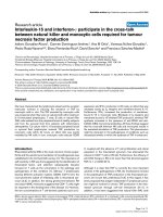
Figure 1 Schematic representation of the mammalian circadian clock mechanism. ROREs are retinoic acid-related orphan nuclear receptor
response elements present in Bmal1 promoter to which REV-ERBs and RORs compete to bind whereas E-boxes are regulatory enhancer
sequences present in the promoter regions of the genes under consideration to which CLOCK-BMAL1 heterodimer binds. Casein kinase (CK)
isoforms phosphorylate PER, CRY and BMAL1 proteins decreasing their stability and critically regulating the time of action of clock proteins.
Similarly, targets of GSK3 (glycogen synthase kinase-3) include PER, REV-ERBa and CRY2. c-Myc, Wee1 and Cyclin D1 are clock-controlled cell cycle
genes.
Rana and Mahmood Journal of Circadian Rhythms 2010, 8:3
/>Page 3 of 13
The post-translational modifications regulating the
circadian clock include acetylation, phosphorylation,
ubiquitination and sumoylation. In terms of phosphory-
lation, Casein kinase 1 epsilon (CK1ε) and Casein kinase
1 delta (CK1ε and CK1δ), Casein Kinase 2 (CKII), glyco-
gen synth ase kinase -3 (GSK3) and adenosine monopho-
sphate-activated protein kinase (AMPK) are c ritical
factors that re gulate the core circadian protein turnover.
It has been shown that mutations in CK1ε and CK1δ
result in altered kinase activities and cause shorter circa-
dian periods in mammals [49]. BMAL1 and C RYs are
reported to be the targets of CKIε [50]. Casein Kinase 2
(CKII) is one of the more recent kinases identified as a
clock component in N. crassa [51,52] and D. melanoga-
ster. [53-55] but its role in regulation of mammalian
clock has yet to be clarified. Changes in GSK3 activity
have been reported to alter period length in mammalian
cells [56]. The targets of GSK3 in mamm als might be
the PER proteins (PER phosphorylation by GSK3 might
prevent nuclear entry of PER proteins), REV-ERBa [57]
and/or CRY2 (for which phosphorylation might control
CRY2 degradation a t the end of night) [58]. Recently,
the n utrient-responsive AMPK has been found to regu-
late circadian clock by phosphorylation and destabiliza-
tion of the clock component CRY1 [59]. Also, AMPKg3
subunit is found to be involved in the regulation of pe r-
ipheral circadian clock function [60]. Likewise kinases,
phosphatases also participate in clock regulation. M ost
recently, the serine/threonine phosphatase PP5 (protein
phosphatase 5) has been found to interact with and be
regulated by CRY proteins [61]. Through its interaction
with CRY, PP5 migh t regulate the phosphorylation state
and so the activity of CK Iε in the clock [62,63]. Thus, it
can be said that phosphorylation by kinases, balanced by
regulated dephosphorylation, sets the stage for protein
degradation.
Phosphorylation is required for the recruitment of ubi-
quitin ligases, which mediate the polyubiquitylation and
the subsequent degradation of these proteins in the pro-
teasome. In mammals, the stability of PER1 and PER2 is
regulated by eith er bTrCP1 or bTrCP2. CKI phosphory-
lates PER 1 and PER2 and this phosphorylation leads to
the recruitment of bTrCP which mediates the ubiquity-
lation and proteasomal degradation of these proteins
[64,65]. Most rec ently, sumoylation has been revealed as
an additional level of regulation within the core
mechanism of the circadian clock. It is a reversible post-
translational modification in which a small ubiquitin-
related modifier p rotein (SUMO) is covalently linked to
lysine residues. It is controlled by an enzymatic pathway
analogous to the ubiquitin pathway. BMAL1 has been
found to be rhythmically sumoylated in vivo through a
process that requires the heterodimerization partner
CLOCK. Sumoylation of BMAL1 regulates the turnover
of the protein, as a mutation in the sumoylation site
(K259R) of BMAL1 lengthens the half-life of BMAL1
[66]. However, SUMO ligases and proteases which may
be involved in controlling this sumoylation and their cir-
cadian regulation are still to be known.
The transcriptional circadian regulation extends
beyond core clock components to include various
clock-controlled genes (CCGs), i.e., genes that are
under the direct or indirect transcriptional control of
the clock transcription factors but are not themselves
part of the clock. Regulation of clock-controlled genes
is a mechanism by which the molecular clockwork
controls physiological processes. The clock-controlled
genes (CCG) constitute about 10% of the expressed
genesinagiventissue(SCNorinperipheraltissues)
to generate rhythmic outputs, and, apart from few
exceptions, most of these clock-con trolled genes are
distinct in different tissues depending upon different
physiological needs [67]. Clock-controlled genes may
encode a variety of proteins including key regulators
for cell cycle.
Circadian clock and cell cycle
Circadian clock and cell cycle are global regulatory sys-
tems found in almo st all organisms. The circadian clock
shares a number of conceptual and molecular similari-
ties with the cell cycle [68]. Both are periodic for ca.
24 hours, and intrinsic to most cells. Simila rly, both are
based on the conceptual device of interlock auto-regula-
tory loops. Moreover, both rely on sequential phases of
transcription, translation and protein modification and
degradation. The circadian clock controls the expression
of cell cy cle- rela ted genes; in co ntrast , circadian clock-
work can oscillate accurately and independently of the
cell cycle, [69]. It is there by highly releva nt that CCGs
include genes that play an essential role in cell cycle
control.
It has been shown that CLOCK-BMAL1 directly regu-
late cell cycle genes such as Wee1 (G2-M t ransition)
[69], c-Myc (G0-G1 transition) and Cyclin D1 (G1-S
transition) [70]. The level of antimitotic WEE1 kinase in
the liver of Cry mutant mice (cryptochromeless mice) is
found to be elevated and consequently, liver regenera-
tion in these m ice following pa rtial hepatec tomy is
delayed relative to wild-type controls [ 69]. The binding
of CLOCK-BMAL1 to the E-boxes of Wee1 prom oter
stimulates the transcription of this gene. The elevation
of WEE1 in the Cry mutant is ascribed to the lack of
inhibition of CLOCK-BMAL1 by CRY [69,71]. WEE1 is
a cell cycle kinase that plays a key role in the G2-M
transition. Ongoing DNA replication or t he presence of
DNA damage activate WEE1, which then phosphorylates
CDC2 (cell division cycle 2)/Cyclin B1 complex, causing
its inactivation and delay of mito sis or arrest of the cell
Rana and Mahmood Journal of Circadian Rhythms 2010, 8:3
/>Page 4 of 13
cycle at the G2-M interface [72,73]. It is conceivable
that elevated WEE1 in Cry mutant mice phosphorylates
CDC2/CYCB1 complex at an increased rate even in
nonstressed cells, slowing down the G2-M transition
and the overall growth rate [69].
Transcription of c-Myc, which plays an important role
in both cell proliferation and apoptosis, is found to be
upregulated, and transcription of p53, which plays a cri-
tical role in the G1-S checkpoint, is downregulated in
Per2 mutant mice (mPer2
m/m
). Also, there is a general
cell cycle dysregulation as the circadian e xpression pat-
tern of genes functioning in cell proliferation and
tumour suppression, such as Cyclin D1, Cyclin A, Mdm-
2 (murine double minute, a negative regulator of p53)
and Gadd45a (growth arrest and DNA damage-induci-
ble protein a), is deregulated. Consequently, these ani-
mals have increased incidence of spontaneous and
ionizing radiation-induced lymphomas and an increased
rate of mortality after ionizing radiation [70]. Normally,
the binding of CLOCK-BMAL1 to the E-boxes of c-Myc
promoter inhibits the transcription of this gene. Upregu-
lation of c-Myc transcription in Per2 mutant is ascribed
to the reduced level of BMAL1 because PER2, in addi-
tion to its inhibitory effect on the CLOCK-BMAL1 com-
plex, stimulates transcription of the Bmal1 gene [28,74].
Oncogenic transformation mediated b y c-Myc must
overcome its proapoptotic activity [75] in which modu-
lation of p53-mediated apo ptosis plays an important
role [76,77]. Overexpression of c-Myc can induce geno-
mic DNA damage and compromise p53 function,
presumably through a reactive oxygen species (ROS)-
mediated mechanism [78]. Following g radiation,
MYC-overexpressing cells are less efficient in G1 arrest
compared to normal cells [79,80], indicating that c-Myc
overexpression could drive cells to progress through cell
cycle in th e presence of genomic DNA damage. Follow-
ing g radiation, the l oss of mPer2 function partially
impairs p53-mediated apoptosis, leading to accumula-
tion of damaged cells. However, the mutant mPer cells,
expressing MYC at elevated levels, could still progress
through cell cycle in the presence of genomic DNA
damage, resulting in the high incidence of tumor devel-
opment after g radiation.
Cyclin D1 (CCND1) is also a clock-controlled cell
cycle gene. Overexpression of CC ND1 induces mam-
mary tumorigenesis, in addition, increased levels of
CCND1 in ERa (estrogen receptor a) -positive breast
cancer is associated with poor prognosis [81]. However,
additional studies are needed to know whether the
rhythmic expression of CCND1 is deregulated in cancer.
Recently, it has been reported that p21 (Waf1/Cip1),
which does not possess an E-box element in its regula-
tory region, is controlled indirectly via CLOCK/BMAL1-
mediated transcriptional regulation of the orphan
nuclear receptor Rev-erb. p21 circadian expression is
dramatically increased and no longer rhythmic in Bmal1
knock-out mice. p21 upregulation in Bmal1
-/-
animals
primarily results from the loss of Rev-erba and Reverbb
expression possibly combined with the increased expres-
sion of RORg [82].Inthiscontext,thereleaseofthe
REV-ERB-dependent inhibition of RORa4 activity is
also likely to play a role. Changes in additional unidenti-
fied positive and negative regulators of p21 expression
may also play an additional r ole. Thus, in liver, the
clock control of p21 high amplit ude oscillation results
from a RORa4-andRORg-dependent activation, which
is rhythmically repressed by REV-ERBa and REV-ERBb.
As p21 negatively regulates cell cycle progression by
inhibiting the activity of CYCE/CDK2 complexes during
G1 phase progression, p21 overexpressing Bmal1
-
/
-
pri-
mary hepatocytes exhibit a decreased proliferation rate
[82].
Circadian clock, DNA damage response and
tumour suppression
The circadian control of an organism’s response to DNA
damage response rests up on circadian p roteins which
play important roles in the processes of cell proliferation
and control of response to genotoxic stress both at the
cellular and organismal levels [83]. DNA damage trig-
gers cellular stress response pathways which may result
in checkpoint cell cycle arrest, apoptosis or DNA repair.
DNA damage leads to activation of critical components
of cellular stress response pathways including ATM/
ATR (ataxia telangiectasia mutated/ataxia telangiectasia
and Rad3-related) and CHK1/2 (checkpoint kinase1/2)
which in turn activates tumour suppressor protein p53
and subsequently causes cell cycle arrest or apoptosis
[84]. It has been shown that Bmal1-deficient human
cells are unable to und ergo growth arrest on p53 activa-
tion by DNA damage. Contrary to in vivo mouse data
connecting BMAL1-dependent delay in G1 progression
to upregulation of p21 [82], radiation induced growth
arrest in Bmal1-deficient human cells correlated with
the decrease in levels of p53 and p21 [85]. This disparity
may be attributable to interspecies variation or differ-
ences between in vitro and in vi vo state and warrants
further investigation.
PER1 seems to function as a tumour suppressor by
regulating cell cycle genes and interacting w ith key
DNA damage-activated checkpoint proteins. Per1 over-
expression in cance r cells increases ionizing radiation-
induced apoptosis, whereas inhibition of Per1 in
similarly treated cells blunts apoptosis. Ionizing radia-
tion leads to PER1 nuclear translocation, the induction
of c-Myc expression and re pression of p21 (Waf1/Cip1).
Moreover, PER1 directly interacts with the DNA dou-
ble-strand break-activated kinases ATM and CHK2.
Rana and Mahmood Journal of Circadian Rhythms 2010, 8:3
/>Page 5 of 13
Thus, PER1 can function as a tumour suppressor by
activating multiple pathways, including the DNA
damage response [86]. Another circadian protein, time-
less (TIM), which is necessary for the robustness of
rhythmicity [87], has been shown to interact with the
cell cycle checkpoint proteins ATR, CHK1 and ATRIP
(ATR-interacting protein). This interaction is also sti-
mulated by DNA damage, and TIM seems to functio n
as a mediator between sensors and effectors of the DNA
damage response [88].
PER2 protein has also been proposed to function as a
tumor suppressor. Per2 mutant mice develop g radiation-
induced lymphomas at a higher rate than wild-type con-
trols due to partial impairment of p53-mediated apoptosis
[70]. Moreover, crossing these mice with polyp formation-
prone adenomatosis polyposis coli (Apc)
Min/+
animals
increases the frequency of formation of intestinal and
colonic polyps in Apc
Min/+
Per2
m/m
mice compared to Apc-
Min/+
mice. Following downregulation of Per2, Cyclin D,
which is a cir cadian regulated and b-catenin target gene,
has been shown to increase in human colon cancer cell
lines, as does cell proliferation. Thus, Pe r2 loss during
intestinal tumorigenesis may, in part, act through upregu-
lation of b-catenin, increasing intest inal b-cat enin signal-
ing and cell proliferation. Also, increase in small-intestinal
mucosa b-catenin in Per2
m/m
mice is associated wi th an
increase in MYC protein, again a circadian regulated and
b-catenin target gene [89]. Furthermore, accelerated
b-catenin expression is associated with PER2 protein
instability and lower PER2 levels as a result of increased
b-TrCP protein levels as it has been observed that overex-
pression of wild type or mutant b-catenin protein
decreases the stability of PER2 protein, and this PER2
instability i s reversed when the induction of b-TrCP is
prevented [90]. It has also been reported that mP er2 may
play an important role in tumor suppressio n by inducing
apoptotic cell death. Overexpression of Per2 in the mouse
Lewis lung carcinoma cell line (LLC) and mammary carci-
noma cell line (EMT6) results in reduced cellular prolif-
eration and rapid apoptosis, but not in non-tumorigenic
NIH3T3 cells. This is attributable to enhanced proapopto-
tis signaling and attenuated anti-apoptosis processes as
overexpressed mPER2 downregulate the mRNA and pro-
tein levels of c-Myc, Bcl-XL and Bcl-2, and upregulate the
expression of p53 and bax in mPer2-overexpressing LLC
cells [91]. Similarly, the intratumoral expression of mPer2
in C57Bl/6J mice transplanted with Lewis lung carcinoma
shows a significant antitumor effect [92]. All this evidence
indicates that Per2 has a ro le in tumour suppress ion, but
further research is needed to asc ertain whether Per2 is in
factatumoursuppressorgeneorwhetheraparticular
mutation of Per2 acquires oncogenetic properties.
In contrast to Per2 mutants, Cry double mutant (Cry1
-/-
Cry2
-/-
) mice are indistinguishable from the wild-type
controls with respect to radiation-induced morbidity and
mortality. Similarly, the Cry1
-/-
Cry2
-
/
-
mutant fibroblasts
are indistinguishable from the wild -type controls with
respect to their sensitivity to ionizing radiation and UV
radiation, and ionizing radiation-induced DNA damage
checkpoint response [93]. In another study, mice deficient
in the core circadian gene Bmal1 show reduce d lifespan
and various symptoms of premature aging but none of the
Bmal1
-/-
mice develop tumors in the c ourse of their life-
span [94]. Similarly, Clock/Clock mutant mice do not dis-
play predisposition to tumor formation either during their
normal lifespan or when exposed to a low dose of g-radia-
tion that is able to initiate and promote neoplastic pro-
gression [95]. Instead, exposure of Cl ock
-/-
mice to
ionizing radiation results in the development of pathologi-
cal conditions similar to those of premature aging
described for Bmal1
-/-
mice [94]. Recently, Ozturk et al.
reported the effect of the Cry mutation on carcinogenesis
in a mouse strain prone to cancer because of a p53 muta-
tion. Contrary to the expectation that clock disruption in
this sensitized background would further increase cancer
risk, they found that the Cry mutation protects p53
mutant mice from the early onset o f cancer and extends
their median lifespan ~50%, in part by sensitizing p53
mutant cells to apoptosis i n response to genotoxic stress
[96]. These studies suggest that disruption of the circadian
clock in itself d oes not comprom ise mammalian DNA
repair and DNA damage checkpoints and does not predis-
pose animals to spontaneous and ionizing radiation-
indu ced cancers. The effect of circadian clock disruption
on cellular response to DNA damage and cancer predispo-
sition may depend on the mechanism by which the clock
is disrupted, and elucidation of this mechanism warrants
further investigation.
Another aspect of DNA damage response is D NA
repair. Cells have evolved a number of mechanisms to
repair damaged DNA. One such repair mechanism,
nucleotide excision repair, is a multicomponent system
that replaces a short single stranded region encompass-
ing a DNA lesion. Recently, the effect of the circadian
clock on nucleotide excision repair has been investigated
in mice. Nucleotide excision repair is found to display
prominent circadian oscillations in mouse brain reach-
ing at its maximum in the afternoon/early evening
hours and minimum in the night/early morning hours.
The circadian oscillation of the repair capacity is caused
at least in part by the circadian oscillations in the
expression of DNA damage recognition protein xero-
derma pigmentosum A (XPA) [97].
Iterative alterations of lifestyle: clock -cancer
connection
The clock-cancer connection has been investigated in
studies of pilots, flight attendants, and shift workers
Rana and Mahmood Journal of Circadian Rhythms 2010, 8:3
/>Page 6 of 13
who are more likely to have disrupted circadian cy cles
due to abnormal work hours. Incidence of breast cancer
increases significantly in women working nightshifts,
being higher among individuals who spend more years
and hours per week working at night [98]. Exposure to
light-at-night, including disturbance of the circadian
rhythm, possibly mediated via the melatonin synthesis
and clock ge nes, has been suggested as a contributing
cause of breast cancer. Since working nightshifts is pre-
valent and increasing in modern societies, this exposure
may be of public health concern, and contribute to the
ongoing elevation in breast cancer risk [99-101]. Keith
et al. propose that circadian rhythms could be more
important than family history in determining breast can-
cer risk [102]. A pilot study in India showe d that the
risk of developing breast cancer in menopausal visually
challenged women is very much lower as compared to
sighted women in the similar age group suggesting a
relationship between visible light and breast cancer risk
[103]. Another study revealed that women working
more than 20 years of rotating night shifts have a signif-
icantly increased risk of endometrial cancer. In stratified
analyses, obese women working rotating night shifts had
doubled their baseline risk of endometrial c ancer com-
pared with obese women who did no night work,
whereas no significant increase was seen among non-
obese women [104]. Observations from a cohort study
of Air Canada pilots showed a significantly increased
incidence rate of prostate cancer when compared with
the respective Canadian p opulation rates [105]. A simi-
lar cohort of Nordic pilots demonstrated that the rela-
tive risk of prostate cancer increases as the number of
flight hours in long distance aircraft increases [106].
A significant association between rotating-shift work
and prostate cancer incidence among Japanese male
workers has also been found [107]. Incidence rate of
acute myeloid leukemia (AML) has been reported to be
significantly increased in a cohort of Air Canada pilots
in comparison to respective Canadian population rates
[108]. It has also been found that working a rotating
nightshift at least three nights per month for 15 or
more years may increase the risk of color ectal cancer in
women [109]. Colon cancer patients who have main-
tained a regular pattern of rest and activity rhythms
have shown a fivefold higher survival time than those
who have chaotic circadian rhythms [110].
Aberrant expression of clock genes in cancer
Several reports have revealed that clock genes are found
to be deregulated in various cancers. In comparison
with nearby non-cancerous cells, more than 95% of
breast cancer cells reveal disturbances in the expression
of the three Per genes attributable to methylation of the
per gene promoters [111]. Moreover, a structural
variation of the Per3 gene has been identified as a
potential biomarker for breast cancer in pre-menopausal
women [112]. Significantly decreased expres sion of Per1
has been observed between sporadic breast tumors and
normal samples, as well as a further significant decrease
between familial and sporad ic breast tumors for both
Per1 and Per2 suggesting a role for both in normal
breast function [113]. It has been demonstrated recently
that Per2 is endogenously expressed in human breast
epithelial cell lines but is not expressed or is expressed
at significantly reduced level in human breast cancer
cell lines. Expression of Per2 in these breast cancer cells
results in inhibition of cell growth and induction of
apoptosis demonstrating thetumorsuppressivenature
of PER2. Moreover, PER2 activity is found to be signifi-
cantly enhanced in the presence of its normal cloc k
partner CRY2. Furthermore, Per2 expression in cancer
cell l ines is associated with a significant decrease in the
expression of Cyclin D1 and an up-regulation of p53
[114]. The proliferation in ovarian cancer cells has been
found to follow a cyclical pattern of peaks and troughs
that is out of phase with the circadian rhythm in prolif-
eration of normal tissues [115,116]. Recently, it has been
reported that expression levels of Per 1, Per2, Cry2,
Clock,andCKIε in ovarian cancers are significantly
lower than those in normal ovaries. On the contrary,
Cry1 expression is highest followed by Per3 and Bmal1
[117]. Similarly, significantly decreased expression levels
of Per1 as compared to paired non-tumour tissues, have
been reported in endometrial carcinoma (EC). The
decreased Per1 expression in EC is partly due to inacti-
vation of the Per1 gene by DNA methylation of the
promoter and partly due to other facto rs. This downre-
gulation of the Per1 gene disrupts the circadian rhythm,
which might fav our the survival of endometrial cancer
cells [118]. In another study, the promoter methylation
in the Per1, Per2,orCry1 circadian genes has been
detected in about one-third of EC and one-fifth of non-
cancerous endometrial tissues of 35 paired specimens
indicating possible disruption of the circadian clock in
the development of EC [119]. Serum-shocked synchro-
nized prostate cancer cells have been found to display
disrupted circadian r hythms compared with the normal
prostate tissue. Per1 is down-regulated in human pros-
tate cancer samples compared to normal prostates.
Moreover, over-expression of Per1 in prostate cancer
cells has resulted in significant growth inhibition and
apoptosis [120].
CCAAT/enhancer-binding proteins (C/EBPs) are a
family of transcription factors that regulate cell growth
and differentiation in numerous cell types. The results
from a recent study suggest that Per2 is a dow nstream
C/EBPa-target gene invol ved in acute myeloid leukemia
(AML). Its disruption might be involved in initiation
Rana and Mahmood Journal of Circadian Rhythms 2010, 8:3
/>Page 7 of 13
and/or progression of AML, as significantly reduced
expression of Per2 has been noted in lymphoma cell
linesaswellasinAMLpatientsamples[121].The
expression of Per1, Per2, Per3, Cry1, Cry2,andBmal1 is
significantly impaired in both chronic phase and blast
crisis of chronic myeloid leukemia (CML) samples com-
pared with those in normal samples. Although no muta-
tions have been detected within the coding region of
Per3, the CpG islands in its promoter are methylated in
all the CML samples. Likewise, the CpG islands of Per2
are also methylated in 40% of cases [122]. Recently,
Cryptochrome1 has been found to be a valuable predic-
tor of disease progression in early-stage chronic lympho-
cytic leukemia (CLL) [123]. More recently, it has been
reported that CRY1: PER2 expression ratio is indepen-
dent prognostic marker in chronic lymphocytic leukemia
[124]. There is also a case report showing that a patient
with primary cerebral B-cell non-Hodgkin’ slymphoma
(NHL) has lost circadian control of sleep [ 125]. More-
over, genetic association and functional analyses suggest
that the circadian gene Cry2 might play an impo rtant
role in NHL development [126]. Several circadian
related genes have been found to be under-expressed in
pancreatic cancer indicating that pancreatic tumors have
altered circadian rhythms [127]. Recently, Per1 has been
identified as a candidate tumor suppressor, epigeneti-
cally silenced in nonsmall-cell lung cancer (NSCLC).
Per1 expression has been found to be low in a large
panel of NSCLC patient samples and in NSCLC cell
lines compared to normal lung tissue. The down-regula-
tion of Per1 expression is associat ed with hypermethyla-
tion of the Per1 promoter. Moreover, the study reveals
that aberrant acetylation of Per1 promoter is also a
potential m echanism for silencing Per1 in cancer [128].
More recently, Bmal1 has been reported to be transcrip-
tionally silenced by promoter CpG island hypermethyla-
tion in hematologic malignancies, such as diffuse large
B-cell lymphoma and a cute lymphocytic and myeloid
leukemias. It has been shown that BMAL1 epigenetic
inactivation impairs the characteristic circadian clock
expression pattern of certain genes including c-Myc,in
association with a loss of BMAL1 occupancy in their
respective promoters. Furthermore, the DNA hyper-
methylation-associated loss of BMAL1 also prevents the
recruitment of its natural partner, the CLOCK protein,
to their common targets, further enhancing the per-
turbed circadian rhythm of the malignant cells [129].
Epigenetic technologies in cancer studies are helping
increase the number of cancer candidate genes and
allow us to examine changes in 5-methylcytosine DNA
and histone modifications at a genome-wide level. In
fact, all the various cellular pathways contributing to the
neoplastic phenotype are affected by epigeneti c genes in
cancer. They are being explored as biomarkers in
clinical use fo r early detection of disease, tumor classifi-
cation and response to treatment with classical che-
motherapy agents, target compounds and epigenetic
drugs [130]. The discovery of cancer-relevant gene silen-
cing by epigenetic mechanisms is closely linked to epi-
genetic drug design and development. Application of
epigenetic therapies in terms of developing drugs that
block epigenetic events in cancer is one of the major
cours es of action that can influence the epigenetic yield.
Demethylating agents namely 5-azacytidine ( 5-aza-CR)
and 5-aza-2’-deoxycytidine (5-Aza-CdR) are the only
cytidine analogues that have been approved by the U.S.
Food and Drug Administration (FDA) for hematological
malignancies in non-toxic doses [131,132].
Cancer as a circadian rhythm related disorder
Different lines of evidence in mice and humans suggest
that cancer may be a circadian-related disorder
[133,134]. A number of studies by Filipski et al. indicate
that the circadian clock of the host might play an
important role in the endogenous control of tumor pro-
gression. SCN ablation or exposure to experimental
chronic jetlag (CJL) caused alterations in circadian phy-
siology and significantly accelerated tumor growth. CJL
suppressed or altered the rhythms of clock gene and cell
cycle gene expression in mouse liver. I t increased p53
and decreased c-Myc expression, a result in line with
the promotion of diethylnitrosamine-initiat ed hepatocar-
cinogenesis in jet-lagged mice. The accelerating effect of
CJL on tumor growth is counterbalanced by the regular
timing of food access over the 24 hours. Meal timing
prevented the circadian disruption produced by CJL and
slowed down tumor growth. In synchronized mice, meal
timing reinforced host circadian coordination, phase-
shifted the transcriptional rhythms of clock genes in the
liver of tumor-bearing mice and slowed down cancer
progression [135].
Recent findings suggest that circadian genes may func-
tion as tumor suppressors [133,136] at the systemic, cel-
lular and molecula r levels due to their involvement in
cell proliferation [82,89], apoptosis [85,91], cell cycle
control [69,70,82], and DNA damage response
[70,86,97]. The genetic or functional disruption of the
molecular circadian clock may result in genomic
instability and accelerated cellular proliferation, two
conditions that favor carcinogenesis [137]. Thus, aber-
rant expression of circadian clock genes could have
important consequences on the transactivation of down-
stream targets that control the cell cycle and on the
ability of cells to undergo apoptosis thus potentially pro-
moting carcinogenesis.
It must be noted that contrary to epidemiological data,
genetic data do not always show a positive correlation
between the disruption of circadian clock and
Rana and Mahmood Journal of Circadian Rhythms 2010, 8:3
/>Page 8 of 13
manifestation o f cancer. A direct and simple answer to
the question, why disturbances in circadian rhythms can
cause cancer in some cases and not others, could be
that desynchronization of phases attributable to abnor-
mal working hours may produce a more profound and
generalized effect on the pathophysiology of cancer than
a single mutation. Thus, shift work and circadian
mutation may have different impacts on physiological
processes. Abnormal working hours leading to desyn-
chronization of endogenous clock with the environment
can affect overall clock-controlled physiological pro-
cess es that can result in partial or complete phase shifts
between physiology and behaviour depending upon the
circumstances. On the other hand, a single mutation of
anyclockgenemaydisruptthe system at a particular
state not always producing such a drastic effect as can-
cer because of the compensatory and redundant role o f
other genes. Another possibl e answer to the abovemen-
tioned question is based on the fact that a cell has a
variety of o ptions to illicit DNA damage response. The
cell may go through growth arrest to permit DNA
repair, and if damage is removed, the cell may restore
its normal state. If the cell fails to repair the DNA
damage, it can undergo apoptosis. Simultaneo usly, the
cell can proliferate without elimination of m utations
which will lead to neoplasia and tumorigenesis. In case
of considerable damage, massive apoptosis may lead to
disruption of tissue integrity, thus, the cell undergoes
senescence while retaining its metabolic activity. The
final outcome will depend on the type of the cell, var-
ious extracellular signals, a nd the functional status o f
the relevant intracellular pathways. The circadian genes
may control some important steps of these pathways,
thus, insufficiency of any particular clock gene will affect
thespecificpathwayinwhichitisinvolved.This,in
turn, will determine the final outcome of exposure to
DNA damage. Evidence is being generated to show that
deficiency of certain clock proteins favors the trigger to
senescence. Bmal1
-/-
and Clock
-/-
mice show signs of
premature aging, Bmal1
-/-
mice naturally in life [138]
and Clock
-/-
mice after exposure to ionizing radiation
[139]. Moreover, Per2 mutant mice show an increased
number of senescent cells in vasculature deve loping
early in life [140]. As stress-induced senescence has
been proposed as one of the mechanisms for tumour
suppression [141], the delay in tumorigenesis seen in
p53
-/-
Cry1
-/-
Cry
-/-
mice may indica te a switch of DNA
damage response to senescence, which in these com-
pound mutants is attributable to insufficiency of
Cryptochromes.
Cancer chronotherapy
Research in chronotherapy, which takes into considera-
tion the biological time to improve treatments, plays an
important role in devising new therapeutic approaches
for the treatment of cancer [142]. The circadian timing
system controls cellular proliferation as well as drug
metabolism over 24 hours through molecular clocks, cir-
cadian physiology, and the SCN [143]. That is why both
the toxicity and efficacy of more than 30 anticancer
agents vary by more than 50% as a function of dosing
time in experimental models [144]. The administration
of a drug at a circadian time when it is best tolerated
usually achieves best antitumor activity. This has been
reported for antimetabolites, such as arabinofuranosylcy-
tosine, 5-fluorouracil (5-FU), or 5-fluorouracil deoxyri-
bonucleoside (FUDR); for intercalating agents such as
doxorubicin; and for alkylating drugs such as melphalan
or cisplatin [145]. The results obtained by numerical
simulations o f automaton model for the cell cycle indi-
cate that the least cytotoxic patterns of 5-FU and l-OHP
(oxaliplatin) circadian administration match those used
clinically. The model also shows that continuous admin-
istration of 5-FU and l-OHP has the same effect as the
most cytotoxic circadian pattern of drug delivery.
Furthermore, the model helps to identify factors that
may contribute to explain why temporal patterns corre-
sponding to minimum cytotoxicity for a population of
healthy cells could at the same time prove more cyto-
toxic toward a population of tumour cells [146]. The
clinical relevance of c hronotherapy is currently being
investigated along these lines for the outcome of
patients suffering from metastatic breast and pancreatic
cancers. Multicenter clinical trials comparing chrono-
modulated versus conventional therapy are being
planned for the adjuvant treatment of colorectal cancer
and for head and neck and biliary duct cancers [146].
More phase III trials will be needed to firmly establish
chronotherapy in medical oncology.
A recent study finds that wild-type and circadian
mutant mice demonstrate striking differences in their
response to the anticancer drug cyclophosphamide (CY).
While the sensitivity of wild-type mice varies greatly,
depending on the time of drug administration, Clock
mutant and Bmal1 knockout mice are highly sensitive
to treatment at all times tested. On the contrary, mice
with loss-of-function mutations in Cryptochrome
(Cry1
-/-
Cry2
-/-
double knockouts) were more resistant to
CY compared with their wild-type littermates. This indi-
cates that sensitivity of chemotherapeutic drug cyclo-
phosphamide (CY ) is directly correlated with the
functional status of the major circadian transactivation
complex, suggesting that molecular determinants of
sensitivity to CY may be directly regulated by CLOCK-
BMAL1, which is based on a CLOCK-BMAL1-
dependent modulation of target B cell responses to
drug-induced toxicity [147]. As discussed earlier,
nucleotide excision repai r is found to display prominent
Rana and Mahmood Journal of Circadian Rhythms 2010, 8:3
/>Page 9 of 13
circadian oscillations in mouse brain reaching at its peak
in the afternoon/early evening attributable to circadian
oscillation of XPA [97]. It is interesting to note that the
peak of DNA repair activity coincide with the previously
determined peak of animal’s resistance to CY forming a
background for important clinical applications. However,
further research is required to know whether the
damage cause d by CY is repaired by nucleotide excision
repair mechanism in vivo. Anticancer agents generally
produce their cytotoxic effect in both malignant and
normal tissues. If we know the circadian rhythm of
DNA repair capacity of cancer and normal tissues, we
can extrapolate that the most favorable time for
drug administration will be when the excision repair
activity is low in cancer tissues and when the repair
activity is high in normal tissues. Multiple preclinical
models with different clock proper ties are needed for
the personalization of cancer chronotherapeutics and
the prophecy of optimal chronomodulated drug delivery.
The stages where chronothera peutics will be incorpo-
rated into the development of new anticancer drugs will
have to be defined, ranging from screening to clinical
phases.
Conclusion
Circadian regulation is important to maintain normal
cellular functions, and a disruption of core clock genes
can be damaging to the organism’s overall well-being.
The work is in progress to explicate the cascading inter-
actions of networks of CCGs that connect the clockwork
to the expressed rhythms. Results from several epide-
miological and genetic studies have shown that disrup-
tion of circadian rhythm may lead to cancer. Contrary
to this, some genetic data have also sho wn negative
resultsfortumorigenesisinclockmutantswhenchal-
lenged with genotoxic stress. This indicates that the
effect of circadian clock disruption on cellular response
to DNA damage and cancer predisposition may depend
on the mechanism by which the clock is disrupted and
not on circadian dysregulation itself. However, overall
the clock-cancer connection has gained some limited
but consistent support from previous studies. Further
Research is needed to reveal the mechanism behind the
loss of circadian control which contributes to disease
states at the organ and systemic levels. Finally, the circa-
dian system may serve as a unique system for studying
the mechanisms of cancer and for developing novel
chronotherapeutic strategies to facilitate the treatment
of cancer.
Acknowledgements
We wish to thank Dr. Muhammad Jawad Hassan, Department of
Biochemistry, University of Health Sciences, Khayaban-e-Jamia Punjab,
Lahore, Pakistan, for his valuable comments and suggestions.
Author details
1
Department of Physiology & Cell Biology, University of Health Sciences,
Lahore, Pakistan.
2
Department of Human Genetics & Molecular Biology,
University of Health Sciences, Lahore, Pakistan.
Authors’ contributions
SR and SM contributed equally for this review article (literature search,
systematization and writing).
Competing interests
The authors declare that they have no competing interests.
Received: 20 January 2010 Accepted: 31 March 2010
Published: 31 March 2010
References
1. Panda S, Hogenesh JB, Kay SA: Circadian rhythms from flies to humans.
Nature 2002, 417:330-335.
2. Reppert SM, Weaver DR: Coordination of circadian timing in mammals.
Nature 2002, 418:935-941.
3. Hastings MH, Reddy AB, Maywood ES: A clockwork web: circadian timing
in brain and periphery, in health and disease. Nat Rev Neurosci 2003,
4:649-661.
4. Morse D, Sassone-Corsi P: Time after time: inputs to and outputs from
the mammalian circadian oscillators. Trends Neurosci 2002, 25:632-637.
5. Lowrey PL, Takahashi JS: Mammalian circadian biology: elucidating
genome-wide levels of temporal organization. Annu Rev Genomics Hum
Genet 2004, 5:407-441.
6. Kondratov RV, Gorbacheva VY, Antoch MP: The role of mammalian
circadian proteins in normal physiology and genotoxic stress responses.
Curr Top Dev Biol 2007, 78:173-216.
7. Weaver DR: The suprachiasmatic nucleus: a 25-year retrospective. J Biol
Rhythms 1998, 13:100-112.
8. Welsh DK, Logothetis DE, Meister M, Reppert SM: Individual neurons
dissociated from rat suprachiasmatic nucleus express independently
phased circadian firing rhythms. Neuron 1995, 14:697-706.
9. Liu C, Weaver DR, Strogatz SH, Reppert SM: Cellular construction of a
circadian clock: period determination in the suprachiasmatic nuclei. Cell
1997, 91:855-860.
10. Czeisler CA, Shanahan TL, Klerman EB, Martens H, Brotman DJ, Emens JS,
Klein T, Rizzo JF: Suppression of melatonin secretion in some blind
patients by exposure to bright light. N Engl J Med 1995, 332:6-11.
11. Freedman MS, Lucas RJ, Soni B, von Schantz M, Munoz M, David-Gray ZK,
Foster RG: Regulation of mammalian circadian behavior by non-rod, non-
cone, ocular photoreceptors. Science 1999, 284:502-504.
12. Lucas RJ, Freedman MS, Munoz M, Garcia-Fernandez JM, Foster RG:
Regulation of the mammalian pineal by non-rod, non-cone, ocular
photoreceptors. Science 1999, 284:505-507.
13. Gooley JJ, Lu J, Chou TC, Scammell TE, Saper CB: Melanopsin in cells of
origin of the retinohypothalamic tract. Nature Neurosci 2001, 4:1165.
14. Hattar S, Liao HW, Takao M, Berson DM, Yau KW: Melanopsin-containing
retinal ganglion cells: architecture, projections, and intrinsic
photosensitivity. Science 2002, 295:1065-1070.
15. Panda S, Sato TK, Castrucci AM, Rollag MD, DeGrip WJ, Hogenesch JB,
Provencio I, Kay SA: Melanopsin (Opn4) requirement for normal light-
induced circadian phase shifting. Science 2002, 298:2213-2216.
16. Cermakian N, Sassone-Corsi P: Environmental stimulus perception and
control of circadian clocks. Curr Opin Neurobiol 2002, 12:359-365.
17. Schibler U, Ripperger J, Brown SA: Peripheral circadian oscillators in
mammals: time and food. J Biol Rhythms 2003, 18:250-260.
18. Yoo SH, Yamazaki S, Lowrey PL, Shimomura K, Ko CH, Buhr ED, Siepka SM,
Hong HK, Oh WJ, Yoo OJ, Menaker M, Takahashi JS: PERIOD2::LUCIFERASE
real-time reporting of circadian dynamics reveals persistent circadian
oscillations in mouse peripheral tissues. Proc Natl Acad Sci USA 2004,
101:5339-5346.
19. Damiola F, Le Minh N, Preitner N, Kornmann B, Fleury-Olela F, Schibler U:
Restricted feeding uncouples circadian oscillators in peripheral tissues
from the central pacemaker in the suprachiasmatic nucleus. Genes Dev
2000, 14:2950-2961.
20. Stokkan KA, Yamazaki S, Tei H, Sakaki Y, Menaker M: Entrainment of the
circadian clock in the liver by feeding. Science 2001, 291:490-493.
Rana and Mahmood Journal of Circadian Rhythms 2010, 8:3
/>Page 10 of 13
21. Balsalobre A, Marcacci L, Schibler U: Multiple signalling pathways elicit
circadian gene expression in cultured rat-1 fibroblasts. Curr Biol 2000,
10:1291-1294.
22. Brown S, Zumbrunn G, Fleury-Olela F, Preitner N, Schibler U: Rhythms of
mammalian body temperature can sustain peripheral circadian clocks.
Curr Biol 2002, 12:1574-1583.
23. Bartness TJ, Song CK, Dernas GE: SCN efferents to peripheral tissues:
implications for biological rhythms. J Biol Rhythms 2001, 16:196-204.
24. Kalsbeek A, Buijs RM: Output pathways of the mammalian
suprachiasmatic nucleus: coding circadian time by transmitter selection
and specific targeting. Cell Tissue Res 2002, 309:109-118.
25. Buijs RM, Wortel J, Van Heerikhuize JJ, Feenstra MGP, Ter Horst GJ,
Romijn HJ, Kalsbeek A: Anatomical and functional demonstration of a
multisynaptic suprachiasmatic nucleus adrenal (cortex) pathway. Eur J
Neurosci 1999, 11:1535-1544.
26. Schibler U, Sassone-Corsi P: A web of circadian pacemakers. Cell 2002,
111:919-22.
27. Yagita K, Tamanini F, Horst van Der GT, Okamura H: Molecular mechanisms
of the biological clock in cultured fibroblasts. Science 2001, 292:278-281.
28. Shearman LP, Sriram S, Weaver DR, Maywood ES, Chaves I, Zheng B,
Kume K, Lee CC, Horst van der GT, Hastings MH, Reppert SM: Interacting
molecular loops in the mammalian circadian clock. Science 2000,
288:1013-1019.
29. Takahashi JS: Finding new clock components: past and future. J Biol
Rhythms 2004, 19:339-347.
30. Gekakis N, Staknis D, Nguyen HB, Davis FC, Wilsbacher LD, King DP,
Takahashi JS, Weitz CJ: Role of the CLOCK protein in the mammalian
circadian mechanism. Science 1998, 280:1564-9.
31. Ueda HR, Hayashi S, Chen W, Sano M, Machida M, Shigeyoshi Y, Iino M,
Hashimoto S: System-level identification of transcriptional circuits
underlying mammalian circadian clocks. Nat Genet 2005, 37:167-192.
32. Doi M, Hirayama J, Sassone-Corsi P: Circadian regulator CLOCK is a
histone acetyltransferase. Cell 2006, 125:497-508.
33. Kume K, Zylka MJ, Sriram S, Shearman LP, Weaver DR, Jin X, Maywood ES,
Hastings MH, Reppert SM: mCRY1 and mCRY2 are essential components
of the negative limb of the circadian clock feedback loop. Cell 1999,
98:193-205.
34. Sato TK, Yamada RG, Ukai H, Baggs JE, Miraglia LJ, Kobayashi TJ, Welsh DK,
Kay SA, Ueda HR, Hogenesch JB: Feedback repression is required for
mammalian circadian clock function. Nat Genet
2006, 38:312-319.
35. Hirayama J, Sahar S, Grimaldi B, Tamaru T, Takamatsu K, Nakahata Y,
Sassone-Corsi P: CLOCK-mediated acetylation of BMAL1 controls
circadian function. Nature 2007, 450:1086-1090.
36. Nakahata Y, Kaluzova M, Grimaldi B, Sahar S, Hirayama J, Chen D,
Guarente LP, Sassone-Corsi P: The NAD+-dependent deacetylase SIRT1
modulates CLOCK-mediated chromatin remodeling and circadian
control. Cell 2008, 134:329-340.
37. Asher G, Gatfield D, Stratmann M, Reinke H, Dibner C, Kreppel F,
Mostoslavsky R, Alt FW, Schibler U: SIRT1 regulates circadian clock gene
expression through PER2 deacetylation. Cell 2008, 134:317-328.
38. Guillaumond F, Dardente H, Giguere V, Cermakian N: Differential control of
Bmal1 circadian transcription by REV-ERB and ROR nuclear receptors.
J Biol Rhythms 2005, 20:391-403.
39. Sato TK, Panda S, Miraglia LJ, Reyes TM, Rudic RD, McNamara P, Naik KA,
FitzGerald GA, Kay SA, Hogenesch JB: A functional genomics strategy
reveals Rora as a component of the mammalian circadian clock. Neuron
2004, 43:527-537.
40. Akashi M, Takumi T: The orphan nuclear receptor RORa regulates
circadian transcription of the mammalian core-clock Bmal1. Nat Struct
Mol Biol 2005, 12:441-448.
41. Preitner N, Damiola F, Lopez-Molina L, Zakany J, Duboule D, Albrecht U,
Schibler U: The orphan nuclear receptor REV-ERBa controls circadian
transcription within the positive limb of the mammalian circadian
oscillator. Cell 2002, 110:251-260.
42. Triqueneaux G, Thenot S, Kakizawa T, Antoch MP, Safi R, Takahashi JS,
Delaunay F, Laudet V: The orphan receptor Rev-erba gene is a target of
the circadian clock pacemaker. J Mol Endocrinol 2004, 33:585-608.
43. Reick M, Garcia JA, Dudley C, McKnight SL: NPAS2: an analog of clock
operative in the mammalian forebrain. Science 2001, 293:506-509.
44. Amir S, Lamont EW, Robinson B, Stewart J: A circadian rhythm in the
expression of PERIOD2 protein reveals a novel SCN-controlled oscillator
in the oval nucleus of the bed nucleus of the stria terminalis. J Neurosci
2004, 24:781-790.
45. Lamont EW, Robinson B, Stewart J, Amir S: The central and basolateral
nuclei of the amygdala exhibit opposite diurnal rhythms of expression
of the clock protein Period2. Proc Natl Acad Sci USA 2005, 102:4180-4184.
46. Harms E, Kivimae S, Young MW, Saez L: Posttranscriptional and
posttranslational regulation of clock genes. J Biol Rhythms 2004,
19:361-373.
47. Meyer P, Saez L, Young MW: PER-TIM interactions in living Drosophila
cells: an interval timer for the circadian clock. Science 2006, 31:226-229.
48. Dunlap JC: Running a clock requires quality time together. Science 2006,
311:184-186.
49. Xu Y, Padiath QS, Shapiro RE, Jones CR, Wu SC, Saigoh N, Saigoh K,
Ptacek LJ, Fu YH: Functional consequences of a CKI delta mutation
causing familial advanced sleep phase syndrome. Nature 2005,
434:640-644.
50. Eide EJ, Vielhaber EL, Hinz WA, Virshup DM: The circadian regulatory
proteins BMAL1 and Cryptochromes are substrates of casein kinase Iε.
J Biol Chem 2002, 277:17248-17254.
51. Yang Y, Cheng P, Liu Y: Regulation of the Neurospora circadian clock by
casein kinase II. Genes Dev 2002, 16:994-1006.
52. Yang Y, Cheng P, He Q, Wang L, Liu Y: Phosphorylation of FREQUENCY
protein by casein kinase II is necessary for the function of the
Neurospora circadian clock. Mol Cell Biol 2003, 23:6221-6228.
53. Lin JM, Kilman VL, Keegan K, Paddock B, Emery-Le M, Rosbash M, Allada R:
A role for casein kinase 2a in the Drosophila circadian clock. Nature
2002, 420:816-820.
54. Lin JM, Schroeder A, Allada R: In vivo circadian function of casein kinase 2
phosphorylation sites in Drosophila PERIOD. J Neurosci 2005,
25:11175-11183.
55. Akten B, Jauch E, Genova GK, Kim EY, Edery I, Raabe T, Jackson FR: Arole
for CK2 in the Drosophila circadian oscillator. Nature Neurosci 2003,
6:251-257.
56. Iitaka C, Miyazaki K, Akaike T, Ishida N: A role for glycogen synthase
kinase-3b in the mammalian circadian clock. J Biol Chem 2005,
280:29397-29402.
57. Yin L, Wang J, Klein PS, Lazar MA: Nuclear receptor Rev-erba is a critical
lithium-sensitive component of the circadian clock. Science 2006,
311:1002-1005.
58. Harada Y, Sakai M, Kurabayashi N, Hirota T, Fukada Y: Ser-557-
phosphorylated mCRY2 is degraded upon synergistic phosphorylation
by glycogen synthase kinase-3b. J Biol Chem 2005,
280:31714-31721.
59. Lamia KA, Sachdeva UM, DiTacchio L, Williams EC, Alvarez JG, Egan DF,
Vasquez DS, Juguilon H, Panda S, Shaw RJ, Thompson CB, Evans RM: AMPK
regulates the circadian clock by cryptochrome phosphorylation and
degradation. Science 2009, 326:437-440.
60. Vieira E, Nilsson EC, Nerstedt A, Ormestad M, Long YC, Garcia-Roves PM,
Zierath JR, Mahlapuu M: Relationship between AMPK and the
transcriptional balance of clock-related genes in skeletal muscle. Am J
Physiol Endocrinol Metab 2008, 295:1032-1037.
61. Partch CL, Shields KF, Thompson CL, Selby CP, Sancar A: Posttranslational
regulation of the mammalian circadian clock by cryptochrome and
protein phosphatase 5. Proc Natl Acad Sci USA 2006, 103:10467-10472.
62. Rivers A, Gietzen KF, Vielhaber E, Virshup DM: Regulation of casein kinase
Iε and casein kinase Iδ by an in vivo futile phosphorylation cycle. J Biol
Chem 1998, 273 :15980-15984.
63. Cegielska A, Gietzen KF, Rivers A, Virshup DM: Autoinhibition of casein
kinase Iε (CKIε) is relieved by protein phosphatases and limited
proteolysis. J Biol Chem 1998, 273:1357-1364.
64. Eide EJ, Woolf MF, Kang H, Woolf P, Hurst W, Camacho F, Vielhaber EL,
Giovanni A, Virshup DM: Control of mammalian circadian rhythm by CKIε-
regulated proteasome-mediated PER2 degradation. Mol Cell Biol 2005,
25:2795-2807.
65. Shirogane T, Jin J, Ang XL, Harper JW: SCFb- TRCP controls clock-
dependent transcription via casein kinase 1-dependent degradation of
the mammalian period-1 (Per1) protein. J Biol Chem 2005,
280:26863-26872.
66. Cardone L, Hirayama J, Giordano F, Tamaru T, Palvimo JJ, Sassone-Corsi P:
Circadian clock control by SUMOylation of BMAL1. Science 2005,
309:1390-1394.
Rana and Mahmood Journal of Circadian Rhythms 2010, 8:3
/>Page 11 of 13
67. Duffield GE: DNA microarray analyses of circadian timing: the genomic
basis of biological time. J Neuroendocrinol 2003, 15:991-1002.
68. Hunt T, Sassone-Corsi P: Riding tandem: Circadian clocks and the cell
cycle. Cell 2007, 129:461-464.
69. Matsuo T, Yamaguchi S, Mitsui S, Emi A, Shimoda F, Okamura H: Control
mechanism of the circadian clock for timing of cell division in vivo.
Science 2003, 302:255-259.
70. Fu L, Pelicano H, Liu J, Huang P, Lee C: The circadian gene Period2 plays
an important role in tumor suppression and DNA damage response in
vivo. Cell 2002, 111:41-50.
71. Oishi K, Miyazaki K, Kadota K, Kikuno R, Nagase T, Atsumi G, Ohkura N,
Azama T, Mesaki M, Yukimasa S, Kobayashi H, Iitaka C, Umehara T,
Horikoshi M, Kudo T, Shimizu Y, Yano M, Monden M, Machida K, Matsuda J,
Horie S, Todo T, Ishida N: Genome-wide expression analysis of mouse
liver reveals CLOCK-regulated circadian output genes. J Biol Chem 2003,
278:41519-41527.
72. Nyberg KA, Michelson RJ, Putnam CW, Weinert TA: Toward maintaining
the genome: DNA damage and replication checkpoints. Annu Rev Genet
2002, 36:617-656.
73. Sancar A, Lindsey-Boltz LA, Unsal-Kacmaz K, Linn S: Molecular mechanisms
of mammalian DNA repair and the DNA damage checkpoints. Annu Rev
Biochem 2004, 73:39-85.
74. Hogenesch JB, Panda S, Kay S, Takahashi JS: Circadian transcriptional
output in the SCN and liver of the mouse. Novartis Found Symp 2003,
253:171-180.
75. You Z, Saims D, Chen S, Zhang Z, Guttridge DC, Guan KL, MacDougald OA,
Brown AM, Evan G, Kitajewski J, Wang CY: Wnt signaling promotes
oncogenic transformation by inhibiting c-Myc-induced apoptosis. J Cell
Biol 2002, 157:429-440.
76. Pucci B, Kasten M, Giordano A: Cell cycle and apoptosis. Neoplasia 2000,
2:291-299.
77. Evan GI, Vousden KH: Proliferation, cell cycle and apoptosis in cancer.
Nature 2001, 411:342-348.
78. Vafa O, Wade M, Kern S, Beeche M, Pandita TK, Hampton GM, Wahl GM:
c-Myc can induce DNA damage, increase reactive oxygen species, and
mitigate p53 function: a mechanism for oncogene-induced genetic
instability. Mol Cell 2002, 9:1031-1044.
79. Sheen JH, Dickson RB: Overexpression of c-Myc alters G1-S arrest
following ionizing radiation. Mol Cell Biol 2002, 22:1819-1833.
80. Vafa O, Wade M, Kern S, Beeche M, Pandita TK, Hampton GM, Wahl GM:
c-Myc can induce DNA damage, increase reactive oxygen species, and
mitigate p53 function: a mechanism for ontogeny-induced genetic
instability. Mol Cell 2002, 9:1031-1044.
81. Roy PG, Thompson AM: Cyclin D1 and breast cancer. Breast 2006,
15:718-727.
82. Gréchez-Cassiau A, Rayet B, Guillaumond F, Teboul M, Delaunay F: The
circadian clock component BMAL1 is a critical regulator of p21
WAF1/CIP1
expression and hepatocyte proliferation. J Biol Chem 2008, 283:4535-4542.
83. Antoch MP, Kondratov RV: Circadian proteins and genotoxic stress
response. Circ Res 2010, 106:68-78.
84. Riley T, Sontag E, Chen P, Levine A: Transcriptional control of human p53-
regulated genes. Nat Rev Mol Cell Biol 2008, 9:402-412.
85. Mullenders J, Fabius AW, Madiredjo M, Bernards R, Beijersbergen RL: A large
scale shRNA barcode screen identifies the circadian clock component
ARNTL as putative regulator of the p53 tumor suppressor pathway. PloS
One 2009, 4:e4798.
86. Gery S, Komatsu N, Baldjyan L, Yu A, Koo D, Koeffler HP: The circadian
gene per1 plays an important role in cell growth and DNA damage
control in human cancer cells. Mol Cell 2006, 22:375-382.
87. Barnes JW, Tischkau SA, Barnes JA, Mitchell JW, Burgoon PW, Hickok JR,
Gillette MU: Requirement of mammalian Timeless for circadian
rhythmicity. Science 2003, 302:439-442.
88. Unsal-Kacmaz K, Mullen TE, Kaufmann WK, Sancar A: Coupling of human
circadian and cell cycles by the timeless protein. Mol Cell Biol 2005,
25:3109-3116.
89. Wood PA, Yang X, Taber A, Oh EY, Ansell C, Ayers SE, Al-Assaad Z,
Carnevale K, Berger FG, Pena MM, Hrushesky WJ: Period 2 mutation
accelerates Apc
Min
/
+
tumorigenesis. Mol Cancer Res 2008, 6:1786-1793.
90. Yang X, Wood PA, Ansell CM, Ohmori M, Oh EY, Xiong Y, Berger FG,
Pena MM, Hrushesky WJ: Beta-catenin induces beta-TrCP-mediated PER2
degradation altering circadian clock gene expression in intestinal
mucosa of Apc
Min
/
+
mice. J Biochem 2009, 145:289-297.
91. Hua H, Wang Y, Wan C, Liu Y, Zhu B, Yang C, Wang X, Wang Z, Cornelissen-
Guillaume G, Halberg F: Circadian gene mPer2 overexpression induces
cancer cell apoptosis. Cancer Sci 2006, 97:589-596.
92. Hua H, Wang Y, Wan C, Liu Y, Zhu B, Wang X, Wang Z, Ding JM: Inhibition
of tumorigenesis by intratumoral delivery of the circadian gene mPer2
in C57BL/6 mice. Cancer Gene Ther 2007, 14:815-818.
93. Gauger MA, Sancar A: Cryptochrome, Circadian Cycle, Cell Cycle
Checkpoints, and Cancer. Cancer Res 2005, 65:6828-6834.
94. Kondratov RV, Kondratova AA, Gorbacheva VY, Vykhovanets OV, Antoch MP:
Early aging and age-related pathologies in mice deficient in BMAL1, the
core component of the circadian clock. Genes Dev 2006, 20:1868-1873.
95. Antoch MP, Gorbacheva VY, Vykhovanets O, Toshkov IA, Kondratov RV,
Kondratova AA, Lee C, Nikitin AY: Disruption of the circadian clock due to
the Clock mutation has discrete effects on aging and carcinogenesis.
Cell Cycle 2008, 7:1197-1204.
96. Ozturk N, Lee JH, Gaddameedhi S, Sancar A: Loss of cryptochrome
reduces cancer risk in p53 mutant mice. Proc Natl Acad Sci USA 2009,
106:2841-2846.
97. Kang TH, Reardon JT, Kemp M, Sancar A: Circadian oscillation of
nucleotide excision repair in mammalian brain. Proc Natl Acad Sci USA
2009, 106:2864-2867.
98. Schernhammer ES, Laden F, Speizer FE, Willett WC, Hunter DJ, Kawachi I,
Colditz GA: Rotating night shifts and risk of breast cancer in women
participating in the nurses ’ health study. J Natl Cancer Inst 2001,
93:1563-1568.
99. Hansen J: Risk of breast cancer after night- and shift work: current
evidence and ongoing studies in Denmark. Cancer Causes Control 2006,
17:531-537.
100. Stevens RG: Artificial lighting in the industrialized world: Circadian
disruption and breast cancer. Cancer Causes Control 2006, 17:501-507.
101. Davis S, Mirick DK: Circadian disruption, shift work and the risk of cancer:
a summary of the evidence and studies in Seattle. Cancer Causes Control
2006, 17:539-545.
102. Keith LG, Oleszczuk JJ, Laguens M: Circadian rhythm chaos: a new breast
cancer marker. Int J Fertil Womens Med 2001, 46:238-247.
103. Pushkala K, Gupta PD: Prevalence of breast cancer in menopausal blind
women. IJMMS 2009, 1:425-431.
104. Viswanathan AN, Hankinson SE, Schernhammer ES:
Night Shift Work and
the Risk of Endometrial Cancer. Cancer Res 2007, 67:10618-10622.
105. Band PR, Le ND, Fang R, Deschamps M, Coldman AJ, Gallagher RP,
Moody J: Cohort study of Air Canada pilots: mortality, cancer incidence,
and leukemia risk. Am J Epidemiol 1996, 143:13-43.
106. Pukkala E, Aspholm R, Auvinen A, Eliasch H, Gundestrup M, Haldorsen T,
Hammar N, Hrafnkelsson J, Kyyrönen P, Linnersjö A, Rafnsson V, Storm H,
Tveten U: Cancer incidence among 10,211 airline pilots: a Nordic study.
Aviat Space Environ Med 2003, 74:699-706.
107. Kubo T, Ozasa K, Mikami K, Wakai K, Fujino Y, Watanabe Y, Miki T, Nakao M,
Hayashi K, Suzuki K, Mori M, Washio M, Sakauchi F, Ito Y, Yoshimura T,
Tamakoshi A: Prospective Cohort Study of the Risk of Prostate Cancer
among Rotating-Shift Workers: Findings from the Japan Collaborative
Cohort Study. Am J Epidemiol 2006, 164:549-555.
108. Band PR, Le ND, Fang R, Deschamps M, Coldman AJ, Gallagher RP,
Moody J: Cohort study of Air Canada pilots: mortality, cancer incidence,
and leukemia risk. Am J Epidemiol 1996, 143:13-43.
109. Schernhammer ES, Laden F, Speizer FE, Willett WC, Hunter DJ, Kawachi I,
Fuchs CS, Colditz GA: Night-Shift Work and Risk of Colorectal Cancer in
the Nurses’ Health Study. J Natl Cancer Inst 2003, 95 :825-828.
110. Mormont MC, Waterhouse J, Bleuzen P, Giacchetti S, Jami A, Bogdan A,
Lellouch J, Misset JL, Touitou Y, Levi : Marked 24-h rest/activity rhythms
are associated with better quality of life, better response, and longer
survival in patients with metastatic colorectal cancer and good
performance status. Clin Cancer Res 6:3038-3045.
111. Chen ST, Choo KB, Hou MF, Yeh KT, Kuo SJ, Chang JG: Deregulated
expression of the PER1, PER2 and PER3 genes in breast cancers.
Carcinogenesis 2005, 26:1241-1246.
112. Zhu Y, Brown HN, Zhang Y, Stevens RG, Zheng T: Period3 structural
variation: a circadian biomarker associated with breast cancer in young
women. Cancer Epidemiol Biomarkers Prev 2005, 14:268-270.
Rana and Mahmood Journal of Circadian Rhythms 2010, 8:3
/>Page 12 of 13
113. Winter SL, Bosnoyan-Collins L, Pinnaduwage D, Andrulis IL: Expression of
the Circadian Clock Genes Per1 and Per2 in Sporadic and Familial Breast
Tumors. Neoplasia 2007, 9:797-800.
114. Xiang S, Coffelt SB, Mao L, Yuan L, Cheng Q, Hill SM: Period-2: a tumor
suppressor gene in breast cancer. Journal of Circadian Rhythms 2008, 6:4.
115. Klevecz RR, Shymko RM, Blumenfeld D, Braly PS: Circadian gating of S
phase in human ovarian cancer. Cancer Res 1987, 47:6267-6271.
116. Klevecz RR, Braly PS: Circadian and ultradian rhythms of proliferation in
human ovarian cancer. Chronobiol Int 1987, 4:513-523.
117. Tokunaga H, Takebayashi Y, Utsunomiya H, Akahira J, Higashimoto M,
Mashiko M, Ito K, Niikura H, Takenoshita S, Yaegashi N: Clinicopathological
significance of circadian rhythm-related gene expression levels in
patients with epithelial ovarian cancer. Acta Obstetricia et Gynecologica
2008, 87:1060-1070.
118. Yeh KT, Yang MY, Liu TC, Chen JC, Chan WL, Lin SF, Chang JG: Abnormal
expression of period I (PERI) in endometrial carcinoma. J Pathol 2005,
206:111-120.
119. Shih MC, Yeh KT, Tang KP, Chen JC, Chang JG: Promoter methylation in
circadian genes of endometrial cancers. Mol Carcinog 2006, 45:732-740.
120. Cao Q, Gery S, Dashti A, Yin D, Zhou Y, Gu J, Koeffler HP: A Role for the
Clock Gene Per1 in Prostate Cancer. Cancer Res 2001, 69:7619-7625.
121. Gery S, Gombart AF, Yi WS, Koeffler C, Hofmann WK, Koeffler HP:
Transcription profiling of C/EBP targets identifies Per2 as a gene
implicated in myeloid leukemia. Blood 2005, 106:2827-2836.
122. Yang MY, Chang JG, Lin PM, Tang KP, Chen YH, Lin HY, Liu TC, Hsiao HH,
Liu YC, Lin SF: Downregulation of circadian clock genes in chronic
myeloid leukemia: Alternative methylation pattern of hPER3. Cancer Sci
2006, 97:1298-1307.
123. Lewintre EJ, Martín CR, Ballesteros CG, Montaner D, Rivera Rf, Mayans JR,
García-Conde J: Cryptochrome-1 expression: a new prognostic marker in
B-cell chronic lymphocytic leukemia. Haematologica| 2008, 94:280-284.
124. Eisele L, Prinz R, Klein-Hitpass L, Nückel H, Lowinski K, Thomale J,
Moeller LC, Dührsen U, Dürig J: Combined PER2 and CRY1 expression
predicts outcome in chronic lymphocytic leukemia. Eur J Haematol 2009,
83:320-327.
125. Spathis A, Morrish E, Booth S, Smith IE, Shneerson JM: Selective circadian
rhythm disturbance in cerebral lymphoma. Sleep Med 2003, 4:583-586.
126. Hoffman AE, Zheng T, Stevens RG, Ba Y, Zhang Y, Leaderer D, Yi C,
Holford TR, Zhu Y: Clock-Cancer Connection in Non-Hodgkin’s
Lymphoma: A Genetic Association Study and Pathway Analysis of the
Circadian Gene Cryptochrome 2. Cancer Res 2009, 69:3605-3613.
127. Pogue-Geile KL, Lyons-Weiler J, Whitcomb DC: Molecular overlap of fly
circadian rhythms and human pancreatic cancer. Cancer Lett 2006,
243:55-57.
128. Gery S, Komatsu N, Kawamata N, Miller CW, Desmond J, Virk RK,
Marchevsky A, McKenna R, Taguchi H, Koeffler HP: Epigenetic silencing of
the candidate tumor suppressor gene Per1 in non-small cell lung
cancer. Clin Cancer Res 2007, 13:1399-1404.
129. Taniguchi H, Fernández AF, Setién F, Ropero S, Ballestar E, Villanueva A,
Yamamoto H, Imai K, Shinomura Y, Esteller M: Epigenetic inactivation of
the circadian clock gene BMAL1 in hematologic malignancies. Cancer Res
2009, 69:8447-8454.
130. Mulero-Navarro S, Esteller M: Epigenetic biomarkers for human cancer:
the time is now. Crit Rev Oncol Hematol 2008, 68:1-11.
131. Kaminskas E, Farrell A, Abraham S, Baird A, Hsieh LS, Lee SL, Leighton JK,
Patel H, Rahman A, Sridhara R, Wang YC, Pazdur R, FDA: Approval
summary: azacitidine for treatment of myelodysplastic syndrome
subtypes. Clin Cancer Res 2005, 11:3604-3608.
132. Issa JP, Gharibyan V, Cortes J, Jelinek J, Morris G, Verstovsek S, Talpaz M,
Garcia-Manero G, Kantarjian HM: Phase II study of low-dose decitabine in
patients with chronic myelogenous leukemia resistant to imatinib
mesylate. J Clin Oncol 2005, 23:3948-3956.
133. Fu L, Lee CC: The circadian clock: pacemaker and tumour suppressor.
Nat Rev Cancer 2003, 3:350-361.
134. Sahar S, Sassone-Corsi P: Metabolism and cancer: the circadian clock
connection. Nat Rev Cancer 2009, 9:886-896.
135. Filipski E, Lévi F: Circadian disruption in experimental cancer processes.
Integr Cancer Ther 2009, 8(4):298-302.
136. Lee CC: Tumor suppression by the mammalian Period genes. Cancer
Causes Control 2006, 17:525-530.
137. Hanahan D, Weinberg RA: The hallmarks of cancer: A review. Cell 2000,
100:57-70.
138. Kondratov RV, Kondratova AA, Gorbacheva VY, Vykhovanets OV, Antoch MP:
Early aging and age-related pathologies in mice deficient in BMAL1, the
core component of the circadian clock. Genes Dev 2006, 20:1868-1873.
139. Gauger MA, Sancar A: Cryptochrome, Circadian Cycle, Cell Cycle
Checkpoints, and Cancer. Cancer Res 2005, 65:6828-6834.
140. Wang CY, Wen MS, Wang HW, Hsieh IC, Li Y, Liu PY, Lin FC, Liao JK:
Increased vascular senescence and impaired endothelial progenitor cell
function mediated by mutation of circadian gene Per2. Circulation 2008,
118:2166-2173.
141. Campisi J: Senescent cells, tumor suppression, and organismal aging:
good citizens, bad neighbors. Cell 2005, 120:513-522.
142. Lévi F, Okyar A, Dulong S, Innominato PF, Clairambault J: Circadian timing
in cancer treatments. Annu Rev Pharmacol Toxicol 2010, 50:377-421.
143. Lévi F, Schibler U: Circadian rhythms: Mechanisms and therapeutic
implications. Annu Rev Pharmacol Toxicol 2007, 47:593-628.
144. Mormont MC, Lévi F: Cancer chronotherapy: Principles, applications, and
perspectives. Cancer 2003, 97:155-169.
145. Lévi F: Chronopharmacology of anticancer agents. Physiology and
Pharmacology of Biological Rhythms Berlin: Springer-VerlagRedfern P,
Lemmer B 1997, 299-33.
146. Altinok A, Lévi F, Goldbeter A: Identifying mechanisms of chronotolerance
and chronoefficacy for the anticancer drugs 5-fluorouracil and
oxaliplatin by computational modeling. Eur J Pharm Sci 2009, 36:20-38.
147. Gorbacheva VY, Kondratov RV, Zhang R, Cherukuri S, Gudkov AV,
Takahashi JS, Antoch MP: Circadian sensitivity to the chemotherapeutic
agent cyclophosphamide depends on the functional status of the
CLOCK/BMAL1 transactivation complex. Proc Natl Acad Sci USA 2005,
102:3407-3712.
doi:10.1186/1740-3391-8-3
Cite this article as: Rana and Mahmood: Circadian rhythm and its role in
malignancy. Journal of Circadian Rhythms 2010 8:3.
Submit your next manuscript to BioMed Central
and take full advantage of:
• Convenient online submission
• Thorough peer review
• No space constraints or color figure charges
• Immediate publication on acceptance
• Inclusion in PubMed, CAS, Scopus and Google Scholar
• Research which is freely available for redistribution
Submit your manuscript at
www.biomedcentral.com/submit
Rana and Mahmood Journal of Circadian Rhythms 2010, 8:3
/>Page 13 of 13