Báo cáo y học: "Weak evidence of bright light effects on human LH and FSH" ppt
Bạn đang xem bản rút gọn của tài liệu. Xem và tải ngay bản đầy đủ của tài liệu tại đây (658.01 KB, 9 trang )
Kripke et al. Journal of Circadian Rhythms 2010, 8:5
/>Open Access
RESEARCH
BioMed Central
© 2010 Kripke et al; licensee BioMed Central Ltd. This is an Open Access article distributed under the terms of the Creative Commons
Attribution License ( which permits unrestricted use, distribution, and reproduction in
any medium, provided the original work is properly cited.
Research
Weak evidence of bright light effects on human LH
and FSH
Daniel F Kripke*
1
, Jeffrey A Elliott
1
, Shawn D Youngstedt
2
, Barbara L Parry
1
, Richard L Hauger
1,3
and Katharine M Rex
1
Abstract
Background: Most mammals are seasonal breeders whose gonads grow to anticipate reproduction in the spring and
summer. As day length increases, secretion increases for two gonadotropins, luteinizing hormone (LH) and follicle
stimulating hormone (FSH). This response is largely controlled by light. Light effects on gonadotropins are mediated
through effects on the suprachiasmatic nucleus and responses of the circadian system. There is some evidence that
seasonal breeding in humans is regulated by similar mechanisms, and that light stimulates LH secretion, but primate
responses seem complex.
Methods: To gain further information on effects of bright light on LH and FSH secretion in humans, we analyzed urine
samples collected in three experiments conducted for other goals. First, volunteers ages 18-30 years and 60-75
commenced an ultra-short 90-min sleep-wake cycle, during which they were exposed to 3000 lux light for 3 hours at
balanced times of day, repeated for 3 days. Urine samples were assayed to explore any LH phase response curve.
Second, depressed participants 60-79 years of age were treated with bright light or dim placebo light for 28 days, with
measurements of urinary LH and FSH before and after treatment. Third, women of ages 20-45 years with premenstrual
dysphoric disorder (PMDD) were treated to one 3-hour exposure of morning light, measuring LH and FSH in urine
before and after the treatments.
Results: Two of the three studies showed significant increases in LH after light treatment, and FSH also tended to
increase, but there were no significant contrasts with parallel placebo treatments and no significant time-of-day
treatment effects.
Conclusions: These results gave some support for the hypothesis that bright light may augment LH secretion. Longer-
duration studies may be needed to clarify the effects of light on human LH and FSH.
Background
Several generations of scientists have studied photoperi-
odism extensively throughout the biological world. One
of the most dramatic photoperiodic responses in birds is
a massive increase in luteinizing hormone (LH) appear-
ing within a few hours of stimulatory light exposure at a
critical time of night [1]. Likewise, in small mammals,
light at critical times of night produces large increases in
LH and follicle stimulating hormone (FSH), resulting in
growth of gonads [2-4]. These increases in gonadotropins
prepare animals for seasonal breeding [5]. There is a pre-
ponderance of evidence that light at night functions to
decrease the duration of melatonin secretion, causing the
nights to be interpreted as shorter (complementing a lon-
ger summer-like day) [6-8]. However, some evidence for
critical time intervals for light (or melatonin) sensitivity
at night suggests a more complex mechanism [9,10].
Moreover, in hamsters, light may stimulate LH at times
outside the normal interval of melatonin secretion [4]. In
mammals, neurobiologic responses to light are intrinsi-
cally bound to the circadian timing system [4,5,11].
The relationships between melatonin peak duration
and LH stimulation are species-specific and may be
somewhat different in large mammals, whose gestation
period may require autumn-winter breeding for births to
occur in the spring. LH is regulated photoperiodically in
sheep, but short days (longer nights) stimulate LH in
order to implement autumn-winter breeding, which
results in spring births [12]. Nonhuman primates are also
photoperiodic breeders, but the situation is complex, as
* Correspondence:
1
Department of Psychiatry, University of California, San Diego, La Jolla,
California 92093, USA
Full list of author information is available at the end of the article
Kripke et al. Journal of Circadian Rhythms 2010, 8:5
/>Page 2 of 9
some primates breed (and show LH peaks) in autumn or
winter [13]. In female rhesus monkeys, light increased
multiunit activity of hypothalamic neurons (which were
presumably those GnRH neurons which stimulate LH
release) [14].
Although seasonal elevations of LH may occur at vari-
ous times of year, depending on the seasonal breeding
adaptation, prolactin elevations occur in summer in both
summer-breeding and most winter-breeding mammals
[15]. Humans may be an exception [16].
Much progress has been made in understanding the
mechanisms of mammalian photoperiodism. The light
stimulus is probably transmitted from retinal neurons
containing the photopigment melanopsin and supplying
the axons of the retino-hypothalamic tract [17-20], which
synapse within the suprachiasmatic nucleus (SCN),
releasing neurotransmitters PACAP, glutamate, and per-
haps acetylcholine [21]. Accessory pathways to the SCN
may be involved, including pathways from the serotonin-
releasing cells of the midbrain and the intergeniculate
leaflet cells releasing NPY and GABA. SCN cell-surface
receptors regulate the transcription of circadian clock
genes through complex pathways [20,22]. The SCN
appears to be a crucial center of photoperiodic control
[23]. The timing of dawn and dusk influences SCN func-
tional circadian organization, apparently through differ-
ential entrainment effects on morning and evening
circadian oscillator components [24-26]. It is possible
that separate evening and morning components are rep-
resented in the molecular circadian clock, e.g., by PER1,
PER2, and the cryptochromes, the circadian phase rela-
tionships of which, somehow mediate photoperiodism
[27-30]. Thus, the timing of light exposure may influence
the dynamics of the SCN molecular circadian clock. An
important output of the SCN controls the nocturnal
release of melatonin from the pineal. The secretion of
melatonin is longer in long nights (short days), but may
be abbreviated by brief light exposures during the night
[6]. There is much evidence that the pineal and melatonin
are necessary for the inhibition of gonadal function pro-
duced by short days. It appears that melatonin effects on
hypothalamic synthesis of active T3 and reverse T3 may
mediate some of these responses [31,32].
Regulation of LH and FSH is responsive to the circa-
dian timing of light, to the absolute photoperiod, and also
to the history of change in photoperiod [4,33]. Pituitary
release of LH is largely governed by the frequency of pul-
satile releases of gonadotropin releasing hormone
(GnRH) into the portal circulation [34]. Part of the
response involves sensitivity to negative feedback of
gonadal hormones (such as estrogen) upon GnRH cells
[35]. There is considerable interspecies variation in the
anatomy and connections of GnRH cells. GnRH cells
tend to be anterior to the SCN in rodents, but in humans,
GnRH is released mainly from cells of the arcuate nucleus
posterior to the SCN. The neurophysiologic pathways
controlling GnRH are not as clear as with prolactin. In
sheep, melatonin seems to influence neurons in the pos-
terior or median hypothalamus which use several neu-
rotransmitters to modulate the GnRH neurons [12,35].
There are also direct synaptic connections to GnRH neu-
rons from SCN cells, which might transmit the photope-
riodic regulatory message [14,36,37]. A possible SCN
neurotransmitter or diffusable messenger is prokineticin
2, for which a dense receptor supply is found in the arcu-
ate [38]. Kisspeptin is also thought to be released by the
SCN [39-41]. The exact mechanisms by which day length
may be transduced to control arcuate nucleus pulsatile
GnRH release remain somewhat a mystery.
FSH pulsatile release is mediated to some extent by
GnRH release. However, there is also a chemically-related
peptide more specific for releasing FSH, called FSHRF
[42]. The cells releasing FSHRF in the rat have somewhat
distinct anatomic distributions compared to the GnRH
cells.
There is intriguing evidence that humans are photope-
riodic, but the data are not entirely consistent. Human
melatonin secretion has a longer duration in winter than
summer [13,43-46]. Human reproduction varies some-
what by season; moreover, seasonal effects interact with
latitude [47,48]. There are two peaks of births per year in
some human population data sets. In hot climates, air
temperature may be a factor [49]. Regarding the repro-
ductive hormones, one study found a small June LH peak
in young men from the west coast of America [50]. Simi-
larly, in elderly Romanian men and women, LH was
higher in spring and summer [51]. In contrast, February-
March and August-September LH peaks were found in
male and female Italian children ages 6-10 years [52]. In
Finnish women, 6-hour midfollicular LH was greater in
December than April-May [53]. Some discrepancies
between studies may be due to failure to control ade-
quately for circadian effects in hormones which undergo
both circadian and seasonal modulation.
In a small study of 11 healthy young men, our labora-
tory found that 1000 lux bright light from 0500-0600 in
the morning for 5 mornings could increase daily LH pro-
duction as much as 65% [54]. This dramatic augmenta-
tion of LH production by bright light needed replication.
It also raised many additional questions: What would
happen with light treatment at other times of day? What
would happen with longer durations of daily treatment?
What would happen after several weeks of treatment?
Will similar responses occur in older men who may be
more in need of LH-testosterone augmentation? Would
similar responses occur in women, in whom there is pre-
liminary evidence that light may augment LH, FSH, and
Kripke et al. Journal of Circadian Rhythms 2010, 8:5
/>Page 3 of 9
ovulation [55]? Would the effect in women depend upon
menstrual cycle phase or upon menopause?
To seek data relevant to these questions, we assayed LH
and FSH in samples from three human studies under-
taken for other goals.
Methods
Study 1, PRCs
A series of experiments were performed to examine light
circadian phase-response curves (PRCs), contrasting
adult women and men of young and older ages [56]. Base-
line circadian phase was assessed by monitoring subjects
for 30-48 hours in the laboratory while they underwent
an ultra-short 90-min sleep-wake cycle, consisting of a 30
min. lights-out-sleep period followed by a 60 min. lights-
on-wake interval in 50 lux. Urine samples collected by the
participants were frozen every 90 min. (2 ml) for later
assays, to measure urinary aMT6s (6-sulfatoxymela-
tonin), the primary metabolite of melatonin. After aMT6s
was assayed, the refrozen samples were subsequently
rethawed for urinary LH assays.
Using stratified randomization, subjects were assigned
to receive 3000 lux light treatment for 3 hours on each of
3 consecutive days at one of 8 times of day, while continu-
ing the 90-min ultra-short sleep-wake cycle. Because of
variability in baseline circadian phase adjustment, light
treatments were effectively administered randomly
throughout the 360-degree circadian cycle. Then a 30-
hour follow-up assessment was made with further urine
collections every 90 minutes.
LH was assayed in rethawed baseline and follow-up
samples with the DSL-10-460 Active
R
LH Elisa, an enzy-
matically amplified "one-step" sandwich-type immunoas-
say (Diagnostic System Laboratories, Inc., Webster, TX.)
Standards (0 to 100 mIU/ml), controls and unknowns
were incubated with an anti-LH antibody in micro plate
wells coated with another anti-LH antibody. After incu-
bating and washing, the wells were incubated with
tetramethylbenzidine (TMB) substrate and the timed
reaction stopped with an acidic solution. Finally, enzy-
matic turnover of the substrate was quantified by dual
wavelength (450 and 630 nm) absorbance measurement
in a micro plate reader. With the above protocol, the DSL
LH EIA displays a sensitivity of 0.1 mIU/ml with intra-
assay and inter-assay coefficients of variation ranging
with mean dose (2.8 to 69.2) from 5.3 to 7.6%. Urine sam-
ples were typically measured by diluting 1:1 with zero
standard. The concentration of LH or FSH was multiplied
by the urine volume per time to obtain excretion during
each sampling interval expressed as mIU/h.
By assaying LH in the same urine specimens in which
melatonin had been measured (aliquots of which
remained frozen at -70
ο
C), we produced LH-response
curves resembling light phase-response curves [56], to
indicate increases or decreases in LH following light
treatment at contrasting circadian phases. The LH
response was the 24-hour excretion rate for LH during
the follow-up divided by the 24-hour excretion rate dur-
ing the baseline. Because these responses formed a highly
skewed distribution, the responses were then normalized
as the Log
10
of this ratio, the response index. These
logged normalized responses formed a normal distribu-
tion. The circadian times of optimal response were
sought. A dead zone for LH stimulation was predicted
near mid-day, similar to that observed in light responses
[56]. Such a stimulation dead zone might incidentally
serve the function of a control-placebo condition.
Responses of older and younger adults were compared.
Advantages of this design were that the responses of
subjects ages 18-30 years and 60-75 years could be
directly contrasted in the same protocol, and LH
responses to light could be observed at all circadian
times. A limitation was that although an attempt was
made to study all women ages 18-30 years during the fol-
licular menstrual phase, there were various operational
scheduling problems. Unfortunately, the menstrual
phases were not physiologically verified, so studies with
better control of menstrual phase are needed for young
women.
Study 2, Light treatment of depression
Our group tested the value of bright light treatment for
depression in a controlled clinical trial, recruiting partici-
pants 60-79 years of age [57]. Subjects were selected for a
Geriatric Depression Scale [58] score of at least 11 at
baseline, which indicated probable major depressive dis-
order. An actual research diagnosis of major depression
was not required, because many older people suffer sub-
stantial handicaps from depression without meeting for-
mal diagnostic criteria for major depression [59].
Participants were studied in their own homes and com-
munity, while they continued with any ongoing treat-
ment, which sometimes included stable utilizations of
antidepressant drugs and psychotherapy. Light was tested
as an augmenting therapy along with antidepressants and
psychotherapy. Usage of drugs which distort melatonin
secretion was an exclusion criterion, in order not to inval-
idate aMT6s measurements. At the end of a baseline
week during which all subjects received mid-day placebo
treatment, subjects collected all fractional urine speci-
mens for 24 hours. Based on actigraphy, subjects were
phase typed to predict whether they were somewhat
phase-advanced or phase-delayed, and therefore might
best respond to early morning light (administered imme-
diately after awakening), mid-day light (no phase adjust-
ment desired), or evening light (administered from 1-2
hours before bedtime). After the phase-typing to prede-
termine treatment timing, using structured randomiza-
Kripke et al. Journal of Circadian Rhythms 2010, 8:5
/>Page 4 of 9
tion, subjects were then randomly assigned either to
bright light treatment (10,000 lux for 1 hour) or dim red
(<10 lux for 1 hour) placebo light treatment, to continue
for 28 days. Wrist actigraphic monitoring, sleep logs, and
self-ratings were carried out throughout the study. At the
end of the 28 days, urine collections were repeated for 24
hours, as well as mood ratings. LH was assayed as
described above, using the DSL-10-460 Active
R
LH Elisa.
Since we only saw tentative actigraphic and mood evi-
dence of light effects in the morning-light group, we lim-
ited our LH assays to that group.
Study 3, PMDD
The bright light responses of menstruating women were
studied, contrasting those with and without premenstrual
dysphoric disorder (PMDD), as an extension of previous
work [60]. The women with PMDD who were selected for
this study regularly became depressed during the luteal
menstrual phase. In pilot studies, adult women with
PMDD exhibited an abnormal phase shift to early morn-
ing light, suggesting some disorder of the circadian sys-
tem. This study contrasted the phase-shifting responses
to early-night and early-morning bright light in women
with DSM-IV PMDD with the responses of normal con-
trols. The age range was 20-45 years. First, the women
underwent extensive diagnostic evaluations and prelimi-
nary Clinical Research Center admissions to determine
their dim light melatonin onset (DLMO) in the follicular
and luteal menstrual phases. DLMO is that time of eve-
ning when the onset of melatonin secretion occurs, an
excellent circadian timing marker, which is used to insure
that each subject can be exposed to bright light at stan-
dardized phases of the circadian rhythm, even though
circadian rhythms differ in phase-timing from one sub-
ject to another. Then, in separate months, each woman
underwent two Clinical Research Center admissions to
test bright light responses, once in the follicular men-
strual phase (8 ± 2 days after the onset of menses) and
once in the luteal menstrual phase (2-4 days before the
date of next expected menses, over a month later).
Each of these Clinical Research Center admissions
included a baseline urine-sampling night, a bright light
treatment night, and an additional follow-up night for
urine sampling to determine hormonal results of light
treatment. Apart from the treatment, subjects were main-
tained in dim light or darkness. Fractional urine speci-
mens were measured and sampled both during the
baseline (6 PM to 11 AM) and during the follow-up (6
PM to 11 AM). Part of each group (PMDD and controls)
received bright 6000 lux light for 3 hours early in the
night, starting 3 hours after the DLMO (approximately
24:00-0300). Also, the other half of each group (PMDD
and controls) received similar bright light early in the
morning starting 8 hours after the DLMO (approximately
05:00-08:00). Blood and urine samples were assayed for
melatonin, to determine the phase of the circadian sys-
tem, which is indicated well by the nocturnal rise and
morning fall of melatonin. The bright light administered
starting 3 hours after the DLMO was expected to be
phase-delaying. The bright light starting 8 hours after the
DLMO was expected to be phase-advancing in controls,
but possibly not among PMDD patients.
Urinary LH and FSH data were analyzed in SPSS 12.0
with a multivariate repeated-measures general linear
model. The main within-subjects factor was the baseline
to after-light-treatment change within each CRC admis-
sion. The secondary within-subjects factor was the follic-
ular phase and luteal phase repetitions of the light
treatment protocol. A between-subjects factor was light
treatment in the morning or evening. Since hormonal
results were highly skewed, the log
10
transformations of
excretion were utilized.
Results
Study 1, PRCs
The participants were 25 young women with a mean age
of 24.0 years (range 18-31), 17 young men with a mean
age of 23.6 years (range 18-30), 28 older women with a
mean age of 66.2 years (range 60-74), and 28 older men
with a mean age of 67.8 years (range 59-75).
The mean 24-hour LH excretion rate before light treat-
ment was 597 mIU/h. The young females secreted more
than 4 times as much LH as young males, but older par-
ticipants of either gender were intermediate and about
equal (ANOVA gender effect P = 0.028, age effect NS,
age/gender interaction P = 0.03). LH excretion was more
than twice as high in the April-June quarter as in January-
March (with July-December intermediate), but the
ANOVA for season effect controlled for age and gender
was not significant, nor did the LH response after light
treatment differ significantly by season of the year.
After treatment, mean excretion was 719 mIU/ml. The
gender effect persisted post treatment (P < 0.04), but the
interaction with age did not. The mean ratio of excretion
after/before treatment was 1.62 ± SE 0.19. The increase in
log
10
(LH) post treatment, controlled for age group and
gender, was significant (P = 0.03). Thus, the Log
10
mean
excretion index was .063. The timing groups did not dif-
fer significantly by log
10
(LH response), which averaged
close to zero for all groups, independent of time of light
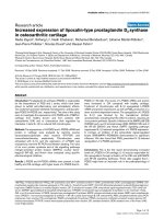
stimulation. As shown in Figures 1 and 2, the moving
average responses were close to 0 at all times of day. Nei-
ther for the 98 participants as a whole nor for any of the
age-gender subgroups (not shown) was there a consis-
tently elevated response at a particular time of day.
Kripke et al. Journal of Circadian Rhythms 2010, 8:5
/>Page 5 of 9
Study 2, Light treatment of depression
Effects of the light treatment on mood and sleep, which
were quite minimal and equivocal, were reported else-
where [57]. There were 7 subjects assigned to bright
morning light and 7 assigned to dim morning light who
had provided adequate urine samples which could be
assayed satisfactorily.
The Spearman rank-order correlation of LH and FSH
excretion before light treatment was R
s
= 0.62 (P < 0.02)
among 14 participants. The correlation of LH excretion
before and after light (dim control or bright) was R
s
= 0.72
among 14 participants (P = 0.004). The correlation of
FSH excretion before and after light was only R
s
= 0.34
(NS). Excretion per hour of LH and FSH did not differ
significantly between bright-light and dim-light treated
groups, either before or after treatment. Both LH and
FSH tended to increase after treatment, but the trend was
for a greater LH increase after dim light and a greater
FSH increase after bright light (NS).
Study 3, PMDD
Complete measurements of urinary LH and FSH before
and after light treatment in the both the follicular and
luteal phases were available for only 4 women given
morning light and 2 women given evening light.
The multivariate contrast of hormone measures before
and after the bright light stimulation was significant (P =
0.044). Log
10
(LH) excretion increased from 1.047 on the
baseline night to 1.264 after the bright light treatment
(univariate P = 0.027). Log
10
(FSH) excretion increased
from 0.030 on the baseline night to 0.196 on the night
after the bright light treatment (univariate P = 0.125). The
follicular vs. luteal phase hormonal excretion contrasts
were not significant, nor were the morning vs. evening
light treatment contrasts, but the interaction of the pre-
post hormonal excretion contrast with time of treatment
was significant (P = 0.008). For LH, the increase after
morning light treatment was consistently higher than
with evening light in both the follicular and luteal phases.
For FSH, the increase with morning light treatment was
higher than with evening light in the follicular phase, but
in the luteal phase, FSH excretion was extremely reduced,
and the very small increase was greater with evening than
with morning light. There were no other significant inter-
actions.
Discussion
To summarize, assays of urinary LH and FSH before and
after bright light treatments gave weak and somewhat
equivocal support to the hypothesis that bright light
stimulates these gonadotropic hormones. Light stimula-
tion of LH and FSH would be potentially useful, if a
robust effect could be more clearly demonstrated. In chil-
dren, regulation of light exposures might influence
Figure 1 Change in LH in relation to time of light exposure. Change in urinary LH excretion (mIU/ml) is expressed as the LH stimulation index. This
is a change index calculated by computing the ratio of the post-treatment LH excretion rate divided by the baseline (pre stimulus) rate, and then tak-
ing the log (base 10) of this fraction. In this way, a decrease producing a ratio of < 1.0 gives a negative log value. Each point is plotted relative to the
internal circadian time of the midpoint of the 3 h light stimulus. Zero h represents circadian midnight, defined as 3.52 h prior to the time of the aMT6s
acrophase in the pre-stimulus phase assessment. The key to symbols identifies the points by age and gender of the participants, N = 98. The black line
presents a 3-hour moving average of the points, with dotted lines showing 95% confidence intervals of the mean.
-12 -8 -4 0 4 8 12
-2.5
-2.0
-1.5
-1.0
-0.5
0.0
0.5
1.0
1.5
2.0
2.5
Young Females
Young Males
Elder Females
Elder Males
Lower 95% CL
Moving Average
Upper 95% CL
Internal Time (h)
LH stimulation Index
Kripke et al. Journal of Circadian Rhythms 2010, 8:5
/>Page 6 of 9
puberty and menarche. In men, an increase in testoster-
one production might lead to increased fertility in some
younger men, and palliation of loss of libido, erectile dys-
function, and muscle wasting among older men. In
women, light stimulation might promote fertility as well
as regularize the menstrual cycle [55,61]. In both genders,
bright light augmentation of gonadotropins might be one
aspect of the antidepressant benefits [62-64].
In the first and third studies, LH was significantly
increased after bright light treatment, but in neither case
was a significant contrast with any parallel control treat-
ment demonstrated. Therefore, we cannot exclude that
the increases in LH were due to various placebo and non-
specific experimental effects, as well as the passage of
time. For example, suggestion and hope often produce
positive placebo responses in a wide variety of trials. If a
Figure 2 LH responses grouped by age group and times of light stimulus. The log LH response index (as described for Figure 1) is shown sepa-
rately for young and older subjects versus the time of stimulus (in reference to the acrophase-peak of 6-sulfatoxymelatonin). Thick horizontal lines are
medians. Blue and green boxes are interquartile ranges. The thin bars represent the range for each time bin.
-10.00 -6.00 -2.00 2.00 6.00 10.00
4h bin stimulus time,
relative to baseline aMT6s acrophase
-1.50
-1.00
-0.50
0.00
0.50
1.00
1.50
LH ratio tr ansformed to base 10 log
young (18-30
yrs)
older (60-75
yrs)
Kripke et al. Journal of Circadian Rhythms 2010, 8:5
/>Page 7 of 9
clinical trial commenced with subjects in a heightened
state of anxiety or depression, spontaneous remission
might lead to endocrine changes such as enhancements
of LH and FSH production. In a separate study, our group
demonstrated a significant increase in FSH after early
morning stimulations with bright green light, as com-
pared to dim light placebo, but this effect was not very
robust [65]. We are left with tantalizingly suggestive evi-
dence that bright light stimulates LH and FSH in human
subjects, but there is a need for much more convincing
evidence.
These studies had a number of limitations. In the first
study, the light stimuli were administered for only 3 days
at a level of only 3000 lux, whereas, a longer duration of
brighter stimulation might have produced a greater
gonadotropic response. In the second study, though
10,000 lux bright light stimuli were randomized for 4
weeks, we were uncertain of the compliance of these very
depressed and elderly study participants, particularly
since there was only weak evidence for circadian phase-
shifting effect in the experimental groups and no persua-
sive evidence of a favorable mood effect. Only data from
the morning light treatments were analyzed because of
lack of evidence that the evening treatments had any
physiologic or mood effects. In the third study, the num-
bers of subjects were extremely small, and the light stim-
ulation was given only on a single day, which may have
been insufficient to produce a large effect. A light stimu-
lus intended to produce a phase shift may not be optimal
for stimulating LH and FSH. Though we were able to
obtain data from these studies which had been organized
for other goals, the numbers of participants yielding
usable data was unexpectedly small in the second and
third studies. There is also concern that the several years
that the urine samples were frozen, and the freezing and
thawing processes, might have introduced inaccuracy
into the assays.
Conclusions
Larger studies organized specifically to measure bright
light effects on gonadotropins may be needed to verify
the potential of bright light regulation for the reproduc-
tive endocrine system. Longer durations of exposure
should be tested.
Competing interests
The authors declare that they have no competing interests.
Authors' contributions
DFK participated in design, acquisition of data, interpretation and statistics, and
manuscript preparation. JAE provided intellectual background, performed the
assays, and contributed to design, interpretation, statistics, and manuscript
preparation. SDY contributed to design, performance, analysis, and interpreta-
tion of the first study. BLP designed and performed the third study and contrib-
uted to manuscript preparation. RLH consulted on the assays, provided
laboratory facilities, and helped edit the manuscript. KMR managed human
subjects consents and reimbursement, helped perform the first study, and
contributed to manuscript preparation. All authors read and approved the final
manuscript.
Acknowledgements
Supported by NIH R01 grants MH068545, HL61280, AG12364, MH63462, and
NIH Clinical Research Center (CRC) grant M01-RR-00827. RLH was supported by
NIH R01 grants AG022982 and MH074697 and by the VA Center of Excellence
for Stress and Mental Health (CESAMH). The NIH and the VA had no role in the
design, collection, analysis, or interpretation of data, in writing the manuscript,
or in submission of the manuscript. Richard T. Loving, D.N.Sc. and Nancy Knick-
erbocker collected data for Study 2. Charles J. Meliska, Ph.D. coordinated Study
3 and Luis F. Martinez assisted with data collection. The Sunbox Company,
Gaithersburg, MD contributed light treatment boxes for this research. Bio-Light
by Enviro-Med and Apollo Light Systems (now part of Philips Electronics) have
also contributed light treatment boxes for the laboratory's research.
Author Details
1
Department of Psychiatry, University of California, San Diego, La Jolla,
California 92093, USA,
2
Department of Exercise Science, University of South
Carolina, Columbia, South Carolina, USA and
3
San Diego VA Healthcare System,
Psychiatry Service, San Diego, CA 92161, USA
References
1. Dawson A, King VM, Bentley GE, Ball GF: Photoperiodic control of
seasonality in birds. J Biol Rhythms 2001, 16:365-380.
2. Hastings MH, Walker AP, Powers JB, Hutchison J, Steel EA, Herbert J:
Differential effects of photoperiodic history on the responses of
gonadotrophins and prolactin to intermediate daylengths in the male
Syrian hamster. J Biol Rhythms 1989, 4:335-350.
3. Goldman BD: Mammalian photoperiodic system: Formal properties
and neuroendocrine mechanisms of photoperiodic time
measurement. J Biol Rhythms 2001, 16:283-301.
4. Elliott JA: Circadian rhythms and photoperiodic time measurement in
mammals. Fed Proc 1976, 35:2339-2346.
5. Karsch FJ, Foster DL, Legan SJ, Hauger RL: On the control of tonic LH
secretion in sheep: a new concept for regulation of the estrous cycle
and breeding season. In Endocrinology - Proceedings of the V International
Congress of Endocrinology (Hamburg, Germany) July 18-24, 1976 Volume 1.
Edited by: James VHT. Amsterdam-Oxford: Excerpta Medica;
1976:192-198.
6. Elliott JA, Tamarkin L: Complex circadian regulation of pineal melatonin
and wheel-running in Syrian hamsters. J Comp Physiol A 1994,
174:469-484.
7. Goldman B: The circadian timing system and reproduction in
mammals. Steroids 1999, 64:679-685.
8. Bartness T, Powers B, Hastings M, Bittman E, Goldman B: The timed
infusion paradigm for melatonin delivery: what has it taught us about
the melatonin signal, its reception, and the photoperiodic control of
seasonal responses? J Pineal Res 1993, 15:161-190.
9. Gündüz B, Stetson MH: A test of the coincidence and duration models
of melatonin action in Siberian hamsters: the effects of 1-hr melatonin
infusions on testicular development in intact and pinealectomized
prepubertal Phodopus sungorus. J Pineal Res 2001, 30:97-107.
10. Gündüz B, Stetson MH: A test of the coincidence and duration models
of melatonin action in Siberian hamsters. II. The effects of 4- and 8-hr
melatonin infusions on testicular development of pinealectomized
juvenile Siberian hamsters (Phodopus sungorus). J Pineal Res 2001,
30:56-64.
11. Elliott JA, Stetson MH, Menaker M: Regulation of testis function in
golden hamsters: a circadian clock measures photoperiodic time.
Science 1972, 178:771-773.
12. Malpaux B, Migaud M, Tricoire H, Chemineau P: Biology of mammalian
photoperiodism and the critical role of the pineal gland and
melatonin. J Biol Rhythms 2001, 16:336-347.
13. Wehr TA: Photoperiodism in humans and other primates: Evidence and
implications. J Biol Rhythms 2001, 16:348-364.
14. O'Byrne KT, Thalabard JC, Chiappini SE, Chen MD, Hotchkiss J, Knobil E:
Ambient light modifies gonadotropin-releasing hormone pulse
Received: 27 February 2010 Accepted: 11 May 2010
Published: 11 May 2010
This article is available from: 2010 Kripke et al; licensee BioMed Central Ltd. This is an Open Access article distributed under the terms of the Creative Commons Attribution License ( which permits unrestricted use, distribution, and reproduction in any medium, provided the original work is properly cited.Journal of Circadian Rhythms 2010, 8:5
Kripke et al. Journal of Circadian Rhythms 2010, 8:5
/>Page 8 of 9
generator frequency in the rhesus monkey. Endocrinology 1993,
133:1520-1524.
15. Lincoln GA, Andersson H, Hazlerigg D: Clock genes and the long-term
regulation of prolactin secretion: Evidence for a photoperiod/
circannual timer in the pars tuberalis. J Neuroendocrinol 2003,
15:390-397.
16. Haus E, Lakatua DJ, Halberg F, Halberg E, Cornelissen G, Sackett LL, Berg
HG, Kawasaki T, Ueno M, Uezono K, Matsuoka M, Omae T:
Chronobiological studies of plasma prolactin in women in Kyushu,
Japan, and Minnesota, USA. J Clin Endocrinol & Metab 1980, 51:632-640.
17. Hannibal J, Hindersson P, Knudsen SM, Georg B, Fahrenkrug J: The
photopigment melanopsin is exclusively present in pituitary adenylate
cyclase-activating polypeptide-containing retinal ganglion cells of the
retinohypothalamic tract. J Neurosci 2002, 22(RC191):1-7.
18. Provencio I, Rollag MD, Castrucci AM: Photoreceptive net in the
mammalian retina. Nature 2002, 415:493.
19. Hattar S, Liao H-W, Takao M, Berson DM, Yau K-W: Melanopsin-containing
retinal ganglion cells: Architecture, projections, and intrinsic
photosensitivity. Science 2002, 295:1065-1070.
20. Berson DM, Dunn FA, Takao M: Phototransduction by retinal ganglion
cells that set the circadian clock. Science 2002, 295:1070-1074.
21. Gillette MU, Mitchell JW: Signaling in the suprachiasmatic nucleus:
selectively responsive and integrative. Cell Tissue Res 2002, 309:99-107.
22. Reppert SM, Weaver DR: Coordination of circadian timing in mammals.
Nature 2002, 418:935-941.
23. Schwartz WJ, de la Iglesia HO, Zlomanczuk P, Illnerova H: Encoding Le
Quattro Stagioni within the mammalian brain: Photoperiodic
orchestration through the suprachiasmatic nucleus. J Biol Rhythms
2001, 16:302-311.
24. Jagota A, de la Iglesia HO, Schwartz WJ: Morning and evening circadian
oscillations in the suprachiasmatic nucleus in vitro. Nature Neurosci
2000, 3:372-376.
25. vanderLeest HT, Houben T, Michel S, Deboer T, Albus H, Vansteensel MJ,
Block GD, Meijer JH: Seasonal encoding by the circadian pacemaker of
the SCN. Curr Biol 2007, 17:468-473.
26. Naito E, Watanabe T, Tei H, Yoshimura T, Ebihara S: Reorganization of the
suprachiasmatic nucleus coding for day length. J Biol Rhythms 2008,
23:140-149.
27. Daan S, Albrecht U, Horst GTJ van der, Illnerova H, Roenneberg T, Wehr TA,
Schwartz WJ: Assembling a clock for all seasons: Are there M and E
oscillators in the genes? J Biol Rhythms 2001, 16:105-116.
28. Hastings M: Modeling the molecular calendar. J Biol Rhythms 2001,
16:117-123.
29. Oster H, Maronde E, Albrecht U: The circadian clock as a molecular
calendar. Chronobiol Intl 2002, 19:507-516.
30. Steinlechner S, Jacobmeier B, Scherbarth F, Dernbach H, Kruse F, Albrecht
U: Robust circadian rhythmicity of Per1 and Per2 mutant mice in
constant light, and dynamics of Per1 and Per2 gene expression under
long and short photoperiods. J Biol Rhythms 2002, 17:202-209.
31. Bechtold DA, Loudon AS: Hypothalamic thyroid hormones: mediators
of seasonal physiology. Endocrinology 2007, 148:3605-3607.
32. Nakao N, Ono H, Yamamura T, Anraku T, Takagi T, Higashi K, Yasuo S, Katou
Y, Kageyama S, Uno Y, Kasukawa T, Iigo M, Sharp PJ, Iwasawa A, Suzuki Y,
Sugano S, Niimi T, Mizutani M, Namikawa T, Ebihara S, Ueda HR,
Yoshimura T: Thyrotrophin in the pars tuberalis triggers photoperiodic
response. Nature 2008, 452:317-322.
33. Gorman MR, Freeman DA, Zucker I: Photoperiodism in Hamsters: abrupt
versus gradual changes in day length differentially entrain morning
and evening circadian oscillators. J Biol Rhythms 1997, 12:122-135.
34. Knobil E, Hotchkiss J: The circhoral gonadotropin releasing hormone
(GnRH) pulse generator of the hypothalamus and its physiological
significance. In Ultradian Rhythms in Physiology and Behavior Edited by:
Schulz H, Lavie P. Berlin: Springer-Verlag; 1985:32-40.
35. Lehman MN, Goodman RL, Karsch FJ, Jackson GL, Berriman SJ, Jansen HT:
The GnRH system of seasonal breeders: Anatomy and plasticity. Brain
Res Bull 1997, 44:445-457.
36. Beek EM van der, Wiegant VM, Donk HA van der, Hurk R van den, Buijs RM:
Lesions of the suprachiasmatic nucleus indicate the presence of direct
vasoactive intestinal polypeptide-containing projection to
gonadotrophin-releasing hormone neurons in the female rat. J
Neuroendocrinol 1993, 5:137-144.
37. Leak RK, Moore RY: Topographic organization of suprachiasmatic
nucleus projection neurons. The Journal of Comparative Neurology 2001,
433:312-334.
38. Cheng MY, Bullock CM, Li C, Lee AG, Bermak JC, Belluzzi J, Weaver DR,
Leslie FM, Zhou Q-Y: Prokineticin 2 transmits the behavioural circadian
rhythm of the suprachiasmatic nucleus. Nature 2002, 417:405-410.
39. Popa SM, Clifton DK, Steiner RA: The Role of Kisspeptins and GPR54 in
the Neuroendocrine Regulation of Reproduction. Annu Rev Physiol
2008, 70:213-238.
40. Quaynor S, Hu L, Leung PK, Feng H, Mores N, Krsmanovic LZ, Catt KJ:
Expression of a functional g protein-coupled receptor 54-kisspeptin
autoregulatory system in hypothalamic gonadotropin-releasing
hormone neurons. Mol Endocrinol 2007, 21:3062-3070.
41. Revel FG, Masson-Pevet M, Pevet P, Mikkelsen JD, Simonneaux V:
Melatonin controls seasonal breeding by a network of hypothalamic
targets. Neuroendocrinology 2009, 90:1-14.
42. McCann SM, Karanth S, Mastronardi CA, Dees WL, Childs G, Miller B, Sower
S, Yu WH: Control of gonadotropin secretion by follicle-stimulating
hormone-releasing factor, luteinizing hormone-releasing hormone,
and leptin. Arch Med Res 2001, 32:476-485.
43. Vondrasova D, Hajek I, Illnerova H: Exposure to long summer days affects
the human melatonin and cortisol rhythms. Brain Res 1997,
759:166-170.
44. Tuunainen A, Kripke DF, Elliott JA, Assmus JD, Rex KM, Klauber MR, Langer
RD: Depression and endogenous melatonin in postmenopausal
women. J Affect Dis 2002, 69:149-158.
45. Wehr TA, Duncan WC, Sher L, Aeschbach D, Schwartz PJ, Turner EH,
Postolache TT, Rosenthal NE: A circadian signal of change of season in
patients with seasonal affective disorder. Arch Gen Psychiatry 2001,
58:1108-1114.
46. Wehr TA: Effect of seasonal charges in daylength on human
neuroendocrine function. Horm Res 1998, 49:118-124.
47. Roenneberg T, Aschoff J: Annual rhythm of human reproduction: I.
Biology, sociology, or both? J Biol Rhythms 1990, 5(3):195-216.
48. Roenneberg T, Aschoff J: Annual rhythm of human reproduction: II.
Environmental correlations. J Biol Rhythms 1990, 5:217-239.
49. Lam DA, Miron JA: Global patterns of seasonal variation in human
fertility. Ann NY Acad Sci 1994, 709:9-28.
50. Meriggiola MC, Noonan EA, Paulsen CA, Bremner WJ: Annual patterns of
luteinizing hormone, follicle stimulating hormone, testosterone and
inhibin in normal men. Hum Repro 1996, 11:248-252.
51. Nicolau GY, Haus E, Bogdan C: Chronobiology and aging. Romanian J
Geront Geriatrics 1988, 9(2):149-171.
52. Bellastella A, Criscuolo T, Mango A, Perrone L, Sinisi AA, Faggiano M:
Circannual rhythms of plasma luteinizing hormone, follicle-stimulating
hormone, testosterone, prolactin and cortisol in prepuberty. Clin
Endocrinol 1983, 19:453-459.
53. Martikainen H, Ruokonen A, Tomas C, Kauppila A: Seasonal changes in
pituitary function: Amplification of midfollicular luteinizing hormone
secretion during the dark season. Fertil Steril 1996, 65:718-720.
54. Yoon I-Y, Kripke DF, Elliott JA, Youngstedt SD: Luteinizing hormone
following light exposure in healthy young men. Neurosci Let 2003,
341:25-28.
55. Danilenko KV, Samoilova EA: Stimulatory effect of morning bright light
on reproductive hormones and ovulation: results of a controlled
crossover trial. PLoS Clin Trials 2007, 2:e7.
56. Kripke DF, Elliott JA, Youngstedt SD, Rex KM: Circadian phase response
curves to light in older and young women and men. J Circadian
Rhythms 2007, 5:1.
57. Loving RT, Kripke DF, Elliott JA, Knickerbocker NC, Grandner MA: Bright
light treatment of depression for older adults [abstract]. Chronobiol Intl
2006, 23:720.
58. Yesavage JA, Brink TL, Rose TL, Lum O, Huang V, Adey M, Leirer VO:
Development and validation of a geriatric depression screening scale:
a preliminary report. J Psychiatr Res 1983, 17:37-49.
59. Lyness JM, King DA, Cox C, Yoediono Z, Caine ED: The importance of
subsyndromal depression in older primary care patients: prevalence
and associated functional disability. J Am Geriatr Soc 1999, 47:647-652.
60. Parry BL, Udell C, Elliott JA, Berga SL, Klauber MR, Mostofi N, LeVeau B,
Gillin JC: Blunted phase-shift responses to morning bright light in
premenstrual dysphoric disorder. J Biol Rhythms 1997, 12:443-456.
Kripke et al. Journal of Circadian Rhythms 2010, 8:5
/>Page 9 of 9
61. Putilov AA, Danilenko KV, Protopopova AY, Kripke DF: Menstrual phase
response to nocturnal light. Biological Rhythm Research 2002, 33:23-38.
62. Tuunainen A, Kripke DF, Endo T: Light Therapy for Non-Seasonal
Depression. Cochrane Database Syst Rev 2004:CD004050.
63. Golden RN, Gaynes BN, Ekstrom RD, Hamer RM, Jacobsen FM, Suppes T,
Wisner KL, Nemeroff CB: The efficacy of light therapy in the treatment of
mood disorders: a review and meta-analysis of the evidence. Am J
Psychiatry 2005, 162:656-662.
64. Even C, Schroder CM, Friedman S, Rouillon F: Efficacy of light therapy in
nonseasonal depression: A systematic review. J Affect Disord 2008,
108:11-23.
65. Grandner M, Kripke DF, Elliott J, Cole R: Effects of nocturnally-
administered green light on luteinizing hormone and follicle-
stimulating hormone in young men [abstract]. Sleep 2007, 30:A62.
doi: 10.1186/1740-3391-8-5
Cite this article as: Kripke et al., Weak evidence of bright light effects on
human LH and FSH Journal of Circadian Rhythms 2010, 8:5