Báo cáo y học: "The importance of localizing pulmonary veins in atrial septal defect closure" ppsx
Bạn đang xem bản rút gọn của tài liệu. Xem và tải ngay bản đầy đủ của tài liệu tại đây (771.58 KB, 3 trang )
CAS E REP O R T Open Access
The importance of localizing pulmonary veins
in atrial septal defect closure!
Ahmad Ali Amirghofran
1
, Ashkan Karimi
2*
, Gholam Hossein Ajami
3
and Alireza Rasekhi
4
Abstract
An 8-year-old girl was admitted for a simple closure of echocardiographically diagnosed Atrial Septal Defect (ASD).
During the operation the right pulmonary veins orifices were not detected in the left atrium and attempt to
localize them led to the discovery of three additional anomalies, namely Interrupted Inferior Vena Cava (IIVC),
Scimitar syndrome, and systemic arterial supply of the lung. Postoperatively these finding were confirmed by CT
angiography. This case report emphasizes the need for adequate preoperative diagnosis and presents a very rare
constellation of four congenital anomalies that to the best of our knowledge is not reported before.
Background
The need for adequate preoperative diagnosis in the
field of congenital heart surgery cannot be overempha-
sized. To this end many centers routinely use Intrao-
perative Trans-Esophageal Echocardiography (ITEE).
Mayo clinic group in a study of 1002 congenital heart
disease patients demonstrated that ITEE had major
impact in 13.4% of cases defined as revealing any unde-
tected pre or intaoperative information requiring an
otherwise non-performed procedure during the surgery;
however, the ASD secundum subset (67 cases) was the
only primary diagnosis in this study that ITEE had zero
major impact on and routine ITEE did not seem to be
cost effective in this group [1]. In our e xperience also a
comprehensive preoperative Trans-Thoracic Echocardio-
graphy (TTE) is considered adequate for delineation of
simple cardiac defects such as ASD secundum unless
the cardiologist is not satisfied with the quality of the
study in which case preoperative TEE, cardiac MRI or
ITEE is considered. In contrast to what was just men-
tioned the following case report serves as an example of
inadequate TTE that failed to detect other major conco-
mitant congenital heart defects accompanying an ASD
secundum in an 8-year-old girl which had major impact
on her operation.
Case presentation
An 8-year-old girl referred with the complaint of mild
exertional dyspnea. TTE revealed a 16 mm ASD secun-
dum, moderate enlargemen t of the right atrium and ven-
tricle, and an estimated Qp/Qs ratio of 2.3. Since the ASD
did not have enough rim inferiorly to be closed by device,
she was scheduled for surgical closure. Cardiopulmonary
bypass was established by aorto-bicaval cannulatio n, and
the ASD was approached through right atriotomy. Intrao-
peratively there was no trace of the right pulmonary veins
orifices within either atria. Intrapericardial exploration of
the distal Superior Vena Cava (SVC), as a common site for
Partial Anomalous Pulmonary Venous Connection
(PAPVC), failed to identify them and subsequently the
right pleura was opened to look for them. Two vessels
were discovered: a 12 mm vessel which ran between the
right lung a nd the central pa rt of the dia phragm and a
7 mm vessel which arose next to it and passed through
the right dome of the diaphragm. Under the impression of
Scimitar Syndrome, the Inferior Vena Cava (IVC) was
decannulated to identify where these vessels drained into
below the diaphragm. To our surprise, the IVC was inter-
rupted bearing just few small orifices for the hepatic veins.
Next the SVC was decannulated and dissected more
superiorly to explore the enlarged Azygos vein which car-
ries most of the subdiaphragmatic venous return to the
heart in the setting of IIVC. Before transferring these two
vessels as an omalous pulmonary veins to the left atrium
we decided to confirm their drainage into the systemic
venous circulation. A blood sample from the 12 mm vessel
revealed 95% oxygen saturation and its baseline pressure
* Correspondence:
2
University of Florida, Division of Thoracic and Cardiovascular Surgery,
Gainesville, FL, USA
Full list of author information is available at the end of the article
Amirghofran et al. Journal of Cardiothoracic Surgery 2011, 6:41
/>© 2011 Amirghofran et al; licensee BioMed Central Ltd. This is an Open Access article distributed under the terms of the Creative
Commons Attribution License ( which permits unrestricted use, distribution, and
reproduction in any medium, provide d the original work is properly cited.
was measured at 6-7 mmHg, then the A zyos vein was
clamped immediately before its drainage into the SVC and
the pressure readings gradually increased and established
at 25 mmHg, implying that this vessel emptied into the
systemic venous circulation under the diaphragm. Subse-
quently this vessel was cut at the level of the diaphragm to
be transferred to the left atrium, but due to its short length
wefixedittotherightatriumadjacenttotheASDin
order to use an intraatrial baffle later for directing its flow
to the left atrium (Figure 1). we were about to transfer the
7 mm vessel as another anomalous pulmonary vein that
we noticed a considerable amount of bright red blood
coming out from the site of new anastomosis, suggesting
an aortopulmonary connection. A blood sample from the
7 mm vessel showed 97% oxygen saturation and upon its
clamping the flow through the anastomosis stopped
implying that the 7 mm vessel was a systematic artery
which was supplying part of the right lung. Obviously the
visible amount of shunt could not be left unattended; how-
ever, it w as not clear whether this vessel was supplying
normal lung tissue or an intralobar pulmonary sequestra-
tion. The patient had negative history for repeated pneu-
monia to suggest sequestration and the preoperative chest
x-ray was normal. Given the small size of this vessel, we
ultimately decided to ligate it without doing any resection
and follow the patient closely after the operation in light
of possible pulmonary necrosis and infection. In the end a
pericardial patch was used to close the ASD and as an
intraatrial baffle to direct flow from the anastomosed pul-
monary vein to the left atrium. Fortunately the patient tol-
erated the procedure well and was discharged after 7 days
without any pulmonary complication. Subsequent CT
angiography 3 weeks later confirmed the intraoperative
findings and showed homogenous lung parenchyma with
no evidence of sequestration or necrosis (Figure 2).
Discussion
This case report merits special consideration not only
because of the very rare constellation of ASD, IIVC, Sci-
mitar Syndrome, and anomalous systemic arterial supply
of the lung, but also what the appropriate management
Figure 1 Intraoperative view of the two anomalous blood
vessels. A - The long arrow shows the 12 mm vessel originating
below the hilum of the right lung (hollow arrow) after being
transferred to the right atrium. The small arrow shows the 7 mm
vessel passing through the right dome of the diaphragm (*).
B - Inside the right atrium is shown. The large arrow depicts the
orifice of the redirected 12 mm vessel, which is fixed to the right
atrium just at the right side of the ASD (small arrow). A pericardial
patch is used later to redirect flow from this new orifice towards
the left atrium.
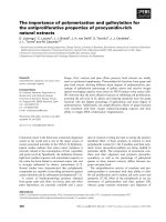
Figure 2 A - Frontal projection of the venous phase of 3D CT
angiography with volume rendering, which is obtained after
the operation. Annotated structures are: short solid arrow =
enlarged azygos vein; long solid arrow = IVC, which is interrupted at
the hepatic level; short hollow arrow = SVC; long hollow arrow =
Redirected anomalous pulmonary vein; arrow head = hepatic vein,
which drains into right atrium. B - The arterial phase depicts the
anomalous systemic artery (arrow) arising from the celiac trunk and
intending to supply the base of the right lung, which is ligated at
the level of the diaphragm.
Amirghofran et al. Journal of Cardiothoracic Surgery 2011, 6:41
/>Page 2 of 3
should be while three of these four anomalies were dis-
covered during the operation. It is not unheard-of for
surgeons to come across new pathologies during the
operation, but it is very unlikely for these new findings
to change the nature of the procedure. In current prac-
tice TTE is considered adequate for the preoperative
evaluation of ASD secundum and a detailed comprehen-
sive echocardiography is expected to obviate the need
for routine invasive diagnostic tests such as cardiac
catheterization or even ITEE [1,2]. Preoperative TTE
should include evaluati on of all pulmonary veins. Occa-
sionally they are not visualized due to poor acoustic
window; in which case preoperative TEE, cardiac MRI
or ITEE should be considered if not routinely performed
[3]. Subcostal view in TTE should delineate Scimitar
syndrome [3] and IIVC with Azygos continuation should
also be readily diagnosed from this window [3]. Unfortu-
nately these pathologies were missed in the preoperative
TTE and were first diagnosed d uring the surgery. After
IIVC was discovered intraoperatively the subject was
raised to abort the operation and perform cardiac cathe-
terization to accurately describe concomitant cardiac
anomalies before proceeding further, but we ultimately
decided to continue the operation and not to impose
the risk of another surgery. Several surgi cal techniques
are available to correct scimitar syndrome which are
best summarized in the paper by Gudjonsson et al [4].
We applied the surgical technique introduced by Shu-
macker and Judd, which includes transfer of the anoma-
lous pulmonary vein to the right atrium adjacent to the
ASD and then baffling its flow across towards the left
atrium [5]. Several other congenital anomalies are also
reported in patients with IIVC including visceral hetero-
taxy and polysplenia which were absent in our case [6].
Conclusions
Although preoperative TTE is considered adequate for
the delineation of ASD secundum and its associated
cardiac anomal ies, this case report shows how an inade-
quate TTE can complicate the operation. Accordingly
cardiologists should attempt to identify the site of
drainage for all four pulmonary veins in the preoperative
TTE and if there is any doubt about the quality of the
study preoperative TEE, cardiac MRI or ITEE should be
requested especially in centers where ITEE is not routi-
nely performed for simple congenital heart surgeries
such as ASD secundum closure.
Consent
Written informed consent was obtained from the patient
for publication of this case report and any accompanying
images. A copy of the written consent is available for
review by the Editor-in-Chief of this journal.
Author details
1
Shiraz University of Medical Sciences, Division of Cardiovascular Surgery,
Shiraz, Iran.
2
University of Florida, Division of Thoracic and Cardiovascular
Surgery, Gainesville, FL, USA.
3
Shiraz University of Medical Sciences, Division
of Pediatric Cardiology, Shiraz, Iran.
4
Shiraz University of Medical Sciences,
Department of Radiology, Shiraz, Iran.
Authors’ contributions
AAA performed the surgery and supervised the manuscript. AK wrote the
article and gathered the data. GHA contributed to the patient’s care. AR
interpreted radiographic images. All authors read and approved the final
manuscript.
Competing interests
The authors declare that they have no competing interests.
Received: 10 February 2011 Accepted: 30 March 2011
Published: 30 March 2011
References
1. Randolph GR, Hagler DJ, Connolly HM, Dearani JA, Puga FJ, Danielson GK,
Abel MD, Pankratz VS, O’Leary PW: Intraoperative transesophageal
echocardiography during surgery for congenital heart defects. J Thorac
Cardiovasc Surg 2002, 124(6):1176-82.
2. Allen HD, Driscoll DJ, Shaddy RE, Feltes TF: Moss and Adams’ Heart
Disease in Infants, Children, and Adolescents: Including the Fetus and
Young Adult. Philadelphia, Lippincott Williams & Wilkins; 2008.
3. Huhta JC, Smallhorn JF, Macartney FJ: Cross-sectional echocardiographic
diagnosisof azygos continuation of the inferior vena cava. Cathet
Cardiovasc Diagn 1984, 10(3):221-32.
4. Gudjonsson U, Brown JW: Scimitar syndrome. Semin Thorac Cardiovasc
Surg Pediatr Card Surg Annu 2006, 56-62.
5. Shumacker HB Jr, Judd D: Partial anomalous pulmonary venous return
with reference to drainage into the inferior vena cava and to an intact
atrial septum. J Cardiovasc Surg (Torino) 1964, 45:271-278.
6. Van Praagh S, Santini F, Sanders SP: Cardiac malpositions with special
emphasis on visceral heterotaxy (asplenia and polysplenia syndromes).
In Nadas’ Pediatric Cardiology. Edited by: Fyler DC. Philadelphia, PA: Hanley
and Belfus; 1992:589-608.
doi:10.1186/1749-8090-6-41
Cite this article as: Amirghofran et al.: The importance of localizing
pulmonary veins in atrial septal defect closure!. Journal of Cardiothoracic
Surgery 2011 6:41.
Submit your next manuscript to BioMed Central
and take full advantage of:
• Convenient online submission
• Thorough peer review
• No space constraints or color figure charges
• Immediate publication on acceptance
• Inclusion in PubMed, CAS, Scopus and Google Scholar
• Research which is freely available for redistribution
Submit your manuscript at
www.biomedcentral.com/submit
Amirghofran et al. Journal of Cardiothoracic Surgery 2011, 6:41
/>Page 3 of 3