Báo cáo y học: "tromal Vascular Fraction Transplantation as an Alternative Therapy for Ischemic Heart Failure: Anti-inflammatory Role" doc
Bạn đang xem bản rút gọn của tài liệu. Xem và tải ngay bản đầy đủ của tài liệu tại đây (1.59 MB, 10 trang )
RESEARCH ARTIC LE Open Access
Stromal Vascular Fraction Transplantation as an
Alternative Therapy for Ischemic Heart Failure:
Anti-inflammatory Role
Goditha U Premaratne
1*
, Li-Ping Ma
1,2
, Masatoshi Fujita
3
, Xue Lin
3
, Entela Bollano
1
and Michael Fu
1
Abstract
Background: The aims of this study were: (1) to show the feasibility of using adipose-derived stromal vascular
fraction (SVF) as an alternative to bone marrow mono nuclear cell (BM-MNC) for cell transplantation into chronic
ischemic myocardium; and (2) to explor e underlying mechanisms with focus on anti-inflammation role of
engrafted SVF and BM-MNC post chronic myocardial infarction (MI) against left ventricular (LV) remodelling and
cardiac dysfunction.
Methods: Four weeks after left anterior descending coronary artery ligation, 32 Male Lewis rats with moderate MI
were divided into 3 groups. SVF group (n = 12) had SVF cell transplantation (6 × 10
6
cells). BM-MNC group (n =
12) received BM-MNCs (6 × 10
6
) and the control (n = 10) had culture medium. At 4 weeks, after the final
echocardiography, histological sections were stained with Styrus red and immunohistochemical staining was
performed for a-smooth muscle actin, von Willebrand factor, CD3, CD8 and CD20.
Results: At 4 weeks, in SVF and BM-MNC groups, LV diastolic dimension and LV systolic dimension were smaller
and fractional shortening was increased in echocardiography, compared to control group. Histology revealed
highest vascular density, CD3+ and CD20+ cells in SVF transplanted group. SVF transplantation decreased
myocardial mRNA expression of inflammatory cytokines TNF-a, IL-6, MMP-1, TIMP-1 and inhibited collagen
deposition.
Conclusions: Transplantation of adipose derived SVF cells might be a useful therapeutic option for angiogenesis in
chronic ischemic heart disease. Anti-inflammation role for SVF and BM transplantation might partly benefit for the
cardioprotective effect for chronic ischemic myocardium.
Background
Cell transplantation is an effective treatment of repairing
ischemically damaged hearts [1,2]. The use of stem cells
emerged as a reasonable alternative treatment and two
general types of stem cells are being used for this aspect
[3,4]. Although theoretically highly applicable, there are
some potential limitations of cell regulation and ethical
considerations for the practical use of embryonic stem
cells [4]. Bone marrow mono nuclear cells (BM-MNCs)
have been the most commonly used stem cells for
ischemic myocardium, probably due to the availability of
multipote ntial progenitor cells. Mesenchymal stem cells
(MSCs) are multipotent adult stem cells that reside
within the bone marrow microenvironment. Although
mesenchymal stem cells derived from bone marrow
have been used experimentally [2,3] and clinically [5,6],
bone marrow aspirat ion is very painful and s ometimes
requires the use of general or s pinal anaesthesia. There-
fore, an autologous pluripotent mesenchymal stem cell
source that allows harvesting in large numbers with
minimal discomfort would be ideal for transplantation.
Adipose tissue is derived from embryonic mesoderm
and contains a heterogeneous stromal cell population
that can be easily harvested from the patients by a sim-
ple, minimally invasive method, and they can be easily
cultured. Several studies have demonstrated the pre-
sence of uncommitted MSCs within the adipose tissue
of animals and humans [7,8], that have the ability to
* Correspondence:
1
Wallenberg Laboratory for Cardiovascular Research, Sahlgrenska University
Hospital, University of Gothenburg, Gothenburg, Sweden
Full list of author information is available at the end of the article
Premaratne et al. Journal of Cardiothoracic Surgery 2011, 6:43
/>© 2011 Premaratne et al; licensee BioMed Central Ltd. This is an Open Acces s article distributed under the terms of the Creative
Commons Attribution License (http://creativecommon s.org/licenses/by/2 .0), which permits unrestricted use, distribution, and
reprodu ction in any medium, provided the original work is properly cited.
regenerate damaged organs. In addition, it has been
reported that MSCs derived from adipose tissue are
multipotent cells that can differentiate into cardiomyo-
cytes [9,10] and vascular endothelial cells [11,12]. There-
fore, adipose-derived stromal v ascular fraction (SVF)
emerging as a better option to replace bone marrow for
implantation into ischemic my ocardium using easy and
non-invasive procedures.
Although, the effects of adipose-derived SVF trans-
plantation into ischemic myocardium have been recently
reported [13], underline mecha nisms of adipose-derived
cells transplanted into chronic ischemic myocardium
have not yet been established. Therefore, this study
investigated the therapeutic efficacy of adipose-derived
SVF cells or freshly isolated BM-MNCs in a rat model
of chronic myocardial infarction and the anti-inflammatory
role of engrafted SVF and BM-MNC in post chronic
myocardial infarctio n.
Methods
Experimental Animals
Adult male syngeneic Lewis rats weighing 250-290 g
were used as recipients and donors in this study. All
experimental procedures were approved by the regional
Animal Ethic Committee of Gothenburg University,
Gothenburg, Sweden and conducted in accordance with
the Guide for the Care and Use of Laboratory Animals
published by the US National Institute of Health (NIH
publication no.85-23, revised 1996).
Stromal Vascular Fraction (SVF) Isolation
Stromal vascular fraction was isolated as Zuk et al.
described with some modifications [14]. Adipose tissue
was obtained from the inguinal region of syngeneic
Lewis rats under sterile conditions, kept in the tissue
culture media on ice, washed extensively with phos-
phate-buffered saline (PBS) to remove contaminating
blood cells, dissected from vessels and minced with scis-
sors. Minced adipose tissue was enzymatically digested
using PBS containing 2% BSA and collagenase (0.2%) at
37°C for 45 minutes; the enzyme reaction was inacti-
vated by the addition of D MEM/Ham’ sF-12(PAA
Laboratories GmbH, H aidmannweg, Pasching, Austria)
containing 10% newborn calf serum (NCS) and centri-
fuged at 800 g for 10 minutes to obtain a high density
SVF pellet. The pellet was resuspended in 160 mM
NH
4
Cl for 15 minutes at room temperature to lyse red
blood cells, added equal volume of DMEM/Ham’sF-12
containing 10% NCS, centrifuged at 800 g for 10 minutes.
The cell suspension was filtered through a 100 μmnylon
mesh to remove undispersed tissue elements and plated
(30 000 cells/cm
2
) in DMEM-F12 containing 10% NCS.
Six hours after incubation, the plates were washed exten-
sively with PBS to remove residual non-adhe rent red
blood cells. Cells were labeled with a fluorescent dye
using PKH26 (PKH26 Red Fl uorescent Cell Linker Mini
Kit, for General Cell Membrane Labeling, SIGMA-
ALDRICH Inc.) [15]. Cells were suspended at a concen-
tration of 6 × 10
7
/mL in 0.1 mL culture medium (without
serum) for transplantation.
Bone marrow mononuclear cell (BM-MNC) Isolation
BMCs were harvested from 8-week syngeneic Lewis rats
by flushing the femurs and tibias with PBS supplemen-
ted with 2% fetal bovine serum. To isolate mononuclear
cells, the g radient centrifugation method with Percoll
wasused[16].AfterthecellswerewashedinPBSfor
3 times, labeled with a fluorescent dye using PKH26,
before suspended in 0.1 mL of culture medium (without
serum) at a concentration of 6 × 10
7
/mL cells for
transplantation.
Chronic myocardial infarction model
The animal model, which was employed in this study,
has been described previously [17]. Male Lewis rats
weighing 250-290 g were anesthetized with isoflurane,
orally intubated into the trachea and anesthesia was
maintai ned with 1.5% to 2.5% isoflurane during the liga-
tion procedure. They underwent a left lateral thoracot-
omy, the left anterior descending coronary (LAD) artery
was ligated with a 6-0 polypropylene suture (Ethicon,
Inc, Somerville, NJ). As a result, ST-segment elevation
on electrocardi ogram and color changes in the left ven-
tricular (LV) myocardium were observed in all rats.
Experimental Groups
Four weeks after LAD ligation, infarction size was evalu-
ated by echocardiography and rats w ith moderate-sized
infarction (infarct size, 20% to 40%) were randomized
into 3 groups. In SVF Group (n = 11), SVF 6 million
cells suspended in culture medium were subepicardially
implanted at 2 points of the border zone. In BM-MNC
group (n = 11), 6 × 10
6
bone marrow mono nuclear
cells were transpl anted. Control group (n = 10) received
culture medium injection. Fresh DMEM culture med-
ium without serum was used for all the injections. Thus,
all the 32 rats had repeat thoracotomy for the myocar-
dial injection.
Echocardiography
Rats were anesthetized with isoflurane. Left ventricular
function was studied just before transplantation and
followed-up 2 and 4 weeks later, by echocardiography with
an ultrasound machine (HDI 5000 ultrasound system,
ATL, Philip Medical System, Best, Netherlands) equipped
with a 12 MHz phased-array transducer. A two-dimen-
sional short-axis view of the LV was obtained at the level
of the papillary muscles, M-mode images were recorded at
Premaratne et al. Journal of Cardiothoracic Surgery 2011, 6:43
/>Page 2 of 10
the same plane and LV end-diastolic dimension (EDD) and
end-systolic dimension (ESD) were measured. In addition,
the percentage of fractional shortening (FS) was calculated.
All measurements were performed in a blind fashion
according t o the Am erican Society for Echocardiology, a nd
averaged over 3 c onsecutive cardiac c ycles.
Histology
After echocardiographic assessment, all rats were sacri-
ficed, hearts from each group were cryo-embedded and
the whole left ventricle was sectioned in 4 μm thickness
along the short axis. They were microscopically exam-
ined with the use of fluorescence microscopy for PKH26
dye. The sections were stained for hematoxylin and
eosin. Immunohistochemistry was performed for a-
sarcomeric actin, von Willebrand factor (Dako Cytoma-
tion Inc, Glostrup, D enmark), Interleukin-6 (IL-6)
(Abcam plc., UK), CD3 (Santa Cruz Biotechnology, Inc.,
Europe), CD8 (Santa Cruz Biotechnology, Inc., Europe)
and CD20 (Santa Cruz Biotechnology, Inc., Europe).
In addition, Sirus red staining was performed to exam-
ine the fibrosis percentage in the infarct area with an
image analysis software (Scion Image Beta 4.02 Win,
Photoshop 6.0, San Jose, CA, USA).
Analysis of Vascular Density
The number of vessels was counted in each heart using
immunohistochemistry for von Willebrand factor [15].
The vessels per 1 mm
2
in the peri-infarct zone were
counted in 5 randomly cho sen fields per slide in a
blinded manner in 5 sections from each heart and aver-
aged for statistical analysis.
Analysis of Fibrotic Area
The percentage of fibrotic area in the infarct and peri-
infarct zone was calculated in each heart using the
image analysis software (Scion Image Beta 4.02 Win,
Scion Corporation) in a representative preparation for
Sirius Red staining, with the red areas regarded as fibro-
tic. The percentage of fibrotic area was analyzed in 5
randomly chosen fields per slide in the infarct and peri-
infarct zone in a blinded manner in 5 sections from
each heart and averaged for statistical analysis.
Isolation of RNA and real time RT-PCR
Total RNA was isolated from left ventricular myocar-
dium using SV total RNA Isolation System (Promega,
Madison, WI, USA) according to the manufacturer’s
recommendations. Reverse transcriptase reaction using
TaqMan High capacity cDNA Archive Kit (Applied Bio-
systems, Foster City, CA, USA) was performed for
cDNA synthesis. The cycling parameters were 25°C for
10 minutes and 37°C for 2 hours.
Real time RT-PCR analyses were used to determine
mRNA expressions of tumor necrosis factor alpha
(TNFa), Interleukin-6 (IL-6), tissue inhibitor of matrix
metalloproteinase-1 (TIMP-1), matrix metalloproteinase-
1 (MMP-1), brain natriuretic peptide (BNP) and vascular
endothelial growth factor (VEGF), and were performed
with TaqMan Assay-on-Demand on ABI 7700 sequence
Detection System (ABI), according to the manufacturer’s
recommend ations. The expression data were normalized
to an endogenous control, b-glucuronidase (Gus B). The
reactions for TNFa, IL-6, TIMP-1, MMP-1, BNP and
VEGF were analyzed in duplicates and the relative
expression levels were calculated according to the stan-
dard curve method. The logarithm of the RNA concen-
tration was calculated from standard curves. The
expression was determined as the ratio of the RNA
target
/
RNA
GusB
.
Positive cells for CD3, CD8 and CD 20
Immunohistochemical staining was performed on left
ventricular sections using anti-CD3, anti-CD8 and anti-
CD20. The diffusely scattered positive cells were
counted in each image. The visual field area of the x20
objective of the light microscope used; the positive cells
in four consecutiv e fields of representative areas were
counted i n 5 sections from each heart and averaged for
statistical analysis.
Quantification of IL6 positive immunohistochemical
staining
For quantification of IL6 positive area, the immunoposi-
tive components from the images from each section
were dissected using the property of color recognition
of BioPix iQ 2.1.6 softaware. The percentage of IL6
positive area was analyzed in 4 rand omly chosen fields
per slide in the infarct area in a blinded manner in 5
sections from each heart and averaged for statistical
analysis.
Data Analysis
All data are expressed as the mean ± SEM. Comparison s
of echocardiographic data among the groups were per-
formed by 2 wa y repeated measures analysis of vari ance
(ANOVA) including time, group, and group-by-time inter-
action terms. If significance was recognized for the group
effect or the group-by-time interaction, post hoc compari-
sons among the groups or among the groups at each time
point were performed, and if significance was found for
the time effect or the group-by-time interaction, post hoc
comparisons among the time points in each group were
made, when appropriate, using Fisher’sprotectedleast
significant difference method. Comparisons of vascular
density data, fibrosis and mRNA expressions among
Premaratne et al. Journal of Cardiothoracic Surgery 2011, 6:43
/>Page 3 of 10
the groups were conducted by one-way factorial ANOVA.
All statistical analyses were performed with using compu-
ter software (Stat View for Windows version 5.0, SAS
Institute Inc, Cary, NC, USA). A probability value < 0.05
was considered statistically significant.
Results
Mortality
The mortality rate due to coronary artery ligation was
20%. There was no intraoperative or postoperative death
concerning treatment procedures.
Echocardiography
Echocardiographic data are shown in Table 1. There
were no differences among the 3 groups regarding pre-
treatment LVDd, LVDs and FS. Four weeks after each
treatment, both LVDd and LVDs in the SVF and BM-
MNC groups were significantly smaller than those in
the control group (P < 0.05). The SVF and BM-MNC
groups had better fractional shortening and ejection
fraction than the control group.
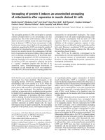
Cell transplants
PKH26 labelled transplanted cells were detected in host
myocardium by their intense red fluorescence, 4 week
after cell implantation. (Figure 1).
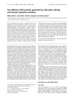
Effects of cell therapy on vascular density
Microscopic examination showed the following findings.
There were many neovessels in and around the sc ar tis-
sue 4 weeks after the injections of SVF and BM-MNC.
Representative images are shown in Figure 2a. The vas-
cular density of vessels larger than 30 μm in diameter in
the peri-MI area was highest in the group with SVF
(SVF, BM-MNC, Control: 6.88 ± 2.03, 4.45 ± 1.45 and
1.95 ± 1.19/mm2, respectively; p < 0.001). The vascu lar
density in the groups with SVF and BM-MNC were sig-
nificantly higher than the control group. Microvessel
(<30μm) numbers were significantly lower in control
rats than the SVF implanted group. (SVF, BM-MNC,
Control: 28.78 ± 3.5, 25.17 ± 2. 54 and 17.11 ±
4.18/mm2, respectively; p < 0.05). Results of post hoc
analysis were shown with symbols in Figure 2b.
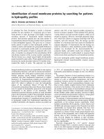
Fibrotic area inside the infarct and peri-infarct zone
Thepercentageoffibroticareainsidetheinfarctarea
was less in SVF and BM-MNC groups than that of
control group (SVF, BM-MNC, Control: 31.84 ± 6.2,
42.88 ± 3.1 and 65.11 ± 7.86%, respectively; p < 0.01;
Figure 3A). The percentage o f fibrotic area inside the
peri-infarct area was directionally similar to that of the
infarct area. (SVF, B M-MNC, Control: 30.30 ± 2.35,
29.14 ± 5.5 and 56.39 ± 6.3%, respectively; p < 0.01;
Figure 3B).
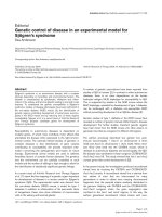
SVF transplantation decreased gene expression of
inflammatory cytokines TNFa and IL6
Expression of TNFa and IL6 mRNA was lower in the
LV myocardium from the SVF group than th e culture
medium-injected control group following cell/culture
medium treatment (P < 0.05; Figure 4A, and 4B). In the
BM-MNC injected LV tissue, no significant differences
were observed in TNFa or IL-6, mRNA levels, either
with SVF or culture medium-injected LV myocardium.
A high decrease in mRNA expression was noted in
TNFa and IL-6 in the BM-MNC group rats compared
with the control group, although these results did not
reach statistical significance.
SVF transplantation reduced MMP1 and TIMP1 gene
expression
The mRNA analysi s demonstrated decreased expression
of MMP-1 and TIM P-1 in the SVF group as compared
with the c ontrol group (P < 0.05; Figure 4C, and 4D).
A high decrease in mRNA expression was noted in
MMP-1 in the BM-MNC group rats compared with the
control group, although these results did not reach
statistical significance.
BNP and VEGF mRNA expression
AsshowninFigure4Eand4F,theexpressionofBNP
mRNA was lower and the expression of VEGF mRNA
was higher in the LV myocardium from the SVF group
compared with the culture medium-injected control
group (P < 0.05), following 4 weeks treatment.
Immunohistochemical studies for CD3, CD8 and CD 20
The mean number of cells positive for CD3 was
decreased significantly in SVF transplanted rats com-
paredtocontrols(p<0.05;Figure5).Themean
Table 1 Echocardiographic data at pretreatment and
4 Weeks after cell or culture medium transplantation in
3 Groups
SVF BMMNC Control
Pre treatment
LVDd (cm) 0.92 ± 0.02 0.91 ± 0.02 0.92 ± 0.02
LVDs (cm) 0.68 ± 0.03 0.66 ± 0.03 0.68 ± 0.03
FS (%) 26.7 ± 1.6 28.5 ± 2 26.1 ± 2.6
EF (%) 57.0 ± 2.6 59.5 ± 3.1 55.2 ± 3.9
After treatment
LVDd (cm) 0.88 ± 0.02* 0.93 ± 0.03* 1.02 ± 0.09
LVDs (cm) 0.60 ± 0.03* 0.65 ± 0.03* 0.78 ± 0.15
FS (%) 31.6 ± 2.6* 30.3 ± 1.7* 23.3 ± 1.1
EF (%) 63.8 ± 3.5* 62.5 ± 2.7* 51.2 ± 1.9
Values are shown as the mean ± SEM. LVDd, left ventricular end-diastolic
dimension; LVDs, left ventricular end-systolic dimension; FS, fractional
shortening; EF, ejection fraction. * p < 0.05 versus Control group.
Premaratne et al. Journal of Cardiothoracic Surgery 2011, 6:43
/>Page 4 of 10
number of CD20+ cells in the infarct was dec reased sig-
nificantly in SVF and BM transplanted rats compared to
controls (p < 0.001, p < 0.01 respectively; Figure 6). In
the cell transplanted groups, the number of CD8+ cells
was not significantly different from the culture medium
injected controls.
Presence of IL-6 protein in the heart
The percentage of area positive for IL-6 inside the LV
myocardium 4 weeks after treatment was less in SVF
and BM-MNC groups than that of control group (SVF,
BM-MNC, C ontrol: 0.38 ± 0.27, 1.33 ± 0.4 and 11.83 ±
2.41%, respectively; p < 0.001; Figure 7).
Discussion
Cell therapy may be an alternative treatment for heart
failure. The optimal cell for transplantation and the
source of the cells to be isolated are important consid-
erations. It has led to the investigations of different
types of stem c ell therapy for therapeutic angiogenesis.
Several recent studies in animals [2,3] as well as humans
[5,6] have repeatedly demonstrated that the transplant a-
tion of adult bone marrow derived cells can improve left
ventricular function and inhibit adverse remodeling after
myocardial infarction. The cardioprotective benefits may
be mainly derived from the enhancement of neovascu-
larization by BM cells, either by their ability to supply
large amounts of angiogenic, anti-apop totic and m ito-
genic factors [18] or by differentiating into vascular cells
[11] and cardiomyocyte-like cells [12,19]. Unfortunately,
the positive initial results of phase I/II studies remains
highly controversial [20]. Moreover, bone marrow can
only be obtained by bone marrow biopsy, a potentially
painful procedure. Therefore, alternative source of stem
cells or progenitors for therapeutic angiogenesis has
been desired.
In this study, we focused on the prote ctive effects of
SVF transplantation compared to those of BM-MNC
transplantation and the anti-inflammatory role of
transplanted cells after implanted into a rat chronic
myocardial infarction. Survived donor cells in host myo-
cardium were clearly visualized with red fluorescence in
SVF and BM-MNC implanted groups (Figure 1).
Major findings of the present study are summarized as
follows. (1) Intramyocardial injection of SVF was more
effective than that of BM-MNC or culture medium in
enhancing neovascularization, inhibiting collagen deposi-
tion and reducing gene expression of inflammatory cyto-
kines TNF-a, IL-6, TIMP-1 and BNP as well as
inflammatory cells CD3, in rat chronic ischemic myocar-
dium.; (2) Both the SVF and BM-MNC transplantation
improved cardiac function, attenuated LV dilation, and
thus prevented further myocardial remodelling.
Injection of SVF into ischemic myocardium was not
associated with any side effects; specially, there were no
casualties or arrhythmias due to cell implantation and
there was no evidence of local infection. In this report, we
demonstrated that direct intramyocardial injection of adi-
pose derived SVF was more effective than BM-MNC or
culture medium in enhancing neovascularization and
improvement of LV function in chronic ischemic myocar-
dium. By the ability o f the other subpopulations of SVF
and BM, including hematopoietic stem cells and mesench-
ymal stem cells to supply large amounts of angiogenic,
anti-apoptotic and mitogenic factors [18,21], cell trans-
planted groups may have increased neoangiogenesis via a
paracrine effect in the ischaemic myocardium. On the
other hand, t he culture medium injection group showed
deleterious effects on angiogenesis, probably, due to an
increased amount of various unfavorable cytokines such as
TNFa and IL-6 that impair new vessel formation. It has
been demonstrated that bone marrow cells strong ly sup-
press T-lymphocyte proliferation [22,23]. In the present
study, direct intramyocardial injection of SVF and
BM-MNC to the ischemic myocardium substantially sup-
pressed CD3 cell (T lymphocyte) and CD20 cell prolifera-
tion (Figure 5 and 6, respectively) and down regulated the
production of inflammatory cytokines, such as TNFa,IL-6
50μm
50μm
50μm
BMSVF Control
Figure 1 Transplanted cells. P KH2 6 labeled donor cells (re d fluorescence, x200) in SVF and BM-MNC transplanted groups. Bars rep resent a
distance of 50 μm.
Premaratne et al. Journal of Cardiothoracic Surgery 2011, 6:43
/>Page 5 of 10
A
B
Figure 2 Vascular density. 2a (A-C) Immunohistochemistry for von Willebrand factor (brown, x100). Representat ive pictures in the peri-MI area
from SVF, BM-MNC and Control groups, respectively. (D-F) Immunohistochemistry with a-smooth muscle actin antibody (brown, x100).
Representative pictures in the peri-MI area from SVF, BM-MNC and Control groups, respectively. Scale bars indicate distances of 100 μm.
2b Graphs: the number of vessels (number/mm
2
) in the peri-MI area, micro-vessel density (density of vessels <30 μm in diameter) (A), and
large-vessel density (density of vessels >30 μm in diameter) (B). Data are given as the mean ± SEM. *p < 0.05 vs. Control group, **p < 0.05 vs.
BM-MNC group,
†
p < 0.001 vs. Control group.
Premaratne et al. Journal of Cardiothoracic Surgery 2011, 6:43
/>Page 6 of 10
BM
SVF
100μm 100μm
100μm
Control
(A) Central-MI
*
†
Fibrotic area (%)
(B) Peri-MI
†
†
Fibrotic area (%)
Figure 3 Fibrotic area.RepresentativepicturesfromgroupsSVF,
BM-MNC and Control, respectively. Bars represent a distance of
100 μm. Graphs: Percentage of fibrotic area inside the infarct
(A) and peri-infarct area (B). Data are given as the mean ± SEM.
*p < 0.05 vs. Control group,
†
p < 0.01 vs. Control group.
TNFα
/
G
us
*
(A) (B)
IL6 / Gus
*
*
(C)
MMP1 / Gus
*
(D)
TIMP1 / Gus
BNP / Gus
*
*
(E)
VEGF / Gus
*
**
†
(F)
Figure 4 Expression of mRNA. Expression of mRNA levels of tumor necrosis factor a (A, TNFa); interleukin 6 (B, IL-6); matrix metalloproteinase
1 (C, MMP-1); tissue inhibitor of metalloproteinase 1 (D, TIMP-1), brain natriuretic peptide (E, BNP) and vascular endothelial growth factor (F,
VEGF) in the left ventricular myocardium as measured by reverse transcription polymerase chain reaction in the rat left ventricular myocardium,
4 weeks after treatment. mRNA expressions were calculated via a standard curve and normalized to an endogen control. Data are given as the
mean ± SEM. *p < 0.05 vs. Control group, **p < 0.01 vs. BM-MNC group, †p < 0.001 vs. Control group.
*
CD3 (number of cells/mm
2
)
BMSVF
Control
100μm 100μm
100μm
Figure 5 Immunohistochemistry for CD3+ (T lymphocytes),
(brown, × 100). Representative pictures in the infarct area from
SVF, BM-MNC and Control groups, respectively. Bars represent a
distance of 100μm. Graph: the number of CD3+ (number/mm
2
)in
the infarct area. Data are given as the mean ± SEM. *p < 0.05 vs.
Control group.
Premaratne et al. Journal of Cardiothoracic Surgery 2011, 6:43
/>Page 7 of 10
and TIMP-1 (Figure 4; A, B and D respectively). These
cytokines may be involved in the pathogenesis of heart
failure or LV remodelling [24,25]. It has been previously
shown that TNFa released from ischemic heart after acute
MI, has been shown to reduce contractility, increases the
production of other cytokines such as IL-1, IL-6 and
TIMP-1, induces pathophysiological hypertrophy, pro-
motes apoptosis of cardiomyocytes and other alterations
of the extracellular matrix which finally accelerates LV
remodeling [26]. In a ddition, serum levels as well as the
local concentrations of inflammatory cytokines, especially,
TNFa, are significantly increased in patients with chronic
heart failure and these levels correlate with the degr ee of
functional impairment [27,28]. Repeated TNFa infusion
may lead to a permanent decrease in myocardial contracti-
lity [29]. An increasing number of experimental observa-
tions suggests that IL-6 is also capable of modulating
cardiovascular function, exerting a negative inotrophic
function. IL-6 can be expressed in myocardium under var-
ious forms of stress and, also, it has the ability to induce
apoptosis, cardiac hypertrophy and fibrosis in myocardium
SVF
50μm
BM
50μm
Control
50μm
†
**
CD20 (number of cells/mm
2
)
Figure 6 Immunohi stochemistry for CD20+ (B lymphocytes),
(brown, × 100). Representative pictures in the infarct area from
SVF, BM-MNC and Control groups, respectively. Bars represent a
distance of 50μm. Graph: the number of CD20+ (number/mm
2
)in
the infarct area. Data are given as the mean ± SEM.
†
p < 0.001 vs.
Control group, **p < 0.01 vs. Control group.
BMSVF
Control
100μm
100μm
100μm
IL-6 (%)
†
†
Figure 7 Localization of IL-6 (brown) by immunohistochemical analysis in cell transplanted and control hearts. Magnification × 100.
Representative pictures in the infarct area from SVF, BM-MNC and Control groups, respectively. Bars represent a distance of 100μm. Graph:
Percentage of IL-6 positive area inside the infarct. Data are given as the mean ± SEM.
†
p < 0.001 vs. Control group.
Premaratne et al. Journal of Cardiothoracic Surgery 2011, 6:43
/>Page 8 of 10
[29]. Therefore, in the present experiment, IL-6 in the
myocardium of the culture medium injected animals may
have been upreg ulated by relative isch emia in the hyper-
trophied myocyte itself.
We focused on the role of MMP-1 activation for sev-
eral reasons. It has pre viously been shown that BM
mesenchymal stem cell transplantation reduces gene
and protein expression of MMP-1 and TIMP-1, inhibits
collagen deposition in the ischemic myocardium [30].
MMP-1 has been shown to play an importan t role in
myocardi al matrix degradation, which is associated with
ischemic heart disease [31]. W e observed that SVF
transplantation inhibited gene expression of MMP-1 and
TIMP-1, which might have influenced the collagen
degradation in the myocardium. We noticed that severe
fibrosis developed in the infarcted area in the control
group with culture medium injection, whereas only lim-
ited fibrosis was seen in the groups receiving SVF and
BM-MNC.
The results implicate the mechanisms and efficiency of
using SVF as an alternative to BM in treating cardiac
dysfunction. Our findings on the expression of inflam-
matory cytokines in the myocardium add another
dimension to u nderstanding the anti-inflammation role
of SVF and BM-MNC transplantation in cardiac dys-
function. The potential anti-inflammation role of both
SVF and BM-MNC transplantatio n is well recognized
but needs t o be fur ther studied. It is obvious that the
failed clinical trials [20,32] were carried out before we
had sufficient understanding of how inflammation is
involved and regulated following cell transplantation in
heart disease.
Conclusions
In conclusion, our data suggest that transplantation of
SVF might be a useful therapeutic option for angiogen-
esis in chronic ischemic heart disease. Given the feasibil-
ity and the lower invasiveness required to obtain adipose
tissues from patients, freshly isolated SVF could be
widely used to treat pa tients with ischemic heart dis-
eases along with other sources of stem cells such as
BM-MNC. Although our study has provided data sup-
porting the usefulness of SVF implantation into the
ischemic myocardium, further studies are required to
improve the reproducibility and to monitor long-term
effects in larger animal models.
Acknowledgements
This work was supported by grants from Swedish Medical Research Council,
Swedish Heart-Lung Foundation and Sahlgrenska University Hospital.
Author details
1
Wallenberg Laboratory for Cardiovascular Research, Sahlgrenska University
Hospital, University of Gothenburg, Gothenburg, Sweden.
2
Department of
Cardiology, Shanghai Second Military Medical University, Shanghai, PR China.
3
Department of Human Health Sciences, Graduate School of Medicine, Kyoto
University, Kyoto, Japan.
Authors’ contributions
GUP performed all the cell culture procedures, surgical procedures, histology
and design of the manuscript. LPM participated in the animal studies. MF
(Professor Masatoshi Fujita) performed critical review of the concepts, read
and approved the final version. XL contributed to the histology and
statistical analysis. EB participated in echocardiography. MF (Professor
Michael Fu) participated in its design and coordination. All authors read and
approved the final manuscript.
Competing interests
The authors declare that they have no competing interests.
Received: 1 November 2010 Accepted: 31 March 2011
Published: 31 March 2011
References
1. Shabbir A, Zisa D, Suzuki G, Lee T: Heart failure therapy mediated by the
trophic activities by the bone marrow mesenchymal stem cells: a
noninvasive therapeutic regimen. Am J Physiol Heart Circ Physiol 2009,
296(6):1888-1997.
2. Tse H-F, Siu C-W, Zhu S-G, Songyan L, Zhang Q-Y, Lai W-H, Kwong Y-L,
Nicholls J, Lau C-P: Paracrine effects of direct intramyocardial
implantation of bone marrow derived cells to enhance
neovascularization in chronic ischaemic myocardium. Eur J Heart Fail
2007, 9(8):747-753.
3. Makkar RR, Price MJ, Lill M, Frantzen M, Takizawa K, Kleisli T, Zheng J, Kar S,
McClelan Miyamota T, Bick-Forrester J, Fishbein MC, Shah PK, Forrester JS,
Sharifi B, Cheng P-S, Qayyum M: Intramyocardial injection of allogenic
bone marrow-derived mesenchymal stem cells without
immunosuppression preserves cardiac function in a porcine model of
myocardial infarction. J Cardiovasc Pharmacol Ther 2005, 10(4):225-233.
4. Baba S, Heike T, Yoshimoto M, Umeda K, Doi H, Iwasa T, Lin X, Matsuoka S,
Komeda M, Nakahata T: Flk1+ cardiac stem/progenitor cells derived from
embryonic stem cells improve cardiac function in a dilated
cardiomyopathy mouse model. Cardiovasc Res 2007, 76(1):119-131.
5. Tse H-F, Thamber S, Kwong Y-L, Rowlings P, Bellamy G, McCrohon J, Thomas P,
Bastian B, Chan JKF, Lo G, Ho C-L, Chan W-S, Kwong RY, Parker A, Hauser
Chan J, Fong DYT, Lau C-P: Prospective randomized trial of direct
endomyocardial implantation of bone marrow cells for treatment of severe
coronary artery diseases (PROTECT-CAD trial). Eur Heart J 2007, 28:2998-3005.
6. Dill T, Schächinger V, Rolf A, Möllmann S, Thiele H, Tillmanns H, Assmus B,
Dimmeler S, Zeiher AM, Hamm C: Intracoronary administration of bone
marrow-derived progenitor cells improves left ventricular function in
patients at risk for adverse remodeling after acute ST-segment elevation
myocardial infarction: Results of the Reinfusion of Enriched Progenitor
cells And Infarct Remodeling in Acute Myocardial Infarction study
(REPAIR-AMI) cardiac magnetic resonance imaging substudy. Am Heart J
2009, 157(3):541-547.
7. Yamamoto N, Akamatsu H, Hasegawa S, Yamada T, Nakata S, Ohkuma M,
Miyachi EI, Marunouchi T, Matsunaga K: Isolation of multipotent stem cells
from mouse adipose tissue. J Dermatol Sci 2007, 48(1):43-52.
8. Astori G, Vignati F, Bardelli S, Tubio M, Gola M, Albertini V, Bambi F, Scali G,
Castelli D, Rasini V, Soldati G, Moccetti : “In vitro” and multicolor
phenotypic characterization of cell subpopulations identified in fresh
human adipose tissue stromal vascular fraction and in the derived
mesenchymal stem cells. J Transl Med 2007, 5(5):844-851.
9. Palpant NJ, Yasuda S, MacDougald O, Metzger JM: Non-canonical Wnt
signaling enhances differentiation of Sca+/c-kit+ adipose-derived
murine stromal vascular cells into spontaneously beating cardiac
myocytes. J Mol Cell Cardiol 2007, 43:362-370.
10. Planat-Bénard V, Menard C, André M, Puceat M, Perez A, Garcia-Verdugo JM,
Pérnicaud L, Casteilla L: Spontaneous cardiomyocyte differentiation from
adipose tissue stroma cells. Circ Res 2004, 94:1-10.
11. Planat-Bénard V, Silvestre JS, Cousin B, Andre M, Nibbelink M, Tamarat R,
Clergue M, Manneville C, Saillan-Barreau C, Duriez M, Tedgui A, Levy B,
Pérnicaud L, Casteilla : Plasticity of human adipose lineage cells toward
endothelial cells: physiological and therapeutic perspectives. Circulation
2004, 109:656-663.
Premaratne et al. Journal of Cardiothoracic Surgery 2011, 6:43
/>Page 9 of 10
12. Nagaya N, Kangawa K, Itoh T, Iwase T, Murakami S, Miyahara Y, Fujii T,
Uematsu M, Ohgushi H, Yamagishi M, Tokudome T, Mori H, Miyatake K,
Kitamura S: Transplantation of mesenchymal stem cells improves cardiac
function in a rat dilated cardiomyopathy. Circulation 2005, 112:1128-1135.
13. Mazo M, Planat-Bénard V, Abizanda G, Pelacho B, Léobon B, Gavira JJ,
Penuelas I, Cemborain A, Pérnicaud L, Laharrague P, Joffre C, Boisson M,
Ecay M, Collantes M, Barba J, Casteilla L, Prosper F: Transplantation of
adipose derived stromal cells is associated with functional improvement
in a rat model of chronic myocardial infarction. Eur J Heart Fail 2008,
10:454-462.
14. Zuk PA, Zhu M, Mizuno H, Huang J, Futrell JW, Katz AJ, Benhaim P,
Lorenz HP, Hedrick MH: Multilineage cells from human adipose tissue:
implications for cell-based therapies. Tissue Eng 2001, 7:211-228.
15. Tambara K, Premaratne GU, Sakaguchi G, Kanemitsu N, Lin X, Nakajima H,
Sakakibara Y, Kimura Y, Yamamoto M, Tabata Y, Ikeda T, Komeda M:
Administration of control-released hepatocyte growth factor enhances
the efficacy of skeletal myoblast transplantation in rat infarcted hearts
by greatly increasing both quantity and quality of the graft. Circulation
2005, 112(Suppl I):129-134.
16. Nakajima H, Sakakibara Y, Tambara K, Marui A, Yoshimoto M,
Premaratne GU, Lin X, Kanemitsu N, Sakaguchi G, Ikeda T, Nishimura K,
Nakahata T, Komeda M: Delivery route in bone marrow cell
transplantation should be optimized according to the etiology of heart
disease. Circ J 2008, 72:1528-1535.
17. Premaratne GU, Tambara K, Fujita M, Lin X, Kanemitsu N, Tomita S,
Sakaguchi G, Nakajima H, Ikeda T, Komeda M: Repeated implantation is a
more effective cell delivery method in skeletal myoblast transplantation
for rat myocardial infarction. Circ J 2006, 70:1184-1189.
18. Fazel S, Cimini M, Chen L, Li S, Angoulvant D, Fedak P, Verma S, Weisel RD,
Keating A, Li RK: Cardioprotective c-kit+ cells are from the bone marrow
and regulate the myocardial balance of angiogenic cytokines. J Clin
Invest 2006, 116(7):1865-1877.
19. Wang JS, Shum-Tim D, Galipeau J, Chedrawy E, Eliopoulos N, Chiu RC:
Marrow stromal cells for cellular cardiomyoplasty: feasibility and
potential clinical advantages. J Thorac Cardiovasc Surg 2000, 120:999-1005.
20. Meyer GP, Wollert KC, Lotz J, Steffens J, Lippolt P, Fichtner S, Hecker H,
Schaefer A, Arseniev L, Hertenstein B, Ganser A, Drexler H: Intracoronary
bone marrow cell transfer after myocardial infarction: Eighteen months’
follow-up data from the randomized, controlled BOOST (Bone marrow
transfer to enhance ST-elevation infarct regeneration) Trial. Circulation
2006, 113:1287-1294, 4.
21. Miyahara Y, Nagaya N, Kataoka M, Yanagawa B, Tanaka K, Hao H, Ishino K,
Ishida H, Shimizu T, Kangawa K, Sano S, Okano T, Kitamura S, Mori H:
Monolayered mesenchymal stem cells repair scarred myocardium after
myocardial infarction. Nature Med 2006, 12:459-465.
22. DiNicola M, Carlo-Stella C, Magni M, Milanesi M, Longoni PD, Matteucci P,
Grisanti S, Gianni AM: Human bone marrow stromal cells suppress T-
lymphocyte proliferation induced by cellular or nonspecific mitogenic
stimuli. Blood 2002, 99:3838-3843.
23. Tse WT, Pendleton JD, Beyer WM, Egalka MC, Guinan EC: Suppression of
allogeneic T-cell proliferation by human marrow stromal cells:
implications in transplantation. Transplantation 2003, 75:389-397.
24. Prabhu SD: Cytokine-induced modulation of cardiac function. Circ Res
2004,
95:1140-1153.
25. Torre-Amione G, Kapadiya S, Lee J, Durans JB, Bies RD, Young JB, Mann DL:
Tumor necrosis factor-α and tumor necrosis factor receptors in the
failing human heart. Circulation 1996, 93:704-711.
26. Ono K, Matsumori A, Shioi T, Furukawa Y, Sasayama S: Cytokine gene
expression after myocardial infarction in rat hearts. Possible implication
in left ventricular remodeling. Circulation 1998, 98:149-156.
27. Testa M, Yeh M, Lee P, Fanelli R, Loperfido F, Berman JW, LeJemtel TH:
Circulating levels of cytokines and their endogenous modulators in
patients with mild to severe congestive heart failure due to coronary
artery disease or hypertension. J Am Coll Cardiol 1996, 28:964-971.
28. Torre-Amione G, Kapadiya SR, Benedict C, Oral H, Young JB, Mann DL:
Proinflammatory cytokine levels in patients with depressed left
ventricular ejection fraction: a report from the Studies of Left Ventricular
Dysfunction (SOLVD). J Am Coll Cardiol 1996, 27:1201-1206.
29. Haugen E, Chen J, Wikström J, Grönros J, Gan LM, Fu M: Parallel gene
expressions of IL-6 and BNP during cardiac hypertrophy complicated
with diastolic dysfunction in spontaneously hypertensive rats. Int J
Cardiol 2007, 115(1):24-28.
30. Guo J, Lin G, Bao C, Hu Z, Hu M: Anti-inflammation role for mesenchymal
stem cells transplantation in myocardial infarction. Inflammation 2007,
30:97-104.
31. Kerckhoven V, Kalkman REA, Sexena PR, Schoemaker RG: Altered cardiac
collagen and associated changes in diastolic function of infarcted rat
hearts. Cardiovasc Res 2000, 46:316-323.
32. Assmus B, Walter DH, Lehmann R, Honold J, Martin H, Dimmeler S,
Zeiher AM, Schächinger V: Intracoronary infusion of progenitor cells is
not associated with aggravated restenosis development or
atherosclerotic disease progression in patients with acute myocardial
infarction. Eur Heart J 2006, 27(24):2989-2995.
doi:10.1186/1749-8090-6-43
Cite this article as: Premaratne et al .: Stroma l Vascular Fraction
Transplantation as an Alternative Therapy for Ischemic Heart Failure:
Anti-inflammatory Role. Journal of Cardiothoracic Surgery 2011 6:43.
Submit your next manuscript to BioMed Central
and take full advantage of:
• Convenient online submission
• Thorough peer review
• No space constraints or color figure charges
• Immediate publication on acceptance
• Inclusion in PubMed, CAS, Scopus and Google Scholar
• Research which is freely available for redistribution
Submit your manuscript at
www.biomedcentral.com/submit
Premaratne et al. Journal of Cardiothoracic Surgery 2011, 6:43
/>Page 10 of 10