Báo cáo y học: " Outcome of left heart mechanical valve replacement in West African children - A 15-year retrospective study." pdf
Bạn đang xem bản rút gọn của tài liệu. Xem và tải ngay bản đầy đủ của tài liệu tại đây (460.64 KB, 8 trang )
RESEARCH ARTICLE Open Access
Outcome of left heart mechanical valve
replacement in West African children - A 15-year
retrospective study
Frank Edwin
*
, Ernest Aniteye, Mark Mawutor Tettey, Martin Tamatey and Kwabena Frimpong-Boateng
Abstract
Background: The West African sub-region has poor health infrastructure. Mechanical valve replacement in children
from such regions raises important postoperative concerns; among these, valve-related morbidity and
complications of lifelong anticoagulation are foremost. Little is known ab out the long-term outcome of mechanical
valve replacement in West Africa. We sought to determine the outcome of mechanical valve replacement of the
left heart in children from this sub-region.
Method: We conducted a retrospective review of all consecutive left heart valve replacements in children (< 18
years old) from January 1993 - December 2008. The study end-points were mortality, valve-related morbidity, and
reoperation.
Results: One hundred and fourteen patients underwent mitral valve replacement (MVR), aortic valve replacement
(AVR) or mitral and aortic valve replacements (MAVR). Their ages ranged from 6-18 years (13.3 ± 3.1 years). All
patients were in NYHA class III or IV. Median follow up was 9.1 years. MVR was performed in 91 (79.8%) patients,
AVR in 13 (11.4%) and MAVR in 10 (8.8%) patients. Tricuspid valve repair was performed concomitantly in 45
(39.5%) patients.
There were 6 (5.3%) early deaths and 6 (5.3%) late deaths. Preoperative left ventricular dysfunction (ejection fraction <
45%) was the most important factor contributing to both early and late mortality. Actuarial survival at 1 and 15 years
were 98.1% and 94.0% respectively. Prosthetic valve thrombosis occurred in 5 patients at 0.56% per patient-year. There
was 1(0.9%) each of major bleeding event and prosthetic valve endocarditis. Two reoperations were performed at
0.22% per patient-year. Actuarial freedom from reoperation was 99.1% at 1 and 10 years, and 85.1% at 15 years.
Conclusion: Mechanical valve replacement in West African children has excellent outcomes in terms of mortality,
valve-related events, and reoperation rate. Preoperative left ventricular dysfunction is the primary determinant of
mortality within the first 2 years of valve replacement. The risk of valve-related complications is acceptably low.
Anticoagulation is well tolerated with a very low risk of bleeding even in this socioeconomic setting.
Keywords: mechanical valve replacement rheumatic heart disease, mitral valve, aortic valve, anticoagulation, West
Africa, children
Background
The West African sub-region has poor health infrastruc-
ture similar to most of the developing countries in
Africa. Gross National Product (GNP) in Africa is less
than $1800 per inhabitant compare d to $24000-$ 31000
in North America and Western Europe [1]. The
worldwide distribution of cardio-thoracic surgeons (and
cardiothoracic surgery) very closely follows the distribu-
tion of GNP; Africa has only 1% of the world’spopula-
tion of cardiothoracic surgeons [1]. Cardiac surgery and
subsequent long term postoperative management raise s
seri ous concerns. Consequently, mechanical valve repla-
cement in Africa is un dertaken with some disquiet.
Mechanical valve replacement commits the patient to
lifelong anticoagulation and its associated risks of
* Correspondence:
National Cardiothoracic Center, Korle Bu Teaching Hospital, P. O. Box KB 846,
Accra, Ghana
Edwin et al. Journal of Cardiothoracic Surgery 2011, 6:57
/>© 2011 Edwin et al; licensee BioMed Central Ltd. This is an Open Access article distributed under the terms of the Creative Commons
Attribution License ( /by/2.0), which permits unrestricted use, distribution, and reproduction in
any medium, provided the original work is properly cited.
bleeding and embryopathy in pregna ncy. Valve-related
morbidity such as prosthetic valve thrombosis, endocar-
ditis, and non-structur al dysfunction are additional con-
cerns that may require reoperation. Reoperation in
developing countries is often an insurmountable eco-
nomic hurdle for most families.
The present study was prompted by the lack of data
on the outcome of mechan ical valve replacement in
children from the West African sub-region. We report
early and late outcomes of mechanical valve replace-
ment of the left heart in West African children over a
15-year period.
Methods
Study design
We retrospectively reviewed results of consecutive chil-
dren undergoing mechanical valve replacement of the
left heart in the study period. Hospital records of the
selected patients were obtained for the purpose of the
review.
Study setting
Established in 1989, Ghana’ s National Cardiothoracic
Center is a referral center and the only tertiary institu-
tion in the country for cardiothoracic pathology. It also
serves as a cardiothoracic referral base for many of the
West African countries where cardiothora cic surgery is
not actively practiced due to lack of facilities.
Patients
Between J anuary 1993 and December 2008, 114 conse-
cutive patients of age ≤18 years underwent left heart
mechanical valve replacement at our institution. Opera-
tion records and patients’ case notes were retrospec-
tively reviewed. The study end-points included
mortality, valve-related events, and reoperation. Valve-
related events studied were prosthet ic valve thrombosis,
thromboembolism, prosthetic valve endocarditis, non-
structural dysfunction, and major bleeding events.
Procedures performed include mitral valve replace-
ment (MVR), aortic valve replacement (AVR), or mitral
and aortic valve replacements (MAVR) using mechanical
prostheses.
Operative Technique
In all patients median sternotomy was used to establish
full cardiopulmonary bypass and moderate systemic
hypothermia. Myocardial protection was employed by
infusion of cold crystalloid cardioplegia (St. Thomas’
Hospital solution) throu gh the root or coronary ostia of
the cross-clamped ascending aorta. This was repeated
every 20-25 minutes and augmented by the use of topi-
cal hypothermia with saline at 4°C.
The mitral valve was approached through the inter-
atrial septum in most cases. The aortic valve was
approached through a standard aortotomy. The type of
valve implanted is shown in table 1. Implanted valve
sizes ranged from 27-31 for MVR and 19-23 for AVR.
The modified De Vega annuloplasty technique [2] was
used for repair of non-structural tricuspid regurgitation
(TR).
Anticoagulation protocol
Post-operative anticoagulation is initiated with unfrac-
tionat ed heparin as a continuous infusion (300unit s/kg/
day) adjusted to an activated partial thromboplastin
time of twice the control value after postoperative bleed-
ing has come under control, usually on post-operative
day (POD) one. Oral warfarin is begun concomitantly
on the 2
nd
POD. Heparin infusion is discontinued when
the target international normalized ratio (INR) is
attained. The target INR was 2.0-3.0 for AVR, and 2.5-
3.5 for MVR and MAVR. The INR is adjusted upward
by 0.5 units in the presence of atrial fibrillation or left
ventricular dilatation/dysfunction. In our earlier experi-
ence (before 2001), a target INR of 2.5-3.0 was used for
all patients.
Discharge and Follow up
Follow up consists of clinical evaluation, anticoagulation
control, prophylaxis for rheumatic fever, and antibiotic
cover for endocarditis-prone procedures. Transthoracic
echocardiogram is performed at least once a year.
Patients were usually discharged with an INR in the
target range and followed up on a patient-specific 4-12
week interval at our institution’ s out-patient clinics.
Non-Ghanaian West African patients were followed up
3 m onthly at the Center and by telephone contact after
INR testing using local laboratory facilities.
We emphasize patient/parental education before dis-
charge. This effort is directed by a clinical pharmacist-
led team. We focus attention on the need for regular
monitoring and control of anticoagul ation, food and
drug interactions with warfarin, and prophylaxis for
both endocarditis and rheumatic fever. We gen erally
require patients to report for follow up every 4 weeks.
Patients living beyond 100 km of our institution
Table 1 Implanted Valves.
VALVE Number
Bileaflet mechanical
Sorin (Sorin Biomedica, Sallugia, Italy) 97
St. Jude Medical (St. Jude Medical; St. Paul, MN) 11
Monoleaflet mechanical
Sorin (Sorin Biomedica, Sallugia, Italy) 6
Edwin et al. Journal of Cardiothoracic Surgery 2011, 6:57
/>Page 2 of 8
(including our non-Ghanaian West African patients)
may be seen once every 8-12 weeks. For patients with
such proximity problems, telephone consultation regard-
ing dosage adjustment for warfarin and advi ce on endo-
carditis prophylaxis is an important adjunct to the
clinical follow up.
Statistical Analysis
Statistical analyses were performed using SPSS 16.0 soft-
ware. Continuous variables are expressed as mean ±
standard deviation. Actuarial curves were computed
using the Kaplan-Meier survival analysis technique. The
incidence of multiple events in individual patients is
reported as a linearized event rate. Morbidity and mor-
tality reporting are in keeping with t he guidelines pro-
posed by the 2008 ad hoc Liaison Committee for
standardizing definitions of prosthetic valve morbidity
and mortality [3].
Results
The 114 patients who qualified for inclusion showed a
female preponderance o f 57.9%; they included 13
(11.4%) non-Ghanaian West Africans. Their ages ranged
from 6-18 years (13.3 ± 3.1 years). Seventy-nine (69.3%)
patients were in NYHA Class III; the remaining patients
were in NYHA Class IV. Advanced cardiac disability
(reflected in the high NYHA class) was attributable to
prolonged illness without appropriate treatment. In
most cases, financial c onstraint was responsible for the
delay in seeking treatment resulting in progressive car-
diac dysfunction. Even after diagnosis and recommenda-
tion for surgery, less than 20% could afford surgery
within one year of diagnosis.
The nutritional status of the patients was acceptable
except for some patients in NYHA IV who had preo-
perative cardiac cachexia and muscle wasting most evi-
dent after diuresis as part of their medical optimization
for surgery. For hospital survivors, preoperative nutri-
tional depletion sometimes manifested as prolonged
mechanical ventilation (extra 4-7 days) and Intensive
Care Unit stay. Nutritional status however improved
promptly with restoration of cardiac function and ent-
eral feeding after surgery.
Complete follow up was available for 108 patients
(94.7% complete). Follow up ranged from 0.2-15.9 years
(median 9.1 years).
The etiology of valve pathology (Table 2) was rheu-
matic in 1 04 (91.2%) patients. In rheumatic valve dis-
ease, valvar regurgitation was the dominant
hemodynamic abnormality in both isolated and double
valve involvement (Table 3).
MVR was performed in 91 (79.8%) patients, AVR in
13 (11.4%) and MAVR in 10 (8.8%) patients.
Four patients required AVR at the time of ventricular
septal defect (VSD) closure; the VSD was associated
with aortic cusp prolapse and irreparable degeneration
in these patients. Three of the four patients presented
late in their teens having suffered previous endocarditis
resulting in deformed, unsalvageable aortic valve leaflets.
The fourth patient had a bicuspid valve with shallow
sinuses; a durable repair was not deemed feasible. Preo-
perative echocardiography and right heart catheteriza-
tion excluded hypertensive pulmonary vascular disease
in these patients (ages 9, 17, 17, and 18 years).
Tricuspid valve annuloplasty using the modified De
Vega technique was performed concomitantly in 45
(39.5%) cases. Forty-one of these (91%) were associated
with isolated mitral valve disease and 4 (9%) were due
to mitral and aortic valve pathology. None was asso-
ciated with isolated aortic valve pathology.
On echocardiogra phic follow up, there was one docu-
mented case of moderate tricuspid regurgitation in the
patients who had undergone tricuspid valve repair using
themodifiedDeVegaannuloplasty.Thispatienthad
rheumatic mitral regurgitation and non-structural TR
with poor pre-operative left ventricular function (ejec-
tion fraction of 42%). LV dysfunction persisted post-
operatively with evolution into dilated cardiomyopathy
and moderate TR despite successful MVR. Tricuspid
regurgitation was mild or absent in the remainder of the
patients. We have had acceptable results with the modi-
fied De Vega technique in a prior experience [4].
Preoperative left ventric ular dysfunction (ejection frac-
tion < 45%) was the most important factor contributing
to both early and late mortality; it was present in 5 of 6
(83%) early deaths and in 3 of 6 (50%) late deaths. Most
Table 2 Etiology of valve pathology.
Etiology Number Percentage
Rheumatic 104 91.2
VSD 4 3.5
Endocarditis (Aortic) 2 1.7
SAS + Valvar AS 1 0.9
MV prolapse (Marfan’s) 1 0.9
PVT 1 0.9
Annulo-aortic ectasia 1 0.9
AS - aortic stenosis; MV - mitral valve; PVT - prosthetic valve thrombosis; SAS -
subaortic stenosis; VSD - ventricular septal defect.
Table 3 Valvar hemodynamics in rheumatic heart
disease.
Regurgitation Stenosis Total
Isolated mitral valve involvement 83 (96.6%) 4 (3.4%) 87
Isolated aortic valve involvement 7 0 7
Double valve pathology 10 0 10
Edwin et al. Journal of Cardiothoracic Surgery 2011, 6:57
/>Page 3 of 8
survivors, however, demonstrated improvement in LV
dysfunction on long-term follow up. Two patients
(including the one with moderate TR) showed persistent
LV dysfunction and dilated cardiomyopathy with atrial
fibrillation after 2 and 3 years of follow up respectively.
They are on appropriate decongestive medication and
antiarrhythmic drug therapy.
Mortality
The overall 30-day mortality was 5.3% (6 patients, Table
4); late mortality occurred in 6 patients (Table 5) at a
linearized rate of 0.67% per patient-year. The actuarial
survival was 98.1% at 1 year, 97.0% at 5 years, and
94.0% at 10 and 15 years.
Valve-related events
1) Prosthetic Valve Thrombosis (PVT) and thromboembolism
PVT occurred in 5 patients (Table 6) at 0.56% per
patient-year. Four occurred in the mitral position and
one in the aortic position. T hrombosis was r esponsible
for all four prosthetic mitral valve obstructions. In the
fifth patient, coexistent thrombus and pannus caused
aortic valve obstruction. Only two of the four mitral
PVTs survived. Of the two survivors, thrombolysis with
streptokinase was successful in one; the other required
reoperation to replace the thrombosed valve.
The only aortic PVT in the series responded partially
to thrombolysis; the obstruction was due to a combina-
tion of thrombus and pannus. She underwent elective
replacement of the aortic prosthesis.
Actuarial freedom from PVT was 98.7% a t 1 and 5
years, 96.9% and 94.0% at 10 and 15 years respectively.
2) Thromboembolism
There were no documented postoperative embolic
events in this series.
3) Prosthetic Valve Endocarditis (PVE)
Early PVE occurred in one patient (0.9%) and was con-
tributory to the patient ’ s death. This was a six year old
boy who had left ventricular failure from rheumatic
mitral incompetence complicated by preoperative bac-
terial endocarditis. Failing medical management, MVR
was arranged as an urgent measure. Postoperatively, he
remained ill in a low cardiac output state until his
demise on the 16
th
POD. The postmortem examination
showed peri-prosthetic micro-abscesses.
4) Major Bleeding Event
There was 1 (0.9%) major bleeding event in thi s series.
Severe upper gastrointestinal bleeding occurred in a 14
year-old boy nine days after MVR when the INR was
2.0. He required a laparotomy the next day when con-
servative measures failed. A bleeding ulcer in the first
part of the duodenum was found and hemostasis was
secured by suture.
Reoperation
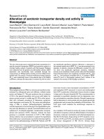
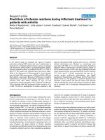
Reoperation was necessary in 2 patients (0.22% per
patient-year). The actuarial free dom from reoperation
(Figure 1) was 99.1% at 1, 5, and 10 years and 85.1% at
15 years.
The first reoperation wa s for PVT in an 8 year-old
boy 6 weeks after MVR with a Sorin monoleaflet valve
(Sorin Biomedica, Sallugia, Italy). The INR at the time
of PVT diagnosis was 2.0. The thrombosed valve was
urgently replaced with a bileaflet valve from the same
manufacturer. The second reoperation was for another
PVT that was only partly responsive to thrombolytic
therapy after 11 years of AVR.
Discussion
Rheumatic heart disease was responsible for more than
90% of patients undergoing mechanical valve replace-
ment in the present study. Late presentation of patients
for surgery was characteristic. Advanced rheumatic valve
pathology made valve replacement the preferred option.
Cardiologists may delay the recommendation of valve
replacement in children until greater clinical disability
Table 4 Early mortality.
Causes of early death Number Comment
Postoperative low cardiac output 4 Preoperative LV dysfunction (EF<45%).
Cerebral re-infarction 1 Preoperative cerebral embolism.
Prosthetic valve endocarditis and CHF 1 Preoperative endocarditis and CHF.
CHF - congestive heart failure, EF - ejection fraction.
Table 5 Late mortality.
Late mortality Number Comment
Prosthetic valve thrombosis 2 Died 9 and 14 years respectively after MVR.
Non-compliance with follow-up.
Progressive LV dysfunction postoperatively 3 All died within 2 years of valve replacement.
Sudden cardiac death 1 Progressive rheumatic aortic valve regurgitation post-MVR. Parents declined reoperation.
LV - left ventricular.
Edwin et al. Journal of Cardiothoracic Surgery 2011, 6:57
/>Page 4 of 8
and failed medical therapy occur. This practice has been
based on the belief that children tolerate anticoagulation
poorly and that valve-related complica tions are far too
common in children. We have shown in th is study the
excellent tolerability of anticoagulation in West African
children and the acceptable valve-related morbidity. In
addition, our results demonstrate that delay in the insti-
tution of surgical treatment allows progressive deteriora-
tion of ventricular function and contributes primaril y to
both early and late mortality.
Mortality
Preoperative left ventricular dysfunction was the primary
contributor to both early and late mortality. Patients
with preoperative left ventricular dysfunction experi-
enced early postoperative low cardiac output syndrome
or relentlessly progressive cardiomyopathy of mitral
incompetence after discharge from hospital. In develo p-
ing countries, rheumatic heart valves tend to deteriorate
rapidly due to repeated episodes of acute rheumatic car-
ditis that lead to severe debilitating disease, ventricular
dysfunction, and premature death [5,6]. Such advanced
disease allows progressive deterioration of LV function
even after valve replacement. Delay in seeking treatment
as a result of inadequate health infrastructure and poor
health care financing contributes to creating a pool of
children who present with preoperative LV dysfunction.
In Ghana, state medical insurance is available for regis-
tered citizens but gives limited coverage for primary
health care, anticoagulation testing, and medications
(including warfarin and generic decongestive drugs for
heart failure). There i s no health insurance cover for
open heart surgery. The Ghana Heart Foundation
(GHF), a non-governmental organization set up by the
senior author (KF-B) at the inception of our institution
ass ists Ghanaian patients to the t une of 50% of the cost
of open heart surgery. In dire emergencies, the GHF
may bear the full cost . Unfortunately, if the patient is
unable to afford the cost of surgery, appeals to the pub-
lic and philanthropist s for financial assistance becomes
the only option with sometimes unfortunate conse-
quences. This situation, common in much of West
Africa, contributes to the situation where children are
kept waiting unti l severe preoperati ve LV dysfunction
occurs before presentation for surgery. As our results
show, preope rative LV dysfunction may progress relent-
lessly even after successful valve replacement. Workers
from other developing countries have reported similar
outcomes attributable to late presentation of rheumatic
heart disease and poor preoperative ventricular function
[7,8], with comparable early mortality. This underscores
the importance of early intervention in rheumatic heart
disease before i rreversible deterioration of ventricular
function occurs.
The presence of active rheumatic carditis ( suspected
in 2 early deaths) and preoperative infective endocarditis
(confirmed in 1 early death) were additional factors con-
tributing to early mortality.
Late death occurred in 6 (5.3%) patients at a linearized
rate of 0.67% per patient-year. The actuarial survival at 1
and 15 years were 98.1% and 94.0% r espectiv ely. WHO
figures from 2008 suggest that the 15-year life expec-
tancy of adolescent male and female Ghanaians with
median age similar to the study group (13 years) is
about 96.5% [9]. Mechanical valve replacement is thus
Table 6 Development of PVT.
Patient Surgery INR at PVT diagnosis Mechanism of
PVT
Surgery to PVT Interval
(years)
1 MVR 2.1 Thrombus 0.3
2 MVR ? Thrombus 0.5
3 AVR 2.0 Thrombus and Pannus 9.1
4 MVR INR not done for > 8 months Thrombus 9.0
5 MVR INR not done for > 18 months Thrombus 15.0
Time from surgery to PVT: Mitral - 6.2 ± 7.3 years; All PVT cases - 6.8 ± 6.3 years. AVR - aortic valve replacement, MVR - mitral valve replacement, PVT - prosthetic
valve thrombosis.
Figure 1 Actuarial freedom from reoperation. Kaplan-Meier
estimates of freedom from reoperation after 15 years’ follow up
showing actuarial figures of 99.1% and 85.1% at 10 and 15 years
respectively.
Edwin et al. Journal of Cardiothoracic Surgery 2011, 6:57
/>Page 5 of 8
associated with a near-normal 15-year life expectancy in
these patients.
In a similar p atient cohort, Akhtar and colleagues [7]
reported 11 (12.4%) late deaths; patient survival at 1 and
10 years was 87.5% and 82.9% respectively. Barnard and
colleagues [8] recently reported 14.3% late death with
actuarial survival at 5 years of 80%. These workers [7,8]
confirm the results of the current study that the risk of
on-going death for hospital survivors of valve replace-
ment for rheumatic heart disease can be significant.
This may be attributed to a combination of progressive
deterioration of ventricular function in late presenters
and poor anticoagulation control.
Compared to these reports [7,8], valve-related mortal-
ity was relatively low in this study (3 of 114, or 2.7%).
This is probably a reflection of higher compliance with
postoperative management protocols. Akta r and collea-
gues[7]reportedthatmostof their patients suffering
fatal events had erratic follow-up with sub-therapeutic
anticoagulation. Because of the formidable financial hur-
dle of ‘out-of-pocket’ financing of open heart procedures
in West Africa, most patients and parents have a high
motivation to avoid reoperation. With a ppropriate gui-
dance most tend to comply with follow-up management
protocols. Other factors may be operative in the lower
valve-related mortality in the present study. The use of
3
rd
generation bileaflet mechanical prostheses with
lower thrombogenicity in the majority of our patients
may be contributory. Additionally, out-patient follow-up
by our team of surge ons and clinical pharmacists using
patient-specific anticoagulation regimens probably
played a role as well [10].
The impact of age and underlying etiology for
mechanical valve replacement must not be overlooked.
The report of Ackermann and coworkers [11] demon-
strate that replacement of the systemic atrioventricular
valve with a mechanical prosthesis in children aged less
than 6 years in whom the underlying etiology is non-
rheumatic may portend a somewhat higher mortality.
They showed a survival of 73% at 1 year and 65% at 5,
10, and 15 years. The youngest patient in our study was
6 years of age; rheumatic heart disease was the underly-
ing etiology i n close to 91% of our cases. Outcomes for
mechanical valve replacement of the left heart in chil-
dren therefore are dependent on the etiology and age of
the patient cohort.
PVT and thromboembolism
PVT and thromboembolism after mechanical valve
replacement are both related to the balanc e between
thrombogenicity and the adequacy of anticoagulation.
Two forms of PVT are usually described - obstructive
and non-obstructive [12].
Non-obstructive PVT commonly occurs in the highly
unstable early postoperative period [12]. It causes mini-
mal local symptoms but predisposes the patient to sys-
temic thromboembolic phenomena at a rate of 0.7-6%
per patient-year [13]. From the developing world, John
and colleagues [5] reported a thromboembolic rate of
2.8% or 0.8% per patient-year. In a subsequent report,
their thromboembolic rate dropped to 0.41% per
patient-year for MVR [14]. During the first six post-
operative months, the thromboembolic risk is up to
seven times greater than in the months and years after-
ward. After AVR, thromboembolic risk falls from 16%
per patient-year in the early postoperative period to
1.4% per patient-year at 5 years. Similarly, after MVR,
the risk falls from 21% per patient-year to 2.5% per
patient-year [15]. We did not document any episode of
thromboembolism in our study.
Unlike non-obstructive PVT, obstructive PVT is an
acute life-threatening complication demanding urgent
intervention. The incidence of obst ructive PVT for
mechanical valves varies between 0.3-1.3 percent per
patient-year [13]; our experience compares favorably.
The first postoperative year is marked by a 24% inci-
dence of o bstructive thrombosis, with a stable incidence
between the second to fourth years of approximately
15%, and a subsequent decrease thereafter [16]. Poor
health infrastructure in most developing nations w ould
predict a higher rate of obstructive and non-obstructive
PVT but our results and those of others [5,14] indicate
that this may be unfounded.
Among the commonest precipitating factors for
mechanical valve thrombosis are inadequate anticoagula-
tion and poor patient compliance. In this study, obstruc-
tive PVT (4.4%) occurred at a linearized rate of 0.56%
per patient-year with an overall mean time to develop-
mentof6.8±6.3years(Table6).Theincidenceof
acute thrombotic occlusion of a mechanical replacement
device has been reported to average 0.03% to 8% per
patient -year [17,18]. From the Montr eal Heart Institute,
Durrleman and coworkers [17] found the time interval
from first valve replacement to prosthetic valve throm-
bosis was 3.25 ± 3.50 years. At presentation of PVT, the
INR was less than 2.5 in 54% of their patients, with
inadequate anticoagulation management in 26% and
poor compliance in another 26%. Two of our patients
who developed obstructive PVT were notoriously non-
compliant for several years without apparent adverse
effects. Incidentally, these were the only two who suf-
fered late death from PVT after 9 and 15 years of MVR.
Patient-related factors therefore heavily influence the
occurrence of PVT and high patient motivatio n to com-
ply with postoperative anticoagulation management i s a
sine qua non to improved long-term outcome.
Edwin et al. Journal of Cardiothoracic Surgery 2011, 6:57
/>Page 6 of 8
Reoperation
In terms of freedom from reoperation, mechanical valve
replacement for rheumatic heart disease is superior to
valve repair. A reoperation rate of 23% after valve repair
for rheumatic heart disease has been reported [19,20]
with actuarial freedom from reoperation of 78% at 5
years, 65% at 10 years, and 49% at 15 years [20]. Free-
dom from moderate or severe mitral regurgitation may
be as low as 32 ± 3.9% 10 years after valve repair for
rheumatic heart disease [21]. In our experience, reopera-
tion occurred at 0.22% per patient-year with freedom
from reoperation of 99.1% and 85.1% at 10 and 15 years
respectively (Figure 1).
The financial implication of reoperation in our popu-
lation is considerable. In 2005, 47 children (≤15 years)
were diagnosed by echocardiography as rhe umatic heart
surgical candidates at our institution. Of this number,
only 7 (14.9%) underwent surgical treatment that same
year. Lack of funding was the primary reason for the
delay. Where health financing is limited, a major finan-
cial hurdle is imposed by the necessity of reoperation
for a failed valve repair, most of which is dependent on
progression of t he underlying rheumatic cardiac pathol-
ogy and not on surgical technique [22]. Mechanical
valve replacement in this setting confers a significant
advantage in populations where funding for open-heart
procedures is deficient. The drawback to this manage-
ment strategy is the impact and consequences of lifelong
anticoagulation.
The need for reoperation resulting from patient-pros-
thesis mismatch in growing children was not realiz ed in
our experience. The presence of gross cardiomegaly in
most of our patients, all of whom presented with long-
standing disease in NYHA class III or IV, allowed the
implantation of adult-sized prostheses which ostensibly
curtailed the development of this complication.
PVE
PVE occurred at 0.11% per patient-year and was respon-
sible for one early death in this series. This was of the
culture-negative variety detected pre-operatively in a six
year-old boy. Mechanical prostheses predispose to
device-related infections especially those caused by coa-
gulase-negative staphylococci, which are able to adhere
to a variety of surfaces an d produce an antibiotic-resis-
tant biofilm [23,24]. The risk for early PVE is higher in
patients undergoing valve replacement surgery during
active infective endocarditis, especially if the causal
organism is unknown. Once established, PVE carries a
mortality rate that may be as high as 70% [25]. The
established treatment for PVE is rigorous intravenous
antimicrobial therapy, although this has extremely lim-
ited success. The majority of cases require surgical
removal and replacement of the infected prosthesis.
Unfortunately, in our patient this could not be carried
out before he succumbed.
Major Bleeding Event
After mechanical valve replacement, major bleeding and
thromboembolic complications are notably commoner
in patients with a high variability in anticoagulation con-
trol [26]. Major bleeding events occur in 2.4-4.6% per
patient-year after warfarin anticoagulati on for mechani-
cal valve replacements [27]. We believe the low rate of
major bleeding events in our study (0.11% per patient-
year) testifies to the excellent tolerability of warfarin in
our pediatric population. Our experience is similar to
other workers from developing countries [14].
Prospects for Selected Patients
This patient cohort included two categories of notable
patients . First is the female with child-bearing potential.
Four in the group have successfully borne children with-
out complications. Our anticoagulant protocol involves
substitution of warfarin with subcutaneous unfractio-
nated heparin in the first trimester and at elective Cae-
sarian section through a collaborativ e effort with the
attending obstetricians. Three were delivered by elec tive
Caesarean section at 37 weeks gestation. The fourth had
a spontaneous vaginal term delivery at a peripheral hos-
pital without complications. We did not encounter war-
farin embryopathy o r fetal wastage in this p atient
cohort.
The second category consists of two patients who
have homozygous (SS genotype) sickle cell hemoglobi-
nopathy. Notably, the frequency of sickling crises has
drastically reduced in both patients since undergoing
mechanical valve replacement with institution of antic-
oagulation. Some workers have previously pointed out
that warfarin may protect against sickling crises [28,29].
Study limitations
Inherent limitations in all retrospective analysis apply to
this study. Because complete follow-up was not available
for 6 patients, the possibility of missed events must be
kep t in mind in the interpretation of the results. Diff er-
ences in socio-cu ltural behavior of different populations
with regard to compliance with post-operative manage-
ment protocols impact on the generalizability of the
study results.
Conclusion
Mechanical valve replacement in West African children
has excellent outcomes in terms of early and l ate mor-
tality, valve-related events, and reoperation rate.
Preoperative left ventricular dysfunction is the primary
determinant of mortality within the first 2 years of valve
replacement.
Edwin et al. Journal of Cardiothoracic Surgery 2011, 6:57
/>Page 7 of 8
Anticoagulation is well tolerated with a v ery low risk
of bleeding.
The risk of valve-related complications is acceptably
low even in this socioeconomic setting.
Efforts at improving long-term surgical outcomes for
mechanical valve replacement should focus on early sur-
gical intervention and improved control of anticoagula-
tion for mechanical valves.
Abbreviations
AS: aortic stenosis; AVR: aortic valve replacement; EF: ejection fraction; GNP:
gross national product; INR: international normalized ratio; LV: left ventricle;
MAVR: mitral and aortic valve replacements; MV: mitral valve; MVR: mitral
valve replacement; NYHA: New York Heart Association; POD: postoperative
day; PVT: prosthetic valve thrombosis; SAS: subaortic stenosis; TR: tricuspid
regurgitation; VSD: ventricular septal defect.
Acknowledgements
The authors acknowledge Dr. Philip Amu for providing the statistical
software for the data analysis.
Authors’ contributions
FE conceived the study, performed literature search, collected data,
performed statistical analysis, and drafted the manuscript. EA contributed to
the data analysis, editing and reviewing the manuscript. MMT contributed to
the study design, editing and reviewing the manuscript. MT participated in
data collection and editing of the manuscript. KF-B provided study
background information, supervised the work, and reviewed the manuscript.
All authors read and approved the final manuscript.
Competing interests
The authors declare that they have no competing interests.
Received: 5 February 2011 Accepted: 19 April 2011
Published: 19 April 2011
References
1. Turina MI: European Association for Cardio-Thoracic Surgery: carrying the
torch. Eur J Cardiothorac Surg 2002, 22:857-863.
2. Aoyagi S, Tanaka K, Hara H, Kumate M, Oryoji A, Yasunaga H, Kosuga K,
Ohishi K: Modified De Vega’s annuloplasty for functional tricuspid
regurgitation–early and late results. Kurume Med J 1992, 39:23-32.
3. Akins CW, Miller DC, Turina MI, Kouchoukos NT, Blackstone EH,
Grunkemeier GL, Takkenberg JJ, David TE, Butchart EG, Adams DH,
Shahian DM, Hagl S, Mayer JE, Lytle BW: Guidelines for reporting mortality
and morbidity after cardiac valve interventions. Eur J Cardiothorac Surg
2008, 33:523-528.
4. Frimpong-Boateng K: Valve replacement surgery at the Korle Bu
Teaching Hospital - A review. Ghana Med J 2002, 36:145-148.
5. John S, Bashi VV, Jairaj PS, Muralidharan S, Ravikumar E, Sathyamoorthy I,
Babuthaman C, Krishnaswamy S, Cherian G, Sukumar IP: Mitral valve
replacement in the young patient with rheumatic heart disease. Early
and late results in 118 subjects. J Thorac Cardiovasc Surg 1983, 86:209-216.
6. John S, Ravikumar E, John CN, Bashi VV: 25-year experience with 456
combined mitral and aortic valve replacement for rheumatic heart
disease. Ann Thorac Surg 2000, 69:1167-1172.
7. Akhtar RP, Abid AR, Zafar H, Sheikh SS, Cheema MA, Khan JS: Prosthetic
Valve Replacement in Adolescents with Rheumatic Heart Disease. Asian
Cardiovasc Thorac Ann 2007, 15:476-481.
8. Barnard BJ, le Roux PJ, van Wyk HWJ: Mitral valve replacement at
Tygerberg Hospital: a 5 year follow-up. SAHeart 2010, 7:30-37.
9. WHO Global Health Observatory Database - Database; Mortality and
burden of disease - Life expectancy: Life tables (Ghana). [.
int/ghodata/?vid=60630#.], Accessed March 26th, 2011.
10. Chiquette E, Amato MG, Bussey HI: Comparison of an anticoagulation
clinic with usual medical care. Arch Intern Med 1998, 158:1644-1647.
11. Ackermann K, Balling G, Eicken A, Günther T, Schreiber C, Hess J:
Replacement of the systemic atrioventricular valve with a mechanical
prosthesis in children aged less than 6 years: late clinical results of
survival and subsequent replacement. J Thorac Cardiovasc Surg 2007,
134:750-756.
12. Laplace G, Lafitte S, Labèque JN, Perron J-M, Baudet E, Deville C, Roques X,
Roudaut R: Clinical significance of early thrombosis after prosthetic
mitral valve replacement. J Am Coll Cardiol 2004, 43:1283-1290.
13. Horstkotte D, Burckardt D: Prosthetic valve thrombosis. J Heart Valve Dis
1995, 4:141-153.
14. John S, Ravikumar E, Jairaj PS, Chowdhury U, Krishnaswamy S: Valve
replacement in the young patient with rheumatic heart disease. J Thorac
Cardiovasc Surg 1990, 99:631-638.
15. Kulik A, Rubens FD, Wells PS, Kearon C, Mesana TG, van Berkom J, Lam BK:
Early postoperative anticoagulation after mechanical valve replacement:
a systematic review. Ann Thorac Surg 2006, 81:770-781.
16. Roudaut R, Serri K, Lafitte S: Thrombosis of prosthetic heart valves:
diagnosis and therapeutic considerations. Heart 2007, 93:137-142.
17. Dürrleman N, Pellerin M, Bouchard D, Hébert Y, Cartier R, Perrault LP,
Basmadjian A, Carrier M: Prosthetic valve thrombosis: twenty-year
experience at the Montreal Heart Institute. J Thorac Cardiovasc Surg 2004,
127:1388-1392.
18. Kogon B, Kirshbom PH, Forbess JM, Kanter KR: Thrombolytic therapy for
prosthetic valve thrombosis in children: Two case reports and review of
the literature. J Thorac Cardiovasc Surg 2004, 127:1519-1522.
19. Duran CM, Gometza B, Saad E: Valve repair in rheumatic mitral disease:
an unsolved problem. J Card Surg 1994, 9(2 Suppl):282-285.
20. Hillman ND, Tani LY, Veasy LG, Lambert LL, Di Russo GB, Doty DB,
McGough EC, Hawkins JA: Current status of surgery for rheumatic
carditis. Ann Thorac Surg 2004, 78:1403-1408.
21. Kumar AS, Talwar S, Saxena A, Singh R, Velayoudam D: Results of mitral
valve repair in rheumatic mitral regurgitation. Interact CardioVasc Thorac
Surg 2006, 5:356-361.
22. Gillinov AM, Cosgrove DM, Lytle BW, Taylor PC, Stewart RW, McCarthy PM,
Smedira NG, Muehrcke DD, Apperson-Hansen C, Loop FD: Reoperation for
failure of mitral valve repair. J Thorac Cardiovasc Surg 1997, 113:467-473.
23. Horstkotte D, Weist K, Rueden H: Better understanding of the
pathogenesis of prosthetic valve endocarditis-recent perspectives for
prevention strategies. J Heart Valve Dis 1998, 7:313-315.
24. Hyde JAJ, Darouiche RO, Costeron JW: Strategies for prophylaxis against
prosthetic valve endocarditis. A review article. J Heart Valve Dis 1998,
7:316-326.
25. Hyde JA, Darouiche RO, Costerton JW: Strategies for prophylaxis against
prosthetic valve endocarditis: a review article. J Heart Valve Dis 1998,
7:316-326.
26. Wołkanin-Bartnik J, Zieli ński T, Pogorzelska H, Browarek A, Leszek P,
Korewicki J: Thromboembolic and haemorrhagic complications in
patients treated with anticoagulants after artificial heart valve
replacement. Risk factors dependent on quality of anticoagulant
treatment. Pol Arch Med Wewn 2005, 114:673-680.
27. Arom KV, Nicoloff DM, Kersten TE, Northrup WF, Lindsay WG: Six years of
experience with the St. Jude Medical valvular prosthesis.
Circulation 1985,
72(3 Pt 2):II153-158.
28. Frimpong-Boateng K, Aniteye E, Amoah AGB, Amuzu VA, Konotey-
Ahulu FID: Cardiopulmonary Bypass Surgery in Sickle Cell Disease: An
Update. Ghana Med J 2001, 35:194-197.
29. Craenen J, Kilman J, Hosier DM, Weinberger M: Mitral valve replacement
in a child with sickle cell anemia. J Thorac Cardiovasc Surg 1972,
63:797-799.
doi:10.1186/1749-8090-6-57
Cite this article as: Edwin et al.: Outcome of left heart mechanical valve
replacement in West African children - A 15-year retrospective study.
Journal of Cardiothoracic Surgery 2011 6:57.
Edwin et al. Journal of Cardiothoracic Surgery 2011, 6:57
/>Page 8 of 8