Báo cáo y học: "Acute heart failure caused by a giant hepatocellular metastatic tumor of the right atrium" pps
Bạn đang xem bản rút gọn của tài liệu. Xem và tải ngay bản đầy đủ của tài liệu tại đây (2.09 MB, 4 trang )
CAS E REP O R T Open Access
Acute heart failure caused by a giant
hepatocellular metastatic tumor of the right
atrium
Panagiotis Dedeilias
1
, Ioannis Nenekidis
1
, Ioannis Koukis
2*
, Vania Anagnostakou
3
, Niki Paparizou
4
,
Spyros Zompolos
5
and Efstratios Apostolakis
6
Abstract
We present a symptomatic 40-year-old cirrhotic man who presented with sudden onsets of syncope.
Echocardiography revealed right ventricular outflow track obstruction caused by a huge right atrial mass. The
tumor was surgically excised under cardiopulmonary bypass. Although no primary cancerous lesion in the liver was
detected, histopathology revealed that the mass was a metastatic hepatocellular carcinoma. The aim of this report
is to show the value of urgent preoperative computed tomography and its contribution in the operative strategy.
The importance of urgent surgical treatment with tricuspid valve sparing tumor resection is emphasized even
though the prognosis for such patients is dismal. We also discuss the further management options of such rare
cases
Background
Hepatocellular metastatic carcinomas to the heart are
uncommon malignant tumors that are usually located to
the right atrium. Prompt diagnosis of their presence is
of major clinical importance because although rare they
can cause obstructive phenomena, heart failure and even
sudden cardiac death [1-3]. Herein, we present a patient
with a metastatic hepatocellular c arcinoma located in
the right atrium and invading the right ventricle, the pre
op workout and the subsequent management
Case report
A 40 year old cirrhotic male was admitted to the cardi-
ology emergency department due to sudden onsets of
syncope. He also presented with exertional dyspnoea
accompanied by continuous chest pain a nd cough. His
medical history included hepatitis B marker positive.
Clinical examination revealed cyanotic and swollen head
and neck with distended jugular veins up to the angle of
the mandible. His blood pressure was 98/62 mmHg and
the oxygen saturation on room air was 90%. Cardiac
rhythm was normal but the rate was increased. Electro-
cardiogram (ECG) showed sinus tachycardia (145 beats/
min). Urgent cardiac ultrasound revealed a giant mass
that pa rtially occupied the right atrium. A subsequent
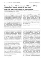
urgent chest CT angiography presented a huge non-
homogenous tumour occupying almost the entire right
atrium and partially invading the right ventricle. The
CT showed no liver tumour or other subdiaphragmatic
tumour extension. (Figure 1)
The patient underwent urgent surgical treatment due
to worsening of his clinical condition. The findings of
the CT guided our surgical strategy as follows: Initially
femoro-femoral cannulation was installed in order to
commence cardiopulmonary bypass (CPB). Thus the
pericardial cavity co uld be approached with safety. After
median sternotomy, the superior vena cava was also
cannulated and transfixed and then antegrade cardiople-
gia was administered. The heart was cooled down to 30°
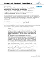
C, the right at rium was incised and the large tumor was
carefullyandcopiouslydissectedfromthesurrounding
tissues due to its friability. (F igure 2) The tumor origi-
nated mostly from the inferior vena cava and its term-
inal end w as inside the right ventricle. The tricuspid
valve was also invaded. The tumor was removed using a
valve sparing technique. It was cautiously dissected from
* Correspondence:
2
Department of Cardiothoracic Surgery, 401 Army General Hospital, Athens ,
Greece
Full list of author information is available at the end of the article
Dedeilias et al. Journal of Cardiothoracic Surgery 2011, 6:102
/>© 2011 Dedeilias et al; licensee BioMed Central Ltd. This is an Open Access article distributed under the terms of the Creative
Commons Attribution Li cense ( y/2.0), which permits unrestricted use, distribution, and
reproduction in any medium, provided the original work is properly cited.
the tricuspid valve and the right ventricular e ndocar-
dium ensuring that no remnants were left behind both
on the tricuspid valvular cusps and within the vicinity of
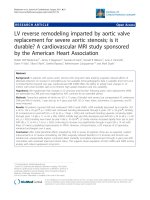
the right ventricle. The specimen was histopathologically
investigated and eventually diagnosed as a metastatic
hepatocellular carcinoma (HCC). (Figure 3)
Recovery was uneventful. Follow up echocardiography
and cardiac MRI two months after surgery did not
demonstrate any tumor recurrence or tricuspid regurgi-
tation. Additionally PET CT and abdominal MRI
showed no primary hepatoma or metastasis elsewhere in
the body. No further adjuvant therapy was c onsidered
necessary in this stage by our consulting oncologists.
The patient was suggested to be under closed follow-up
with rad iology studies (MRI, CT, and PET) in order to
promptly detect solitary masses might be respectable
but unfortunately he refused any other treatment option
and returned to his country.
Discussion
Primary liver cancer is the fifth most common neoplasm
with an incidence of 5.5-14.9% of all tumours. Predis-
posing factors for orthotopic primary Hepatocellular
Carcinoma (HCC) generation are chronic hepatitis B or
C, infection and cirrhosis secondary to other chronic
liver disease. Worldwide, the majority of patients with
HCC have underlying cirrhosis, and it is uncommon to
find HCC in patients without cirrhosis. Among rare
cases with HCC without underlying cirrhosis, HCV
infection accounted for 3-54%, HBV infection for 4-29%,
and hea vy alcohol intake for 0-28% [ 1]. Although HCC
has a very aggressive metastatic profile, i ts tendency to
spread towards the heart is unusual but well documen-
ted through several published case reports which define
an incidence of cardiac metastasis at 0.67-3% [2,3].
However very few cases of giant metastatic HCC within
the r ight cardiac cavities that cause significant occlusion
of the tricuspid valve are described in the current
literature.
Therefore an interesting feature of HCC can be its
varied and sometimes bizarre presentation [4]. This
report describes an unusual presentation of HCC. The
patient appeared with symptoms of acute heart failure
caused by a giant right atrial malignant obstructive
hepatocellular mass without any detectab le cancerous
lesions in the liver. There was no radiological, clinical or
laboratory suspicion of HCC. Metastatic HCC was only
apparent on histology examination of the right atrial
tumor. Metastatic disease as the initial presentation of
HCC appears in less than 5% of cases [5]. In addition
histological investigation defines whether the mass
Figure 1 CT angiogram verifying the presence of a mass inside
the right atrium occupying almost the whole cavity.
Figure 2 The right atrium incised and the exposed tumor.
Figure 3 The tumor specimen.
Dedeilias et al. Journal of Cardiothoracic Surgery 2011, 6:102
/>Page 2 of 4
derives from an occult HCC or is presented as an ecto-
pic one with no liver involvement.
Regarding the symptoms, there is a variety of clinical
manifestations caused by the atrial neoplasm and those
are mainly tumor-size dependent. Patients may have no
symptoms, dyspnea due to pulmonary embolism, syn-
cope, or heart failure. Physical findings include edema,
pan systolic murmur with diastolic rumble over the tri-
cuspid valve, and improvement of symptoms with left
lateral decubitus position [6].
Extracardiac tumours involvin g inferior vena cava and
right atrium include renal cell tumour (4-10%) [7], thyr-
oid carcinoma, testicular tumours and HCC. In m ost
cases of advanced HCC the extent of the disease is veri-
fied with presence of me tastasis at the lungs, peritoneum,
adrenal glands and bones. Generally hepatocellular carci-
noma appears to ha ve a tendency to invade vascular
structures [8]. Extension to the portal vein system is
common as opposed to extension into the inferior vena
cava or the right atrium which is uncommon [2]. When
this occurs, it mostly happens through the hepatic veins
and the inferior vena cava towards the right atrium. A
right atrial intracavitary massisthenformattedwhich
causes significant hemodynamic instability. In addition,
left atrium, right ventricle, and intramyocardial involve-
ment of the left ventricle have also been reported as rare
sites of HCC metastasis as well as spreading of the cancer
to the left chambers through pulmonary metastasis or
patent foramen ovale [9].
Regarding the case described here, the appearance of a
metastatic HCC tumor inside the right atrium as the
only manifestation and without apparent primary focus
is unique. The chest C T angiography was the most
important diagnostic modality in this case. It was very
helpful in identifying the borders of the tumor. There
are various sub diaphragmatic tumors including renal
cell tumors and HCC which extend from below the dia-
phragm up to the right chambers of the heart either
through the venous system or through diaphragm inva-
sion. It is important to know the t umor location before
bicaval cannulation to prevent fragmentation and embo-
lisation of the tumor. The understanding that the tumor
was well confined inside the righ t atrium was important
for the correct planning of the procedure. CT angio-
gram allowed for correct placement of the arterial and
venous cannulas. Thus, the arterial and one venous can-
nula were placed inside the femoral vessel s and the uti-
lity o f femoro femoral by pass circuit allowed opening
of the chest with optimal safety. The other venous can-
nula was placed inside the superior vena cava. The right
atrium was left without any cannulas in o rder to avoid
any contact with the friable mass and minimise the risk
for pulmonary embolism [10].
Intra atrial manifestation of the HCC constitutes a life
threatening condition. The major causes of death are
either sudden pulmonary embolism of the thrombus or
acute obstruction of the tri cuspid valve or bot h. Resec-
tion can provide relatively good mid-term survival
regarding this clinical situation but not more than 2
years [11]. Standard treatment is hepatic resection with
removal of the intracardial mass usually under cardiopul-
monary bypass with deep hypothermia and circulatory
arrest which seems to be the optimal option in most
cases. A few reports describe the successful removal of
HCC from the right atrium without extracorporeal circu-
lation as an alternative [12]. However, both curative
resection treatments have a dismal prognosi s, with a 5
years reported survival around 12-39% [3].
After resection of a hepatocellular carcinoma, tumour
recurrence exceeds up to 70% at 5 years, including
recurrence due to dissemination and de novo tumours
of the liver. The most important statistically predictor of
recurrence seems t o be the presence of micro vascular
invasion and/or additional tumour sites besides the pri-
mary lesion [13]. There is no effective adjuvant therapy
that can reduce t he recurrence rates . (Recommendation
level II) [13,14]
Internal radiation and adoptive immunotherapy by
activated lymphocytes may have some anti-tumor efficacy
but the early results have not been statistically powered
as yet [15]. There are no adequate published data to indi-
cate proper treatment of recurrences. Solitary recurrent
masses might benefit from repeat resection but in the
majority of cases recurrence appears to be multifocal and
so further treatment is impossible [13].
Conclusion
In conclusion, w hen a patient with a history of chronic
hepatic disease presents with symptoms of right heart
failure one must be cautious and should bear in mind
that right heart involvement from a malignant tumour
may be present [16]. Echocardiography computed tomo-
graphy and magnetic resonance imaging are the stan-
dard imaging modalities to determine the nature of
tumors presented as secondary cardiac neoplasms
[16,17]. Urgent Computed tomography can easily and
quickly be performed prior to surgical treatment of
emergency cases. Ultrasound w ith liver-specific micro
bubbles and PET CT can be helpful in certain cases of
occult HCC [18,19].
Consent
Written informed consent was obtained from the patient
for publication of this Case report and any accompany-
ing images. A copy o f the written consent is available
for review by the Editor-in-Chief of this journal.
Dedeilias et al. Journal of Cardiothoracic Surgery 2011, 6:102
/>Page 3 of 4
Author details
1
1
st
Department of Cardiac Surgery, Evangelismos General Hospital, Athens,
Greece.
2
Department of Cardiothoracic Surgery, 401 Army General Hospital,
Athens, Greece.
3
Radiology Department, Evangelismos General Hospital,
Athens, Greece.
4
Anaesthesiology Department, Evangelismos General
Hospital, Athens, Greece.
5
Cardiology Department, Kalamata General
Hospital, Kalamata, Greece.
6
Cardiothoracic Department, University Hospital
of Ioannina, Ioannina, Greece.
Authors’ contributions
PD: Has made substantial contributions to conception and design,
acquisition of data and analysis and interpretation of data. Also, has given
final approval of the version to be published. IN: Has made substantial
contributions to acquisition of data. IK: Has been involved in drafting the
manuscript and revising it critically for important intellectual content. VA:
Has made substantial contributions to acquisition of data. NP: Has made
substantial contributions to acquisition of data. SZ: Has made substantial
contributions to acquisition of data. EA: Has made substantial contributions
to conception and design.
All authors read and approved the final manuscript.
Competing interests
The authors declare that they have no competing interests.
Received: 21 February 2011 Accepted: 26 August 2011
Published: 26 August 2011
References
1. Llovet JM, Bourroughs A, Bruix J: Hepatocellular carcinoma. Lancet 2003,
363:1907-1917.
2. Masci G, Magagnoli M, Grimaldi A, Covini G, Carnaghi C, Rimassa L,
Santoro A: Metastasis of Hepatocellular Carcinoma to The Heart: a Case
Report and Review of The Literature. Tumori 2004, 90:345-7.
3. Afonso DV, Laranjeira A, Galrinho A, Fragata J: Metastatic hepatocellular
carcinoma: right atrial tumor as primary clinical manifestation. Case
report. Rev Port Cir Cardiotorac Vasc 2008, 15(2):79-81.
4. Sabir AA, Banoo T, Al Haj OB, Fouad Sedky AA, Hamid TA, Mahrous AR:
Metastatic hepatocellular carcinoma with occult primary presentation: A
case report. Saudi J Gastroenterol 1997, 3:49-52.
5. Karim RZ, Greenberg ML: Occult hepatocellular carcinoma metastasising
to the left temple diagnosed by fine-needle aspiration biopsy. Diagn
Cytopathol 2006, 34(6):430-3.
6. Hayashi P, Trotter JF, Everson GT: Hepatocellular carcinoma extension into
the right atrium. Liver Transpl 2003, 9(11):1225-6.
7. Zini L, Haulon S, Decoene C, Amara N, Villers A, Biserte J, Leroy X, Koussa M:
Renal Cell Carcinoma Associated with tumor Thrombus in The Inferior
Vena Cava: Surgical Strategies. Ann Vasc Surg 2005, 19:522-8.
8. Kojiro M, Nakahara H, Sugihara S, Murakami T, Nakashima T, Kawasaki H:
Hepatocellular carcinoma with intra-atrial tumor growth. A
clinicopathologic study of 18 autopsy cases. Arch Pathol Lab Med 1984,
108:989-92.
9. Shyu KG, Chiang FT, Kuan PL, Lien WP, Chen CL, How SW: Cardiac
metastasis of hepatocellular carcinoma mimicking pericardial effusion
on radionuclide angiocardiography. Chest 1992, 10:261-262.
10. Dedeilias P, Koletsis E, Rousakis A, Kouerinis I, Zaragkas S, Grigorakis A,
Leivaditis V, Malovrouvas D, Apostolakis E: Deep hypothermia and
Circulatory arrest in the surgical management of renal tumors with
cavoatrial extention. J Cardiac Surg 2009, 24(6):617-23.
11. Elod Papp, Zsuzsanna Keszthelyi, Nagy Karoly Kalmar, Lajos Papp,
Csaba Weninger, Tamas Tornoczky, Endre Kalman, Kalman Toth,
Tamas Habon: Pulmonary embolization as primary manifestation of
hepatocellular carcinoma with intracardiac penetration: A case report.
World J Gastroenterol 2005, 11(15):2357-235.
12. Georgen M, Regimbeau JM, Kianmenesh R, Marty J, Farges O, Belghiti J:
Removal of hepatocellular carcinoma extending in the right atrium
without extracorporeal bypass. J Am Coll Surg 2002, 195:892-894.
13. Bruix J, Sherman M: Management of hepatocellular carcinoma. Hepatology
2005, 42:1208-36.
14. Schwartz JD, Schwartz M, Mandeli J, Sung M: Neoadjuvant and adjuvant
therapy for resectable hepatocellular carcinoma: review of the
randomized clinical trials. Lancet Oncol 2002, 3:593-603.
15. Muto Y, Moriwaki H, Shiratori Y: Prevention of secondary primary tumors
by an acyclic retinoid, polyprenoic acid, in patient with hepatocellular
cancer. N Engl J Med 1996, 334:1561-67.
16. Agelopoulou P, Kapatais A, Varounis C, Grassos C, Kalkandi E, Kouris N,
Pierakeas N, Babalis D: Hepatocellular carcinoma with invasion into the
right atrium. Report of two cases and review of the literature.
Hepatogastroenterology 2007, 54(79):2106-8.
17. Taoli B, Losada M, Holland A, Krinsky G: Magnetic resonance imaging of
hepatocellular carcinoma. Gastroenterology 2004, 127:144-52.
18. Harvey CJ, Lim AK, Blomley MJ, Taylor-Robinson SD, Gedroyc WM,
Cosgrove DO: Detection of an occult hepatocellular carcinoma using
ultrasound with liver-specific microbubbles. Eur Radiol 2002, 12(Suppl 3):
S70-3, Epub 2002 Aug 2.
19. Liangpunsakul S, Agarwal D, Horlander JC, Kieff B, Chalasani N: Positron
emission tomography for detecting occult hepatocellular carcinoma in
hepatitis C cirrhotics awaiting for liver transplantation. Transplant Proc
2003, 35(8):2995-7.
doi:10.1186/1749-8090-6-102
Cite this article as: Dedeilias et al.: Acute heart failure caused by a giant
hepatocellular metastatic tumor of the right atrium. Journal of
Cardiothoracic Surgery 2011 6:102.
Submit your next manuscript to BioMed Central
and take full advantage of:
• Convenient online submission
• Thorough peer review
• No space constraints or color figure charges
• Immediate publication on acceptance
• Inclusion in PubMed, CAS, Scopus and Google Scholar
• Research which is freely available for redistribution
Submit your manuscript at
www.biomedcentral.com/submit
Dedeilias et al. Journal of Cardiothoracic Surgery 2011, 6:102
/>Page 4 of 4