Báo cáo y học: "Total thoracoscopic lung segmentectomy of anterior basal segment of the right lower lobe (RS8) for NSCLC stage IA (case report)" doc
Bạn đang xem bản rút gọn của tài liệu. Xem và tải ngay bản đầy đủ của tài liệu tại đây (1.41 MB, 5 trang )
CAS E REP O R T Open Access
Total thoracoscopic lung segmentectomy of
anterior basal segment of the right lower lobe
(RS8) for NSCLC stage IA (case report)
Masahiro Miyajima, Atsushi Watanabe
*
, Mayuko Uehara, Takuro Obama, Junji Nakazawa, Tomohiro Nakajiima,
Keishi Ogura and Tetsuya Higami
Abstract
A 69-year-old woman with a pulmonary nodule in anterior basal segment of the right lower lobe (RS8) was
referred to our department. The diameter of the tumor was 12 mm, and it had increased over a few months. First,
video-assisted thoracoscopic lung surgery (VATS) biopsy of the pulmonary nodule was carried out. Frozen section
examination of this nodule confirmed the diagnosis of bronchioloalveolar carcinoma (BAC). Segmentectomy of RS8
with lower mediastinal node dissection (ND2a-1) was performed. The intersegmental plane was identified using the
intersegmental veins as landmarks and the demarcation between the resected (inflated) and preserved (collapsed)
lungs. Electrocautery at 70 watts was used to divide the intersegmental plane. A vessel sealing system was used to
seal and cut the pulmonary arteries. Postoperative histopathological examination revealed that the tumor was
T1aN0M0 BAC, and the minimal distance between the surgical margin and the tumor edge was 15 mm. The
patient was discharged from hospital on postoperative day 5 without any complications.
Background
Segmentectomy for non small cell lung cancer (NSCLC)
stage I patients still remains controversial. The late out-
comes of lung function and underlying diseases are
unclear. We are cu rrently loo king forward to the results
of the two ongoing randomized, controlled studies: a
study conducted by the Cancer and Leukemia Group B
(CALGB14053), a phase III randomized trial of lobect-
omy versus sublobar resection for small (< 2 cm)
NSCLC; and a similar phase III randomized study con-
ducted by the Japan Clinical Oncology Group (JCOG)
and the West Japan Oncology Group (WJOG)
(JCOG0802/WJOG4607L) [1]. There are few reports on
VATS segmentectomy [2,3]. The procedure has some
drawbacks: it is technically demanding, and it is difficult
to comprehend the anatomical relations among the
bronchus, pulmonary arteries and pulmonary veins. Pre-
operative three-dimensional contrast-enhanced com-
puted tomography (3D-CT) simulation and the use of a
vessel sealing system (VSS) to cut the vessels and dissect
the parenchyma make this complicated surgery easier
and more practical [4,5]. The case of a patient with
VATS anterior basal segment of the right lower lob e
(RS8) segmentectomy for stage IA NSCLC is presented.
Case Presentation
A 69-year-old woman who was diagnosed with a lung
tumor was admitted to our hospital. The greatest dia-
meter of the tumor was 12 mm, and it had increased
over several months. Bronchoscopy did not yield a defi-
nitive diagnosis, so Thoracoscopic surgery for diagnosis
and treatment was scheduled. Past medical history
included mitral valve insufficiency treated with oral
medication. Family history was unremarkable, and she
had never been a smoker. The physical examination was
normal. The results of the laboratory investigations,
including a complete blood count, liver and renal func-
tion tests, coagulation studies and the serum cancer
antigens, were within the normal range.
Pulmonary function tests showed that vital capacity
(VC) was 2590 ml, percentage of predicted VC was
109.3%, forced expiratory volume in 1 s (FEV1) was
2120 ml and FEV percentage in 1 s was 81.9%. Chest
* Correspondence:
Department of Thoracic and Cardiovascular Surgery, Sapporo Medical
University Chuo-ku, S1W16, Sapporo, Hokkaido0608543, Japan
Miyajima et al. Journal of Cardiothoracic Surgery 2011, 6:115
/>© 2011 Miyajima et al; licensee BioMed Central Ltd. This is an Open Access article distributed under the terms of the Creative
Commons Attribution License ( whi ch permits unrestricted us e, distribution, and
reproduction in any medium, provided the ori ginal work is prope rly cited.
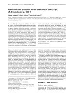
computed tomography demonstrated a partilally serrated
border 12-mm-diameter pulmonary nodule in anterior
basal segment of the right lower lobe (RS8)(Figure 1).
To guide the surgeons in simulating the operation, preo-
perative three-dimensional (3D)-CT was performed.
Using 3D volume rendering, a solid image was con-
structed from 0.65-mm data slices of the contrast-
enhanced CT images. A colored map was used to high-
light the blood vessels of the lung. The 3D rendered
images were magnified, de-magnified, and rotated to
examine these measurements (Figures 2, 3). To secure
an adequate margin from the tumor, preoperative needle
marking was performed under CT guidanc e on the day
before surgery. The needle marker (Guiding Marker Sys-
tem; Hakko Medical Products, Tokyo, Japan) was put
around the tumor [6].
VATS needle biopsy was then planned, with subse-
quent total thoracoscopic segmentectomy of anterior
basal segment of the right lower lobe (RS8) if the diag-
nosis was malignancy. Our indication criteria for seg-
mentectomy are clinical T1aN0M0 peripheral NSCLC.
The segments for resection are determined based on
tumor size and peripheral location in order to critically
secure a segmental margin free of tumor cells. Segmen-
tectomy is co nverted to lobectomy when the intraopera-
tive node sampling shows node involvement. Under
general anesthesia with single lung ventilation and thor-
acic epidural anesthesia, the patient was placed in the
left decub itus position. The surgeon was posit ioned on
the anterior side of the patient. Two thoracoports were
placed in the sixth intercostal space (ICS) at the anterior
axillary line and the seventh ICS at the posterior axillary
line. The anterolateral 30-mm access port was placed in
the fourth ICS. A Lap Protector Mini (Hakko Medical
Co., Tokyo, Japan) was placed on the site of the access
port. A 30-degree scope was used. The interlobula r f is-
sure was almost complete. Frozen section e xamination
of the needle bio psy specimen confirmed the diag nosis
Figure 1 Chest CT revealed a tumor (arrow) in anterior b asal
segment of the right lower lobe (RS8).
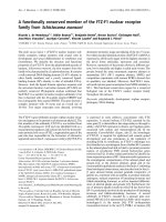
Figure 2 Images of the right lower pulmonary veins. On this image, the relationships of the intersegmental veins (V
8
a and V
8
b) that demark
anterior basal segment (S8) and lateral basal segment (S9) and the veins (V
7
and V
9
b) that should be preserved can be clearly demarcated.
Miyajima et al. Journal of Cardiothoracic Surgery 2011, 6:115
/>Page 2 of 5
of BAC, and VATS segmentectomy of RS8 with node
dissection was performed. First, the intermediate pul-
monary artery, the middle lobe pulmonary artery, A6,
A7, and A8 were exposed in the interlobar site. Proxi-
mal A8 was ligated with 2-0 silk, and distal A8 was
sealed and then divided with the VSS. The
intersegmental plane was identified using the interseg-
mental veins as landmarks and the demarcation between
the resected (inflated) and preserved (collapsed) lungs.
This status of the lung was induced by the following
three steps: temporarily re-inflating the whole lung,
ligating the resected segmental bronchus (B8), and
deflating preserved lung. After making the demarcation,
ligated B8 was then stapled and divided with an endo-
scopic stapler. The intersegmental plane of the parench-
yma was divided by electrocautery by 70 Watts from the
pleural surface of the lung[7,8]. VSS was also used to
dissect along the intersegmental veins. The intersegmen-
tal veins (V
8
aandV
8
b) that demark S8 and lateral basal
segment (S9) were resected with RS8 after sealing and
division with the VSS, while the intersegmental vein
(V7) that demarks medial basal segment (S7) and S8
was preserved (Figure 4). Finally, air leaks were repaired
with several 4-0 PDS horizontal mattress sutures with
absorbable pledgets (Medifit Felt, JMS Co., Hiroshima,
Japan). Absorbable polyglycol acid sheets (NEOVEIL
®
sheet, Gunze, Ayabe, Japan) were applied to the inter-
segmental plane with fibrin glue to obviate further air
leaks.
The operative time was 221 min, and blood loss was
30 ml. The chest drainage tube was removed on pos t-
operative day 2. The patient was discharged from
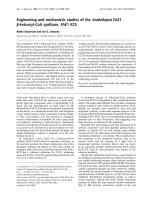
Figure 3 Images from three-dimensional computed tomographic angiography of the right lower lobe of the lung.Thecomplicated
anatomy of the pulmonary arteries (red), pulmonary veins (blue), and bronchus (green) is precisely depicted. From this image, the
intersegmental plane between medial basal segment (S7) and anterior basal segment (S8) can be easily imagined.
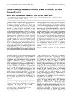
Figure 4 Operative view. On this view, the intersegmental veins
(V
8
a and V
8
b) that demark anterior basal segmet (S8) and lateral
basal segment (S9) can be seen to have been resected with the
VSS. The intersegmental vein (V
9
b) that should be preserved is well
identified.
Miyajima et al. Journal of Cardiothoracic Surgery 2011, 6:115
/>Page 3 of 5
hospital on postoperative day 5 without any complic a-
tions. Postoperative histopathological examination
revealed that the tumor was T1aN0M 0 BAC. The mini-
mal distance between the surgical margin and the tumor
edge was 15 mm, and the margin was free of
malignancy.
Discussion
Along with the recent deve lopment of radiographic
devices such as high-resolution computed tomography
and the widespread practice of low-dose helical com-
puted tomography for screening, early detection of ever-
smaller NSCLS has increased.
It remains to be established whether segmentectomy is
an appropriate procedure for NSCLC patients who can
tolerate lobectomy. The potential advant age of segmen-
tectomy compared with lobectomy is the preservation of
pulmonary function, whereas in comparison with wedge
resection, an improved oncologic outcome is noted with
segmentectomy [9]. This concept may be addressed by a
study conducted by the Cancer and Leukemia Group B
(CALGB14053), a phase III randomized trial of lobect-
omy versus sublobar resection for small (< 2 cm)
NSCLC.
Our indication criteria for segmentectomy are clinical
T1aN0M0 peripheral NSCLC. The segments for resec-
tion are determined based on tumor size and peripheral
location in order to critically se cure a segmental margin
free of tumor cells. The segmental, lobar and mediast-
inal nodes should be carefully sampled and confirmed
to be cancer negative by frozen section examination.
Segmentectomy is converted to lobectomy when the
intraoperative node sampling shows node involvement.
Reports on thoracoscopic segmentec tomy were limited
to segments that can be easily excised. In case of such
segments, i t is possible to simultaneously separate the
lung parenchyma from both the hilum and the periph-
ery by using staplers. Segmentectomy used to be diffi-
cult for the other segments. In such segments, digital
dissection of the segments had been necessary. Owing
to the recent development of the pre-operative 3D-CT
and the improved quality and resolution of scans thus
obtained, total thoracoscopic lung segmentectomy of
these segments have been reported[4,8].
In the present case, as a result of the pre-operative
3D-CT simulation, we could comprehend the precise
anatomy of the complicated vesse ls and the bronchi.
Especially, it is important to identify t he intersegmental
veins for total thoracoscopic lung segmentectomy[4].
In the limited working space, usage of the VSS was
very safe and useful to ex pose and divide pulmonary
vessels. During a total thoracoscopic lung segmentect-
omy, suture ligation of PA and the treatmen t of intrao-
perative bleeding can be more challenging than during
segmentectomy by a video-assisted mini-thoracotomy. It
is assumed that energy-based vessel sealing and cutting
instruments reduced difficulties in dividing pulmonary
blood vessels in total thoracoscopic lung segmentect-
omy. Compared to VSS, the ultrasonic device consis-
tently generates higher temperatures (200°C vs 94.3°C).
It has been reported that compared to the VSS, the
ultrasonic device required twice as long to cool off, and
the mean Burst Pressure was lower[10]. Therefore, for
use in limited thoracic space, and for sealing and cutting
of pulmonary blood vessels, VSS is more suitable.
Once the intersegmental plane has been determined,
the last issue is the choice of the segmental division
method. Some including us use a combination of elec-
trocautery and application of fibrin sealant[7,8]. But
most use staplers[2,3,9]. The application of stapling can
often compromise adjacent pulmonary parenchyma,
restricting full expansion of the residual segments and
thus pulmonary function. On the other hand, post
operative air leakage was the major problem when using
an electric cautery. In patients with severe emphysema-
tous changes, stapling device may be applied for strin-
gent control of air leaks.
We believe that this technique will contribute to
improved outcomes in selected lung cancer patients.
Conclusion
With the help of pre-ope rative 3D-CT simulation of the
complicated vesse ls and the bronchi, as well as VSS to
expose and divide pulmonary vessels, total thoracoscopic
segmentectomy of anterior b asal segment of t he right
lower lobe (RS8) was safely performed.
Acknowledgements
The authors received no financial support.
Authors’ contributions
MM conceived of the study, drafted the manuscript, and participated in its
design and coordination.
AW conceived of the study, and participated in its design and coordination.
MU participated in this surgical operation and took care of the patient.
TO participated in this surgical operation and took care of the patient.
JN participated in this surgical operation and took care of the patient.
TN participated in this surgical operation and took care of the patient.
KO carried out the pre-operative 3D-CT imaging.
TH participated in its design and coordination.
All authors read and approved the final manuscript.
Competing interests
The authors declare that they have no competing interests.
Received: 6 June 2011 Accepted: 24 September 2011
Published: 24 September 2011
References
1. Nakamura K, Saji H, Nakajima R, et al: A phase III randomized trial of
lobectomy versus limited resection for small-sized peripheral non-small
cell lung cancer (JCOG0802/WJOG4607L). Jpn J Clin Oncol 2010,
40:271-274.
Miyajima et al. Journal of Cardiothoracic Surgery 2011, 6:115
/>Page 4 of 5
2. Oizumi H, Kanauchi N, Kato H, et al: Total thoracoscopic pulmonary
segmentectomy. Eur J Cardiothorac Surg 2009, 36:374-377.
3. Gossot D, Ramos R, Braian E, et al: A totally thoracoscopic approach for
pulmonary anatomic segmentectomies. Interact Cardiovasc Thorac Surg
2011, 12:529-32.
4. Oizumi H, Endoh M, Takeda S, et al: Anatomical Lung Segmentectomy
Simulated by Computed Tomographic Angiography. Ann Thorac Surg
2010, 90:1382-1383.
5. Nakamoto K, Omori K, Nezu K: Superselective segmentectomy for deep
and small pulmonary nodule under the guidance of three-dimensional
reconstructed computed tomographic angiography. Ann Thorac Surg
2010, 89:877-884.
6. Nakashima S, Watanabe A, Obama T, et al: Need for preoperative
computed tomography-guided localization in video-assisted
thoracoscopic pulmonary resection of metastatic pulmonary nodules.
Ann Thorac Surg 2010, 89:212-218.
7. Watanabe A, Ohori S, Nakashima S, et al: Feasibility of video-assisted
thoracoscopic surgery segmentectomy for selected peripheral lung
carcinomas. Eur J Cardiothorac Surg 2009, 35:775-780.
8. Okada M, Mimura T, Ikegaki J, et al: A novel video-assisted anatomic
segmentectomy technique: selective segmental inflation via
bronchofiberoptic jet followed by cautery cutting. J Thorac Cardiovasc
Surg 2007, 133:753-758.
9. Atkins B, Harpole D, Mangum J, et al: Pulmonary segmentectomy by
thoracotomy or thoracoscopy: reduced hospital length of stay with a
minimally-invasive approach. Ann Thorac Surg 2007, 84 :1107-1113.
10. Kim J, Chammas F, Gewehr E, et al: Temperature safety profile of
laparoscopic devices: Harmonic ACE (ACE), Ligasure V (LV), and plasma
trisector (PT). Surg Endosc 2008, 22:1462-1469.
doi:10.1186/1749-8090-6-115
Cite this article as: Miyajima et al.: Total tho racoscopic lung
segmentectomy of anterior basal segment of the right lower lobe (RS8)
for NSCLC stage IA (case report). Journal of Cardiothoracic Surgery 2011
6:115.
Submit your next manuscript to BioMed Central
and take full advantage of:
• Convenient online submission
• Thorough peer review
• No space constraints or color figure charges
• Immediate publication on acceptance
• Inclusion in PubMed, CAS, Scopus and Google Scholar
• Research which is freely available for redistribution
Submit your manuscript at
www.biomedcentral.com/submit
Miyajima et al. Journal of Cardiothoracic Surgery 2011, 6:115
/>Page 5 of 5