Báo cáo y học: " Surgical treatment of aortobronchial fistula after thoracic endograft failure" pptx
Bạn đang xem bản rút gọn của tài liệu. Xem và tải ngay bản đầy đủ của tài liệu tại đây (938.66 KB, 4 trang )
CAS E REP O R T Open Access
Surgical treatment of aortobronchial fistula after
thoracic endograft failure
Angelo Maria Dell’Aquila
1*
, Stefano Mastrobuoni
2
, Alina Gallo
1
, Isidro Olavide
3
and Alejandro Martin-Trenor
2
Abstract
Endovascular stent grafting has been recently considered as a less invasive alternative to either medical therapy or
open surgical treatment for many patients with descending thoracic aortic disease. Late complications are rarely
described in literature. Herein, we described the occurrence of an aorto-bronchial fistula and a retro-A dissection in
a 73-year-old man after stent-graf ting for a penetrating atherosclerotic ulcer (PAU) of the descending thoracic aorta
and the successful surgical technique adopted in order to remove the stent-graft.
Keywords: bronchial fistula, aortic dissection, aortic ulcer, endovascular stent
Background
Endovascular stent grafting has been considered as a
less invasive alternative to either medical therapy or
open surgical treatment for many patients with descend-
ing thoracic aortic disease. However, the Expert Consen-
sus Document on the Treatment of Descending
Thoracic Aortic Disease Using Endovascular Stent-
Grafts has recently declared t hat, despite reasonably low
early operative morbidity and mortality, late complica-
tions of thoracic aortic stent grafting are much more
common than those reported for the open aortic surgery
[1]. Thus, it is not clear at this time whether the trend
toward more aggressive endovascular stent-graft man-
agement will affect prognosis, freedom from aortic com-
plications and survival, compared with conventional
open surgical repair or medical management alone. To
date, late complications described in literature after
endovascular stent grafting include endoleaks, graft
migration, stent f ractures and a neurysm-related death
(such a s aneurysm rupture and fistulation). Nowadays,
the lack of standard surgical protocols and a poor litera-
ture raise concerns about how to deal with these com-
plications. Herein, we described a case of aorto-
bronchial fistula after endovascular stent implantation
and the successful surgical strategy in order to remove
the stent.
Case presentation
A 73-year-old man with a history of smoking and hyper-
tension was admitted to his referring hospital with ches t
pain and dyspnea. Computed tomogra phy (CT) revealed
a penetrating atherosclerotic ulcer (PAU) with intra-
mural hematoma in t he distal part of t he aortic arch
and left hemothorax. Antihypertensive therapy was
promptly instituted. A bypass between the left and right
carotid arteries was performed and the intimal ulcer was
covered by the stent-graft (Zenith Cook 36 mm) in
supra-subclavian landing zones; its exclusion was con-
firmed by the postoperative angiography.
The postoperative course was uneventful and the
patient was discharged home on postoperative day 8.
Threemonthsafterhisdischarge,theonsetofnausea
and hemoptysis required emergent hospitalization.
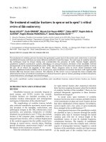
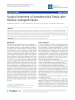
CT scan showed a retro-A dissection with partially
thrombosed false lumen in ascending aorta [Figure 1],
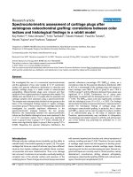
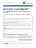
extravasation o f contrast into perigraft space with a big
periaortic hematoma in the area of the distal portion of
the stent graft [Figure 2], left apical lung hemorrhage
and hemothorax.
The patient was referred to our hospital for an emer-
gent surgical approach.
The operation was performed with a single-stage
approach via bilateral anterior thoracosternotomy. Car-
diopulmonary bypass was established using the r ight
axillary artery and right atrium. A clamp was placed on
the distal ascending aorta and the ascending aorta was
incised. No entry tear was found; the false lumen was
* Correspondence:
1
Department of Cardiac Surgery, San Martino University Hospital, l.go R.
Benzi 10, 16132, Genova, Italy
Full list of author information is available at the end of the article
Dell’Aquila et al. Journal of Cardiothoracic Surgery 2011, 6 :134
/>© 2011 Dell’Aquila et al; licensee BioMed Central Ltd. This is an Open Access article distributed under the terms of the Creative
Commons Attribution License ( which permits unrestricted use, distribution, and
reproduction in any medium, provided the original work is properly cited.
partially thrombosed. Cold intermittent blood cardiople-
gia was delivered antegradely. Once the aortic valve was
resuspended and proximal anastomosis was performed
with a 30 mm Dacron graft (Hemashield Gold; Boston
Scientific Medi-Tech. Wayne, NJ, USA), cooling was
initiated in case of circulatory arrest. Once a deep
hypothermia (20° C) was reached, brachiocephalic trunk,
the left common carotid artery and the descending aorta
at level of the diaphragm were clamped and a modified
cardiopulmonary bypass was performed starting the flow
also through a second femoral artery line. After the left
phrenic and left vagus nerves were identified, the aortic
arch and the descending aorta were incised and the
stent graft was removed. After the completion of the
distal anastomosis with a Dacron graft (Hemashield
Gold 26 mm), the two grafts were end-to-end sutured.
The distal clamp was removed and coronary perfusion
was reestablished through the femoral artery line. Perfu-
sate flow was increased and rewarming was initiated. A
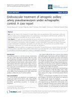
20 × 10 mm bifurcated Dacron graft was anastomosed
in an end-to-side f ashion to the ascending aorta, the
brachiocephalic trunk, and the left common carotid
artery. Antegrade cardiopulmonary bypass was restarted
[Figure 3].
The postoperative period was uneventful excepted for
the presence of prolonged pulmonary air leakage. The
patient was discharged on postoperative day 35. At 3
month follow up, a contrast-enhanced thoracic CT
showed the image of a pseudoaneurysm with a maxi-
mum diameter of 75 mm developed at the l evel of the
distal anastomosis. The patient underwent aortic stent
grafting (William Cook Europe) without complications.
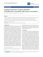
At 2 years follow up a CT showed the occlusion of the
by-pass between the two carotids [Figure 4]. At this
Figure 1 Three-dimensional computed tomographic
reconstruction demonstrating the retro-A dissection.
Figure 2 Computed tomographic showing the periaortic
hematoma.
Figure 3 Picture showing the operative strategy adopted in
order to remove the endograft and to replace the ascending
aorta, aortic arch, and descending aorta avoiding circulatory
arrest.
Dell’Aquila et al. Journal of Cardiothoracic Surgery 2011, 6 :134
/>Page 2 of 4
time, the patient was in optimal state of health and no
neurological episodes were reported.
Discussion
Despite recent l iterature suggesting a significant
improvement in outcomes with open surgical repair [2],
a less invasive approach for high-risk groups of patients
offers the potential for lower morbidity and mortality.
Stevenson et al report a significantly lower perioperative
mortality and complications rate in the endograft versus
the open-surgery control cohort [1]. A lthough results of
endovascular repair are promising, the authors stress the
importance of randomized long-term studies also
because the use of stent grafts is associated with early
and late unique complications that can be difficult to
manage [3].
These late complications often require different and
difficult approaches that have been partially faced by
surgeons using the frozen elephant-trunk via the tec hni-
que of median sternotomy in deep hypothe rmia and cir-
culatory arrest or via left thoracotomy using left heart
by-pass technique [4-6]. However, in presence of an
aorto-esophaegeal or an aorto-bronchial fistula the treat-
ment options are very limited [7,8].
In the present case report, considering the limited mobi-
lity of the patient due to knee arthrodesis and the advanced
age, a less invasive procedure was chosen as the best alter-
native to manage the PAU. The stent graft sealed the PAU
but two serious complications occurred: an aorto-bronc hial
fistula and a r etro-A dissection. W e believe that, because of
the poor flexibility of the stent graft, the distal uncovered
bare stent eroded the aortic w all causing the intramural
hematoma [Figure 2]. The haemoptysis observed three
months later was due to the con tinuous stress produced by
the expansive force of the stent against the intimal mem-
brane, resulting in leaking bloo d into the hema toma and
the left main stem bronchus. This hematoma partially lim-
ited the lo ss of b lood by covering th e leak.
In our case, the bilateral thoracosternotomy provided
an optimal exposure of as cending aorta, aortic arch and
epiaortic vessels.
The simultaneous cannulation of right axilary and
fem oral arteries facilitate the sequential clam p of differ-
ent aorta portions and avoid circulatory arrest maintain-
ing an optimal brain, renal and spinal cord perfusion.
Exceptionally, no selective brain perfusion was required
thanks to the previous carotid-carotid bypass.
Long-term durability of endografts remains unan-
swered; we think that patients with endoprosthesis must
be strictly followed-up and new standard protocols in
managem ent of complications need in order to establish
an optimal surgical approach.
Consent
Written informed consent was obtained from the patient
for publication of this case report and accompanying
images. A copy of the written consent is available for
review by the Editor-in- Chief of this journal.
Abbreviations
CT: Computed tomography; PAU: Penetrating atherosclerotic ulcer.
Author details
1
Department of Cardiac Surgery, San Martino University Hospital, l.go R.
Benzi 10, 16132, Genova, Italy.
2
Department of Cardiovascular Surgery,
University of Navarra, Clinica Universitaria, Avenida Pio XII, Pamplona, Spain.
3
Department of Anesthesiology. University of Navarra, Clinica Universitaria,
Avenida Pio XII, Pamplona, Spain.
Authors’ contributions
AMD conceived, supervise, literature research, wrote the article. AG
participated in its design, writing process and bibliography. AMT, SMT
Figure 4 Three-dimensional computed tomographic
reconstruction (2 years follow-up) demonstrating the occlusion
of the by-pass between the two carotids.
Dell’Aquila et al. Journal of Cardiothoracic Surgery 2011, 6 :134
/>Page 3 of 4
participated in its coordination and correction on the surgical part. IO, SMT;
AMT conceived participated in its coordination on the anesthesiologic and
extracorporal assistance part. All authors read and approved the final
manuscript
Competing interests
The authors declare that they have no competing interests.
Received: 3 July 2011 Accepted: 11 October 2011
Published: 11 October 2011
References
1. Svensson LG, Kouchoukos NT, Miller DC, Bavaria JE, Coselli JS, Curi MA,
Eggebrecht H, Elefteriades JA, Erbel R, Gleason TG, Lytle BW, Mitchell RS,
Nienaber CA, Roselli EE, Safi HJ, Shemin RJ, Sicard GA, Sundt TM, Szeto WY,
Wheatley GH, Society of Thoracic Surgeons Endovascular Surgery Task
Force: Expert consensus document on the treatment of descending
thoracic aortic disease using endovascular stent-grafts. Ann Thorac Surg
2008, 85:S1-41.
2. Coselli JS, LeMaire SA, Conklin LD, Adams GJ: Left heart bypass during
descending thoracic aortic aneurysm repair does not reduce the
incidence of paraplegia. Ann Thorac Surg 2004, 77:1298-303, discussion
1303.
3. Coady MA, Ikonomidis JS, Cheung AT, Matsumoto AH, Dake MD,
Chaikof EL, Cambria RP, Mora-Mangano CT, Sundt TM, Sellke FW, American
Heart Association Council on Cardiovascular Surgery and Anesthesia and
Council on Peripheral Vascular Disease: Surgical management of
descending thoracic aortic disease: open and endovascular approaches:
a scientific statement from the American Heart Association. Circulation
2010, 121:2780-2804.
4. Grabenwoger M, Fleck T, Ehrlich M, Czerny M, Hutschala D, Schoder M,
Lammer J, Wolner E: Secondary surgical interventions after endovascular
stent-grafting of the thoracic aorta. Eur J Cardiothorac Surg 2004,
26:608-613.
5. Neuhauser B, Greiner A, Jaschke W, Chemelli A, Fraedrich G: Serious
complications following endovascular thoracic aortic stent-graft repair
for type B dissection. Eur J Cardiothorac Surg 2008, 33:58-63.
6. Duebener L, Hartmann F, Kurowski V, Richardt G, Geist V, Erasmi A,
Sievers HH, Misfeld M: Surgical interventions after emergency
endovascular stent-grafting for acute type B aortic dissections. Interact
Cardiovasc Thorac Surg 2007, 6:288-292.
7. Isasti G, Gomez-Doblas JJ, Olalla E: Aortoesophageal fistula: an uncommon
complication after stent-graft repair of an aortic thoracic aneurysm.
Interact Cardiovasc Thorac Surg 2009, 9:683-684.
8. Yassin S, Marek J, Schwartz J, Wernly J, Dietl C, Pett S, Langsfeld M: Should
large mediastinal hematomas be drained after endovascular repair of
ruptured descending thoracic aorta? J Thorac Cardiovasc Surg 2007,
134:1040-1041.
doi:10.1186/1749-8090-6-134
Cite this article as: Dell’Aquila et al.: Surgical treatment of
aortobronchial fistula after thoracic endograft failure. Journal of
Cardiothoracic Surgery 2011 6:134.
Submit your next manuscript to BioMed Central
and take full advantage of:
• Convenient online submission
• Thorough peer review
• No space constraints or color figure charges
• Immediate publication on acceptance
• Inclusion in PubMed, CAS, Scopus and Google Scholar
• Research which is freely available for redistribution
Submit your manuscript at
www.biomedcentral.com/submit
Dell’Aquila et al. Journal of Cardiothoracic Surgery 2011, 6 :134
/>Page 4 of 4