Báo cáo y học: "Acute pressure overload of the right ventricle. Comparison of two models of right-left shunt. Pulmonary artery to left atrium and right atrium to left atrium: experimental study" doc
Bạn đang xem bản rút gọn của tài liệu. Xem và tải ngay bản đầy đủ của tài liệu tại đây (4.05 MB, 10 trang )
RESEARCH ARTIC LE Open Access
Acute pressure overload of the right ventricle.
Comparison of two models of right-left shunt.
Pulmonary artery to left atrium and right atrium
to left atrium: experimental study
Mihalis Argiriou
1*
, Dimitrios Mikroulis
2
, Timothy Sakellaridis
1
, Vasilios Didilis
2
, Apostolos Papalois
3
and
George Bougioukas
2
Abtract
Background: In right ventricular failure (RVF), an interatrial shunt can relieve symptoms of severe pulmonary
hypertension by reducing right ventricular preload and increasing systemic flow. Using a pig model to determine if
a pulmonary artery - left atrium shunt (PA-LA) is better than a right atrial - left atrial shunt (RA-LA), we compared
the hemodynamic effects and blood gases between the two shunts.
Methods: Thirty, male Large White pigs weighting in average 21.3 kg ± 0.7 (SEM) were divided into two groups
(15 pigs per group): In group 1, banding of the pulmonary artery and a pulmonary artery to left atrium shunt with
an 8 mm graft (PA-LA) was performed and in group 2 banding of the pulmonary artery and right atrial to left atrial
shunt (RA-LA) with a similar graft was performed. Hemodynamic parame ters and blood gases were measured from
all cardiac chambers in 10 and 20 minutes, half and one hour interval from the baseline (30 min from the
banding). Cardiac output and flow of at the left anterior descending artery was also monitored.
Results: In both groups, a stable RVF was generated. The PA-LA shunt compared to the RA-LA shunt has better
hemodynamic performance concerning the decreased right ventricle afterload, the 4 fold higher mean pressure of
the shunt, the better flow in left anterior descending artery and the decreased systemic vascular resistance.
Favorable to the PA-LA shunt is also the tendency - although not statistically significant - in relation to central
venous pressure, left atrial filling and cardiac output.
Conclusion: The PA-LA shunt can effectively reverse the catastrophic effects of acute RVF offering better
hemodynamic characteristics than an interatrial shunt.
Keywords: Right ventricular failure, Right ventricle overload, Pulmonary hypertension, Pulmonary artery banding,
Right to left shunt
Background
Pulmonary hypertension and right ventricular dysfunc-
tion are associated with poor survival. Management of
patients with acute decompensate RV failure is largely
empiric and targeted towards treating underlying preci-
pitants while optimizing conditions of RV preload, after-
load and contractility.
However, right-sided heart failure remains a major
problem in the long-term follow-up, leading to impair-
ment of functional status, severe arrhythmia, and pre-
mature death. Treatment consists of pulmonary
vasodilator therapy, long-term oxygen therapy, anticoa-
gulation, and lung transplantation, or, at times, heart-
lung transplantation. Management strategies for patients
who develop acute refractory right ventricular failure
are:
1. Mechanical support to the failing right ventricle,
* Correspondence:
1
Second Cardiac Surgery Department, Evaggelismos General Hospital, 45-47
Ipsilantou, 10676, Athens, Greece
Full list of author information is available at the end of the article
Argiriou et al. Journal of Cardiothoracic Surgery 2011, 6:143
/>© 2011 Argiriou et al; licensee BioMed Central Ltd. This is an Open Access article distributed und er the terms of the Creative C ommons
Attribution License ( which permits unrestricted use, distribution, and reproduction in
any medium, provide d the original work is properly cited.
2. Conventional pulmonary vasodilators,
3. Cavopulmonary diversion in select cases, and
4. Maintenance of an adequate left ventricular per-
formance throughout the recovery period [1].
In recent years, percutaneous balloon atrial septostomy
(BAS) has been established as a palliative treatment or
bridge to transplantation in patients with severe right-
heart failure refra ctory to conventional therapy [2-5].
BAS aims at creating a “safety valve” by unlo ading the
rightheartandincreasingleftventricularpreloadand
output, peripheral perfusion, net oxygen tissue delivery,
exercise tolerance, and prognosis. However, this proce-
dure is not always successful because the size of the
opening made with standard balloon septostomy techni-
ques is imprecise and variable from patient to patient.
The mortality rate is relatively high and sometimes
related to severe hypoxemia from excessive right-to-left
shunting through an overly large defect. Procedural mor-
tality varies w idely from 5 to 50% from single center
reports. Beside this procedure has been proposed a
“fontanisation” -right ventricular exclusion of the circula-
tion- as a surgical option of RVF [6,7]. Nevertheless the
presence of pulmonary hypertension is a contraindication
for this procedure. Neither experimental nor clinical data
are available regarding the effects of a shunt not at the
atrial level but from the pulmonary artery to the left
atrium. The purpose of this study was to examine the
effects of right ventricle overload of two different shunts
in a porcine model.
Materials and methods
Surgical Preparation
The animal research protocol was approved by the local
authorities (A.Π. 3940/6-10-2008) in Athens. All animals
used in this study were treated according to the “Guide for
the care and use of Laboratory animals” published by the
US National Institutes of Health (National Institutes of
Health publication no. 85-23, revised 1996).
Thirty pigs weighing 22 to 35 kg were premedicated
with ketamine hydrochloride (15 mg/kg IM) and midazo-
lam (0.5 mg/kg IM), anesthetized with thiopental sodium
(9 mg/kg IV bolus) and fentanyl citrate (0.5 mg IV bolus),
followed by continuous IV infusions of thiopental sodium
(1 mg/min), fentanyl citrate (4 mg/min), pancuronium
bromide (0.25 mg/min), and lidocaine (2 mg/min),
throughout the experiment. After intubation (8Ch),
respiration was controlled with a Soxitronic volume
respirator (Soxil S.P.A.; Segrate, Italy), supplying oxygen at
100%. No changes of tidal volume, respiratory rate, and
percentage of inspired oxygen were made.
Thechestwasopenedviaamidlinesternotomy,and
the heart was suspended in a pericardial cradle. Cathe-
ters were placed, in the right a trium via the right
external jugular vein which was surgically dissected; a
right side arterial line was inserted under direct vision
by a small incision in the groin, and in the left atrium
directly through the left atrial appendix. To the arterial
line was connected a FloTrac sensor also, (Vigileo moni-
tor, Edwards Lifesciences) to measures parameters such
asCCO,SVV/SV,SVR.Thissensorisachievingmea-
surements by pulse contour analysis based on arterial
pressure waveform. In this w ay it is possible to avoid
the use of Swan Ganz and consequently interactions
with tricuspid valve function. The proximal to mid left
anterior descending (LAD) coronary artery was dissected
free and, a transit time flow-meter probe, (Transonic
Inc. Ithaca New York 400-Series Multichannel Flow-
meter) was applied. The temperature of the animal was
kept within 0.5°C of the baseline value with a heating
blank et and lamp. ECG, for severe rhythm disturbances,
arterial pressure, central venous pressure, pulmonary
artery pressure and left atrial pressure were continuously
monitored. Fluid (Ringers lact ate) was given at a rate of
20 ml/kg.
Right ventricular failure model
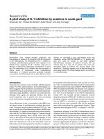
To achieve RVF a banding of the very distal main pulmon-
ary artery was performed. For banding we used a vessel
loop (nylon tape) with a snare (Figure 1). The banding was
persistent until pulmonary artery pressure proximal of the
banding was double than pressure distally of the banding.
RVF following pulmonary artery banding was defined as a
Figure 1 Schemati c diagram of the open-chest preparation.
Note the position of the pulmonary artery (PA) band (arrow). AO =
Aorta, RA = right atrium, LA = Left Atrium, RV = Right Ventricle, LV
= Left Ventricle.
Argiriou et al. Journal of Cardiothoracic Surgery 2011, 6:143
/>Page 2 of 10
profound decrease in systemic blood pressure [mean arter-
ial pressure (MAP) < 2/3 of the beginning], an initial > 1/3
increase of systolic right ventricular pressure (RVP) and a
depressed cardiac output (< 2/3 of the baseline). Addition-
ally, right ventricular function was judged by inspection.
After the completion of the banding, 30 min period was
allowed for the animal t o reach hemodynamic stability
before the baseline recordings of pressures, CO, LAD flow
and b lood gazes measures.
All measurements were taken at end expiration with
the ventilator turned off. Pulmonary artery band tight-
ness was adjusted so as not to allow the systolic arter ial
blood pressure to fall below 60 mmHg at anytime dur-
ing the experiment. With the beginning of the shunt
surgery, the animals were systemically heparinized
(100U/kg).
Experimental protocol
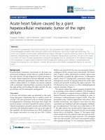
Two different settings of shunts were evaluated. Group
(1)PA-LAshunt(n15)andgroup(2)RA-LAshunt(n
15). A right atrial to the left atrial shunt was created
with an interposition of an 8 mm PTFE graft in group
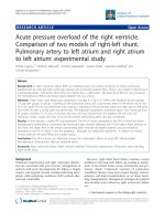
No 2 (Figure 2). By means of partia l vascular clamp a
PTFE 8 mm graft was connected end to side with the
very proximal main pulmonary artery (proximally of
the banding). The other side of the graft was con-
nected end to side with the left auriculum for group
No 1 (Figure 3).
We have chosen to introduce the 14-G hypodermic
needle into the left atrium, shunt, RV, pulmonary artery
proximal and distal directly rather than to introduce a
catheter Swan Ganz through the tricuspid valve because
of the enhanced stability and reproducibility of the pres-
sure and volumet ric data fr om “amorecompleteinter-
rogation of the RV cavity”. Blood gazes samples from
each cavity were selected directly from each cardiac
chamber at 10 and 20 minutes from the baseline.
Statistical Analysis
Data is expressed as mean ± standard deviation (S.D.) or
median (in case of violation of normality) for continuous
variables and as percentages for categorical data. The
Kolmogorov - Smirnov test was utilized for normality
analysis of the parameters. The comparison of variables
at each t ime point was performed using the Indepen-
dent samples t-test or the Mann-Whitney test in case of
violation of normality. One factor Repeated Measures
ANOVA model was used for the com parison of differ-
ent time measurement of varia bles for each group. Pair
wise multiple comparisons were performed using the
method of Tukey critical difference.
To indicate the trend in the first 20 minutes of inter-
vention, the median percentage changes after 10 and 20
minutes respectively are calculated. Comparison of per-
centage change from baseline of parameters during the
observation period between two groups was analyzed
using the Mann-Whitney test because of violation of
normality.
All tests are two-sided, statistical significance was set
at p < 0.05. All analyses were carried out using the sta-
tistical package SPSS ver. 16.00 (Statistical Package for
the Social Sciences, SPSS Inc., Chicago, Ill., USA).
Figure 2 a. Schematic diagram of the open-chest preparation with a right-left atrial shunt. b. Picture of the right-left atrial shunt in the
pig.
Argiriou et al. Journal of Cardiothoracic Surgery 2011, 6:143
/>Page 3 of 10
Results
Hemodynamics
The central venous pressure (mean), the mean pressure of
left atrium, the cardiac output, the pressure of the distal
portion of pulmonary artery at baseline and during 10 and
20 minutes interval were similar in both groups (Table 1).
There is statistically significant difference among the
time measurements of heart rate variable for the PA - LA
shunt, in comparison with the RA - LA shunt, especially
at the 10 minute interval (p < 0.005). Pairwise compari-
sons show statistically significant difference between all
time measurements. The heart rate variable at ba seline
was 95.5 ± 10.45 pulses/min, at 10 minutes interval with
PA - LA shunt was 112.80 ± 9.71 pulses/min and at
20 minutes interval 105.20 ± 16.90 pulses/min, whereas
with the RA - LA shunt the measurements of heart rate
variable at 10 and 20 minutes interval were 106.87 ±
18.31 pulses/min and 103.80 ± 13.52 pulses/min.
There is statistically significant difference among the
time measurements of m ean arterial blood pressure vari-
able for the PA - LA shunt , in comparison with the RA -
LA shunt between al time measurements (p < 0.005).
The mean blood pressure variable at baseline was 64.67 ±
6.72 mmHg, at 10 minutes interval with PA - LA shunt
decreased at a variable of 59.33 ± 14 .02 mmHg and at 20
minutes interval at 49.87 ± 10.08 mmHg, whereas with
the RA - LA shunt the measurements of mean blood
pressure variable at 10 and 20 minutes interval were
59.20 ± 10.71 mmHg and 58.60 ± 13.43 mmHg. Between
the two groups (PA - LA shunt and RA - LA shunt) there
is statistically significant difference of mean blood
pressure variable at 20 min interval (p = 0,054) with the
mean blood pressure of PA - LA shunt at the level of
49.87 ± 10.08 mmHg and of RA - LA shunt at the level
of 58.60 ± 13.43.
As for the mean right ventricular pressure (RVP) vari-
able, there is statistically significant difference among the
time measurements of the RVP variable for the shunt PA-
LΑ (p < 0.005). Pairwise comparisons show statistically
significant differe nce between all time measurements.
Also, between the two groups at 10 minute interval, a sig-
nificant statistically difference (p < 0.022) is observed with
the measurements to be 15.93 ± 4.73 mmHg for the shunt
PA-LΑ and 10.87 ± 3.60 mmHg for the shunt RA-LΑ.
Concerning the percentage change from baseline to 10
min of the mean right ventricular pressure variable, there
is statistical significant difference between the two groups
(p < 0.074), with 50% decrease at the PA-LA shunt and
64% decrease at the RA-LA shunt.
The variable of shunt pressure has statistically difference
between the two groups at 10 minute and 20 minute inter-
val (p < 0.005), whereas there i s a significant statistically
difference between groups concerning the percentage
change from baseline to 20 min (p = 0.023). Comparison
between all time measurements of proximal pulmonary
artery pressure for both groups reveals a statistically differ-
ence (p < 0.005).
TheobserveddecreaseofSVRhasastatisticallydiffer-
ence among the 20 minute interval measurements for the
shunt PA-LΑ andfortheRA-LAshunt(p<0.005).Also
there is statistically difference of SVR variable between
the two groups at 20 minute interval (p = 0.075) and
Figure 3 a. Schematic diagram of the open-chest preparation with a pulmonary artery - left atrial shunt. b. Picture of the pulmonary
artery -left atrial shunt in the pig.
Argiriou et al. Journal of Cardiothoracic Surgery 2011, 6:143
/>Page 4 of 10
Table 1 Hemodynamic measurements and statically analysis
baseline mean ± SD 10 min mean ± SD 20 min mean ± SD % change baseline-10 min median % change baseline-20 min median
Pulse (b/min) Shunt PA -LΑ 95.67 ± 10.45 112.80 ± 9.71** 105.20 ± 16.90 18.75 9.28
Shunt RA -LΑ 95.67 ± 10.45 106.87 ± 18.31* 103.80 ± 13.52 8.69 9.18
p-value NS NS NS NS NS
Arterial Blood pressure (mean) Shunt PA -LΑ 64.67 ± 6,72 59.33 ± 14.02 ** 49.87 ± 10.08 ** -6.34 -20.31
Shunt RA -LΑ 64.67 ± 6,72 59.20 ± 10.71 58.60 ± 13.43 -7.04 -14.23
p-value NS NS 0.054 NS NS
Right Ventricular pressure (mean) Shunt PA -LΑ 30.00 ± 4.42 15.93 ± 4.73** 12.53 ± 4.49** -50.0 -60.0
Shunt RA -LΑ 30.00 ± 4.42 10.87 ± 3.60** 13.5 ± 5.12** -64.0 -65.0
p-value NS 0.022 NS 0.074 NS
Central Venous pressure (mean) Shunt PA -LΑ 6.93 ± 2.40 5.21 ± 2.99* 5.12 ± 3.00 -25.0 0.0
Shunt RA -LΑ 6.93 ± 2.40 6.53 ± 2.72
$
3.46 ± 3.52* 0.0 -50.0
p-value NS NS NS NS NS
Left Atrial pressure (mean) Shunt PA -LΑ 5.67 ± 3.29 5.93 ± 3.51 5.93 ± 3.10 0.0 0.0
Shunt RA -LΑ 5.67 ± 3.29 4.73 ± 3.03 4.47 ± 3.02 0.0 -18.0
p-value NS NS NS NS NS
Pulmonary artery pressure (proximal) Shunt PA -LΑ 36.73 ± 5.28 21.73 ± 8.91** 21.63 ± 92.28** -40.0 -40.0
Shunt RA -LΑ 36.87 ± 4.70 29.03 ± 8.10** 28.90 ± 8.10** -20.0 -21.0
p-value NS NS NS NS NS
Pulmonary artery pressure (distal) Shunt PA -LΑ 12.73 ± 5.28 12.47 ± 4.81 12.40 ± 4.61 0.0 0.0
Shunt RA -LΑ 17.13 ± 5.25 16.80 ± 4.83 17.27 ± 5.36 0.0 0.0
p-value NS NS NS NS NS
Shunt pressure Shunt PA -LΑ 19.60 ± 4.94 19.93 ± 4.83 -5.0
Shunt RA -LΑ 5.53 ± 1.40 4.87 ± 1.36 -14.3
p-value p < 0,0005 p < 0,0005 0.023
CO Shunt PA -LΑ 4.93 ± 0.90 5.19 ± 1.22 5.39 ± 1.34 3.92 14.03
Shunt RA -LΑ 4.93 ± 0.90 4.64 ± 1.02 4.87 ± 1.13 -7.69 -2.33
p-value NS NS NS NS NS
SVR Shunt PA -LΑ 962.02 ± 153.04 847.17 ± 207.17*
$
667.97 ± 207.64** -15.44 -29.90
Shunt RA -LΑ 962.02 ± 153.04 891.98 ± 221.52 815.47 ± 213.14** -6.20 -14.81
p-value NS NS 0.075 NS 0.021
Flow LAD Shunt PA -LΑ 18.43 ± 6.83 16.14 ± 4.28 20.93 ± 7.35 -8.3 9.1
Shunt RA -LΑ 18.43 ± 6.83 14.64 ± 5.37* 10.79 ± 4.98** -28.0 -33.3
p-value NS NS p < 0,0005 NS p < 0,0005
** p < 0.005 vs. baseline, * p < 0.05 vs. baseline, $ p < 0.05 vs. 20 min
Argiriou et al. Journal of Cardiothoracic Surgery 2011, 6:143
/>Page 5 of 10
there is statistical significant difference between the two
groups concerning the percentage change from baseline
to 20 minutes of the SVR variable (p = 0.021).
Another important variable that was measured was the
flow at the LAD. Measurements revealed statistically sig-
nificant difference among the time measurements of the
LAD flow variable for the shunt RA-LΑ (p < 0.005) at 20
minute interval, with a 33.3% decrease. Between the two
groups, at 20 minutes interval, the observed difference is
statistically significant (p < 0.005). Finally, the observed
LAD flow variable between the two groups at 20 minutes
has a significant statistically difference (p < 0.005).
Blood gases
The statistical analysis of blood gases in both groups of
shunt and at all time intervals revealed no statistically
difference for arterial pCO
2
and arterial pO
2
,arterial
O
2
% saturation, pulmonary artery pH, pCO
2
of pulmon-
ary artery and O
2
% saturation of left atrium (Table 2).
The decrease of pO
2
in the pulmonary artery is statisti-
cally significant among the time measurements of the pO
2
variable for the shunt PA-LΑ (p < 0.005). Pairwise com-
parisons show stat istically sig nificant difference between
all time measure ments. The same observations are made
for the decrease of O
2
% saturation of the pulmonary
artery.
pCO
2
of the left atrium increase is statistically significant
between the two group s at 10 minute interval (p = 0.052)
and at 20 minute interval (p = 0.058). Τhere is also a sta-
tistical significant difference between groups concerning
the percentage ch ange from baseline to 10 minute of the
pCO
2
of the left atrium variable (p = 0.016) and the per-
centage change from baseline to 20 minute of the pCO
2
of
the left atrium variable (p = 0.023).
Least, the pO
2
of the left atrium decrease reveals a statis-
tically significant difference among the time measurements
for the shunt PA-LΑ (p < 0.005) and RA-LA shunt. Pair-
wise comparisons show statistically significant difference
between all time measurements. At 10 minute interval,
between the two groups there is a statistically difference
(p = 0.015), and concerning the percentage change from
baselineto10minute,thedifferencebetweenthetwo
groups is statistical significant (p = 0.05).
It is anticipated that the minor fall of PO2 and the
minor increase of PCO2 will not influen ce saturation
because of the morphology of the oxygen-hemoglobulin
dissociation curve. The discrepancy between arterial pO
2
,
pCO
2
and left atria l pO
2
,pCO
2
can be interpreted as a
technical error or as a condition error, probably because
of contiguity of the sample collector to the graft.
Discussion
Right ventricular function is identified to be an indepen-
dent risk factor for mortality in var ious diseases as
chronic obstructive pulmonary disease (COPD), pul-
monary arterial hypertension (PAH) (RV failure is the
end-result of PAH and the cause of at least 70% of all
PAH deaths), adult respiratory distress syndrome
(ARDS), etc [8]. Also pulmonary hypertension secondary
to dilated cardiomyopathy constitutes a risk factor for
heart transplantation procedure because of the dysfunc-
tion of the right ventricle of the graft [9]. Dysfunction of
the right ventricle (RV) can occur in a number of clini-
cal scenarios, including pressure overload, cardiomyopa-
thies, ischemic, congenital, or valvular heart disease,
arrhythmias, and sepsis. Pressure overload can occur in
an acute or chronic setting [10].
Often the development of a RVF exhibits the final phase
of the disease. In cardiothoracic surgery, RVF seems to be
a frequent cause for postoperative cardiogenic shock asso-
ciated with high mortality [11-13]. Different surgical tech-
niques has been proposed for RVF, as atrial septostomy
[3], extracorporeal right to left atrial bypass with a centri-
fuge blood pump and a membrane oxygenator [14], an
experimental atrial septostomy with veno-venous extracor-
poreal membrane oxygenation (VV-ECMO) [15], or a
creation of a peripheral shunt [16]. Nevertheless, the
implantation of a right side assist device is associated with
a high mortality [17].
The first idea of a pulmonary artery to left atrium
shunt was introduced 50 years ago, and belongs to Bilgu-
tay and Lillehei [18]. Gupta evaluate in 1972 a PA-left
atrium shunt in pulmonary hypertension in an experi-
mental model [19]. The most important side effect of
Gupta’s model, but also in recent practice of atrial sep-
tostomy, is severe hypoxemia from excessive right-to-left
shunting. Our recordings confirmed the decrease of
arterial oxygen in both groups,butitwasnotstatistical
significant (Figure 4).
Besides several other mechanisms which l ead to low
cardiac output in RVF, a major feature is a reduced
trans-pulmonary blood flow with a reduced left atrial
respectively ventricular filling result, which is called
serial ventricular interdependence. Our aim was to eval-
uate hemodynamic status of a pulmonary artery to left
atrium shunt which can have many advantages and
comparison of this shunt with an interatrial shunt.
Pulmonary artery banding in pigs reproducibly results
in right side circulatory failure detectable as an increase
in right ventricular and mean pulmonary artery pressures
and a decrease in left ventricular e nd-diastolic pressure.
In our study, in both groups after shunting it was detect-
able an increase in heart rate at 10 and 20 minute and a
decrease of mean arterial pressure but there was statisti-
cally significant difference of mean arteri al pressure
between the two groups at 20 minute (p = 0.054) being
more prominent in group 1 (PA-LA) shunt. This result
can be explained from the concomitant decrease in this
Argiriou et al. Journal of Cardiothoracic Surgery 2011, 6:143
/>Page 6 of 10
Table 2 Blood gases and statistically analysis.
baseline mean ± SD 10 min mean ± SD 20 min mean ± SD % change baseline-10 min median % change baseline-20 min median
pCO
2
arterial Shunt PA -LΑ 32.98 ± 7.61 33.23 ± 6.06 34.25 ± 6.84 0.0 5.1
Shunt RA -LΑ 32.98 ± 7.61 31.28 ± 7.07 32.65 ± 6.20 -1,39 0.30
p-value NS NS NS NS NS
pO
2
arterial Shunt PA -LΑ 377.02 ± 82.72 352.31 ± 76.01 335.65 ± 55.35* -5.18 -8.78
Shunt RA -LΑ 377.02 ± 82.72 362.27 ± 90.92 362.86 ± 90.10 0.0 0.0
p-value NS NS NS NS NS
O
2
Sat arterial Shunt PA -LΑ 99.45 ± 0.94 99.24 ± 0.95 99.32 ± 0.88 -0.10 0.0
Shunt RA -LΑ 99.45 ± 0.94 99.43 ± 0.90 99.59 ± 0.58 0.0 0.0
p-value NS NS NS NS NS
pH pulmonary artery (distal) Shunt PA -LΑ 7.50 ± 0.09 7.44 ± 0.07 7.44 ± 0.08 -0.27 -0.66
Shunt RA -LΑ 7.50 ± 0.09 7.44 ± 0.06 7.42 ± 0.07 -0.27 -0.94
p-value NS NS NS NS NS
pCO
2
pulmonary artery (distal) Shunt PA -LΑ 36.54 ± 8.25 43.22 ± 7.19* 42.78 ± 8.26* 11.11 11.11
Shunt RA -LΑ 36.54 ± 8.25 40.13 ± 8.40 41.25 ± 7.61* 4.06 10.62
p-value NS NS NS NS NS
pO
2
pulmonary artery (distal) Shunt PA -LΑ 39.66 ± 6.40 33.40 ± 5.11** 33.19 ± 6.22** -9.97 -14.65
Shunt RA -LΑ 39.66 ± 6.40 33.48 ± 4.44** 35.66 ± 6.31* -15.21 -5.47
p-value NS NS NS NS NS
O
2
Sat pulmonary artery (distal) Shunt PA -LΑ 76.86 ± 9.63 65.58 ± 9.69** 64.98 ± 10.75** -14.74 -14.28
Shunt RA -LΑ 76.86 ± 9.63 64.75 ± 8.78** 65.83 ± 12.19* -16.86 -7.39
p-value NS NS NS NS NS
pCO2 left atrium Shunt PA -LΑ 36.10 ± 4.07 40.68 ± 4.31** 39.54 ± 5.15* 10.40 3.32
Shunt RA -LΑ 36.10 ± 4.07 37.30 ± 4.80 35.84 ± 5.11 1.07 -6.63
p-value NS 0.052 0.058 0.016 0.023
pO2 left atrium Shunt PA -LΑ 172.85 ± 43.10 99.27 ± 19.83** 118.23 ± 24.57** -42.05 -25.50
Shunt RA -LΑ 172.85 ± 43.10 114.47 ± 10.96** 121.30 ± 17.01** -36.57 -29.94
p-value NS 0.015 NS 0.050 NS
O
2
Sat left atrium Shunt PA -LΑ 99.10 ± 0.97 97.01 ± 2.20* 97.74 ± 1.88 -2.61 -1.00
Shunt RA -LΑ 99.10 ± 0.97 97.46 ± 2.65 98.71 ± 0.89 -0.51 -0.31
p-value NS NS NS NS NS
** p < 0.005 vs. baseline, * p < 0.05 vs. baseline, $ p < 0.05 vs. 20 min
Argiriou et al. Journal of Cardiothoracic Surgery 2011, 6:143
/>Page 7 of 10
group of SVR at 20 minutes. Τhere is statistical signifi-
cant difference between groups concerning the percen-
tage change from baseline to 10 minute of the SVR
variable and a statistically significant differen ce between
the two groups at 20 minute (p = 0 .075). Our recordings
of a low MAP and low SVR in both groups are consistent
with the results described by other investigators [20-22].
The right ventricular pressure was statistically signifi-
cant higher in the group of RA-LA. Right ventricular
overload - pressure lead often to life threatening ventri-
cular tachycardias. From this point of view the PA-LA
shunt has a significant advantage. We observed that right
atrial pressure in both groups was not increased as
expected, because the experiment was acute and the tri-
cuspid valve by epicardial echocardiography had suffi-
cient competence. However, an interatrial shunt is likely
beneficial only if sufficient right-to-left shunting occurs
to increase cardiac output.
The results of lower mean arterial pressure and SVR in
favor of PA-LA shunt insinuate easier manipulation of
heart function in order to optimize heart performance by
simple maneuvers like volume infusion or medical inter-
vention in cases of real conditions of right ventricle
overload.
Atrial septostomy has been associated with a risk of
intraprocedural and postprocedural mortality up to 30%
in sever al series [3,5,23 -25], mos t commonly, secondary
to progressive hypoxia, right heart failure and ventricu-
lar arrhythmias. For this reason, Zierer et al [26] had
tried to determine the qualitative and quantitative
impact of low-flow vs. high-flow shunting. In this study,
low-flow shunting (15% of cardiac output) improved RV
diastolic compliance by 42% and caused a shift o f the
RA reservoir-to-conduit ratio toward physiological con-
ditions. In our study, the cardiac output was not signifi-
cantly different between the two groups. This can be
attributed to the Frank-Starling mechanism. According
to the Frank-Starling mechanism, as the heart is
stretched in response to increased preload, it augments
its contraction force at the expense of increased myocar-
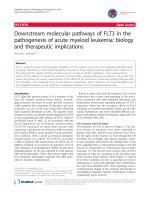
dial oxygen consumption. But in our study we observed
that flow in LAD had statistically significant difference
between the groups concerning the percentage change
from baseline to 10 minutes and statistically significant
difference between the two groups at 20 minutes (p <
0.0005) in favor of the PA-LA shunt (Figure 5, 6).
According the Hagen-Poiseuille law
Q =
π
8
η
Pi − Po
L
R
4
the PA - LA shunt has 10 fold higher volumetric flow
rate, where Q: volumetric flow rate, π: mathematical
constant, h: dynamic fluid viscosity [pascal - second
(Pa·s)], P
i
: inlet pressure, P
o
: outlet pressure, L: total
lengthofthetubeinthex direction (meters), R: is the
radius.
Because of the anatomical contiguity between pulmon-
ary artery and left atrium, the length of the PA-LA graft
is always shorter t han the RA-LA graf t. The pressure
gradient PA-LA is always higher than the RA-LA. These
two issues constitute an inherent advantage of PA-LA
shunt and are rendering PA-LA shunt more effectively
in that it can provide wider range of achievable flows
through the shunt. Given the fact that the current tech-
nology allows the pulmonary artery banding to be adjus-
table, we can assume that in the future we may be able
to calculate the ideal flow in an individualized manner.
Figure 5 Graphic showing the flow in the LAD and t he
changes during the experiment.
Figure 4 Correlation of percentage change from the baseline
of pO
2
arterial between the two shunts.
Argiriou et al. Journal of Cardiothoracic Surgery 2011, 6:143
/>Page 8 of 10
To our surprise, systemic arterial de-saturation following
the PA-LA shunt was not increased dramatically with
devastating consequences such as systemic oxygen deliv-
ery. The advantages of a pulmonary artery to left atrium
shunt are the following:
1. Can be performed without extracorporeal circulation
2. Can be used with a telemetrically controlled
adjustable occlusion device, as the Flo-Wat ch pul-
monary artery banding device (EndoArt, Lausanne,
Switzerland), which has been successfully introduced
in clinical practice of banding [20].
3. Can be easily occluded with the current devices,
as the Gianturco-Grifka vascular occlusion device
which is an appropriate closure system to occlude
the shunt because of the large size (9 mm) [21]
4. Can be easily performed in conjunction with a
pumpless lung assist device as Novalung in parallel
with the PA shunt or in a serial setting [22].
Conclusion
Our experiments have showed that a PA-LA shunt can
more effectively moderate or even partially reverse the
adverse effects of acute right ventricle pressure overload
than an interatrial shunt, offering a decrease in right ven-
tricle afterload, increased flow in left anterior descending
artery with less mean arterial pressure and lower SVR.
Limitations
Our study has some limitations. First of all, all measure-
ments were performed in open chest surgery. Secondly,
the ventilation supplying oxygen was at 100% and not at
room air oxyg en. Finally the measurements were taken at
10 and 20 minute interval. The above parameters may
alter the results of blood gases. Nevertheless all measure-
ments taken together allow for a realistic evaluation of
the overall picture. The use of other acute RVF models
and the determination of long term results are a matter
of further investigations.
Abbreviations
RV: Right Ventricle; RA: Right Atrium; RVF: Right ventricular failure; RVO: Right
ventricular overload; PA-LA: pulmonary artery to left atrium shunt; RA-LA:
Right atrium to left atrium shunt; LAD: left anterior descending artery; CCO:
Continuous Cardiac Output; SV: Stroke Volume; SVV: Stroke Volume Variation;
SVR: Systemic Vascular Resistance; COPD: chronic obstructive pulmonary
disease; PAH: pulmonary arterial hypertension; ARDS: adult respiratory
distress syndrome; ECG: Electrocardiogram; RVP: Right Ventricular Pressure.
Author details
1
Second Cardiac Surgery Department, Evaggelismos General Hospital, 45-47
Ipsilantou, 10676, Athens, Greece.
2
Cardiothoracic Surgery Department,
Democritus University Thrace, University Hospital of Alexandroupolis,
Dragana, 68100, Greece.
3
Surgical Experimental Laboratories ELPEN (AP), 95
Marathonos Avenue, 19009, Pikermi, Athens, Greece.
Authors’ contributions
All authors read and approved the final manuscript.
MA and TS performed all the experiments, collected the data and drafted
the manuscript.
AP is the clinical director of the experimental laboratory, helped out with
the experiments and the data collection.
DM revised it critically for important intellectual content
VD revised it critically for important intellectual content
GB have given final approval of the version to be published
Competing interests - Disclosures
The authors declare that they have no competing interests.
Received: 7 September 2011 Accepted: 19 October 2011
Published: 19 October 2011
References
1. Oz MC, Slater JP, Edwards N, et al: Desaturated venous to arterial
shunting reduces right-sided heart failure after cardiopulmonary bypass.
Journal of Heart and Lung Transplants 1995, 14:172-176.
2. Badesch DB, Abman SH, Simonneau G, et al: Medical therapy for
pulmonary arterial hypertension: updated ACCP evidence-based clinical
practice guidelines. Chest 2007, 131:1918-1928.
3. Reichenberger F, Pepke-Zaba J, McNeil K, et al: Atrial septostomy in the
treatment of severe pulmonary arterial hypertension. Thorax 2003,
58:797-800.
4. Law M, Grifka RG, Mullins CE, Nihill MR: Atrial septostomy improves
survival in select patients with pulmonary hypertension. Am Heart J 2007,
153(5):779-784.
5. Kurzyna M, Dabrowski M, Bielecki D, et al: Atrial septostomy in treatment
of end-stage right heart failure in patients with pulmonary
hypertension. Chest 2007, 131(4):977-983.
6. Takagaki M, Ishino K, Kawada M, Ohtsuki S, Hirota M, Tedoriya T, Tanabe Y,
Nakai M, Sano S: Total right ventricular exclusion improves left
ventricular function in patients with end-stage congestive right
ventricular failure. Circulation 2003, 108(Suppl 1):II226-9.
7. Kaul TK, Kahn DR: Postinfarct refractory right ventricle: right ventricular
exclusion. A possible option to mechanical cardiac support, in patients
unsuitable for heart transplant. J Cardiovasc Surg (Torino) 2000,
41(3):349-355.
8. D’Alonzo GE, Barst RJ, Ayres SM, et al: Survival in patients with primary
pulmonary hypertension: results from a national prospective registry.
Annals of Internal Medicine 1991, 115(5):343-349.
Figure 6 Correlation of percentage change from the baseline
of the LAD between the two shunts.
Argiriou et al. Journal of Cardiothoracic Surgery 2011, 6:143
/>Page 9 of 10
9. Bourge RC, Kirklin JK, Naftal DC, et al: Analysis and predictors of
pulmonary vascular resistance after cardiac transplantation. J Thorac
Cardiovasc Surg 1991, 101:432-435.
10. Simon MA, Pinsky MR: Right ventricular dysfunction and failure in chronic
pressure overload. Cardiol Res Pract 2011, 2011:568095.
11. Dávila-Román VG, Waggoner AD, Hopkins WE, Barzilai B: Right ventricular
dysfunction in low output syndrome after cardiac operations:
assessment by transesophageal echocardiography. Ann Thorac Surg 1995,
60(4):1081-1086.
12. Haddad F, Couture P, Tousignant C, Denault AY: The right ventricle in
cardiac surgery, a perioperative perspective: II. Pathophysiology, clinical
importance, and management. Anesth Analg 2009, 108(2):422-433.
13. Haddad F, Couture P, Tousignant C, Denault AY: The right ventricle in
cardiac surgery, a perioperative perspective: I. Anatomy, physiology, and
assessment. Anesth Analg 2009, 108(2):407-421.
14. Arpesella G, Mikus E, Loforte A, Mikus PM: Right-left atrium by-pass as
salvage treatment for graft failure after heart transplantation. Eur J
Cardiothorac Surg 2007, 32(4):671-673.
15. Camboni D, Akay B, Sassalos P, Toomasian JM, Haft JW, Bartlett RH,
Cook KE: Use of venovenous extracorporeal membrane oxygenation and
an atrial septostomy for pulmonary and right ventricular failure. Ann
Thorac Surg 2011, 91(1):144-149.
16. Slater JP, Goldstein DJ, Ashton RC Jr, Levin HR, Spotnitz HM, Oz MC: Right-
to-left veno-arterial shunting for right-sided circulatory failure. Ann
Thorac Surg 1995, 60(4):978-984.
17. Craig ML: Management of right ventricular failure in the era of
ventricular assist device therapy. Curr Heart Fail Rep 2011, 8(1):65-71.
18. Bilgutay AM, Sanchez LH, Siegal DL, Lillehei CW: Effect of pulmonary
artery-left atrium shunts on ischemic hearts-experimental study and
clinical application. Surg Forum 1961, 12:229-232.
19. Gupta S: Role of pulmonary arterial-left atrial shunt in the treatment of
pulmonary hypertension. Experimental study. J Thorac Cardiovasc Surg
1972, 64(6):949-952.
20. Corno AF, Ladusans EJ, Pozzi M, Kerr S: FloWatch versus conventional
pulmonary artery banding. J Thorac Cardiovasc Surg 2007,
134(6):1413-1419.
21. Zanchetta M, Rigatelli G, Pedon L, Zennaro M, Maiolino P, Onorato E:
Transcatheter amplatzer duct occluder closure of direct right pulmonary
artery-to-left atrium communication. Cath Cardiovasc Interven 2003,
58:107-110.
22. Spillner J, Amerini A, Hatam N, Rex S, Pott F, Goetzenich A, Menon A,
Repas T, Steiner F, Autschbach R, Carpi A, Oster O:
Pulmono-atrial shunt
and lung assist to treat right ventricular failure. Front Biosci 2011,
17:2342-2351.
23. Kerstein D, Levy PS, Hsu DT, Hordof AJ, Gersony WM, Barst RJ: Blade
balloon atrial septostomy in patients with severe primary pulmonary
hypertension. Circulation 1995, 91:2028-2035.
24. Rich S, Dodin E, McLaughlin VV: Usefulness of atrial septostomy as a
treatment for primary pulmonary hypertension and guidelines for its
application. Am J Cardiol 1997, 80:369-371.
25. Thanopoulos BD, Georgakopoulos D, Tsaousis GS, Simeunovic S:
Percutaneous balloon dilatation of the atrial septum: immediate and
midterm results. Heart 1996, 76:502-506.
26. Zierer A, Melby SJ, Voeller RK, Moon MR: Interatrial shunt for chronic
pulmonary hypertension: differential impact of low-flow vs. high-flow
shunting. Am J Physiol Heart Circ Physiol 2009, 296(3):H639-644.
doi:10.1186/1749-8090-6-143
Cite this article as: Argiriou et al.: Acute pressure overload of the right
ventricle. Comparison of two models of right-left shunt. Pulmonary
artery to left atrium and right atrium to left atrium: experimental study.
Journal of Cardiothoracic Surgery 2011 6:143.
Submit your next manuscript to BioMed Central
and take full advantage of:
• Convenient online submission
• Thorough peer review
• No space constraints or color figure charges
• Immediate publication on acceptance
• Inclusion in PubMed, CAS, Scopus and Google Scholar
• Research which is freely available for redistribution
Submit your manuscript at
www.biomedcentral.com/submit
Argiriou et al. Journal of Cardiothoracic Surgery 2011, 6:143
/>Page 10 of 10