Báo cáo y học: " FISH Oracle: a web server for flexible visualization of DNA copy number data in a genomic contex" ppsx
Bạn đang xem bản rút gọn của tài liệu. Xem và tải ngay bản đầy đủ của tài liệu tại đây (3.1 MB, 12 trang )
RESEARC H Open Access
FISH Oracle: a web server for flexible visualization
of DNA copy number data in a genomic context
Malte Mader
1
, Ronald Simon
1
, Sascha Steinbiss
2
and Stefan Kurtz
2*
Abstract
Background: The rapidly growing amount of array CGH data requires improved visualizati on software supporting
the process of identifying candidate cancer genes. Optimally, such software should work across multiple microarray
platforms, should be able to cope with data from different sources and should be easy to operate.
Results: We have developed a web-based software FISH Oracle to visualize data from multiple array CGH
experiments in a genom ic context. Its fast visualization engine and advanced web and database technology
supports highly interactive use. FISH Oracle comes with a convenient data import mechanism, powerful search
options for genomic elements (e.g. gene names or karyobands), quick navigation and zooming into interesting
regions, and mechanisms to export the visualization into different high quality formats. These features make the
software especially suitable for the needs of life scientists.
Conclusions: FISH Oracle offers a fast and easy to use visualization tool for array CGH and SNP array data. It allows
for the identification of genomic regions representing minimal common changes based on data from one or more
experiments. FISH Oracle will be instrumental to identify candidate onco and tumor suppressor genes based on
the frequency and genomic position of DNA copy number changes. The FISH Oracle application and an installed
demo web server are available at />Background
In the recent years, high resolution genomic tiling arrays
and SNP chips have become the standard technolo gy to
analyze copy number varia tions in cancer genomes.
Modern arra ys are inexpensive and allow for determin-
ing copy number changes at th e resolution of individual
genes. Gains or deletions of chromosomal material are
often highly variable in size, ranging from several kilo-
bases to entire chromosomes. One important strategy to
reveal genetic loci containing putative canc er genes is to
perform multiple experiments and identify chromosomal
regions representing minimal common alterations. Since
large alterations spanning many megabases are typically
more common than the small ones containing only a
few genes, a s many experim ents as possible should be
included into such kind of analysis. Public databases like
the Stanford Micro array Database [1], ArrayExpress [2],
the caArray Data P ortal [3], the Cancer Genome Project
[4] or the Gene Expression Omnibus (GEO) [5], provide
an unprecedented source for genomic copy number
data, which may be combined with own data fo r a
meta-analysis. In the following we will use the term
array CGH (array comparative genomic hybridization)
as a synonym for methods generating copy number data
including classical array CGH tiling microarrays or SNP
microarrays. Although a number of software tools for
array CGH analysis and visualization are available —
both from academia and commercial vendors — they
are often limited to a particular data format, cannot be
easily operated, or lack interactivity.
Existing software to ols for the visualization of array
CGH data can be grouped into different ways, i.e.
according to their application type (generic genome
browser or pure array CGH analysis) or according to
their architecture as a desktop or a web-based applica-
tion (Figure 1).
The Integrated Genome Browser (IGB) [6] and Inte-
grative Genomics Viewer (IGV) [7] are gen eral desk-
top-based genome browsers. The IGB software is
based on GenoViz [8], a software library for genome
* Correspondence:
2
Center for Bioinformatics, University of Hamburg, Bundesstrasse 43, 20146
Hamburg, Germany
Full list of author information is available at the end of the article
Mader et al. Journal of Clinical Bioinformatics 2011, 1:20
/>JOURNAL OF
CLINICAL BIOINFORMATICS
© 2011 Mader et al; licensee BioMed Central Ltd. This is an Open Access article distributed under the terms of the Creative Commons
Attribu tion License ( which permits unrestricted use, distribution, and repro duction in
any medium, provided the original work is properly cited.
visualization. IGB is an open-source software allowing
to display gene structure annotations, genomic align-
ments of expression array target sequences and EST/
cDNA genomic alignments. The different kinds of data
loaded from a data source are shown in different sorta-
ble horizontal tracks. IGV is an open source desktop-
based tool for displaying various types of data includ-
ing copy number variation data, loss of heterozygosity
data, gene expression data, significant DNA aberra-
tions, sequence alignments, and mutations. These data
can be displayed using four different types of graphs,
namely heatmaps, bar charts, scatter plots, and line
plots.
By now, a variety of generic web-based genome brow-
sers have been developed. Some, such as GBrowse [9],
the UCSC Genome Browser [10] or the Ensembl Genome
browser [11], are classical server-centered web-based
applications, fetching data and calculating images for a
specific chromosomal region before embedding it in to a
static web page and sending it to the client. One disad-
vantage of this technique is the large amount of data
traffic required for creating and transferring images of
genomic regions with dense information content.
In contrast, recent browsers like AnnoJ [12], the NCBI
Sequence Viewer [13], JBrowse [14], or the University of
Tokyo Genome Browser (UTGB) [15] are Rich Internet
visualization
software
genome
browser
desktop-
based
IGB [6]
IGV [7]
web-
based
web 2.0
JBrowse [14]
UTGB [15]
AnnoJ [12]
NCBI
Sequence
Viewer [13]
pre-web
2.0
GBrowse [9]
UCSC
Genome
Browser [10]
Ensembl [11]
array
CGH
software
desktop-
based
com-
mercial
Affyme-
trix [17]
Illumi-
na [18]
free or
open
source
CGH-
Explo-
rer [19]
Caryo-
scope [20]
CGH-
PRO [21]
CGHAna-
lyser [22]
ChARM-
View [23]
Ideogram-
Brow-
ser [24]
Snoop-
CGH [29]
MD-See-
GH [26]
SEU-
RAT [27]
CHESS [28]
SIGMA
2
[30]
VAMP [25]
web-
based
web server
Array-
CyGHt [31]
array-
CGH-
base [32]
CAP-
web [33]
wavi-
CGH [36]
SIGMA [34]
free or
open
source
FISH
Oracle
ISA-
CGH [35]
Figure 1 Classification tree of software tools for array CGH data analysis. At the root level (black), tools are grouped into general genome
browsers and software specific for array CGH data analysis (gray). At the next level, we distinguish web-based software (red) from desktop-based
software (green). The lowest level divides free or open source software from commercial or closed-source applications or, in the case of web-
based genome browsers, pre-web 2.0 from web 2.0 applications.
Mader et al. Journal of Clinical Bioinformatics 2011, 1:20
/>Page 2 of 12
Applications (R IAs) based on a technology called asyn-
chronous JavaScript and XML (AJAX) [16]. This allows
rendering images at the client si de as well as loading
server side data dynamically without having to refresh
the whole page, thus reducing both the required data
traffic and the server load.
All web-based browsers share the property of being
generic in nature. Although they provide many exten-
sions, it is sometimes not possible or at least difficult to
achieve the desired visualization. For this reason, several
specialized software tools for processing and visualizing
array CGH data have b een developed. The Affymetrix
Genotyping Console [17] and the Illumina GenomeStudio
Software [18] are commercial desktop-based software
products, capable of handling different microarray data,
including array CGH data. Their main disadvantage is
that they are both limited to the respective vendor-spe-
cific array platform.
In academia, several open source or freely available
desktop applications specific for array CGH data have
been developed, including CGH-Explorer [19], Caryo-
scope [20], CGHPRO [21], CGHAnalyzer [22], ChARM-
View [23], IdeogramBrowser [24], VAMP [25], MD-
SeeGH [26], SEURAT [27], CHESS [28], SnoopCGH [29]
and SIGMA2 [30], written in Java or C++. With the
exception of CGHAnalyzer, all offer an interactive dis-
play of array CGH and/or gene expression data. Their
support of additional features varies extensively (see
Tables S1 and S2 in the addition al file 1). The main dis-
advantage of these tools is that each installation of a
program needs to be run on a separate computer,
requiring additional effort to keep the software and data
up-to-date across release updates. Thus they are not
well suited for a distributed, collaborative approach to
genome research.
Finally, the group of web-based software for visualiza-
tion of array CGH data comprises ArrayCyGHt [31],
arrayCGHbase [32], CAPweb [33], SIGMA [34], ISACGH
[35] and WaviCGH [36]. All of these are primarily acces-
sible via static installations on web servers, requiring to
upload the data to be analyzed to external parties. While
this supports collaboration, it may raise problems related
to privacy concerns or a large volume of necessary data
which could become a heavy burden for the server.
Table S3 in the additional file 1 lists the different fea-
tures of existing web-based soft ware tools for visualizing
and analyzing array CGH data. Interestingly, except for
waviCGH, all web-based software tools for array CGH
data analysis have been published in the mid-2000s.
However, waviCGH, published in 2010 and focused on
automatic analysis and visualization of array CGH data
in a genomic context, does not provide a dynamic visua-
lization. Instead it produces static, chromosome-wide
images of the data.
We have developed a software tool called FISH Oracle
combining the most important features of the above
mentioned software tools for visualizing array CGH
data:
First of all, FISH Oracle does not impose a limit on
the number of array CGH experiments to be visualized
at once. This is important since a large number of
experiments is often necessary to obtain accurate
results, a fact confirmed by the large number of avail-
able data. Sec ondly, FISH Oracle provides the relevant
genomic context, i.e. besides the segment data it displays
annotations available in Ensembl [37] at a genomic reso-
lution ranging from ten to 10 million base pairs. This
feature is important because the task of identifying ne w
chromosomal aberrations and single genes overlapping
with copy number variations requires observation of the
relevant data on different scales. Detailed information
about a single gene or other functional elements (e.g.
their UniProt [38] identifier) can be obtained in FISH
Oracle by clicking on the corresponding element. This
feature is important as users quickly want to decide
whether the functional element in question could be a
possible target for further investigation.
FISH Oracle stores its data in a central database. Once
uploaded, it can quickly be accessed for any user of the
system, thus reducing data redundancy (compared to
desktop applications) and allowi ng collaborative work
based on the data. The fast visualization engine in com-
bination with advanced web and database technology
supports highly interactive use. FIS H Oracle comes with
a convenient data import mechanism, powerful search
options for genomic elements (e.g. gene names or karyo-
bands) and mechanisms to export the visualization into
different high quality formats.
We termed our software FISH Oracle because it is
well suited for computational selection of candidate
genes for subsequent fluorescence in situ hybridization
(FISH) experiments.
We tested the application using two different data
sets. One data set consists of SNP microarrays. It
includes our own data, data from the Sanger Cancer
Genome project [4] as well as data from NCBI GEO.
The other data set comprises two channel microarray
data from NCBI GEO.
Results and Disc ussion
User interface
The data import process in FISH Oracle consists of two
steps. In a first step, the data are uploaded to the server
in form of a tab-delimited file. Each line in the uploaded
file specifies a segment by an identifier for the chromo-
some it comes from, its start position, its end position,
its mean intensity value and its numbe r of markers. In
the second step, the user specifies a study name, the
Mader et al. Journal of Clinical Bioinformatics 2011, 1:20
/>Page 3 of 12
tissue type and the microarray type for the uploaded
data. Further information about the pathological state,
as well as a detailed desc ription of the data source, can
optionally be added. Once the annotation file is
uploaded, the data are checked for consistency and
stored in a relational database.
Once the segment data are stored in the database and
the corresponding annotation is available, the user decides
which region of the considered genome is to be displayed.
This can be done by specifying one of the following loca-
tion markers: range of genomic positions, name of a gene
or karyoband, or segment ID (Figure 2). FISH Oracle then
dis plays the genome annotation and segment data at the
specified genomic location or in the region containing the
specified item. The initially displayed genomic region
depends on the extent of found segments. If the search
term is a gene or a karyoband and no segments are found,
the displayed range is equal to the length of the karyoband
or the visualized range is extended by 200% of the gene
size in both chromosomal directions. If only one segment
is found by a segment search, the displayed range equals
the segment size. The maximum initial range is 20 Mbp.
Segments are selected according to a user-specified
threshold for the mean intensity values. This threshold
can be specified in two modes: In the “less than” mode,
all segments whose mean intensity value is less than the
threshold are displayed, allowing to select segments
representing deletions. Similarly, the “greater than”
mode selects segments with a mean intensity value lar-
ger than the threshold. Thus this mode allows to select
segments representing amplifications. In addition to the
threshold, a combo box allows the user to restrict the
selection to segments that originate from experiments
for specific tissues.
Each search delivers an image with up to three tracks.
A track possibly consists of several lines if elements of a
track or their captions o verlap. This makes them mor e
readable. The karyoband track is always shown and it
appears as the top track. The gene track shows, for the
specified region, all genes according to the Ensembl
annotation of the genome. The segment track shows, for
the specified region, all segments according to the cur-
rently chosen thresholds and tissue types. At the top of
the image a genomic scale depicts the shown region of
Figure 2 The FISH Oracle user interface. (1) The search menu is on the left hand side. It allows to specify a region, segment IDs, gene names
and karyobands as search keys. If the user wants to display a certain genomic region, the search box is replaced by three text fields to enter the
chromosome and start and end positions. A threshold for the segment mean intensity values can be specified with the “less than” or “greater
than” option. Typically, the “less than” option is used with a negative threshold to focus the visualization to segments with negative mean
intensity value (deletions). The “greater than” option is used with a positive threshold to focus the visualization to segments with positive mean
intensity value (amplifications). The tissue type filter restricts the display to segments for one ore more specific tissue types. (2) The
administration menu (lower left corner) provides functionality for data import and user account administration, e.g. activation of recently
registered users. (3) Each search opens a new tab that can be identified by the search query appearing as the caption of the tab. Each open tab
has its own toolbar, showing the exact location of the displayed region as well as the current thresholds. The toolbar provides buttons for
zooming into the displayed region, scrolling along a chromosome, and exporting the displayed image for download to the user’s computer. (4)
The visualization, according to the current toolbar settings, is displayed below the toolbar. In this case, the image shows segment data and
annotations in the region of the gene PTEN. (5) Clicking on the symbol representing a gene or a segment triggers a pop-up window containing
corresponding detailed information.
Mader et al. Journal of Clinical Bioinformatics 2011, 1:20
/>Page 4 of 12
the chromoso me. A toolbar shows the exact chromoso-
mal coordinates of the displayed image and contains
control buttons for scrolling over the chromosome and
zooming into or out of the chromosome. Clicking on
the parts of the i mage representing genes or segments
delivers a pop-up window showing additional informa-
tion on the corresponding element (Figure 2).
FISH Oracle also allows for export of the s hown data
(segments and annotation) in a tabular representation to
a file in Microsoft Excel format.
The visualization of the segments and annotatio n can
be exported as PNG bitmaps or in the PDF, PostScript,
or SVG vector graphics format.
Application of FISH Oracle to our own dataset
In a first study, we applied FISH Oracle to our own
array CGH data sets (231 experiments) which w ere
obtained from experiments using different human can-
cer cells and Affymetrix SNP 6.0 microarrays. We also
used parts of the Sanger Cancer Genome Project (CGP)
[4] (Affymetrix SNP 6.0 microarrays, 5 experiments) and
NCBI GEO [39,40] (Affymetrix Mapping 250K Nsp SNP
microarrays, 9 experiments) which were randomly
selected. We will refer to this data set as FISH Oracle
data.
All array CGH data sets (given as CEL files) were nor-
malized based on an internal reference. This means t hat
every intensity value of a specific probe set is divided by
the mean intensity value over the 0.25- and 0.75-quan-
tile of the same probe set of different microarrays. This
allows normalization of the data without reference
arrays. We applied DNAcopy [41] to the normalized
data to calculate breakpoints of intensit y values. The
result is a tab-delimited file with segments characterized
by consecutive positions of similar intensity values. Each
segment is associated with a chromosome number, a
start and end position on the chromosome, the number
of SNP markers covered by the segment and the mean
intensity value of all SNP markers contained in the seg-
ment. A ll resulting tab-del imited files were uploaded to
FISH Oracle.
We show by three examples how the interactive visua-
lization provided by FISH Oracle reve als coincidences
between an accumulation of segments from different
experiments on one side and annotated genes in a speci-
fic region on the other side. The first region (Figure 3)
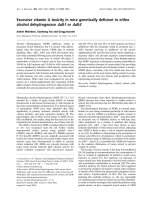
Figure 3 Annotated genes and segment data obtained from different array CGH experiments for esophagus, pancreas, prostate, colon
and multiple myeloma tumor tissue shown at the 12p11.23 locus. The segment data are taken from the FISH Oracle data. All segments
have a mean intensity value greater than 0.5 and therefore represent amplified regions. There are amplifications in the region from about 24.5
Mbp to 25.6 Mbp. The minimal overlapping region covers the area from 25.1 Mbp to 25.35 Mbp. This region contains the genes LRMP, CASC1,
LYRM5 and KRAS. KRAS is a known proto oncogene, which may lead to tumor development if amplified [42]. The minimal region including KRAS
is shown in a red box.
Mader et al. Journal of Clinical Bioinformatics 2011, 1:20
/>Page 5 of 12
is located around the 12p locus of the human genome
with several amplifications overlapping the KRAS gene.
The second region (Figure 4) can be found around t he
10q23 locus with several deletions overlapping the
PTEN gene. The third region (Figure 5) is located
around the 21q22.2/21q22.3 loci with several deletions
overlapping the genes TMPRSS2 and ERG. These coinci-
dences are consistent with previous publications on the
relevance of these genes for cancer: KRAS is a proto
oncogene [42], PTE N is a tumor suppressor gene play-
ing an important role in prostate cancer [43] and
TMPRSS2:ERG is a known fusion gene in prostate can-
cer [44]. The three examples show that FISH Oracle has
the potential to aid a researcher in deriving interesting
new hypotheses about potential cancer genes based on
segment data and corresponding annotations.
Application of FISH Oracle to foreign data
The second study is based on the data of T aylor et al.
[45] who analyzed 231 prostate carcinomas using differ-
ent types of microa rrays, mainly 244K Agilent human
array CGH microa rrays. The data are available as text
files from NCBI GEO (accession number: GSE21035).
We will refer to this data set as Taylor data.Asthe
Taylor data is based on two color microarrays (includ-
ing, for each patient, one tumor tissue sample and one
healthytissuesampleasreference)wehadtouse
another normalization method. The d ata were normal-
ized based on global medians using the method normal-
izeWithinArrays from the R package limma [46].
Segment data were calculated using DNACopy.
Figure 4 and Figure 5 confirm that the Taylor data are
consistent with the FISH Oracle data. As the Taylor
data originate from considerably more experiments than
the FISH Oracle data, the former more clearly reveals
important locations with deletions (like the location
near 10q23 or 21q22.2/21q22.3).
Thresholds for segment mean values
Intensity values for segments originate f rom the loga-
rithmic transformation of sample to reference sample
ratio and can be posi tive or negative [47]. A u ser-speci-
fied mean intensity threshold determines which seg-
ments are displayed. A negative threshold selects
segments with a (negative) mean intensity value not lar-
ger than the threshold. These segments represent
deleted genomic regions. A positive thresholds selects
segments with a (positive)meanintensityvaluenot
smaller than the given threshold. These segments repre-
sent amplified genomic regions. A reasonable threshold
may be adjusted experimentally. This is exemplified in
Figure 6 showing the distribution of the number of seg-
ments d epending on the threshold. The counts refer to
theFISHOracledataandtheTaylordata,focusingon
the regions 10q23 and 21q22.2/21q22.3 already consid-
ered in Figures 4 and 5. The short response time of the
FISH Oracle visualization allows to quickly explore the
effect of different thresholds.
The comparison of the FISH Oracle data with the
Taylor data reveals the in fluence of data quantity: The
segment counts derived from the FISH Oracle data are
approaching zero much faster than the segment counts
derived from the Taylor data. Hence in the FISH Oracle
data it is mor e difficult to spot regions with significant
amplifications or deletions. This problem also became
obvious in the visualization of the FISH Oracle data a t
the 21q22.2/21q22.3 loci where the significant segments
could hardly be d istinguished from noise. In contrast,
the larger Taylor data set shows a much more accurate
picture of interesting regions.
Discussion
We have developed FISH Oracle, an interactive web-
based ap plication to visualize segment data from an
unlimited number of array CGH experiments in the
context of gene annotations. Functional elements and
segments are presented in a clear and concise fashion.
Moreover, the zooming capability of the system makes
it possible to display all elements at the resolution
desired by the user. Easy to use filters allow to select
groups of segments to be visualized. We expect that
the high quality of the visualization and the flexibility
ofthesoftwarewillenablelifescientiststoquickly
derive interesting hypotheses about candidate cancer
genes occurring in amplified or deleted regions. To
communicate their findings, users can quickly export
the generated images in different high qua lity formats,
e.g. for publication or post-processing using standard
graphics software. FISH Oracle is flexible regarding the
underlying g enome as long as th e segment da ta refer
to the same sequence basis as an annotation data set
that is available in Ensembl. For example, segment
data sets from the mouse can be used with FISH
Oracle.
Even though the images in FISH Oracle are gener ated
at the server side of the application, only the image itself
is retransmitted a nd replaced at the client side. Addi-
tional gene annotation information for a specific gene is
loaded from the database when it is needed. In a “classi-
cal” server centered web application all additional gene
annotation information would have to be loaded concur-
rently with the visualization of the data, significantly
increasing the data transfer rates in particular when
visualizing regions with high gene density.
While many of the features of FISH Oracle are avail-
able in general genome purpose browsers, they are not
always available in the software tools specific for array
CGH data.
Mader et al. Journal of Clinical Bioinformatics 2011, 1:20
/>Page 6 of 12
Figure 4 Annotated genes and segment data obtained from different array CGH experiments for prostate tumor tissue shown at the
10q23 locus. The segments colored in orange correspond to the FISH Oracle data. The gray segments are derived from the Taylor data. Both
data sets show an accumulation of segments in the region from 89 Mbp to 91 Mbp. The minimal overlapping region indicated by the FISH
Oracle data extends from about 89.6 Mbp to 90.2 Mbp, containing the genes PTEN and C10orf59. The minimal overlapping region indicated by
the Taylor data overlaps almost exactly with the gene PTEN. The region around PTEN is shown in a red box. It overlaps with several deletions, as
only segments with a mean intensity threshold less than -0.7 for the Taylor data and less than -0.35 for the FISH Oracle data, are shown. PTEN
[43] is a known tumor suppressor gene which can lead to tumor development if it is deleted. Especially in prostate tissue, the deletion of a
chromosomal region containing PTEN leads to tumor development.
Mader et al. Journal of Clinical Bioinformatics 2011, 1:20
/>Page 7 of 12
In contrast to most other web-based applications for
visualizing array CGH data, FISH Oracle is able to
visualize an unlimited number of segments in the cho-
sen chromosomal region at low and high resolution.
Most desktop-based applications also provide the visua-
lization of multiple segments. However, with an increas-
ing number of segments the resulting visualizations of
the desktop t ools become more dense, making it mor e
difficult for the user to maintain an overview. In other
cases, desktop-based software does not provide a high
resolution view of all segments, complicating the search
for single genes overlapping with copy number changes.
FISH Oracle stores the imported data persistently in a
database. In contrast, the desktop-based array CGH software
solutions load the data from text files and store them in
internal data structures. Thus in each session the in put must
be r e-imported. Fo r large d ata sets involving mandatory pre-
processing or manual loading of several data sets (e.g.
SnoopCGH) the import becomes cumbersome for the user.
The web-based appli cations for processing array CGH
data (see introduction) are mainly offered as publicly
available web servers. Additionally CAPweb and
arrayCGHbase can be obtained for local installation by
requesting it from the maintainers. FISH Oracle is avail-
able as a web server and additional ly as an open source
package at racle.
We have made some effort to keep the installation as
easy as possible.
Figure 5 Annotated genes and segment data obtained from different array CGH experiments for prostate tumor tissue shown at the
21q22 locus. The segments below a threshold of -0.55 for the Taylor data (colored gray) and -0.25 for the FISH Oracle data (shown in orange)
indicate interstitial deletions covering a 3 Mbp genomic segment. The breakpoints are located within the genes TMPRSS2 and ERG (indicated by
red boxes), leading to the characteristic TMPRSS2:ERG fusion bond in about 40-60% of prostate cancers [44]. Segment captions are not shown to
make the resulting image more compact.
Mader et al. Journal of Clinical Bioinformatics 2011, 1:20
/>Page 8 of 12
To the best of our knowledge there is no single tool
for processing array CGH data offering a comparable
visualization functionality (see Tables S1-3 in the addi-
tional file 1). In each of the desktop-based software
tools, at least one core functionality is missing when
comparing it to FISH Oracle. Most of the desktop-based
tools do not provide a visualiza tion of the genomic con-
text, do not support alternative genomes, and do not
provide high-quality image export. Not all of them offer
built-in normalization or segmentation procedures. For
some of them, the license conditions are not specified.
MD-SeeGH [26] is probably the desktop-b ased softw are
that comes closest to FISH Oracle in terms of visualiza-
tion capabilities. (see Table S2 in the additional file 1).
However, MD-SeeGH is only available for MS-Win-
dows. Other tools, such as CHESS [28], are apparently
unavailable.
Several of the web-based tools do not provide interac-
tive visualization or genome browsing capabilities (see
Table S3 in the additional file 1). Often the w eb-based
tools are specifically tailored to a fixed set of genomes,
or (as in the case of SIGMA [34]) are restricted to a
specific database and do not provide interfaces to
common data formats. The ISACGH software [35] is no
longer available on its own. Neither is the GEPAS
toolkit it is built upon, and which has been merged into
the Babelomics software suite [48]. The ISACGH soft-
ware also lacks integrated genome browsing funct ional-
ity, and instead provides hyperlinks to Ensembl.
ArrayCGHbase with its “chromosome view” is the
web-based software that comes closest to FISH Oracl e.
While both software-tools have similar capabilities
regarding the visualization of segments data, they differ
in the kinds of additional data displayed: FISH Oracle
focuses on additional gene annotations which are not
handled by arrayCGHbase. On the other hand,
arrayCGHbase allows the display of raw intensity values
which is not displayed by FISH Oracle. Considering the
use of both tools, it becomes apparent that FISH Oracle
pursues a different approach to data visualization than
arrayCGHbase. ArrayCGHbase is centere d on expe ri-
ments, coming with filters to select certain experime nts,
whose data can be visualized using different methods. In
contrast, FISH Oracle is centered on genome annota-
tions. Once logged into the application the user can
immediately search for regions, karyobands or genes of
interest.
In summary, both tools are unique in their own way
and complement each other well.
While FISH Oracle does not contain explicit segmen-
tation, normalization or quality assessment components,
its open input format allows researchers to combine var-
ious specialized tools for these tasks with the visualiza-
tion capabilities of F ISH Oracle. This option makes the
software particularly attractive to life scientists ana lyzing
array CGH data.
On the client side, all software that is needed to access
FISH Oracle is a recent web browser with J avaScript
support enabled. On the server side, the software
requirements are more extensive (see Meth ods section).
With regard to hardware requirements, it is possible to
install the FISH Oracle server software on a standard
Linux workstation with at least 1 GB RAM. The hard
disk space requirement is largely dominated by the size
of the genome annotations. For example, a mirror of the
human genome annotation data from Ensembl r equires
about 14 GB of hard disk space.
Conclusions
Our examples show that FISH Oracle is a powerf ul tool
to detect amplifications and deletions of chromosomal
regions containing proto oncogenes, tumor suppressor
genes and fusion genes. Comprehensive search options,
the dynamic visualization of multiple microarray experi-
ments and export of high quality images are useful func-
tions to cope with todays amounts of data. State of the
art web and database technology facilitate c ollaborative
−6 −5 −4 −3 −2 −10
0 20 40 60 80 100 120 140
Intensity Threshold
Number of Segments
PTEN: Taylor data
PTEN: FISH Oracle data
TMPRSS2:ERG: Taylor data
TMPRSS2:ERG: FISH Oracle data
Figure 6 Segment counts (Y-axes) as a function of the
threshold for the mean intensity value (X-axis). The solid lines
are based on the Taylor data and the dashed lines are based on the
FISH Oracle data. The red colored lines refer to segments in the
minimal chromosomal region containing the gene PTEN (see also
Figure 4) while the black colored lines refer to segments in the
region 21q22.2/21q22.3 (see also Figure 5).
Mader et al. Journal of Clinical Bioinformatics 2011, 1:20
/>Page 9 of 12
work. Altogether FISH Oracle represents a helpful tool
for life scientists in the search of potential candidate
cancer genes.
Methods
Data storage
FISH Oracle uses the MySQL relational database to store
its source data. In particular, two different kinds of data
are stored in two separate databases: genome annotation
data (as available in the Ensembl database [37]) and seg-
mented array CGH data. The segment data are parsed
from text files uploaded to the web-server. Access to the
Ensembl database is established by the EnsJ Java library
[49]. The connection to the desired target database can
be configured by the administrator. For example, it is
possible to obtain the a nnotation information from a
remote database (accessed via the Internet) and the seg-
ment data from a database serv er in a local network.
Splitting the data into two databases has the advantage
that the data sources for the gene a nnotation can easily
be switched or updated without the n eed to change the
database storing the segment data, and vice versa.
User interface and server service
The user interface of FISH Oracle is written in the Java
programming language. This includes both the client
side of the web application (running in the user’sweb
browser) and the server side (running on the web ser-
ver). To reliably integrate both sides, we make use of
the Google Web Toolkit (GWT) [50]. The GWT pro-
gramming framework compiles a unified application
code written in Java into both JavaScript (for client-side
use) and Java servlet bytecode (for use on the server
sid e). It impl ements a conven ient and efficient mechan-
ism for client-server communication. Also, the resulting
web applications are compatible with all common web
browsers. Besides GWT, FISH Oracle is built on the
component library Smart GWT [51], a wrapper library
for the SmartClient [52] JavaScript framework. This fra-
mework provides a large set of convenient software
components (widgets), enabling the programmer to
quickly implement a state-of-the-art user interface that
is efficient, feature-rich and consistent. It should be
noted that SmartClient also offers functionality for cli-
ent-server communication. However, the server side
library requires a commercial license conflicting with
the open-source approach of FISH Oracle. Thus we
used the client-server communication mechanisms pro-
vided by the GWT.
The large user community for GWT, comprising more
than 1200 projects [53] (as of June 2011), and the fact
that Google Inc. uses GWT as their central web devel-
opment tool makes us confident that it will be main-
tained and improved in the remote future, so that
applications depending on it can remain functional. For
importing and exporting tabular data into and from
FISH Oracle, the JExcel [54] and Java CSV [55] software
libraries are used.
Data visualization
For visualization of both segment and annotation data
we used the AnnotationSketch [56] software library, a
portable, fast and space-efficient annotation drawing
solution that allows to display d ata from arbitrary
sources, making it particularly suitable for an interactive
web-based visual ization tool. For efficiency reasons,
AnnotationSketch was implemented in the C program-
ming language. In order to acces s the drawing functions
from FISH Oracle, an additional adapter layer between
the C library and the Java virtual machine is required.
As such an adapter, we used the Java Native Access
library [57] (JNA) which allows to call C functions from
Java programs. This enabled us to create Java counter-
parts for all components of the AnnotationSketch
library, which were then used to i mplement the visuali-
zation functions in FISH Oracle. Our software architec-
ture thus combines the advantage of having the time-
critical image generation step implemented in a fast low
level language (C) with the advantage of using a well-
tested and widely used platform for dynamic web a ppli-
cation development (Java ). Figure S1 in the additional
file 1 shows the data flow in FISH Oracle.
Availability
FISH Oracle is available as a source code package via
the FISH Oracle web site at -ham-
burg.de/fishoracle. It supports many POSIX conforming
UNIX-like target platforms, for example Linux or Mac
OS X.
On the web site we also offer additional documenta-
tion and a screencast video demonstrating the use o f
FISH Oracle.
Additional material
Additional file 1: This PDF file contains additional tables comparing
features of various other array CGH visualization software in detail,
as well as an illustration depicting the data flow during user
interaction with FISH Oracle.
Acknowledgements
This work was supported by a grant from the Werner-Otto-Stiftung to SK
and RS (# 6/73) and a grant from the Federal Ministry of Education and
Research (BMBF), Germany to RS (# FKZ 01GS08189).
Author details
1
Department of Pathology, University Medical Center Hamburg-Eppendorf,
Martinistrasse 52, 20246 Hamburg, Germany.
2
Center for Bioinformatics,
University of Hamburg, Bundesstrasse 43, 20146 Hamburg, Germany.
Mader et al. Journal of Clinical Bioinformatics 2011, 1:20
/>Page 10 of 12
Authors’ contributions
RS and SK conceived of the project. MM, SS and SK developed the software
architecture. MM implemented the software and generated the results. SS
contributed to the implementation of the data visualization. All authors
wrote, read and approved the final manuscript.
Received: 5 April 2011 Accepted: 28 July 2011 Published: 28 July 2011
References
1. Ball CA, Awad IAB, Demeter J, Gollub J, Hebert JM, Hernandez-Boussard T,
Jin H, Matese JC, Nitzberg M, Wymore F, Zachariah ZK, Brown PO,
Sherlock G: The Stanford Microarray Database accommodates additional
microarray platforms and data formats. Nucleic Acids Res 2005, , 33
Database: D580-D582.
2. Parkinson H, Sarkans U, Kolesnik ov N, Abeygunawardena N, Burdett T,
Dylag M, Emam I, Farne A, Hastings E, Holloway E, Kurba tova N, Lukk M,
Malone J, Mani R, Pilicheva E, Rustici G, Sharma A, William s E,
Adamusiak T, Brandizi M, Sklyar N, Brazma A: ArrayExpress update-an
archive of microarray and high-throughput sequencing-base d
functional genomics experiments. Nucleic A cids Res 2011, , 39 Database:
D1002-D1004.
3. caArray - Array Data Management System. [ />caarray/home.action].
4. Futreal PA, Coin L, Marshall M, Down T, Hubbard T, Wooster R, Rahman N,
Stratton MR: A census of human cancer genes. Nat Rev Cancer 2004,
4(3):177-183.
5. Barrett T, Troup DB, Wilhite SE, Ledoux P, Evangelista C, Kim IF,
Tomashevsky M, Marshall KA, Phillippy KH, Sherman PM, Muertter RN,
Holko M, Ayanbule O, Yefanov A, Soboleva A: NCBI GEO: archive for
functional genomics data sets-10 years on. Nucleic Acids Res 2011, , 39
Database: D1005-D1010.
6. Nicol JW, Helt GA, Blanchard SG, Raja A, Loraine AE: The Integrated
Genome Browser: free software for distribution and exploration of
genome-scale datasets. Bioinformatics 2009, 25(20):2730-2731.
7. Robinson JT, Thorvaldsdóttir H, Winckler W, Guttman M, Lander ES,
Getz G, Me sirov J P: Integrative genomics viewer. Nat Biotechnol 2011,
29:24-26.
8. Helt GA, Nicol JW, Erwin E, Blossom E, Blanchard SG, Chervitz SA,
Harmon C, Loraine AE: Genoviz Software Development Kit: Java tool kit
for building genomics visualization applications. BMC Bioinformatics 2009,
10:266.
9. Stein LD, Mungall C, Shu S, Caudy M, Mangone M, Day A, Nickerson E,
Stajich JE, Harris TW, Arva A, Lewis S: The generic genome browser: a
building block for a model organism system database. Genome Res 2002,
12(10):1599-1610.
10. Kent WJ, Sugnet CW, Furey TS, Roskin KM, Pringle TH, Zahler AM,
Haussler D: The human genome browser at UCSC. Genome Res 2002,
12(6):996-1006.
11. Stalker J, Gibbins B, Meidl P, Smith J, Spooner W, Hotz HR, Cox AV: The
Ensembl Web site: mechanics of a genome browser. Genome Res 2004,
14(5):951-955.
12. AnnoJ. [].
13. NCBI Sequence Viewer. [ />14. Skinner ME, Uzilov AV, Stein LD, Mungall CJ, Holmes IH: JBrowse: a next-
generation genome browser. Genome Res 2009, 19(9):1630-1638.
15. Saito TL, Yoshimura J, Sasaki S, Ahsan B, Sasaki A, Kuroshu R, Morishita S:
UTGB toolkit for personalized genome browsers. Bioinformatics 2009,
25(15)
:1856-1861.
16.
Garrett JJ: Ajax: A New Approach to Web Applications. 2005 [http://www.
adaptivepath.com/ideas/essays/archives/000385.php].
17. Affymetrix Genotyping Console. [ />level_seven_software_products_only.jsp?
productId=131535&categoryId=35625#1 1].
18. Illumina GenomeStudio. [ />genomestudio_software.ilmn].
19. Lingjaerde OC, Baumbusch LO, Liestøl K, Glad IK, Børresen-Dale AL: CGH-
Explorer: a program for analysis of array-CGH data. Bioinformatics 2005,
21(6):821-822.
20. Awad IAB, Rees CA, Hernandez-Boussard T, Ball CA, Sherlock G: Caryoscope:
an Open Source Java application for viewing microarray data in a
genomic context. BMC Bioinformatics 2004, 5:151.
21. Chen W, Erdogan F, Ropers HH, Lenzner S, Ullmann R: CGHPRO - a
comprehensive data analysis tool for array CGH. BMC Bioinformatics 2005,
6:85.
22. Margolin AA, Greshock J, Naylor TL, Mosse Y, Maris JM, Bignell G, Saeed AI,
Quackenbush J, Weber BL: CGHAnalyzer: a stand-alone software package
for cancer genome analysis using array-based DNA copy number data.
Bioinformatics 2005, 21(15):3308-3311.
23. Myers CL, Chen X, Troyanskaya OG: Visualization-based discovery and
analysis of genomic aberrations in microarray data. BMC Bioinformatics
2005, 6:146.
24. Müller A, Holzmann K, Kestler HA: Visualization of genomic aberrations
using Affymetrix SNP arrays. Bioinformatics 2007, 23(4):496-497.
25. Rosa PL, Viara E, Hupé P, Pierron G, Liva S, Neuvial P, Brito I, Lair S,
Servant N, Robine N, Manié E, Brennetot C, Janoueix-Lerosey I, Raynal V,
Gruel N, Rouveirol C, Stransky N, Stern MH, Delattre O, Aurias A, Radvanyi F,
Barillot E: VAMP: visualization and analysis of array-CGH, transcriptome
and other molecular profiles. Bioinformatics 2006, 22(17):2066-2073.
26. Chi B, deLeeuw RJ, Coe BP, Ng RT, MacAulay C, Lam WL: MD-SeeGH: a
platform for integrative analysis of multi-dimensional genomic data.
BMC Bioinformatics 2008, 9:243.
27. Gribov A, Sill M, Lück S, Rücker F, Döhner K, Bullinger L, Benner A, Unwin A:
SEURAT: visual analytics for the integrated analysis of microarray data.
BMC Med Genomics 2010, 3:21.
28. Lee M, Kim Y: CHESS (CgHExpreSS): a comprehensive analysis tool for
the analysis of genomic alterations and their effects on the expression
profile of the genome. BMC Bioinformatics 2009, 10:424.
29. Almagro-Garcia J, Manske M, Carret C, Campino S, Auburn S, Macinnis BL,
Maslen G, Pain A, Newbold CI, Kwiatkowski DP, Clark TG: SnoopCGH:
software for visualizing comparative genomic hybridization data.
Bioinformatics 2009, 25(20):2732-2733.
30. Chari R, Coe BP, Wedseltoft C, Benetti M, Wilson IM, Vucic EA, MacAulay C,
Ng RT, Lam WL: SIGMA2: a system for the integrative genomic multi-
dimensional analysis of cancer genomes, epigenomes, and
transcriptomes. BMC Bioinformatics 2008, 9:422.
31. Kim SY, Nam SW, Lee SH, Park WS, Yoo NJ, Lee JY, Chung YJ: ArrayCyGHt:
a web application for analysis and visualization of array-CGH data.
Bioinformatics 2005, 21(10):2554-2555.
32.
Menten B, Pattyn F, Preter KD, Robbrecht P, Michels E, Buysse K, Mortier G,
Paepe AD, van Vooren S, Vermeesch J, Moreau Y, Moor BD, Vermeulen S,
Speleman F, Vandesompele J: arrayCGHbase: an analysis platform for
comparative genomic hybridization microarrays. BMC Bioinformatics 2005,
6:124.
33. Liva S, Hupé P, Neuvial P, Brito I, Viara E, La Rosa P, Barillot E: CAPweb: a
bioinformatics CGH array Analysis Platform. Nucleic Acids Res 2006, , 34
Web Server: W477-W481.
34. Chari R, Lockwood WW, Coe BP, Chu A, Macey D, Thomson A, Davies JJ,
MacAulay C, Lam WL: SIGMA: a system for integrative genomic
microarray analysis of cancer genomes. BMC Genomics 2006, 7:324.
35. Conde L, Montaner D, Burguet-Castell J, Tárraga J, Medina I, Al-Shahrour F,
Dopazo J: ISACGH: a web-based environment for the analysis of Array
CGH and gene expression which includes functional profiling. Nucleic
Acids Res 2007, , 35 Web Server: W81-W85.
36. Carro A, Rico D, Rueda OM, Díaz-Uriarte R, Pisano DG: waviCGH: a web
application for the analysis and visualization of genomic copy number
alterations. Nucleic Acids Res 2010, , 38 Web Server: W182-W187.
37. Flicek P, Amode MR, Barrell D, Beal K, Brent S, Chen Y, Clapham P, Coates G,
Fairley S, Fitzgerald S, Gordon L, Hendrix M, Hourlier T, Johnson N, Kähäri A,
Keefe D, Keenan S, Kinsella R, Kokocinski F, Kulesha E, Larsson P, Longden I,
McLaren W, Overduin B, Pritchard B, Riat HS, Rios D, Ritchie GRS, Ruffier M,
Schuster M, Sobral D, Spudich G, Tang YA, Trevanion S, Vandrovcova J,
Vilella AJ, White S, Wilder SP, Zadissa A, Zamora J, Aken BL, Birney E,
Cunningham F, Dunham I, Durbin R, Fernández-Suarez XM, Herrero J,
Hubbard TJP, Parker A, Proctor G, Vogel J, Searle SMJ: Ensembl 2011.
Nucleic Acids Res 2011, , 39 Database: D800-D806.
38. The UniProt Consortium: Ongoing and future developments at the
Universal Protein Resource. Nucleic Acids Res 2011, , 39 Database:
D214-D219.
39. Ronchetti D, Lionetti M, Mosca L, Agnelli L, Andronache A, Fabris S,
Deliliers GL, Neri A: An integrative genomic approach reveals coordinated
expression of intronic miR-335, miR-342, and miR-561 with deregulated
host genes in multiple myeloma. BMC Med Genomics 2008, 1:37.
Mader et al. Journal of Clinical Bioinformatics 2011, 1:20
/>Page 11 of 12
40. GSE11522. [ />41. Olshen AB, Venkatraman ES, Lucito R, Wigler M: Circular binary
segmentation for the analysis of array-based DNA copy number data.
Biostatistics 2004, 5(4):557-572.
42. Sakakura C, Mori T, Sakabe T, Ariyama Y, Shinomiya T, Date K, Hagiwara A,
Yamaguchi T, Takahashi T, Nakamura Y, Abe T, Inazawa J: Gains, losses, and
amplifications of genomic materials in primary gastric cancers analyzed
by comparative genomic hybridization. Genes Chromosomes Cancer 1999,
24(4):299-305.
43. Cairns P, Okami K, Halachmi S, Halachmi N, Esteller M, Herman JG, Jen J,
Isaacs WB, Bova GS, Sidransky D: Frequent inactivation of PTEN/MMAC1 in
primary prostate cancer. Cancer Res 1997, 57(22):4997-5000.
44. Tomlins SA, Rhodes DR, Perner S, Dhanasekaran SM, Mehra R, Sun XW,
Varambally S, Cao X, Tchinda J, Kuefer R, Lee C, Montie JE, Shah RB,
Pienta KJ, Rubin MA, Chinnaiyan AM: Recurrent fusion of TMPRSS2 and
ETS transcription factor genes in prostate cancer. Science 2005,
310(5748):644-648.
45. Taylor BS, Schultz N, Hieronymus H, Gopalan A, Xiao Y, Carver BS, Arora VK,
Kaushik P, Cerami E, Reva B, Antipin Y, Mitsiades N, Landers T, Dolgalev I,
Major JE, Wilson M, Socci ND, Lash AE, Heguy A, Eastham JA, Scher HI,
Reuter VE, Scardino PT, Sander C, Sawyers CL, Gerald WL: Integrative
genomic profiling of human prostate cancer. Cancer Cell 2010, 18:11-22.
46. Smyth GK, Speed T: Normalization of cDNA microarray data. Methods
2003, 31(4):265-273.
47. Quackenbush J: Microarray data normalization and transformation. Nat
Genet 2002, 32(Suppl):496-501.
48. Medina I, Carbonell J, Pulido L, Madeira SC, Goetz S, Conesa A, Tárraga J,
Pascual-Montano A, Nogales-Cadenas R, Santoyo J, García F, Marbà M,
Montaner D, Dopazo J: Babelomics: an integrative platform for the
analysis of transcriptomics, proteomics and genomic data with
advanced functional profiling. Nucleic Acids Res 2010, , 38 Web Server:
W210-W213.
49. ENSJ. [ />root=ensembl].
50. Google Web Toolkit. [ />51. Smart GWT. [ />52. SmartClient. [].
53. Ohloh. [ />54. JExcel. [].
55. Java CSV. [ />56. Steinbiss S, Gremme G, Schärfer C, Mader M, Kurtz S: AnnotationSketch: a
genome annotation drawing library. Bioinformatics 2009, 25(4):533-534.
57. JNA. [].
58. Fridlyand J, Snijders AM, Pinkel D, Albertson DG, Jain AN: Hidden Markov
models approach to the analysis of array CGH data. Journal of
Multivariate Analysis 2004,
90:132-153.
59. Saeed AI, Bhagabati NK, Braisted JC, Liang W, Sharov V, Howe EA, Li J,
Thiagarajan M, White JA, Quackenbush J: TM4 microarray software suite.
Methods Enzymol 2006, 411:134-193.
60. Myers CL, Dunham MJ, Kung SY, Troyanskaya OG: Accurate detection of
aneuploidies in array CGH and gene expression microarray data.
Bioinformatics 2004, 20(18):3533-3543.
61. Ben-Yaacov E, Eldar YC: A fast and flexible method for the segmentation
of aCGH data. Bioinformatics 2008, 24(16):i139-i145.
62. Price TS, Regan R, Mott R, Hedman A, Honey B, Daniels RJ, Smith L,
Greenfield A, Tiganescu A, Buckle V, Ventress N, Ayyub H, Salhan A,
Pedraza-Diaz S, Broxholme J, Ragoussis J, Higgs DR, Flint J, Knight SJL: SW-
ARRAY: a dynamic programming solution for the identification of copy-
number changes in genomic DNA using array comparative genome
hybridization data. Nucleic Acids Res 2005, 33(11):3455-3464.
63. LaFramboise T, Winckler W, Thomas RK: A flexible rank-based framework
for detecting copy number aberrations from array data. Bioinformatics
2009, 25(6):722-728.
64. Hupé P, Stransky N, Thiery JP, Radvanyi F, Barillot E: Analysis of array CGH
data: from signal ratio to gain and loss of DNA regions. Bioinformatics
2004, 20(18):3413-3422.
65. Hsu L, Self SG, Grove D, Randolph T, Wang K, Delrow JJ, Loo L, Porter P:
Denoising array-based comparative genomic hybridization data using
wavelets. Biostatistics 2005, 6(2):211-226.
66. Marioni JC, Thorne NP, Tavaré S: BioHMM: a heterogeneous hidden
Markov model for segmenting array CGH data. Bioinformatics 2006,
22(9):1144-1146.
doi:10.1186/2043-9113-1-20
Cite this article as: Mader et al.: FISH Oracle: a web server for flexible
visualization of DNA copy number data in a genomic context. Journal of
Clinical Bioinformatics 2011 1:20.
Submit your next manuscript to BioMed Central
and take full advantage of:
• Convenient online submission
• Thorough peer review
• No space constraints or color figure charges
• Immediate publication on acceptance
• Inclusion in PubMed, CAS, Scopus and Google Scholar
• Research which is freely available for redistribution
Submit your manuscript at
www.biomedcentral.com/submit
Mader et al. Journal of Clinical Bioinformatics 2011, 1:20
/>Page 12 of 12