Báo cáo y học: "Augmentation of partially regenerated nerves by end-to-side side-to-side grafting neurotization: experience based on eight late obstetric brachial plexus cases" ppt
Bạn đang xem bản rút gọn của tài liệu. Xem và tải ngay bản đầy đủ của tài liệu tại đây (751.19 KB, 12 trang )
BioMed Central
Page 1 of 12
(page number not for citation purposes)
Journal of Brachial Plexus and
Peripheral Nerve Injury
Open Access
Research article
Augmentation of partially regenerated nerves by end-to-side
side-to-side grafting neurotization: experience based on eight late
obstetric brachial plexus cases
Sherif M Amr*
1
, Ashraf N Moharram
1
and Kamal MS Abdel-Meguid
2
Address:
1
From the Department of Orthopaedics and Traumatology, Cairo University, Cairo, Egypt and
2
From the Department of Orthopaedics
and Traumatology, Fayoum University, Fayoum, Egypt
Email: Sherif M Amr* - ; Ashraf N Moharram - ; Kamal MS Abdel-
Meguid -
* Corresponding author
Abstract
Objective: The effect of end-to-side neurotization of partially regenerated recipient nerves on improving motor
power in late obstetric brachial plexus lesions, so-called nerve augmentation, was investigated.
Methods: Eight cases aged 3 – 7 years were operated upon and followed up for 4 years (C5,6 rupture C7,8T1
avulsion: 5; C5,6,7,8 rupture T1 avulsion:1; C5,6,8T1 rupture C7 avulsion:1; C5,6,7 ruptureC8 T1 compression:
one 3 year presentation after former neurotization at 3 months). Grade 1–3 muscles were neurotized. Grade0
muscles were neurotized, if the electromyogram showed scattered motor unit action potentials on voluntary
contraction without interference pattern. Donor nerves included: the phrenic, accessory, descending and
ascending loops of the ansa cervicalis, 3
rd
and 4
th
intercostals and contralateral C7.
Results: Superior proximal to distal regeneration was observed firstly. Differential regeneration of muscles
supplied by the same nerve was observed secondly (superior supraspinatus to infraspinatus regeneration).
Differential regeneration of antagonistic muscles was observed thirdly (superior biceps to triceps and pronator
teres to supinator recovery). Differential regeneration of fibres within the same muscle was observed fourthly
(superior anterior and middle to posterior deltoid regeneration). Differential regeneration of muscles having
different preoperative motor powers was noted fifthly; improvement to Grade 3 or more occurred more in
Grade2 than in Grade0 or Grade1 muscles. Improvements of cocontractions and of shoulder, forearm and wrist
deformities were noted sixthly. The shoulder, elbow and hand scores improved in 4 cases.
Limitations: The sample size is small. Controls are necessary to rule out any natural improvement of the lesion.
There is intra- and interobserver variability in testing muscle power and cocontractions.
Conclusion: Nerve augmentation improves cocontractions and muscle power in the biceps, pectoral muscles,
supraspinatus, anterior and lateral deltoids, triceps and in Grade2 or more forearm muscles. As it is less expected
to improve infraspinatus power, it should be associated with a humeral derotation osteotomy and tendon
transfer. Function to non improving Grade 0 or 1 forearm muscles should be restored by muscle transplantation.
Level of evidence: Level IV, prospective case series.
Published: 05 December 2006
Journal of Brachial Plexus and Peripheral Nerve Injury 2006, 1:6 doi:10.1186/1749-7221-1-
6
Received: 03 August 2006
Accepted: 05 December 2006
This article is available from: />© 2006 Amr et al; licensee BioMed Central Ltd.
This is an Open Access article distributed under the terms of the Creative Commons Attribution License ( />),
which permits unrestricted use, distribution, and reproduction in any medium, provided the original work is properly cited.
Journal of Brachial Plexus and Peripheral Nerve Injury 2006, 1:6 />Page 2 of 12
(page number not for citation purposes)
Background
Late obstetric brachial plexus palsy serves as a good exam-
ple for studying the outcome of partially regenerated
nerves. Three main types of lesion [1] have been recog-
nized. In a C5-6 lesion, the arm is adducted and internally
rotated at the shoulder and the elbow extended. The fore-
arm is pronated and the wrist (and sometimes fingers)
flexed. In a C5-7 lesion, in addition to the above, the
elbow may be slightly flexed. In a C5-T1 lesion, the arm is
totally flail with a claw hand. In a prospective study of 80
infants with brachial plexus injury followed up for more
than 4 years [2], complete recovery occurred in 66% of
cases; mild weakness persisted in 11%, moderate arm
weakness in 9% and 14% had severe permanent weak-
ness. This unfavourable prognosis was supported by oth-
ers [3]. Several schemes were suggested to establish the
natural history of the injury selecting those cases not
expected to recover for early surgery [1]. Although early
surgery was advocated [4], in C5-7 lesions the shoulder
and elbow did not do as well as in upper-type lesions, the
results at the level of the hand were encouraging, however,
showing 75% with useful function after 8 years [5,6]. In a
further study [7], good results were obtained in 33% of C5
repairs, in 55% of C6, in 24% of C7 and in 57% of oper-
ations on C8 and T1. Posterior dislocation of the shoulder
was observed in 30 cases. All were successfully relocated
after the age of one year. A residual shoulder internal rota-
tion deformity requiring secondary surgery was also noted
by others [8]. Thus, with or without early surgery, a resid-
ual disability remains. This disability increases with age
[9], necessitating surgical correction.
For correcting residual shoulder internal rotation adduc-
tion, humeral derotation osteotomies [10] or tendon
transfers [11] gave good results. Nevertheless, this can
only occur if there is some range of shoulder abduction.
Besides, the early satisfactory results of anterior release
and latissimus dorsi to rotator cuff transfer are not main-
tained. In one study [12], there was loss of active external
rotation, because of gradual degeneration of the trans-
ferred muscles, contracture of the surrounding soft tissues
and degenerative changes in the glenohumeral joint. In
another study [13], children with sequelae of C5-C6 palsy
gained in abduction and external rotation more than chil-
dren with C5-C6-C7 or complete palsy. Patients with mild
preoperative shoulder dysfunction achieved the best
results. The clinical results were related to the type of
paralysis and to preoperative shoulder function, but not
to age at surgery. Progressive deterioration of abduction
began at 6 years despite preserved active external rotation.
In a prospective study of secondary surgery on 183 sublux-
ations or dislocations of the shoulder consequent upon
obstetric brachial plexus palsy [14], 20 failures were
reported. The functional outcome was related to the sever-
ity of the neurological lesion, the duration of the disloca-
tion and onset of deformity.
Apart from the shoulder, corrective surgery would not
benefit a forearm or hand which had regained little func-
tion and might have remained flail.
The conclusion is, in many cases muscle power has to be
improved before embarking on secondary reconstructive
procedures.
The technique of (recipient)end-to-(donor)side neuror-
rhaphy [15] allowed neurotization of injured nerves with-
out affecting donor nerves. Reverse end-to-side
neurotization [16] allowed neurotization of partially
injured recipient nerves without downgrading already
regained recipient muscle power, a technique which we
called nerve augmentation. This was tried out experimen-
tally [17]. It was also carried out in early complete obstet-
ric brachial plexus palsy [18]. In a previous work [19], we
introduced several end-to-side side-to-side neurorrhaphy
techniques, which made it easier to tackle this problem.
In this study and using the latter techniques, we aim to
investigate the effect of nerve augmentation on improving
motor power in late obstetric brachial plexus lesions.
Materials and methods
Patients
8 patients suffering from obstetric brachial plexus palsy
were operated upon from 1996 up to 2001 and followed
up for 4 years.
Their ages at the time of surgery ranged from 3 up to 7
years with a median of 4 years; 1 was male, the rest female.
5 patients were late presentations of a C5,6 rupture
C7,8T1 avulsion, 1 was a late presentation of a C5,6,7,8
rupture T1 avulsion, 1 was a late presentation of a
C5,6,8T1 rupture C7 avulsion; the eighth patient pre-
sented to us 3 years after having been operated upon at the
age of 3 months, when sural and radial nerve grafting had
been carried out for a C5,6,7 rupture, C8 T1 neurolyzed.
The demographic data, clinical and operative findings and
operative procedures are presented in Table 1.
Patient evaluation
All patients were evaluated pre- and postoperatively
(every 6 months) for deformities, muscle function, cocon-
tractions and upper limb growth. To limit intraobserver
and interobserver variability, testing for deformities, mus-
cle function and cocontractions was recorded by digital
photography on both normal and healthy sides. The nor-
mal side was recorded to ensure the patient had complied
Journal of Brachial Plexus and Peripheral Nerve Injury 2006, 1:6 />Page 3 of 12
(page number not for citation purposes)
Table 1: The demographic data of the patients, lesion types, operative procedures, preoperative cocontractions and deformities and the pre- and postoperative evaluation scores.
Pt Age sex Type of Lesion Procedure Cocontractions Deformities Nerve grafts Shoulder
function score
Elbow
function score
Hand function
score
Donor to recipient shoulder elbow forearm Wrist Preop. Postop. Preop. Postop. Preop. Postop.
1 4F C5,6 rupture
C7,8T1 avulsion
Phrenic to suprascapular;
contralateral C7 to all
cords
Cocontractions
of biceps, clav. pect.
major and deltoid
on shoulder
abduction and
elbow flexion
Internal rotation
adduction (+ve
scapular elevation
sign)
Flexion deformity
20 degrees
Supination def. Flexible extension
deformity
Sural and radial
ns.
243412
2 4F C5,6,7,8
rupture T1
avulsion
Phrenic to suprascapular;
contralateral C7 to all
cords
Cocontractions of
biceps and deltoid
on shoulder
abduction and
elbow flexion
Internal rotation
adduction (+ve
scapular elevation
sign)
Flexion deformity
30 degrees
Pronation def. Wrist drop sural 222313
3 7M C5,6 rupture
C7,8T1 avulsion
Ansa cervicalis to
musculocutaneous and
median, phrenic to
axillary, spinal accessory
to suprascapular
- - Flexion deformity
10 degrees
Supination def. Flail wrist sural 244511
4 4 F C5,6 rupture
C7,8T1 avulsion
Spinal accessory to
axillary, Phrenic to ulnar,
Ansa cervicalis to radial
- Internal rotation
adduction (+ve
scapular elevation
sign)
- - - sural 554444
5 6 F C5,6,8T1
rupture C7
avulsion
Phrenic to suprascapular;
contralateral C7 to all
cords
- - Flexion deformity
10 degrees
Supination def. Flexible flexion
deformity
sural 555555
6 3 F C5,6 rupture
C7,8T1 avulsion
Phrenic to suprascapular;
contralateral C7 to all
cords
Cocontractions of
biceps and deltoid
on shoulder
abduction
Internal rotation
adduction (+ve
scapular elevation
sign)
Flexion deformity
20 degrees
Supination def Flail wrist Flexible
extension
deformity
sural 244412
7 4 F C5,6 rupture
C7,8T1 avulsion
Phrenic to suprascapular;
contralateral C7 to all
cords
Cocontractions
of biceps, deltoid
and wrist extensors
on shoulder
abduction and
elbow flexion
Internal rotation
adduction (+ve
scapular elevation
sign)
Flexion deformity
20 degrees
Supination def Flexible extension
deformity
sural 463412
8 3 F sural and radial
nerve grafting
for C5,6,7
rupture, C8 T1
neurolysis; at
the age of 3
months
3
rd
and 4
th
intercostals to
musculocutaneous n.
(intertwining
neurotization); partial
ulnar to radial n.
interwining neurotization
(mod. Oberlin transfer);
Ulnar to median (side-to-
side neurotization);
external rotation
osteotomy and Hoffer
transfer (lat. dorsi and
teres major tendons to
infraspinatus)
Cocontractions
of biceps and
deltoid on elbow
flexion
Internal rotation
adduction (+ve
scapular elevation
sign)
Flexion deformity
10 degrees
- Flexible flexion
deformity 10
degrees
- 465555
Journal of Brachial Plexus and Peripheral Nerve Injury 2006, 1:6 />Page 4 of 12
(page number not for citation purposes)
with the examiner's instructions. Electromyographic stud-
ies and cervical myelography were performed preopera-
tively. Root avulsions were evaluated by CT cervical
myelography [20] and confirmed intraoperatively [21].
Shoulder, elbow and hand functions were scored pre- and
postoperatively using the modified Gilbert shoulder eval-
uation scale, the Gilbert elbow evaluation scale and the
hand evaluation scale according to Raimondi respectively
[22].
Deformities
At the shoulder, 6 patients had an internal rotation adduc-
tion deformity with a positive Putti's scapular elevation
sign. At the elbow, 3 had a 20 degree flexion deformity, 2
a 10 degree flexion deformity, 1 a 30 degree flexion
deformity. At the forearm, 5 had a supination deformity
and 1 a pronation deformity. At the wrist, 2 had a flail
wrist, 2 a flexible flexion deformity with preservation of
some wrist extension, 1 a complete wrist drop and 2 a flex-
ible extension deformity. Deformities in individual
patients are shown in Table 1.
Muscle function
Muscle function was assessed using the system described
in the report of the Nerve Committee of the British Medi-
cal Council in 1954 and previously used by other authors
[23]. Muscle testing was complicated by the presence of
cocontractions and deformities. The highest muscle
power value was taken regardless of cocontractions.
In testing the shoulder muscles, we faced the following
problems. First, the anterior, middle and posterior deltoid
had to be tested separately [24]. The second problem was
testing for the subscapularis, which is usually tested by the
lift-off test and the lift-off lag sign [25-27]. Using both of
the above tests was difficult both because of cocontrac-
tions between the anterior and lateral parts of the deltoid
and the biceps muscle on elbow flexion and because of
the absence of shoulder extension. The belly press (Napo-
leon) test was more applicable in our cases. Identifying a
sensitive test for supraspinatus function was the third
problem. This was done using Jobe's empty can test. Iden-
tifying a sensitive test for infraspinatus function was the
fourth problem. Infraspinatus integrity is usually tested by
the external rotation lag (dropping) sign, by Hornblower's
sign and by the drop arm sign. These tests were modified
to test for muscle power. Although all of the above tests
were reliable, the most sensitive test was the drop arm test
[25]. Some reports questioned its sensitivity, however
[27]. In the current study, when the patient could actively
abduct his shoulder, the drop arm sign was used, as it was
the most sensitive; otherwise, the other two tests were
used.
In testing finger flexors and extensors, both elbows and
wrists were immobilized on a board.
Evaluation for cocontractions
Cocontractions were evaluated by asking the patient to
flex the shoulder without actively abducting, internally or
externally rotating it and without actively moving the
elbow, forearm, wrist or fingers. He was observed if he
could flex the shoulder independently of other move-
ments. The same procedure was repeated for shoulder
abduction, elbow flexion and extension, forearm prona-
tion and supination, wrist and finger flexion and exten-
sion. Cocontractions of the biceps and deltoid both on
shoulder abduction and on elbow flexion were present in
3 cases; in Case2 without any other cocontractions, with
additional cocontractions of the clavicular head of the
pectoralis major in Case1, and with additional cocontrac-
tions of the wrist extensors in Case7 (Table 1). Cocontrac-
tions of the biceps and deltoid on shoulder abduction
only was noted in Case6. Cocontractions of the biceps
and deltoid on elbow flexion only was also noted in
Case8.
Evaluation scales
The Gilbert shoulder scale comprised the following
grades: Grade 0: completely paralysed shoulder or fixed
deformity; Grade 1: abduction = 45 degrees, no active
external rotation; Grade 2: abduction < 90 degrees, bi-
active external rotation; Grade 3: abduction = 90 degrees,
active external rotation < 30 degrees; Grade 4: abduction
< 120 degrees, active external rotation 10–30 degrees;
Grade 5: abduction > 120 degrees, active external rotation
30–60 degrees; Grade 6: abduction > 150 degrees, active
external rotation > 60 degrees).
The Gilbert elbow scale included the following items: flex-
ion (1: no or minimal muscle contraction, 2: incomplete
flexion, 3: complete flexion); extension (0: no extension;
1: weak extension; 2: good extension); flexion deformity
(extension deficit) (0: 0–30 degrees, -1:30–50 degrees, -
2:> 50 degrees). Evaluation was as follows: 4–5 points:
good regeneration; 2–3 points: moderate regeneration; 0–
1 points: bad regeneration
The Raimondi hand evaluation scale comprised the fol-
lowing grades: Grade 0: complete paralysis or minimal
useless finger flexion; Grade 1: useless thumb function, no
or minimal sensation, limitation of active long finger flex-
ors; no active wrist or finger extension, key-grip of the
thumb; Grade 2: active wrist extension; passive long finger
flexors (tenodesis effect); Grade 3: passive key-grip of the
thumb (through active thumb pronation), complete wrist
and finger flexion, mobile thumb with partial abduction,
opposition, intrinsic balance, no active supination; Grade
4: complete wrist and finger flexion, active wrist exten-
Journal of Brachial Plexus and Peripheral Nerve Injury 2006, 1:6 />Page 5 of 12
(page number not for citation purposes)
sion, no or minimal finger extension, good thumb oppo-
sition with active intrinsic muscles (ulnar nerve), partial
pronation and supination; Grade 5: as in Grade 4 in addi-
tion to active long finger extensors, almost complete
thumb pronation and supination.
Selection for surgery
All nerves to muscles with motor power less than 4 were
selected for neurotization. The axillary nerve was neuro-
tized if the anterior deltoid had a motor power 4, but the
lateral and posterior deltoids had motor powers less than
4. The suprascapular nerve was neurotized if the suprasp-
inatus had a motor power 4, but the infraspinatus a motor
power less than 4. Nerves to muscles with motor power 0
were also neurotized, if the electromyogram showed scat-
tered motor unit action potentials on voluntary contrac-
tion without interference pattern. This was arbitrarily
taken as a sign that the muscle bulk had not been com-
pletely replaced by fibrosis and therefore function might
be restored to it.
Operative procedure
In the first 7 cases, the brachial plexus was approached
through a transverse supraclavicular incision with a delto-
pectoral extension, yet without clavicular osteotomy [27].
After cutting the clavicular head of the sternomastoid and
the insertion of scalenus anterior muscle medially, and
the clavicular and part of acromial insertion of the trape-
zius muscle laterally [28,29], exploration of the brachial
plexus proceeded as described elsewhere [21,30-32].
In Cases 1,2, 5, 6, 7, the intranervous intertwining tech-
nique [19] was used to neurotize the phrenic nerve
(donor) to the suprascapular nerve without nerve grafts.
The long length contact technique [19] was used to neu-
rotize the ventral part of contralateral C7 to the lateral and
medial cords and the dorsal part of contralateral C7 to the
posterior cord [21]. Nerve grafts were laid in a pos-
toesophageal premuscular plane [33] to shorten the dis-
tance between contralateral C7 and the recipient plexus.
Both sural nerves and the superficial radial nerve served as
nerve grafts.
In Case 3, the inferior part of the spinal accessory nerve
was located on the anterior surface of the trapezius muscle
after cutting its insertion to the clavicle and acromion
process and reflecting it posteriorly [28,29]. The intraner-
vous intertwining technique [19] was used to neurotize
this donor nerve to the suprascapular nerve without nerve
grafts. The phrenic nerve (donor) was neurotized to the
axillary nerve via closed loop grafting [25]. The descend-
ing and ascending loops of the ansa cervicalis (donor)
were exposed on the anterior surface of the internal jugu-
lar vein, followed to the superior and inferior bellies of
the omohyoid muscle and neurotized to the musculocu-
taneous and median nerves via side grafting neur-
rorhaphy.
Similarly, in Case 4, the intranervous intertwining tech-
nique [19] was used to neurotize the spinal accessory
nerve (donor) to the axillary reinforced by side grafts, and
the phrenic nerve to the ulnar without grafts. The ansa cer-
vicalis (donor) was neurotized to radial nerve via side
grafting neurrorhaphy.
Case 8 had been successfully explored before via the supr-
aclavicular route. To compensate for the residual internal
rotation adduction contracture of the shoulder and its
weak external rotation, an external rotation humeral oste-
otomy and a Hoffer transfer (latissimus dorsi and teres
major tendons to the infraspinatus tendon) were per-
formed. An anterior axillary axillary route was chosen
both for the above procedure and for subsequent neuroti-
zation. The intranervous intertwining technique [19] was
used to neurotize the 3
rd
and 4
th
intercostal nerves
(donors) to the musculocutaneous nerve without nerve
grafts. In a modified Oberlin transfer [34] the dorsolateral
part of the ulnar nerve was intertwined through the radial
nerve. Next side-to-side neurotization of the ulnar to the
median nerve was carried out.
Results
Improvements in motor power are shown in Table 2 and
could be summarized as follows.
Proximal versus distal regeneration
Regeneration of the shoulder and elbow muscles was
superior to that of the forearm, wrist and finger muscles
both before and after surgery. The median muscle powers
of the deltoid, rotator cuff, pectoralis major, latissimus
dorsi, biceps and triceps ranged from Grades0–4 before
surgery and from Grades2–5 after surgery. The median
muscle powers of the pronator teres, supinator, the long
wrist, finger and thumb extensors and flexors and the
intrinsic muscles of the hand ranged from Grades1–2
before surgery and from Grades1–3 after surgery.
Differential regeneration of muscles supplied by the same
nerve
Exemplary for this were the supra- and infraspinatus mus-
cles, both supplied by the suprascapular nerve. Regenera-
tion of the supraspinatus muscle was superior to the
infraspinatus, both before and after surgery. Before sur-
gery, the median motor power of the supraspinatus was
Grade3 (range:3–4), that of the infraspinatus Grade1
(range:0–3). After surgery, the median motor power of the
supraspinatus improved to Grade4 and that of the the
infraspinatus to Grade2 (range:0–4). Improvement was
recorded in 6 supraspinatus muscles versus 4 infraspina-
tus muscles
Journal of Brachial Plexus and Peripheral Nerve Injury 2006, 1:6 />Page 6 of 12
(page number not for citation purposes)
Table 2: The pre- and postoperative motor power grades of the individual muscles in each patient, their median, minimum and maximum values and their range
Pt Bi =
ceps
Deltoid Rotator cuff ms. Pectoralis major Lat.
dorsi
Tricep
s
Fore
= arm
pron.
Fore
= arm
sup.
Wrist extensors
(extrs.)
Wrist flexors Finger
extrs.
Finger flexors Thumb Intrinsic muscles
ant lat post Supra
= Spin
= atus
Infra =
spin =
atus
Sub =
scap =
ularis
Clav.
head
Pect.
head
Pron.
teres
Supi =
nator
Ulnar
(ECU)
Radial
(ECRL
& br.)
Ulnar
(FCU)
Radial
(FCR)
FDS to Ds2-
5
FDP to
Ds2-5
FPL EPL EPbr. Abd.
Poll.
Suppl. by
ulnar n.
Suppl.
by
median
n.
C5,6 C5,6 C5,6 C5,6 C(4),
5,6
C(4),
5,6
C5,6,
(7)
C5,6 C7,8
T1
C6,7 C5,6 C7,8 C6,7–
C7,8
C7,8 C6,7 C7,8 C7,8T1 C8T1 C8T1 C7,8 C7,8 C7,8 C8T1 C7,8
pre/
post
pre/
post
pre/
post
pre/
post
pre/
post
pre/
post
pre/
post
pre/
post
pre/
post
pre/
post
pre/
post
pre/
post
pre/
post
pre/
post
pre/
post
pre/
post
pre/post pre/post pre/post pre/post pre/
post
pre/
post
pre/
post
pre/
post
pre/post pre/
post
1 35352402340224343444230200130 00200 1 3 1 2 1 2 001200000 0 0 0
2 34332300340023343444230200010 02400 0 0 1 3 1 3 000100002 3 1 3
3 35352402340224343444440200000 04433 0 0 3 3 1 1 223300000 0 0 0
4 34242423342223343444220200230 00023 Ds
2,3:
3
Ds
4,5:
0
Ds
2,3:
4
Ds
4,5:
0
Ds
2,3:
3
Ds
4,5:
0
Ds
2,3:
4
Ds
4,5:
0
Ds
2,3:
3
Ds
4,5:
0
Ds
2,3:
4
Ds
4,5:
0
230000000 0 0 0
5 34353424443444444444340222242 42424 2 3 3 3 3 3 333233222 2 2 2
6 55352402342224343444230200130 00000 1 3 1 2 1 2 001200000 0 0 0
7 35352402340324343444230200130 00000 0 1 0 2 0 2 000000000 0 0 0
8 45554422442244444444444444444 44444 4 4 Ds
2,3:
2
Ds
4,5:
4
Ds
2,3:
4
Ds
4,5:
4
Ds
2,3:
2
Ds
4,5:
4
Ds
2,3:
4
Ds
4,5:
4
244444445 5 2 4
Median 353524023412243434442302001300 1311.51 3 1.5 3 1 2.5 111200000 0 0 0
Range 21322124103421101000224244444 44444 4 4 4 4 4 4 344444445 5 2 4
Min 342323003400233434442202000000 0000 0 0 0 0 0 0 000000000 0 0 0
Max 55554424443444444444444444444 44444 4 4 4 4 4 4 344444445 5 2 4
Journal of Brachial Plexus and Peripheral Nerve Injury 2006, 1:6 />Page 7 of 12
(page number not for citation purposes)
Differential regeneration of antagonistic muscles
Exemplary for this were the biceps and triceps and the pro-
nator teres and supinator. Before surgery, the median
motor power of the biceps was Grade3 (range:3–5), that
of the triceps Grade2 (range:2–4). After surgery, the
median motor power of the biceps improved to Grade5
(range:4–5), while that of the triceps became Grade3
(range:2–4).
Before surgery, the median motor power of the pronator
teres was Grade0 (range:0–4), that of the supinator
Grade0 (range:0–4). After surgery, the median motor
power of the pronator teres improved to Grade2 (range:2–
4), while that of the supinator remained Grade0 (range:0–
4).
Differential regeneration of fibres within the same muscle
Exemplary for this was the deltoid muscle, its anterior and
middle fibres regenerating better than its posterior fibres
both before and after surgery. Before surgery, the median
motor power of the anterior fibres was Grade3 (range:2–
5), that of the middle fibres Grade2 (range:2–4) and that
of the posterior fibres Grade0 (range:0–2). After surgery,
the median motor power of the anterior fibres improved
to Grade5 (range:3–5), that of the middle fibres to Grade4
(range:3–4) and that of the posterior fibres to Grade2
(range:0–4) (see Figs 1a and 1b).
Differential regeneration of muscles having different
preoperative motor powers
Exemplary for this were the long wrist, finger and thumb
extensors and flexors and the intrinsic muscles of the
hand. Out of 53 Grade0 muscles, 47 (88.7%) remained
Grade0, 3 (5.7%) improved to Grade1, 3 (5.7%) to
Grade2, none to Grades3 or 4. Out of 15 Grade1 muscles,
1 (6.7%) remained Grade1, 6 (40%) improved to Grade2,
8 (53%) to Grade3, none to Grade4. Out of 16 Grade2
muscles, 3 (18.8%) remained Grade2, 4 (25%) improved
to Grade3 and 9 (56.3%) improved to Grade4. Out of 10
Grade3 muscles, 7 (70%) remained Grade3 and 3 (30%)
improved to Grade4. None of the 11 Grade4 muscles
improved to Grade5. Thus Grade1 muscles had a better
chance of improving to Grades 1 or 3 and Grade2 muscles
to Grades 3 or 4 than Grade0 muscles to Grades 1 or 2.
Improvement of cocontractions
Cocontractions improved in 3 out of 5 cases (Cases 1, 7
and 8). In Case8, they disappeared completely. In Case1,
they disappeared completely on intentional shoulder
abduction and flexion and on elbow flexion but remained
on unintentionally using the limb. In Case 7, elbow flex-
ion decreased from 130 up to 90 degreees on 90 degree
active shoulder abduction (see Fig. 1c); shoulder abduc-
tion increased from 60 up to 90 degrees on 90 degree
active elbow flexion; cocontractions of the wrist extensors
did not improve, however.
Improvement of deformities
At the shoulder, the internal rotation adduction deformity
disappeared in 4 out of 6 patients (Cases1, 6, 7 and 8);
Putti's scapular elevation sign became negative. At the
forearm, the supination deformity disappeared in all of
the 5 cases (Cases1, 3, 5, 6 and 7); the pronation deform-
ity in Case2 persisted, however. At the wrist, due to
improvement in extension, the flail wrist assumed a flexi-
ble extension deformity in 1 of the 2 cases (Case6); in
Case2, the flexor carpi ulnaris, having improved to
Grade4, was transferred to the wrist extensors to correct
the wrist drop deformity.
Evaluation scales
The shoulder score improved from 2 to 4 in 3 cases
(Cases1, 3 and 6), from 4 to 6 in 2 cases (Cases7 and 8);
it remained 2 in 1 case (Case2) and 5 in 2 cases (Cases 4
and 5).
The elbow score improved from 2 to 3 in 1 case (Case2),
from 3 to 4 in 2 cases (Cases 1 and 7), from 4 to 5 in 1 case
(Case3); it remained 4 in 2 cases (Cases 4 and 6) and 5 in
2 cases (Cases5 and 8).
The hand score improved from 1 to 2 in 3 cases (Cases1,
6 and 7) and from 1 to 3 in 1 case (Case2); it remained 1
in 1 case (Case3), 4 in 1 case (Case4) and 5 in 2 cases
(Cases5 and 8).
The pre- and postoperative scores are presented in Table 1.
Discussion
We have presented our experience in augmenting partially
regenerated nerves by end-to-side side-to-side grafting
neurotization in late obstetric brachial plexus palsy cases.
Superior proximal to distal regeneration was the first
observation. Regeneration of the shoulder and elbow
muscles was superior to that of the forearm, wrist and fin-
ger muscles. This was consistent with previous reports on
early repair of brachial plexus lesions [21,28,30-32].
These reports also advised surgery within 5–6 months
after injury. Explanation for this was provided in a mor-
phologic study [35], in which changes within the muscle
cells and the motor endplates were the main cause for the
poor motor recovery after that time. In our series, how-
ever, all but the eighth case were operated upon primarily
3 up to 7 years after injury. The eighth case presented to us
3 years having been operated upon at the age of 3 months.
Our aim was to improve already regained muscle power
and to activate Grade 0 muscles. For this reason, all nerves
to muscles with motor power less than 4 were selected for
Journal of Brachial Plexus and Peripheral Nerve Injury 2006, 1:6 />Page 8 of 12
(page number not for citation purposes)
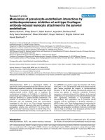
a. Case 1: 1 year after surgery on the right side, no improvement has yet occurredFigure 1
a. Case 1: 1 year after surgery on the right side, no improvement has yet occurred. She was operated upon at the age of 4 for
a C5,6 rupture C7,8T1 avulsion, when phrenic to suprascapular and contralateral C7 to lateral, medial and posterior cord neu-
rotization was carried out. The anterior deltoid was Grade3, the lateral deltoid Grade2, the posterior deltoid Grade0. Note
the supination deformity of the forearm, the extension deformity at the wrist and biceps cocontraction on attempted active
shoulder abduction. At this stage, with that degree of weak shoulder abduction, a humeral external rotation osteotomy or lat-
issimus dorsi to rotatotar cuff transfer will be of no avail. b. Case 1: 2 years after surgery. The anterior deltoid became Grade5,
the lateral deltoid Grade4 and the posterior deltoid Grade2. The wrist extensors improved from Grade1 up to Grade3. Some
degree of pronation has been regained at the forearm. At this stage, a humeral external rotation osteotomy or latissimus dorsi
to rotatotar cuff transfer will also be of no avail, because of extensive biceps cocontraction on attempted shoulder abduction.
c. Case7: 4 years after surgery on the right side. She was also operated upon at the age of 4 for a C5,6 rupture C7,8T1 avul-
sion, when phrenic to suprascapular and contralateral C7 to lateral, medial and posterior cord neurotization was carried out.
In addition to improvement of the deltoid and wrist extensors, some shoulder external rotation has been regained as the infra-
spinatus became Grade3. Biceps cocontraction on attempted shoulder abduction improved. She may therefore benefit from
secondary corrective procedures at the shoulder. In addition, a free functional gracilis transplantation has to be carried out to
power the weak finger flexors.
Journal of Brachial Plexus and Peripheral Nerve Injury 2006, 1:6 />Page 9 of 12
(page number not for citation purposes)
neurotization. Nerves to muscles with motor power 0
were neurotized, if the electromyogram showed scattered
motor unit action potentials on voluntary contraction
without interference pattern. This was arbitrarily taken as
a sign that the muscle bulk had not been completely
replaced by fibrosis and therefore function might be
restored to it. This muscle mass preserving effect was rec-
ognized by other authors [36]. The median muscle power
of the deltoid, rotator cuff, pectoralis major, latissimus
dorsi, biceps and triceps improved from Grades0–4 before
surgery to Grades2–5 after surgery. This was associated
with improved shoulder and elbow scores in 4 out of 8
cases. The median muscle power of the pronator teres,
supinator, the long wrist, finger and thumb extensors and
flexors and the intrinsic muscles of the hand improved
from Grades1–2 before surgery to Grades1–3 after sur-
gery. This was associated with an improved hand score in
4 out of 8 cases. Thus, nerve augmentation might improve
already regained muscle power.
Differential regeneration of muscles supplied by the same
nerve was the second observation. Exemplary for this were
the supra- and infraspinatus muscles, both supplied by
the suprascapular nerve. Regeneration of the supraspina-
tus muscle was superior to the infraspinatus. Superior
supraspinatus to infraspinatus regeneration was also
observed by other authors after suprascapular nerve graft-
ing or neurotization in the treatment of early brachial
plexus lesions [37,38]. In a third study on early repair of
obstetric brachial plexus lesions [39], it was concluded
that the restoration of a fair range of true glenohumeral
external rotation after neurotization of the suprascapular
nerve, whether by grafting from C5 or by nerve transfer of
the accessory nerve, was disappointingly low.
Differential regeneration of antagonistic muscles was the
third observation. Exemplary for this were the biceps and
triceps and the pronator teres and supinator. Superior
biceps to triceps recovery was observed by other authors
[21,40,41]. To account for this, it was noted [42] that
fatigue-sensitive afferents inhibited extensor but not
flexor motoneurons in humans. In a study on end-to-side
neurorrhaphy [43], it was shown that antagonistic nerves
had the ability to induce axonal regeneration, but muscle
incoordination prevented any useful function. With
regard to pronator teres and supinator recovery, in a his-
torical cohort of obstetric brachial plexus lesions, it was
observed that external rotation and supination were the
last to recover and recovered the least [44].
Differential regeneration of fibres within the same muscle
was the fourth observation. Exemplary for this was the
deltoid muscle, its anterior and middle fibres regenerating
better than its posterior fibres both before and after sur-
gery. In a retrospective study of 33 traumatic lesions of the
axillary nerve [45], deltoid muscle strength was noted to
be good or fair in 18 patients and poor in 15. The out-
come seemed to be better in isolated lesions than in com-
plex nerve lesions, in patients younger than 25 years
compared to older patients, in patients treated with neu-
rolysis compared to grafting, and when graft length was.
The outcome was less favourable when associated osteoar-
ticular lesions were present and when surgery was delayed
beyond six months. In another study [46], good or very
good deltoid function was obtained in 23 out of 25 direct
repairs of isolated axillary lesions, and in all 4 patients
with associated injury to the musculocutaneous nerve.
Only 4 good results were obtained in the 8 patients who
also had injuries to the suprascapular nerve. In both of
these studies no mention was made as to the regeneration
of the individual parts of the deltoid muscle. In an ana-
tomic study of the internal topographic features of the
axillary nerve [47], however, the axillary nerve was
divided into three segments. Proximal to the subscapula-
ris muscle, the axillary nerve formed a single nerve trunk.
Nerve fascicles to the deltoid muscle were identified at its
lateral part. In front of the subscapularis muscle, the axil-
lary nerve formed into the lateral and medial fasciculi
groups. Distal to the subscapularis muscle, the nerve
divided into anterior and posterior branches, which were
continuations of the lateral and medial fasciculi groups,
respectively. The anterior branch contained all fibers that
innervated the anterior and middle deltoid muscle. In
90% of cases, the posterior branch containsed part or all
nerve fibers to the posterior deltoid muscle. Nerve fibers
to the teres minor and cutaneous sensory fibers were
found in the posterior branch. It was concluded, that in
neurotization of the deltoid muscle, the best approach
was to match the donor nerve to the lateral fasciculi
group, which would give the highest percentage of rein-
nervation of the deltoid muscle. In a fourth study [48], it
was concluded that secondary compression of the axillary
nerve in the quadrangular space was a separate and com-
mon reason for impairment in children with brachial
plexus birth palsy and persistent weakness of the deltoid
muscle and might provide an important reason for early
intervention.
Differential regeneration of muscles having different pre-
operative motor powers was the fifth observation. Exem-
plary for this were the long wrist, finger and thumb
extensors and flexors and the intrinsic muscles of the
hand. Grade1 muscles had a better chance of improving to
Grades 1 or 3 and Grade2 muscles to Grades 3 or 4 than
Grade0 muscles to Grades 1 or 2. Thus functional
improvement was primarily expected in Grade2 muscles.
This is supported by the experimental observation [35]
that, in long lasting pre-suture denervation intervals,
changes within the muscle cells and the motor endplates
take place and are of outstanding importance for the poor
Journal of Brachial Plexus and Peripheral Nerve Injury 2006, 1:6 />Page 10 of 12
(page number not for citation purposes)
motor recovery. Especially after late nerve sutures the
arrival of axons within the muscle is by no means neces-
sarily followed by a sufficient recurrence of its function.
An interesting speculation is the role of the muscle target
organ as a promoting factor for nerve fibre regeneration in
nerve grafts, whether higher grade muscles are expected to
promote axonal growth more than lower grade muscles.
This was studied in rabbits, sheep and humans [49]. Excel-
lent regeneration of myelinated nerve fibres was observed
without target organ influence through the whole length
of the nerve graft, with an increase in the number of nerve
fibres up to fourfold at the distal end. In the sheep series
the additional contact with a muscle target organ for 6
months had a variable effect on the fibre population in
the distal end of the nerve graft. In humans, however, a
decrease of regenerating nerve fibres arriving at the distal
end of nerve grafts was noted. Interestingly, a possible role
of the muscle target organ as a promoting factor for nerve
fibre regeneration in nerve grafts came from biomaterial
research, where muscle-derived protein with molecular
mass of 77 kDa (MDP77) in artificial nerve grafts was
shown to promote motor nerve regeneration [50,51].
Improvement of cocontractions was the sixth observation.
Cocontractions improved in 3 out of 5 cases. In a clinical
study [52], cocontractions were classified into the follow-
ing types: TypeI involving the deltoid and biceps muscles,
TypeII involving the deltoid, biceps and triceps muscles,
TypeIII involving the biceps and triceps muscles, TypeIV
involving the deltoid, biceps, triceps and forearm muscles,
TypeV involving the deltoid, biceps and forearm muscles,
TypeVI involving the biceps, triceps and forearm muscles
and TypeVII involving the triceps and forearm muscles.
Cocontractions did not improve, but physical therapy or
operative treatment brought improvement in daily activi-
ties. Clinical severity of cross-reinnervation was correlated
to the severity of paralysis and in proportion to the ratio
of normally recovered nerve fibers and cross-reinnervated
nerve fibers. In our study, cocontractions were TypeI in 4
cases, TypeV in 1 case. Both this study and the improve-
ment of cocontractions in our study lend support to the
hypothesis that cocontractions are due to lack of collateral
rather than axial axonal sprouting.
Improvement of deformities was the seventh observation.
At the shoulder, in 4 out of 6 patients the internal rotation
adduction deformity disappeared; Putti's scapular eleva-
tion sign became negative. This observation is consistent
with other reports [53]. At the forearm, the supination
deformity disappeared; the pronation deformity per-
sisted, however. At the wrist, due to improvement in
extension, the flail wrist assumed a flexible extension
deformity in 1 of the 2 cases; in a further case, the flexor
carpi ulnaris, having improved to Grade4, was transferred
to the wrist extensors to correct the wrist drop deformity.
In conclusion, nerve augmentation of late brachial plexus
injuries is expected to improve muscle power in the
biceps, pectoral muscles, supraspinatus, anterior and lat-
eral deltoids, triceps and in forearm muscles with motor
power Grade2 or more. It is also expected to improve
cocontractions. It is less expected to improve infraspinatus
power. Therefore, after recovery of deltoid function,
patients should undergo a humeral derotation osteotomy
and a tendon transfer (see Figs 1a,1b and 1c). As it is less
expected to improve Grade0 or 1 forearm muscles, these
should be powered with a free muscle transfer [54]. But
the surgeon needn't use nerve grafts. The median, ulnar
and radial nerves may act as bridges for neurotization.
This was tried out experimentally [55] and confirmed clin-
ically [56]. For the same reason and contrary to other
reports [54,57], the transplanted muscle can be placed at
the forearm. Inspite of all of the above, the results
obtained are still inferior to those expected clinically.
First, we need to revise our end-to-side techniques. The
channel carrying capacity of the donor nerve, donor-recip-
ient neurorrhaphy and the augmented recipient has to be
increased by cotrophism [58], cotropism [59-62] and
cotransplantation [63-68]. Second, restoration of recipi-
ent muscle mass or regenerative potential should be
aimed at [69-71].
Finally, this study has several limitations. First, the sample
size is small, consisting only of 8 cases. Second, there are
no controls. These are necessary to rule out any natural
improvement of the lesion. Third, although we have tried
to increase muscle testing reliability through document-
ing it on both limbs by digital photographs, there is still
marked intra- and interobserver variability in testing mus-
cle power and cocontractions.
References
1. Kay SPJ: Obstetrical brachial palsy. Review article. Br J Plast
Surg 1998, 51:43-50.
2. Noetzel MJ, Park TS, Robinson S, Kaufman B: Prospective study of
recovery following neonatal brachial plexus injury. J Child Neu-
rol 2001, 16(7):488-492.
3. Hoeksma AF, ter Steeg AM, Nelissen RG, van Ouwerkerk WJ,
Lankhorst GJ, de Jong BA: Neurological recovery in obstetric
brachial plexus injuries: an historical cohort study. Dev Med
Child Neurol 2004, 46(2):76-83.
4. O'Brien DF, Park TS, Noetzel MJ, Weatherly T: Management of
birth brachial plexus palsy. Childs Nerv Syst 2006, 22(2):103-112.
5. Haerle M, Gilbert A: Management of complete obstetric bra-
chial plexus lesions. J Pediatr Orthop 2004, 24(2):194-200.
6. Gilbert A, Pivato G, Kheiralla T: Long-term results of primary
repair of brachial plexus lesions in children. Microsurgery 2006,
26(4):334-342.
7. Birch R, Ahad N, Kono H, Smith S: Repair of obstetric brachial
plexus palsy: results in 100 children. J Bone Joint Surg Br 2005,
87(8):1089-1095.
8. Grossman JA, Price AE, Tidwell MA, Ramos LE, Alfonso I, Yaylali I:
Outcome after later combined brachial plexus and shoulder
surgery after birth trauma. J Bone Joint Surg Br 2003,
85(8):1166-1168.
9. Partridge C, Edwards S: Obstetric brachial plexus palsy: increas-
ing disability and exacerbation of symptoms with age. Physi-
other Res Int 2004, 9(4):157-163.
Journal of Brachial Plexus and Peripheral Nerve Injury 2006, 1:6 />Page 11 of 12
(page number not for citation purposes)
10. Waters PM, Bae DS: The effect of derotational humeral osteot-
omy on global shoulder function in brachial plexus birth
palsy. J Bone Joint Surg Am 2006, 88(5):1035-1042.
11. Aydin A, Ozkan T, Onel D: Does preoperative abduction value
affect functional outcome of combined muscle transfer and
release procedures in obstetrical palsy patients with shoul-
der involvement? BMC Musculoskelet Disord 5:25. 2004 Aug 3
12. Kirkos JM, Kyrkos MJ, Kapetanos GA, Haritidis JH: Brachial plexus
palsy secondary to birth injuries. J Bone Joint Surg Br 2005,
87(2):231-235.
13. Pagnotta A, Haerle M, Gilbert A: Long-term results on abduction
and external rotation of the shoulder after latissimus dorsi
transfer for sequelae of obstetric palsy. Clin Orthop Relat Res
2004:199-205.
14. Kambhampati SB, Birch R, Cobiella C, Chen L: Posterior subluxa-
tion and dislocation of the shoulder in obstetric brachial
plexus palsy. J Bone Joint Surg Br 2006, 88(2):213-219.
15. Viterbo F, Teixeira E, Hoshino K, Padovani CR: End-to-side neur-
orrhaphy with and without perineurium. Sao Paulo Med J 1998,
116(5):1808-1814.
16. Isaacs J, Allen D, Chen LE, Nunley J 2nd: Reverse end-to-side neu-
rotization. J Reconstr Microsurg 2005, 21(1):43-8. discussion 49–50
17. Kerns JM, Sladek EH, Malushte TS, Bach H, El-Hassan B, Kitidumrong-
sook P, Kroin JS, Shott S, Gantsoudes G, Gonzalez MH: End-to-side
nerve grafting of the tibial nerve to bridge a neuroma-in-con-
tinuity. Microsurgery 2005, 25(2):155-164. discussion 164–166
18. Grossman JA, DiTaranto P, Yaylali I, Alfonso I, Ramos LE, Price AE:
Shoulder function following late neurolysis and bypass graft-
ing for upper brachial plexus birth injuries. J Hand Surg [Br]
2004, 29(4):356-358.
19. Amr SM, Moharram AN: Repair of brachial plexus lesions by
end-to-side side-to-side grafting neurorrhaphy: experience
based on 11 cases. Microsurgery 2005, 25(2):126-146.
20. Chow BCL, Blaser S, Clarke HM: Predictive value of computed
tomographic myelography in obstetrical brachial plexus
palsy. Plast Reconstr Surg 2000, 106:971-977.
21. Terzis JK, Papakonstantinou KC: The surgical treatment of bra-
chial plexus injuries in adults. Plast Reconstr Surg 2000,
106:1097-1122.
22. Hierner R, Becker M, Berger A: Indications and results of opera-
tive treatment in birth-related brachial plexus injuries. Hand-
chir Mikrochir Plast Chir 2005, 37(5):323-331.
23. Millesi H, Meissl G, Berger A: Further experience with interfas-
cicular grafting of the median, ulnar and radial nerves. J Bone
Joint Surg (Am) 1981, 58-A:209-218.
24. Arcand MA, Reider B: Shoulder and upper arm. In The orthopaedic
physical examination 2nd edition. Edited by: Reider B. Philadelphia,
Elsevier; 2004:17-66.
25. Tennent TD, Beach WR, Meyers JF: A review of the special tests
associated with shoulder examination. Part I: the rotator
cuff tests. Am J Sports Med 2003, 31(1):154-160.
26. Richards DP, Burkhart SS, Lo IK: Subscapularis tears: arthro-
scopic repair techniques. Orthop Clin North Am 2003,
34(4):485-498. Review
27. McFarland EG, Selhi HS, Keyurapan E: Clinical evaluation of
impingement: what to do and what works. J Bone Joint Surg Am
2006, 88:432-441.
28. Amr SM: Traumatic brachial plexus palsy; a report of 30 cases.
Medical Journal of Cairo University 2000, 68:715-730.
29. Hattori Y, Doi K, Toh S, Baliarsing AS: Surgical approach to the
spinal accessory nerve for brachial plexus reconstruction. J
Hand Surg [Am] 2001, 26(6):1073-1076.
30. Hentz VR: Microneural reconstruction of the brachial plexus.
In Operative hand surgery Edited by: Green DP, Hotchkiss RN. New
York Edinburgh London Melbourne Tokyo: Churchill-Livingstone;
1993:1223-1252.
31. Leffert RD: Brachial plexus. In Operative hand surgery Edited by:
Green DP, Hotchkiss RN. New York Edinburgh London Melbourne
Tokyo: Churchill-Livingstone; 1993:1483-1516.
32. Millesi H:
Chirurgie der peripheren Nerven. In Spezieller Teil, 2.
Plexus brachialis und seine Aeste Muenchen Wien Baltimore:
Urban&Schwarzenberg; 1992:79-112.
33. Mcguiness CN, Kay SP: The prespinal route in contralateral C7
nerve root transfer for brachial plexus avulsion injuries. J
Hand Surg [Br] 2002, 27(2):159-160.
34. Oberlin C, Ameur NE, Teboul F, Beaulieu JY, Vacher C: Restoration
of elbow flexion in brachial plexus injury by transfer of ulnar
nerve fascicles to the nerve to the biceps muscle. Tech Hand
Up Extrem Surg 2002, 6(2):86-90.
35. Richter HP: Impairment of the restoration of motor function
after delayed nerve suture. Fortschr Med 100(10):414-418. 1982
Mar 11
36. Oswald TM, Zhang F, Lei MP, Gerzenshtein J, Lineaweaver WC: Mus-
cle flap mass preservation with end-to-side neurorrhaphy: an
experimental study. J Reconstr Microsurg 2004, 20(6):483-488.
37. Malessy MJ, de Ruiter GC, de Boer KS, Thomeer RT: Evaluation of
suprascapular nerve neurotization after nerve graft or trans-
fer in the treatment of brachial plexus traction lesions. J Neu-
rosurg 2004, 101(3):377-389.
38. Leechavengvongs S, Witoonchart K, Uerpairojkit C, Thuvasethakul P,
Malungpaishrope K: Combined nerve transfers for C5 and C6
brachial plexus avulsion injury. J Hand Surg [Am] 2006,
31(2):183-189.
39. Pondaag W, de Boer R, van Wijlen-Hempel MS, Hofstede-Buitenhuis
SM, Malessy MJ: External rotation as a result of suprascapular
nerve neurotization in obstetric brachial plexus lesions. Neu-
rosurgery 2005, 57(3):530-537.
40. Martin PG, Smith JL, Butler JE, Gandevia SC, Taylor JL: Fatigue-sen-
sitive afferents inhibit extensor but not flexor motoneurons
in humans. J Neurosci 26(18):4796-4802. 2006, May 3
41. Kandenwein JA, Kretschmer T, Engelhardt M, Richter HP, Antoniadis
G: Surgical interventions for traumatic lesions of the brachial
plexus: a retrospective study of 134 cases.
J Neurosurg 2005,
103(4):614-621.
42. Bentolila V, Nizard R, Bizot P, Sedel L: Complete traumatic bra-
chial plexus palsy. Treatment and outcome after repair. J
Bone Joint Surg Am 1999, 81(1):20-28.
43. Lutz BS, Chuang DC, Hsu JC, Ma SF, Wei FC: Selection of donor
nerves – an important factor in end-to-side neurorrhaphy. Br
J Plast Surg 2000, 53(2):149-154.
44. Hoeksma AF, ter Steeg AM, Nelissen RG, van Ouwerkerk WJ,
Lankhorst GJ, de Jong BA: Neurological recovery in obstetric
brachial plexus injuries: an historical cohort study. Dev Med
Child Neurol 2004, 46(2):76-83.
45. Wehbe J, Maalouf G, Habanbo J, Chidiac RM, Braun E, Merle M: Sur-
gical treatment of traumatic lesions of the axillary nerve. A
retrospective study of 33 cases. Acta Orthop Belg 2004,
70(1):11-18.
46. Zhao X, Hung LK, Zhang GM, Lao J: Applied anatomy of the axil-
lary nerve for selective neurotization of the deltoid muscle.
Clin Orthop Relat Res 2001:244-251.
47. Alnot JY, Valenti P: Surgical repair of the axillary nerve. Apro-
pos of 37 cases. Int Orthop 1991, 15(1):7-11.
48. Adelson PD, Nystrom NA, Sclabassi R: Entrapment neuropathy
contributing to dysfunction after birth brachial plexus inju-
ries. J Pediatr Orthop 2005, 25(5):592-597.
49. Frey M, Koller R, Liegl C, Happak W, Gruber H: Role of a muscle
target organ on the regeneration of motor nerve fibres in
long nerve grafts: a synopsis of experimental and clinical
data. Microsurgery 1996, 17(2):80-88.
50. Itoh S, Fujimori KE, Uyeda A, Matsuda A, Kobayashi H, Shinomiya K,
Tanaka J, Taguchi T: Long-term effects of muscle-derived pro-
tein with molecular mass of 77 kDa (MDP77) on nerve
regeneration. J Neurosci Res 81(5):730-738. 2005 Sep 1
51. Itoh S, Uyeda A, Hukuoka Y, Fujimori KE, Matsuda A, Ichinose S,
Kobayashi H, Shinomiya K, Tanaka J, Taguchi T: Muscle-specific
protein MDP77 specifically promotes motor nerve regener-
ation in rats. Neurosci Lett
360(3):175-177. 2004 Apr 29
52. Yagi I: Clinical study of cross-reinnervation in obstetrical
paralysis. Nippon Seikeigeka Gakkai Zasshi 1984, 58(8):761-778.
53. Birch R, Ahad N, Kono H, Smith S: Repair of obstetric brachial
plexus palsy: results in 100 children. J Bone Joint Surg Br 2005,
87(8):1089-1095.
54. Hattori Y, Doi K, Ikeda K, Pagsaligan JM, Watanabe M: Restoration
of prehension using double free muscle technique after com-
plete avulsion of brachial plexus in children: a report of three
cases. J Hand Surg [Am] 2005, 30(4):812-819.
55. Shah MH, Kasabian AK, Karp NS, Kolker AR, Dublin BA, Zhang L,
Sakuma J: Axonal regeneration through an autogenous nerve
bypass: an experimental study in the rat. Ann Plast Surg 1997,
38(4):408-414. discussion 414-5
Publish with BioMed Central and every
scientist can read your work free of charge
"BioMed Central will be the most significant development for
disseminating the results of biomedical research in our lifetime."
Sir Paul Nurse, Cancer Research UK
Your research papers will be:
available free of charge to the entire biomedical community
peer reviewed and published immediately upon acceptance
cited in PubMed and archived on PubMed Central
yours — you keep the copyright
Submit your manuscript here:
/>BioMedcentral
Journal of Brachial Plexus and Peripheral Nerve Injury 2006, 1:6 />Page 12 of 12
(page number not for citation purposes)
56. McCallister WV, Cober SR, Norman A, Trumble TE: Using intact
nerve to bridge peripheral nerve defects: an alternative to
the useof nerve grafts. J Hand Surg [Am] 2001, 26(2):315-325.
57. Doi K: New reconstructive procedure for brachial plexus
injury. Clinics in Plastic Surgery 1997, 24(1):75-85.
58. McConnell MP, Dhar S, Naran S, Nguyen T, Bradshaw RA, Evans GR:
In vivo induction and delivery of nerve growth factor, using
HEK-293 cells. Tissue Eng 2004, 10(9–10):1492-1501.
59. Itoh S, Matsuda A, Kobayashi H, Ichinose S, Shinomiya K, Tanaka J:
Effects of a laminin peptide (YIGSR) immobilized on crab-
tendon chitosan tubes on nerve regeneration. J Biomed Mater
Res B Appl Biomater in press. 2005 Mar 7
60. Belkas JS, Shoichet MS, Midha R: Peripheral nerve regeneration
through guidance tubes. Neurol Res 2004, 26(2):151-160.
61. Fansa H, Keilhoff G: Comparison of different biogenic matrices
seeded with cultured Schwann cells for bridging peripheral
nerve defects. Neurol Res 2004, 26(2):167-173.
62. Nishiura Y, Brandt J, Nilsson A, Kanje M, Dahlin LB: Addition of cul-
tured Schwann cells to tendon autografts and freeze-thawed
muscle grafts improves peripheral nerve regeneration. Tissue
Eng 2004, 10(1–2):157-164.
63. Komiyama T, Nakao Y, Toyama Y, Vacanti CA, Vacanti MP, Ignotz RA:
Novel technique for peripheral nerve reconstruction in the
absence of an artificial conduit. J Neurosci Methods
134(2):133-140. 2004 Apr 30
64. Reier PJ: Cellular Transplantation Strategies for Spinal Cord
Injury and Translational Neurobiology. Neurorx 2004,
1(4):424-451.
65. Mimura T, Dezawa M, Kanno H, Sawada H, Yamamoto I: Peripheral
nerve regeneration by transplantation of bone marrow stro-
mal cell-derived Schwann cells in adult rats. J Neurosurg 2004,
101(5):806-812.
66. Heine W, Conant K, Griffin JW, Hoke A: Transplanted neural
stem cells promote axonal regeneration through chronically
denervated peripheral nerves. Exp Neurol 2004,
189(2):231-240.
67. Tohill M, Terenghi G: Stem-cell plasticity and therapy for inju-
ries of the peripheral nervous system. Biotechnol Appl Biochem
2004,
40(Pt 1):17-24.
68. Dezawa M, Kanno H, Hoshino M, Cho H, Matsumoto N, Itokazu Y,
Tajima N, Yamada H, Sawada H, Ishikawa H, Mimura T, Kitada M,
Suzuki Y, Ide C: Specific induction of neuronal cells from bone
marrow stromal cells and application for autologous trans-
plantation. J Clin Invest 2004, 113(12):1701-1710.
69. Sarig R, Baruchi Z, Fuchs O, Nudel U, Yaffe D: Regeneration and
transdifferentiation potential of muscle-derived stem cells
propagated as myospheres. When inoculated into injured
muscle, myosphere-derived cells participated in regenera-
tion, forming multinucleated cross-striated mature fibers.
Stem Cells 2006, 24(7):1769-1778.
70. Vilquin JT: Myoblast transplantation: clinical trials and per-
spectives. Mini-review Acta Myol 2005, 24(2):119-127.
71. Matziolis G, Winkler T, Schaser K, Wiemann M, Krocker D, Tuischer
J, Perka C, Duda GN: Autologous bone marrow-derived cells
enhance muscle strength following skeletal muscle crush
injury in rats. Tissue Eng 2006, 12(2):361-367.