Báo cáo y học: "urgical correction of unsuccessful derotational humeral osteotomy in obstetric brachial plexus palsy: Evidence of the significance of scapular deformity in the pathophysiology of the medial rotation contracture" ppt
Bạn đang xem bản rút gọn của tài liệu. Xem và tải ngay bản đầy đủ của tài liệu tại đây (586.02 KB, 7 trang )
BioMed Central
Page 1 of 7
(page number not for citation purposes)
Journal of Brachial Plexus and
Peripheral Nerve Injury
Open Access
Research article
Surgical correction of unsuccessful derotational humeral
osteotomy in obstetric brachial plexus palsy: Evidence of the
significance of scapular deformity in the pathophysiology of the
medial rotation contracture
Rahul K Nath*, Sonya E Melcher and Melia Paizi
Address: Texas Nerve and Paralysis Institute, 2201 W. Holcombe Blvd., Houston, TX, USA
Email: Rahul K Nath* - ; Sonya E Melcher - ; Melia Paizi -
* Corresponding author
Abstract
Background: The current method of treatment for persistent internal rotation due to the medial
rotation contracture in patients with obstetric brachial plexus injury is humeral derotational
osteotomy. While this procedure places the arm in a more functional position, it does not attend
to the abnormal glenohumeral joint. Poor positioning of the humeral head secondary to elevation
and rotation of the scapula and elongated acromion impingement causes functional limitations
which are not addressed by derotation of the humerus. Progressive dislocation, caused by the
abnormal positioning and shape of the scapula and clavicle, needs to be treated more directly.
Methods: Four patients with Scapular Hypoplasia, Elevation And Rotation (SHEAR) deformity
who had undergone unsuccessful humeral osteotomies to treat internal rotation underwent
acromion and clavicular osteotomy, ostectomy of the superomedial border of the scapula and
posterior capsulorrhaphy in order to relieve the torsion developed in the acromio-clavicular
triangle by persistent asymmetric muscle action and medial rotation contracture.
Results: Clinical examination shows significant improvement in the functional movement possible
for these four children as assessed by the modified Mallet scoring, definitely improving on what was
achieved by humeral osteotomy.
Conclusion: These results reveal the importance of recognizing the presence of scapular
hypoplasia, elevation and rotation deformity before deciding on a treatment plan. The Triangle Tilt
procedure aims to relieve the forces acting on the shoulder joint and improve the situation of the
humeral head in the glenoid. Improvement in glenohumeral positioning should allow for better
functional movements of the shoulder, which was seen in all four patients. These dramatic
improvements were only possible once the glenohumeral deformity was directly addressed
surgically.
Published: 27 December 2006
Journal of Brachial Plexus and Peripheral Nerve Injury 2006, 1:9 doi:10.1186/1749-7221-1-
9
Received: 06 November 2006
Accepted: 27 December 2006
This article is available from: />© 2006 Nath et al; licensee BioMed Central Ltd.
This is an Open Access article distributed under the terms of the Creative Commons Attribution License ( />),
which permits unrestricted use, distribution, and reproduction in any medium, provided the original work is properly cited.
Journal of Brachial Plexus and Peripheral Nerve Injury 2006, 1:9 />Page 2 of 7
(page number not for citation purposes)
Background
Obstetric brachial plexus injury (OBPI) has been
described as a discrete entity since 1754 [1]. The patho-
physiology of the secondary deformities encountered in
this population was described succinctly in 1905 by Whit-
man who wrote that the large majority of internal rotation
and subluxation deformities of the shoulder in children
with obstetric brachial plexus injuries were caused by
fibrosis and contractures developed as a consequence of
the neurological injury [2]. The medial rotation contrac-
ture (MRC) is the most significant secondary shoulder
deformity in children with severe OBPI, requiring surgery
in more than one third of patients whose injury did not
resolve spontaneously [3].
The current surgical approach to treating persistent MRC
in OBPI patients is derotational humeral osteotomy [4-
12] or anterior capsule release [13]. Humeral osteotomy
attempts to improve the patient's passive range of external
rotation, but ignores the bone deformity at the root of per-
sistent MRC, and does nothing to address the attendant
subluxation of the humeral head within the glenoid fossa.
Anterior capsule release may result in excessive external
rotation positioning of the humerus with attendant loss of
internal rotation and midline functioning [13].
Scapular hypoplasia, elevation and rotation (SHEAR)
deformity [14] is the ultimate bony manifestation of the
muscular fibrosis described by Whitman, and is present in
the majority of OBPI patients exhibiting MRC. The SHEAR
deformity must be accounted for in any surgical correc-
tion of the MRC, and humeral osteotomy as a strategy for
bony correction does not do so (Figure 1).
The presentation of weakness of the deltoid and external
shoulder rotators caused by the common C5 injury seen
in OBPI immediately affects growth of both the muscles
and bones. Formation of contractures and consequent
asymmetric muscle action on the developing bony ele-
ments of the shoulder results in bone deformation of the
scapula and humerus. The scapula not only elevates and
rotates laterally, but also becomes hypoplastic with flat-
tening of the glenoid fossa and hooking of the acromion
process. The clavicle and acromion process impinge upon
the humeral head due to the abnormal positioning of the
scapula and associated acromio-clavicular triangle (ACT),
with its sides defined by the clavicular shaft and the
acromion process and its base by an imaginary line con-
necting their medial ends. Functionally debilitating effects
include medial rotation and posterior and inferior sublux-
ation of the humerus within the glenoid fossa.
The abnormal migration of the scapula disrupts the nor-
mal anatomic relationships of the humeral head, the gle-
noid fossa and the acromio-clavicular triangle.
Impingement of the distal acromio-clavicular triangle
against the humeral head limits external rotation of the
arm and shoulder. Without addressing the joint derange-
ment, procedures such as humeral osteotomy are likely to
fail or have significant rates of recurrence. To our knowl-
edge there is no published method for correcting recur-
rence of the medial rotation contracture other than
repeated humeral osteotomy.
A novel osseous procedure, named the "Triangle Tilt,"
releases and tilts the acromio-clavicular plane back to neu-
tral thus relieving the impingement of the acromio-clavic-
ular triangle on the humeral head. The humeral head may
now reposition passively into the neutral position within
the glenoid fossa. Here we report the use of this technique
to treat 4 children who had undergone unsuccessful
humeral osteotomies.
Methods
During a 10 month period between October 2005 and
August 2006, 73 obstetric brachial plexus patients with
persistent internal rotation underwent Triangle Tilt sur-
gery. Four of these patients had undergone previous
humeral osteotomy (performed by board-certified pediat-
ric orthopedic surgeons) with complete failure of the pro-
cedure. All 4 had residual MRC with SHEAR deformity,
and underwent Triangle Tilt surgery as a salvage procedure
for unsuccessful humeral osteotomy.
The presence of SHEAR deformity was determined by
physical examination and confirmed by 3D-CT (com-
puted tomography) if possible [14]. Elevation of the scap-
ula was estimated clinically. Scapular elevation, defined as
the percentage of scapula visible above the clavicle and
caused by downward and anterior rotation, was quanti-
tated on a 3D-reconstruction of the CT and confirmed the
severity of the underlying SHEAR deformity.
Version and subluxation were measured on axial CT or
MRI images. A scapular line was drawn connecting the
medial margin of the scapula to the middle of the glenoid
fossa on transverse CT or MRI (magnetic resonance imag-
ing) images at the mid-glenoid level. The glenoscapular
angle between the scapular line and a line connecting the
base of the anterior labrum and posterior labrum was
measured according to Friedman et al. [15]. 90° were sub-
tracted from the posteromedial quadrant angle to deter-
mine version. The degree of humeral head subluxation
was determined using the same scapular line and a per-
pendicular line traversing the humeral head at its greatest
diameter. The distance of the scapular line to the anterior
portion of the head and the greatest diameter of the
humeral head were measured. The ratio of these distances
multiplied by 100 determines percent subluxation.
Journal of Brachial Plexus and Peripheral Nerve Injury 2006, 1:9 />Page 3 of 7
(page number not for citation purposes)
Two of the patients were girls, ages 7.9 and 10.4 years, and
2 were boys, ages 10.4 and 11.9 years at the time of sur-
gery. Two patients had undergone nerve surgery in
infancy. Prior to humeral osteotomy, all 4 had undergone
muscle contracture release, tendon transfers, and decom-
pression of the axillary nerve at the quandrangular space
[16-19]. Improvements in abduction from muscle surgery
were maintained at the time of surgery. The medial rota-
tion posture at rest was unaddressed by humeral osteot-
omy and was not responsive to additional therapy and
splinting.
Shoulder movements were assessed preoperatively and
postoperatively by evaluating video recordings of stand-
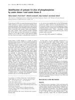
CT images showing SHEAR deformity present after humeral osteotomyFigure 1
CT images showing SHEAR deformity present after humeral osteotomy. Ten year old boy after unsuccessful
humeral osteotomy with right-sided SHEAR deformity demonstrated in 3D CT anterior view (above) and posterior subluxa-
tion demonstrated in axial view (below).
Journal of Brachial Plexus and Peripheral Nerve Injury 2006, 1:9 />Page 4 of 7
(page number not for citation purposes)
ardized movements according to the modified Mallet clas-
sification [20]. Additional measurements were made of
the angle of the humerus to the trunk during the hand-to-
mouth movement (trumpeter sign) and the angle of fore-
arm supination as a more sensitive determination of func-
tional ability. All assessments were made independently
of the surgeon and principal author.
Surgical Procedure
The Triangle Tilt surgery consisted most importantly of
four components. First, osteotomy separated the clavicle
at the junction of the middle and distal thirds. Second,
osteotomy of the acromion process at its junction with the
spine of the scapula was performed. Then, thirdly, ostec-
tomy of the superomedial angle of the scapula was
enacted. Finally, the extremity was splinted in adduction,
5° of external rotation and full forearm supination (90°).
Splinting was maintained for 6 weeks after which time the
splint was worn only at night for an additional 3 months.
Minor elements of the procedure included bone grafting
of the acromion process osteotomy site, and semi-rigid
fixation of the clavicular osteotomy segments to prevent
nonunion. Since all four of these children had proven
shoulder instability, particularly subluxation, diagnosed
by CT or positional MRI imaging, posterior glenohumeral
capsulorrhaphy was performed.
The same surgeon performed all surgical procedures
(RKN).
Results
The preoperative and postoperative Mallet scores for these
patients are presented in Table 1 with representative pho-
tographs in Figure 2. The follow-up periods were 4 to 14
months with two of the four patients still undergoing
nighttime splinting. There were, however, clear improve-
ments in shoulder function which were not previously
achieved with humeral osteotomy. Mallet score before Tri-
angle Tilt surgery was 10, 16, 12 and 13. After surgery,
these patients improved to 17, 19, 18, and 19, respec-
tively. All four children were able to supinate to 60° or
greater and were able to bring their hands to their mouths
with a trumpeter sign of less than 45° postoperatively.
Before surgery, no child was able to supinate to greater
than 30° and the smallest trumpeter sign angle was 70°.
Forearm supination increased secondarily to improved
external rotation at the shoulder, and provided a conven-
ient indicator of changes in external rotation. Improve-
ments were also noticeable in the manner in which the
arm was held at rest (Figure 2C and 2F).
Discussion
The developmental consequences of an obstetric brachial
plexus injury, medial rotation contracture and progressive
posterior dislocation of the shoulder, have serious conse-
quences for shoulder function. Most commonly, the treat-
ment method is humeral osteotomy, which places the
arm in a more functional, externally rotated position.
Though this procedure can give functional improvement,
a significant proportion of children are not helped by this
salvage procedure due to the fact that it does not address
the bone deformities at the root of the progressive poste-
rior dislocation and poor shoulder movement. The pres-
ence of unaddressed SHEAR deformity guarantees the
continued impingement of the acromion upon the
humeral head which can lead to recurrence of the debili-
tating internal rotation. Only in the absence of significant
SHEAR is humeral osteotomy a viable treatment option.
The improvements possible with the Triangle Tilt surgery
are clear from the preoperative and postoperative photo-
graphs shown in Figure 2. Mallet functional scores quan-
titatively show the improvements of all four patients who
had previous humeral osteotomies (Table 1). One patient
improved Mallet score by 3, another by 7 and the remain-
ing two by 6 points. Satisfactory changes in function are
reflected in the measured angles of forearm supination
(improvement by 150, 50, 165 and 90 degrees respec-
tively) and flaring of the elbow during the hand to mouth
movement (80, 60, 35 and 45 degrees). Because of the
apparent pronation deformity due to MRC pre-surgically
the neutral position was inaccessible and so supination
increased by more than 90° in three out of four patients.
The degree of torsion caused by contractures around the
shoulder is manifest during surgery, and observation of
how the bones respond during surgery reveals the forces
still acting on the glenohumeral joint after humeral oste-
otomy. When released by Triangle Tilt, the highly abnor-
mal bony framework around the injured shoulder and the
significant intraosseous torque results in immediate cla-
vicular and acromial movements. Separation of the distal
acromio-clavicular triangle from the abnormal medial
structures relieves the torsion developed over time.
The clavicle is abnormally twisted due to scapular migra-
tion, and the distal and proximal clavicle segments are
intraoperatively observed to rapidly unwind after osteot-
omy. Significant movement also follows osteotomy of the
acromion process, with the body of the acromion process
and the medial margin of the acromion rapidly separat-
ing, and the distal segment moving both inferiorly and
posteriorly. The distal acromio-clavicular triangle
becomes normalized, and so does the humeral head
through its relationship to the lateral structures. With the
release of the abnormal torque and the leveling of the
acromio-clavicular triangle, the glenohumeral axis returns
towards neutral. This improves clinical arm positioning
and movement possibilities.
Journal of Brachial Plexus and Peripheral Nerve Injury 2006, 1:9 />Page 5 of 7
(page number not for citation purposes)
Table 1: Radiographic classifications of glenohumeral deformity
Preoperative values Postoperative values
Patient
no.
Subluxation Version %
Scapula
visible
over
clavicle
Glenohum
eral
deformity*
Age at
surgery
Abduction External
rotation
Hand
to
Neck
Hand
to
Spine
Hand
to
Mouth
Hand
to
Mouth
angle
Supination
angle
Total
Mallet
Abduction External
rotation
Hand
to
Neck
Hand
to
Spine
Hand
to
Mouth
Hand
to
Mouth
angle
Supination
angle
Total
Mallet
Follow-up
(months)
1 13.5 -27 N/A III 10.4 4 1 2 2 1 120 -90
10
433344060
17
6
2 22.2 -24 25 III 11.9 4 3 3 3 3 70 30
16
444341080
19
9
3 45.7 -28 N/A III 7.9 4 1 3 2 2 110 -90
12
433242035
16
14
4 59.7 -42 41 V 10.4 4 2 4 2 1 135 0
13
444341090
19
4
Mallet scores and functional hand to mouth and forearm supination angles in patients who following failed humeral osteotomy recently underwent Triangle Tilt surgery. *Glenohumeral deformity classification according to Waters [21]. N/A data
not available.
Journal of Brachial Plexus and Peripheral Nerve Injury 2006, 1:9 />Page 6 of 7
(page number not for citation purposes)
Functional Improvement with Triangle Tilt surgeryFigure 2
Functional Improvement with Triangle Tilt surgery. Pictures of 10 year old girl who had previously undergone an
unsuccessful humeral osteotomy, pre (a through c) and 6 months post (d through f) Triangle Tilt surgery. Panels a and d show
decreased trumpet sign during the hand to mouth movement. Panels b and e show improved supination. Panels c and f show
the improvement in resting arm position.
Publish with Bio Med Central and every
scientist can read your work free of charge
"BioMed Central will be the most significant development for
disseminating the results of biomedical research in our lifetime."
Sir Paul Nurse, Cancer Research UK
Your research papers will be:
available free of charge to the entire biomedical community
peer reviewed and published immediately upon acceptance
cited in PubMed and archived on PubMed Central
yours — you keep the copyright
Submit your manuscript here:
/>BioMedcentral
Journal of Brachial Plexus and Peripheral Nerve Injury 2006, 1:9 />Page 7 of 7
(page number not for citation purposes)
Conclusion
The four patients presented here demonstrate how impor-
tant it is to recognize and treat the bone deformity. If
SHEAR is present, it must be accounted for in the surgical
plan. The design of the Triangle Tilt procedure aims at
improving the position of the humeral head in the gle-
noid fossa by eliminating the impingement occurring in
the SHEAR deformity. Long-term improved function of
the shoulder is the expected consequence of improved
glenohumeral anatomy. Only months after surgery, these
four patients who had Triangle Tilt surgery to address the
SHEAR as well as the medial rotation contracture show
dramatically improved function.
Competing interests
The authors declare that they have no competing interests.
Authors' contributions
RKN conceived of the study, performed all surgeries, and
edited the manuscript. MP collected and analysed data,
created figures, and edited the manuscript. SEM collected
and analysed data, and drafted the manuscript.
References
1. Smellie W: A collection of cases and observations in mid-
wifery. Volume 2. London , Printed for D. Wilson and T. Durham;
1754.
2. Whitman R: VIII. The treatment of congenital and acquired
luxations at the shoulder in childhood. Ann Surg 1905,
42(1):110-115.
3. Birch R: Medial rotation contracture and posterior disloca-
tion of the shoulder. In Brachial Plexus Injuries First edition. Edited
by: Gilbert A. London , Martin Dunitz, Ltd.; 2001:249-259.
4. Rogers MH: An operation for the correction of the deformity
due to "obstetrical paralysis". Boston Medical and Surgical Journal
1916, 174(5):163-164.
5. Waters PM, Bae DS: The effect of derotational humeral osteot-
omy on global shoulder function in brachial plexus birth
palsy. J Bone Joint Surg Am 2006, 88(5):1035-1042.
6. Akinci M, Ay S, Kamiloglu S, Ercetin O: [External rotation osteot-
omy of the humerus for the treatment of shoulder problems
secondary to obstetric brachial plexus palsy]. Acta Orthop Trau-
matol Turc 2005, 39(4):328-333.
7. Al Zahrani S: Modified rotational osteotomy of the humerus
for Erb's palsy. Int Orthop 1993, 17(3):202-204.
8. Al-Qattan MM: Rotation osteotomy of the humerus for Erb's
palsy in children with humeral head deformity. J Hand Surg
[Am] 2002, 27(3):479-483.
9. Faysse R: [Obstetrical paralysis of the brachial plexus. II.
Therapeutics. Treatment of sequelae. d. Humeral derota-
tion osteotomy in the sequelae]. Revue de chirurgie orthopédique
et réparatrice de l'appareil moteur 1972, 58:Suppl 1:187-92.
10. Goddard NJ, Fixsen JA: Rotation osteotomy of the humerus for
birth injuries of the brachial plexus. J Bone Joint Surg Br 1984,
66(2):257-259.
11. Kirkos JM, Papadopoulos IA: Late treatment of brachial plexus
palsy secondary to birth injuries: rotational osteotomy of the
proximal part of the humerus. J Bone Joint Surg Am
1998,
80(10):1477-1483.
12. Ruhmann O, Gosse F, Schmolke S, Flamme C, Wirth CJ: Osteotomy
of the humerus to improve external rotation in nine patients
with brachial plexus palsy. Scand J Plast Reconstr Surg Hand Surg
2002, 36(6):349-355.
13. Pearl ML: Arthroscopic release of shoulder contracture sec-
ondary to birth palsy: an early report on findings and surgical
technique. Arthroscopy 2003, 19(6):577-582.
14. Nath RK, Paizi M: Scapular Deformity in Obstetric Brachial
Plexus Palsy: A New Finding. Surg Radiol Anat 2007 in press.
15. Friedman RJ, Hawthorne KB, Genez BM: The use of computerized
tomography in the measurement of glenoid version. J Bone
Joint Surg Am 1992, 74(7):1032-1037.
16. Hoffer MM, Phipps GJ: Closed reduction and tendon transfer
for treatment of dislocation of the glenohumeral joint sec-
ondary to brachial plexus birth palsy. J Bone Joint Surg Am 1998,
80(7):997-1001.
17. Safoury Y: Muscle transfer for shoulder reconstruction in
obstetrical brachial plexus lesions. Handchir Mikrochir Plast Chir
2005, 37(5):332-336.
18. Waters PM, Bae DS: Effect of tendon transfers and extra-artic-
ular soft-tissue balancing on glenohumeral development in
brachial plexus birth palsy. J Bone Joint Surg Am 2005,
87(2):320-325.
19. Adelson PD, Nystrom NA, Sclabassi R: Entrapment neuropathy
contributing to dysfunction after birth brachial plexus inju-
ries. J Pediatr Orthop 2005, 25(5):592-597.
20. Mallet J: [Obstetrical paralysis of the brachial plexus. II. Ther-
apeutics. Treatment of sequelae. Priority for the treatment
of the shoulder. Method for the expression of results]. Revue
de chirurgie orthopédique et réparatrice de l'appareil moteur 1972,
58:Suppl 1:166-8.
21. Waters PM, Smith GR, Jaramillo D: Glenohumeral deformity sec-
ondary to brachial plexus birth palsy. J Bone Joint Surg Am 1998,
80(5):668-677.