Báo cáo y học: "Rapid recovery of serratus anterior muscle function after microneurolysis of long thoracic nerve injury" pot
Bạn đang xem bản rút gọn của tài liệu. Xem và tải ngay bản đầy đủ của tài liệu tại đây (499.84 KB, 8 trang )
BioMed Central
Page 1 of 8
(page number not for citation purposes)
Journal of Brachial Plexus and
Peripheral Nerve Injury
Open Access
Research article
Rapid recovery of serratus anterior muscle function after
microneurolysis of long thoracic nerve injury
Rahul K Nath* and Sonya E Melcher
Address: Texas Nerve and Paralysis Institute, Houston, Texas, USA
Email: Rahul K Nath* - ; Sonya E Melcher -
* Corresponding author
Abstract
Background: Injury to the long thoracic nerve is a common cause of winging scapula. When the
serratus anterior muscle is unable to function, patients often lose the ability to raise their arm
overhead on the affected side.
Methods: Serratus anterior function was restored through decompression, neurolysis, and tetanic
electrical stimulation of the long thoracic nerve. This included partial release of constricting middle
scalene fibers and microneurolysis of epineurium and perineurium of the long thoracic nerve under
magnification. Abduction angle was measured on the day before and the day following surgery.
Results: In this retrospective study of 13 neurolysis procedures of the long thoracic nerve,
abduction is improved by 10% or greater within one day of surgery. The average improvement was
59° (p < 0.00005). Patients had been suffering from winging scapula for 2 months to 12 years. The
improvement in abduction is maintained at last follow-up, and winging is also reduced.
Conclusion: In a notable number of cases, decompression and neurolysis of the long thoracic
nerve leads to rapid improvements in winging scapula and the associated limitations on shoulder
movement. The duration of the injury and the speed of improvement lead us to conclude that
axonal channel defects can potentially exist that do not lead to Wallerian degeneration and yet
cause a clear decrease in function.
Background
Scapular winging due to injury of the long thoracic nerve
(LTN) can have significant and debilitating effects on arm
mobility. The serratus anterior muscle, innervated by the
LTN, is responsible for stabilizing the scapula against the
thoracic wall. Additionally, during abduction of the arm,
the scapula is moved and stabilized by the serratus ante-
rior to allow the humeral head to rotate. In studies of
scapulothoracic motion, an increasing angle of humeral
elevation correlates with increasing serratus anterior con-
traction[1,2]. Patients with injury of the LTN may be una-
ble to abduct and flex the arm into upward rotation above
90° at the shoulder, and this is exacerbated when signifi-
cant weight is added. This functional problem does not
always resolve upon conservative treatment with physical
therapy, and the literature is unclear on the role of therapy
alone in recovering from this significant nerve injury.
The LTN is physically delicate and thin and transverses the
middle scalene muscle in the neck where it is susceptible
Published: 9 February 2007
Journal of Brachial Plexus and Peripheral Nerve Injury 2007, 2:4 doi:10.1186/1749-7221-2-
4
Received: 20 November 2006
Accepted: 9 February 2007
This article is available from: />© 2007 Nath and Melcher; licensee BioMed Central Ltd.
This is an Open Access article distributed under the terms of the Creative Commons Attribution License ( />),
which permits unrestricted use, distribution, and reproduction in any medium, provided the original work is properly cited.
Journal of Brachial Plexus and Peripheral Nerve Injury 2007, 2:4 />Page 2 of 8
(page number not for citation purposes)
to stretching and compression[3,4]. A common cause of
LTN injury is sudden lifting of a weight which is generally
heavy, but may be as light as an infant child being carried
by the mother. The mechanism seems to be compression
of the LTN within the scalene muscle exacerbated by a
stretch force along the course of the nerve [3-6]. Direct
blunt force trauma to the upper torso and neck and heavy
lifting can also injure the LTN and cause winging of the
scapula. Other common causes are inadequate intraoper-
ative positioning of a surgical patient and longstanding
compression of the neck and shoulder areas while
obtunded for more than several hours. Onset can some-
times be traced to a single traumatic event, or winging can
also develop more slowly due to repetitive heavy lifting or
overhead movements. The common pathway to injury
seems to be the physical compression of the nerve by the
fibers of the middle scalene muscle, which has been
implicated in similar compression neuropathy of the dor-
sal scapular nerve [3,4].
Decompression and microneurolysis of the long thoracic
nerve has be shown to be an effective treatment solution
for the problem of winging scapula caused by serratus
anterior paralysis in cases where nerve injury exists[4]. It
has good success functionally both in our unpublished
results and in others' experience[3,4]. It is more physio-
logic and carries significantly less morbidity than other
surgical interventions for winging such as pectoralis to
scapula tendon transfers or scapulothoracic fusion. An
interesting phenomenon seen with some of our patients is
the almost immediate improvement in shoulder flexion
and abduction following decompression, microneuroly-
sis and tetanic stimulation of the long thoracic nerve. We
evaluated all cases with video follow-up for early
improvement in active shoulder abduction, and report on
13 instances where serratus anterior function was signifi-
cantly improved within 24 hours of surgery for long tho-
racic nerve neuropathy.
Methods
Patients
Microneurolysis of the long thoracic nerve was performed
107 times over the past 7 years on 98 patients presenting
with winging scapula: 9 patients had bilateral winging
and documented long thoracic nerve injury. All cases with
documented follow-up within 24 hours of surgery were
evaluated for angle of active shoulder abduction. Here we
report data on abduction 24 hours after 13 of these surger-
ies in which an increase in abduction angle of 10% or
greater in one day was recorded.
5 patients were men (1 with bilateral winging) and 7 were
women, for a total of 13 operations. 1 operation was on
the left side and 12 on the right side. The average age of
injury was 2.9 years at the time of surgery, ranging from 2
months to 12 years. The onset is approximate as in some
cases the symptoms were not noticed immediately follow-
ing a traumatic injury or other specific inciting event. The
average patient age was 37 years, ranging from 15 to 62
years old.
Surgery
Patients were placed in the lawn-chair position with a
shoulder roll. A skin incision was created superior and
parallel to the clavicle. Dissection was carried through the
platysma muscle while protecting the underlying supra-
clavicular nerves. Retraction of the omohyoid muscle
allowed access to the scalene fat pad, and elevation of the
fat pad revealed the upper brachial plexus.
Exploration of the upper trunk and its trifurcation into the
anterior and posterior divisions and the suprascapular
nerve typically revealed epineurial scarring. The epineu-
rium was released sharply with microsurgical instruments
and technique under high magnification.
The long thoracic nerve, lateral and posterior to the upper
trunk, was identified within the substance of the middle
scalene muscle. Partial resection of the middle scalene was
performed to reveal the long thoracic nerve and remove
the circumferential muscle fibers. This partial resection of
the middle scalene to decompress the LTN released only
the most superficial fibers compressing the upper trunk,
typically 15%–20% of the thickness of the muscle.
A demarcated area of compression was typically apparent
toward the point of exit of the long thoracic nerve from
the middle scalene muscle, mirroring the experience of
others[4]. The site of compression exhibited narrowing
and surface neovascularization of the epineurium. Exter-
nal and internal neurolysis of the isolated nerve were per-
formed with microsurgical instruments and the operating
microscope because of the nerve's small size (2–3 mm in
diameter) and to reduce surgical scar formation. It should
be noted that the long thoracic nerve is multifascicular at
this level and internal neurolysis is required to achieve the
goals of surgery.
The platysma and two skin layers were reconstructed dur-
ing closure with no drains. Active range of motion of the
shoulder and neck was part of the immediate postopera-
tive management, with a goal of full range of motion at or
beyond preoperative levels by the third day after surgery.
Intraoperative monitoring
Nerve conduction was monitored before and after neurol-
ysis. After induction of anesthesia but prior to surgery
commencing, needle electrodes were placed within the
serratus anterior muscle and resting muscle action poten-
tials were monitored continuously during the operation.
Journal of Brachial Plexus and Peripheral Nerve Injury 2007, 2:4 />Page 3 of 8
(page number not for citation purposes)
Before and after microneurolysis, muscle action potentials
were recorded in response to direct electrical stimulation
of the long thoracic nerve once it had been identified and
exposed. Tetanic stimulation at 47 Hz was also adminis-
tered intraoperatively, after performing decompression
and neurolysis.
Intraoperative muscle action potential testing of the serra-
tus anterior muscle was performed on all patients. The
threshold response for obtaining a signal was measured at
2 intervals: (1) after exposure of the long thoracic nerve
and prior to decompression and microneurolysis (2) after
decompression, microneurolysis and tetanic stimulation
of the nerve. Intensities of stimulation ranged from 0.6 to
16.5 mA (most commonly 2.0–2.6 mA) for 0.5 ms. In all
cases there was a decrease in the threshold stimulation
current required for equivalent measured muscle action
potential amplitudes following microneurolysis and
decompression.
Functional evaluation
The angle of humeral elevation on the day before and the
day after surgical neurolysis of the long thoracic nerve was
captured on video. Stills were taken from video of the
patients performing abduction, and the angle between a
line parallel to the medial line and the humerus was meas-
ured (0° being relaxed at the side and 180° being fully
abducted above the head). In most cases the error on the
angle measurement was determined to be 4°. In a few
cases the error was larger because the patient's attempts at
abduction to the maximum degree involved a certain
degree of flexion anteriorly, which causes a small overes-
timation of the angle of abduction. Overestimation was
more often a problem in the preoperative measurement
angle and gives a minimum determination of the
improvement in those cases.
Measurements were made by the same investigator, inde-
pendent from the surgeon and clinical staff in all cases.
Preoperative and postoperative angles were compared
using a paired, 2-tailed t-test performed in Microsoft
Excel. Averages are given with one standard deviation.
Results
The effect of neurolysis of the long thoracic nerve was
studied in 13 surgeries on 12 patients suffering from wing-
ing scapula and significantly reduced abduction ability.
The ability to abduct the arm on the affected side is
reported in Table 1. The average preoperative angle of
abduction was 105 ± 30°, and the average postoperative
angle of abduction was 164 ± 13°, giving an average
improvement of 59 ± 35° within 24 hours (p < 0.00005).
All patients were able to abduct their arm to over 145°
after neurolysis, whereas before none were able to abduct
over 140°. There was no correlation of improvement with
the number of years of scapular winging (r < .07). Relative
improvement ranged from 10% to 250% above the pre-
operative angle, with the larger gains seen in patients with
the poorest preoperative abduction. Two representative
patients with 71% and 11% improvement are shown in
Figure 1. Scpular winging was also reduced, as observed
clinically, with improved movement of the scapula on the
thoracic cage.
The improvement in abduction is associated with an
intraoperative improvement in nerve conduction and
muscle contraction. Intraoperative monitoring of muscle
action potentials showed definitively improved response
of the serratus anterior to electrical stimulation after
decompression, microneurolysis and electrical stimula-
tion of the long thoracic nerve. The muscle response
increased in amplitude and could also be provoked with
a smaller electrical current than before neurolysis. A repre-
sentative neuromonitoring trace is shown in Figure 2,
Table 1: Abduction angle of the affected arm one day prior to and one day following neurolysis of the long thoracic nerve.
Patient side sex Age (yrs) Age of Injury
(yrs)
Overhead Angle
Pre-surgery
Overhead Angle
Post-surgery
Overhead Angle
Increase
1* l m 23.3 3.0 47° ± 4 163° ± 8 116° ± 9
2 r f 54.1 0.6 63° ± 8 153° ± 8 90° ± 11
3 r f 37.2 1.5 80° ± 8 166° ± 8 86° ± 11
4 r f 46.6 4.0 81° ± 4 170° ± 4 89° ± 6
5 r f 15.5 0.8 90° ± 8 180° ± 4 90° ± 9
6 r f 52.0 4.5 103° ± 4 176° ± 4 73° ± 6
7 r m 24.2 0.2 120° ± 8 180° ± 4 60° ± 9
8 r m 22.0 2.0 124° ± 4 147° ± 4 23° ± 6
9 r m 51.1 12 125° ± 4 170° ± 4 45° ± 6
10* r m 23.4 3.0 129° ± 4 146° ± 8 17° ± 9
11 r f 62.4 4.0 131° ± 4 180° ± 4 49° ± 6
12 r m 17.6 1.5 134° ± 4 149° ± 4 15° ± 6
13 r f 51.8 0.4 138° ± 8 152° ± 8 14° ± 11
*Patients 1 and 10 are the same person, who suffered from bilateral winging.
Journal of Brachial Plexus and Peripheral Nerve Injury 2007, 2:4 />Page 4 of 8
(page number not for citation purposes)
showing pre-neurolysis response to a 3.0 mA stimulation,
and the increased response after neurolysis and tetanic
stimulation. This type of result has previously been sub-
jectively reported as a good/improved contraction of the
serratus anterior by Disa and coworkers[4].
These 13 operations represents 12% of 107 operations to
release and neurolyse the long thoracic nerve performed
by one of the authors (RKN). This constitutes a notewor-
thy proportion of patients who experience an easily meas-
ured improvement within the first day. An additional
number of patients also experienced rapid improvement
following surgery which was not formally quantified by
video analysis or resulted in less than 10% change in
abduction angle. These patients were not included in this
study. The majority of all patients experience improve-
ment within 3 months. The improved overhead move-
ment was retained in all patients at last followup (average
2.3 years, ranging from 8 months to 7 years). Further
improvements can be experienced over time with contin-
ued physical therapy and electrical stimulation.
Discussion
Rapid improvement in functional parameters after neu-
rolysis has been described in humans and experimental
animals[7,8], including anecdotal case reports involving
the long thoracic nerve[4], and ulnar nerve[9]. It is diffi-
cult to measure the outcome of surgical procedures when
relying on the patients' subjective reports or on postoper-
ative electrical testing, which does not always correlate
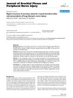
Abduction improvements one day post surgeryFigure 1
Abduction improvements one day post surgery. Video stills showing abduction improvement for two patients in this
series. a, c) Preoperative maximal humeral elevation of 103° and 134° as seen the day before neurolysis. These patients had
been experiencing winging for 4.5 and 1.5 years, respectively. b, d) Postoperative abduction of 176° and 149° documented on
the day after surgery.
Journal of Brachial Plexus and Peripheral Nerve Injury 2007, 2:4 />Page 5 of 8
(page number not for citation purposes)
with function[10,11]. The field remains skeptical of
reports even from experienced surgeons[4]. In many cases
of long thoracic nerve injury, it is possible to quantita-
tively measure improvement by following changes in the
angle at which the arm can be abducted. A small change
in muscle contraction (25% of maximum voluntary con-
traction) can lead to an easily-measured change in abduc-
tion angle (90°)[2]. We used this measurement as a
convenient indicator of early recovery 24 hours post-
microneurolysis. In this retrospective study, 12 patients
undergoing a total of 13 neurolysis surgeries of the long
thoracic nerve increased their angle of abduction by an
average of 59° within one day of the procedure (Table 1).
In complete axonal and demyelinating injuries, the time
to recovery is several weeks to months. In the current
series, therefore, the injury is not axonal and apparently
not demyelinating although preoperative electromyo-
grams were interpreted to suggest some degree of demyeli-
nation in most cases. The nerve must, therefore, be intact,
and the muscle must be able to receive signal, otherwise
the immediate ability to abduct the arm to a higher degree
would not be possible. It is possible that a mixed injury
pattern is present with sufficient numbers of intact axons
present to produce rapid recovery after surgery. Neverthe-
less, rapid return of movement following microneurolysis
is an interesting and real phenomenon that deserves fur-
ther consideration and explanation.
When similar observations of early recovery are made,
they may be dismissed as coincidental, spontaneous
recovery. By directly measuring the nerve conduction
before and after surgical procedures, we determined that
the nerve was not able to function properly until after the
decompression, neurolysis and tetanic stimulation. The
persistence of symptoms in patients experiencing scapular
Intraoperative electrical monitoringFigure 2
Intraoperative electrical monitoring. Intraoperative electrical monitoring of the long thoracic nerve for one, representa-
tive patient in this group. a) Response of the serratus anterior to a 3.0 mA stimulation before neurolysis. b) Response to the
same stimulation after neurolysis and tetanic stimulation.
Journal of Brachial Plexus and Peripheral Nerve Injury 2007, 2:4 />Page 6 of 8
(page number not for citation purposes)
Possible types of neuropathic disorderFigure 3
Possible types of neuropathic disorder. a) Normal motoneuron. b) The sub-threshold injury manifests as a slowing
of electrical conduction over the site of compressive lesion and a reduction in axonal transport. Both factors could result in
reduced muscle response. c) In the local neurapraxic lesion, conduction is eliminated only over the affected portion of the
nerve, and axonal transport is still able to prevent denervation of the muscle. Muscle response is eliminated, since the impulse
is blocked at the site of compression. d) The extended neurapraxic lesion describes a case where the axonal transport is
even further decreased, and distal nerve portions are no longer able to carry electrical impulses. Demyelination may or may
not occur. There is, however, just enough material transported to maintain a connection to the muscle and prevent nerve
degeneration. (adapted from McComas et al., 1974 [15]).
Journal of Brachial Plexus and Peripheral Nerve Injury 2007, 2:4 />Page 7 of 8
(page number not for citation purposes)
winging for longer than 2 years also indicates that recov-
ery was probably not due to the completion of nerve
regeneration coinciding with surgical intervention.
It has been observed in several clinical situations that ana-
tomically intact peripheral nerves may be unable to acti-
vate target muscles. In the ulnar nerve, for example, nerve
conduction is commonly reported to be impeded only at
a site of compression caused by fibrosis[9,12]. Since some
lesions in continuity are able to be reversed even after an
extended period of paralysis, the nerve-muscle connection
is probably maintained by chemical factors. Monitoring
the electrical response of muscles, McComas reports evi-
dence for variations in the excitability at the neuromuscu-
lar junction that appeared to be changes in innervation,
but could not have been [13-15]. He concluded that the
cause was more likely to be a channel defect in the axo-
lemma, similar to the proposed cause of double crush syn-
drome[16,17].
It is convenient for diagnosis and prognosis purposes to
categorize nerve injuries. The mildest category of injury,
neurapraxia, comprises a continuum of increasingly
severe injuries to an intact nerve that do not result in
Wallerian degeneration of the axon. The observed rapid
recovery of muscle function needs to be accounted for
more specifically, since recovery from commonly-
reported neurapraxic injuries involves remyelination,
which takes weeks. It is, therefore, useful to further
describe categories of injury at the very mildest end of the
neurapraxia spectrum, whatever the underlying cause may
be[15,18].
McComas describes several possible types of nerve con-
duction impairment, summarized in Figure 3, that could
be due to slowed axonal transport at the site of compres-
sion[15]. It is not necessary to specify that all axons inner-
vating the paralyzed muscle are affected to the same
extent, and a spectrum of injury severity, with a corre-
sponding spectrum of recovery, is expected to exist within
a single nerve. The sub-threshold (Figure 3b) and local
(Figure 3c) neurapraxic injuries could be rapidly-reversed
once the compression is relieved by neurolysis. If this is
the case for a significant proportion of axons in the
affected nerve, rapid recovery of muscle function is
expected.
A channel defect could easily result in low local concentra-
tions of factors at the motor end plate due to slowed deliv-
ery from the nucleus, as depicted in Figure 3. Impaired
transport or flow of end plate molecular precursors may
result in a situation where the synapse is effectively silent
because it is unable to release enough acetylcholine to
stimulate muscle contraction. The localization of several
neural molecules and cell structures that are affected by
nerve compression has been accomplished. Protein distri-
bution, including tubulin, is affected by compression
injury to the nerve [18-20]. Molecular transport is inhib-
ited by pressure, and leads to a buildup of proteins at both
the proximal and distal sides of the compression, and
swelling is observed proximal to the constriction[21].
Both magnitude of the pressure and duration of compres-
sion affect severity of symptoms and speed of recovery in
experimental models.
There are a large number of proteins and cell components
which are necessary for coordinated acetylcholine release
at the neuromuscular junction: signaling proteins, vesicle
recycling proteins, ATP production and delivery compo-
nents (including mitochondria). Many, but not all, of
these are synthesized exclusively at the soma. The
decreased local concentration of any of these factors at the
end plate could potentially create a nerve that is unable to
stimulate muscle contraction, but is otherwise intact and
healthy. Removing the source of the constriction in these
cases could allow the nerve to quickly resume muscle
stimulation. In the case of this set of surgeries, a rapid
recovery of nerve impulse delivery to the serratus anterior
is observed once the constriction of the long thoracic
nerve is surgically alleviated.
Conclusion
This series of patients demonstrates that even several years
after onset of scapular winging, the long thoracic nerve
and serratus anterior muscle can retain the ability recover
function within a brief period of time. Although the exact
physiological reasons for the phenomenon are unknown,
the observed rapid recovery is real, and good background
research exists to describe potential mechanisms for this
phenomenon. Further inquiry into this area of neuro-
physiology is needed.
Competing interests
The author(s) declare that they have no competing inter-
ests.
Authors' contributions
RKN conceived of the study and performed all surgeries.
SEM analyzed patient movements from video and drafted
the manuscript. All authors read and approved the final
manuscript.
References
1. Ludewig PM, Cook TM, Nawoczenski DA: Three-dimensional
scapular orientation and muscle activity at selected posi-
tions of humeral elevation. J Orthop Sports Phys Ther 1996,
24(2):57-65.
2. Ebaugh DD, McClure PW, Karduna AR: Three-dimensional
scapulothoracic motion during active and passive arm eleva-
tion. Clin Biomech (Bristol, Avon) 2005, 20(7):700-709.
3. Birch R, Bonney G, Wynn Parry CB: Entrapment neuropathy. In
Surgical disorders of the peripheral nerves Edited by: Birch R, Bonney G,
Publish with BioMed Central and every
scientist can read your work free of charge
"BioMed Central will be the most significant development for
disseminating the results of biomedical research in our lifetime."
Sir Paul Nurse, Cancer Research UK
Your research papers will be:
available free of charge to the entire biomedical community
peer reviewed and published immediately upon acceptance
cited in PubMed and archived on PubMed Central
yours — you keep the copyright
Submit your manuscript here:
/>BioMedcentral
Journal of Brachial Plexus and Peripheral Nerve Injury 2007, 2:4 />Page 8 of 8
(page number not for citation purposes)
Wynn Parry CB. New York, NY , Churchill Livingstone;
1998:245-292.
4. Disa JJ, Wang B, Dellon AL: Correction of scapular winging by
supraclavicular neurolysis of the long thoracic nerve. J Recon-
str Microsurg 2001, 17(2):79-84.
5. Tubbs RS, Salter EG, Custis JW, Wellons JC 3rd, Blount JP, Oakes WJ:
Surgical anatomy of the cervical and infraclavicular parts of
the long thoracic nerve. J Neurosurg 2006, 104(5):792-795.
6. Wiater JM, Flatow EL: Long thoracic nerve injury. Clin Orthop
Relat Res 1999:17-27.
7. Kitao A, Hirata H, Morita A, Yoshida T, Uchida A: Transient dam-
age to the axonal transport system without Wallerian
degeneration by acute nerve compression. Exp Neurol 1997,
147(2):248-255.
8. Matsuyama T, Okuchi K, Goda K: Upper plexus thoracic outlet
syndrome case report. Neurol Med Chir (Tokyo) 2002,
42(5):237-241.
9. Sakurai M, Miyasaka Y: Neural Fibrosis and the Effect of Neurol-
ysis. J Bone Joint Surg Br 1986, 68(3):483-488.
10. Choi SJ, Ahn DS: Correlation of clinical history and electrodi-
agnostic abnormalities with outcome after surgery for car-
pal tunnel syndrome. Plast Reconstr Surg 1998, 102(7):2374-2380.
11. Friedenberg SM, Zimprich T, Harper CM: The natural history of
long thoracic and spinal accessory neuropathies. Muscle Nerve
2002, 25(4):535-539.
12. Kim DH, Han K, Tiel RL, Murovic JA, Kline DG: Surgical outcomes
of 654 ulnar nerve lesions. J Neurosurg 2003, 98(5):993-1004.
13. McComas AJ, White CM: Distal dysfunction and recovery in
ulnar neuropathy. Muscle Nerve 1996, 19(12):1617-1619.
14. McComas AJ, DeBruin H, Quartly C: Non-transmitting synapses
in human neuromuscular disorders? In Plasticity of motoneuronal
connections Edited by: Wernig A. Amsterdam ; New York , Elsevier;
1991:223-229.
15. McComas AJ, Jorgensen PB, Upton AR: The neurapraxic lesion: a
clinical contribution to the study of trophic mechanisms.
Can
J Neurol Sci 1974, 1(3):170-179.
16. Upton AR, McComas AJ: The double crush in nerve entrapment
syndromes. Lancet 1973, 2(7825):359-362.
17. Suzuki Y, Shirai Y: Motor nerve conduction analysis of double
crush syndrome in a rabbit model. J Orthop Sci 2003, 8(1):69-74.
18. Lundborg G, Dahlin LB: The pathophysiology of nerve compres-
sion. Hand Clin 1992, 8(2):215-227.
19. Dahlin LB, Nordborg C, Lundborg G: Morphologic changes in
nerve cell bodies induced by experimental graded nerve
compression. Exp Neurol 1987, 95(3):611-621.
20. Dahlin LB, Archer DR, McLean WG: Axonal transport and mor-
phological changes following nerve compression. An experi-
mental study in the rabbit vagus nerve. J Hand Surg [Br] 1993,
18(1):106-110.
21. Lundborg G: Nerve injury and repair. Edinburgh ; New York ,
Churchill Livingstone; 1988.