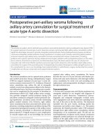
Báo cáo y học: "Postoperative peri-axillary seroma following axillary artery cannulation for surgical treatment of acute type A aortic dissection" pps
Bạn đang xem bản rút gọn của tài liệu. Xem và tải ngay bản đầy đủ của tài liệu tại đây (1.09 MB, 4 trang )
Apostolakis et al. Journal of Cardiothoracic Surgery 2010, 5:43
/>Open Access
CASE REPORT
BioMed Central
© 2010 Apostolakis et al; licensee BioMed Central Ltd. This is an Open Access article distributed under the terms of the Creative Com-
mons Attribution License ( which permits unrestricted use, distribution, and reproduc-
tion in any medium, provided the original work is properly cited.
Case report
Postoperative peri-axillary seroma following
axillary artery cannulation for surgical treatment of
acute type A aortic dissection
Efstratios E Apostolakis*
1
, Nikolaos G Baikoussis
1
, Konstantinos Katsanos
2
and Menelaos Karanikolas
3
Abstract
The arterial cannulation site for optimal tissue perfusion and cerebral protection during cardiopulmonary bypass (CPB)
for surgical treatment of acute type A aortic dissection remains controversial. Right axillary artery cannulation confers
significant advantages, because it provides antegrade arterial perfusion during cardiopulmonary bypass, and allows
continuous antegrade cerebral perfusion during hypothermic circulatory arrest, thereby minimizing global cerebral
ischemia. However, right axillary artery cannulation has been associated with serious complications, including
problems with systemic perfusion during cardiopulmonary bypass, problems with postoperative patency of the artery
due to stenosis, thrombosis or dissection, and brachial plexus injury. We herein present the case of a 36-year-old
Caucasian man with known Marfan syndrome and acute type A aortic dissection, who had direct right axillary artery
cannulation for surgery of the ascending aorta. Postoperatively, the patient developed an axillary perigraft seroma. As
this complication has, not, to our knowledge, been reported before in cardiothoracic surgery, we describe this unusual
complication and discuss conservative and surgical treatment options.
Introduction
The arterial cannulation site for optimal tissue perfusion
and cerebral protection during cardiopulmonary bypass
(CPB) for surgical treatment of acute type A aortic dis-
section remains controversial [1-3]. Avoidance of femoral
artery cannulation may reduce the risk of retrograde
embolic events from atheromatous debris in the thoracic
and abdominal aorta, but direct ascending aorta cannula-
tion can be complicated by the presence of thrombus or
atheromatous debris [4,5]. Right axillary artery cannula-
tion provides antegrade arterial perfusion during CPB
and allows continuous antegrade cerebral perfusion dur-
ing hypothermic circulatory arrest, thereby minimizing
global cerebral ischemia [3,4]. However, right axillary
artery cannulation has been associated with serious com-
plications, including malperfusion problems during CPB,
compromised postoperative patency of the axillary artery
(due to stenosis, thrombosis or dissection) and brachial
plexus injury[6,7]. Perigraft seroma is a rare complication
in vascular surgery and, to our knowledge, has not been
reported after axillary artery cannulation. We herein
describe the case of a 36 year old man with Marfan syn-
drome and acute aortic dissection, who had right axillary
artery cannulation for aortic root and ascending aorta
replacement, and postoperatively developed a seroma in
the right suclavian area.
Case presentation
A 36 year-old Caucasian man with Marfan syndrome was
emergently admitted to our hospital with diagnosis of
acute type A aortic dissection. Transthoracic echocar-
diography and computed tomography revealed aortic
valve regurgitation and aortic dissection extending from
the root of the aorta to the iliac arteries. The dissection
extended into the arch vessels, involving mainly the
innominate and axillary artery (figure 1, 2). The patient
underwent the Bentall procedure under CPB instituted
through direct right axillary artery cannulation, without
interposition of an anastomotic graft. We did not use
total hypothermic circulatory arrest; instead, continuous
antegrade cerebral perfusion was achieved through can-
nulation of the right axillary artery, with the innominate
artery clamped during arch reconstruction, using the
* Correspondence:
1
Cardiothoracic Surgery Department, University of Patras, School of Medicine,
Patras, Greece
Full list of author information is available at the end of the article
Apostolakis et al. Journal of Cardiothoracic Surgery 2010, 5:43
/>Page 2 of 4
"open distal anastomosis" technique. At the end of the
operation, the subclavian artery cannulation site was
repaired using a synthetic patch (Gore-tex Acuseal Car-
diovascular patch, Gore & Associates, Flagstaff, Arizona
86004, USA). Initially we did not observe brachial plexus
injury, bleeding, infection, vessel stenosis or any other
complication related to axillary artery cannulation. How-
ever, local swelling was noted in the right subclavian area
a week later, (figure 3). Needle aspiration revealed 50 ml
of clear yellow transudate (figure 4), and laboratory anal-
ysis was negative for chylous collection (no chylomicrons,
cholesterol/triglycerides >1). Total protein concentration
of the liquid was 3.7 gm/dL, cholesterol 51 mg/dL, trig-
lycerides 14 mg/dL and LDH 174 U/L. As swelling
recurred after fluid aspiration, the patient required
repeated needle aspiration every week for eight weeks.
Three months after the operation, the seroma had disap-
peared, and did not recur. At his last follow-up six
months after the operation, the patient was doing
remarkably well: he had completely recovered from sur-
gery had returned to his previous normal life, and swell-
ing had completely disappeared.
Discussion
Local complications after axillary artery cannulation can
occur either intraoperatively (mostly technical problems,
such as arterial injury with bleeding or malperfusion)
[1,6-9], or postoperatively (mostly neurologic complica-
tions related to brachial plexus injury) [1,4,10]. Compared
to the common femoral artery, the axillary artery is
located deeper in tissues, in the vicinity of the brachial
plexus, and this deep position likely contributes to higher
incidence of cannulation-related complications [6,10].
Strauch et al [1] reported 14 complications among 284
patients who had axillary artery cannulation for surgery
of the proximal aorta, with brachial plexus injury being
the most common complication. Axillary perigraft
seroma was not listed as a complication in this or any
other relevant published clinical study. From the
pathophysiological point of view, perigraft seromas con-
sist of a clear, sterile fluid collection confined within the
non-secreting fibrous pseudomembrane surrounding the
Figure 1 CT scan with contrast reveals ascending aorta dilatation
with intimal flap (arrows) in the ascending and descending aorta
(a). Innominate artery dissection (b) and reconstructed image showing
aortic root dilatation, together with aorta and innominate artery dis-
section (c).
Figure 2 Contrast-enhanced CT scan showing the intimal flap
due to dissection from the aortic root to the ascending aorta, in-
nominate artery and subclavian artery (a). Enhanced reconstructed
CT scan image showing the path of dissection (b).
Figure 3 Local, non-pulsatile swelling in the subclavian area (ar-
row) indicating a subcutaneous collection.
Apostolakis et al. Journal of Cardiothoracic Surgery 2010, 5:43
/>Page 3 of 4
implanted graft, and occur in 1.7% to 2.3% of all graft
implantations in vascular surgery [11]. Knitted Dacron
and polytetrafluorethylene are the materials most com-
monly implicated, with a higher percentage involving
knitted Dacron grafts [11,12]. During the normal incor-
poration process of an implanted vascular graft, firmly
adherent fibrous tissue and healthy wall matrix lining
cover the graft by the 6th postoperative week [13], while
seromas develop when the surrounding connective tissue
fails to incorporate the graft. This failed incorporation
has been well documented histologically as fibrous
pseudomembrane lining the seroma wall and immature
fibroblasts lining the graft [11,13]. When evaluating this
complication, differential diagnosis should include injury
of the minor lymphatic duct or its branches, resulting in
local lymph collection (the so called lymphocele) [1]. In
fact, Strauch et al reported lymphocele in 5 patients, with
2 of these patients requiring aspiration [1]. Lymph is eas-
ily recognized after aspiration, because of its characteris-
tic milky color, while biochemical analysis reveals the
presence of chylomicrons, high triglyceride levels and
cholesterol/triglycerides ratio <1 [14]. In our patient, the
diagnosis of lymphocele was excluded because aspirated
fluid did not have any of the above characteristics. This is
the first reported case of a seroma following axillary
artery repair with a graft, after arterial cannulation for
CPB. Interestingly, seroma in our case was induced by a
small polytetrafluoroethylene (PTFE) patch, indicating
the possible qualitative (rather than quantitative) role of
the synthetic graft. In our opinion, low postoperative
hematocrit, decreased plasma oncotic pressure, hyper-
tension, and presence of fat-rich subcutaneous tissue in
the axillary perigraft space were factors promoting
seroma formation in our patient. Indeed, Dauria et al [11]
claimed that a decrease in hematocrit by one-half
resulted in three-fold increase of graft weeping in renal
patients undergoing arterio-venous graft placement.
Management options for persistent seromas include con-
servative, interventional and surgical therapies. Conser-
vative management consists of repeated aspiration,
topical application of microfibrillar collagen or histoacryl
tissue, wrapping with collagen fleece soaked in fibrin glue
or absorbable collagen, intraluminal injection of hemo-
static fibrin glue, plasmapheresis (10-12 sessions), or
stent implantation [15-17]. However, repeated aspiration
increases graft infection risk to 12% [18] and should be
performed with strict sterile precautions. It is worth not-
ing that, compared to other seroma locations, external
local compression by gauze package has less beneficial
effect in the subclavian area due to deep location of the
cannulation site. Injection of a sclerosing agent can result
in later graft thombosis [16] and is not recommended.
However, case reports of microfibrillar collagen (the end-
product of mature fibroblasts) insertion into the space
surrounding an axillo-bifemoral graft have documented
successful graft incorporation into the surrounding tissue
without fluid re-accumulation [16]. Surgical seroma
treatment is only indicated when conservative manage-
ment has failed, the recurring fluid collection is > 2 cm in
diameter, there is impending skin necrosis, or the graft is
infected [11,18,19]. In such cases, surgical treatment con-
sists of excision of the sac and replacement of the graft
using a new synthetic graft or an umbilical vein or
homograft iliac artery [17,19]. Conservative management
is successful in only 65-70% of cases, due to high rates of
recurrence and infection [16]. In contrast, surgical man-
agement with replacement of the graft and radical exci-
sion of the sac has a cure rate over 92% [11,18,19].
Consent
Written informed consent was obtained from the patient
for publication of this case report and accompanying
images. A copy of the written consent is available for
review by the Editor-in-Chief of this journal.
Competing interests
The authors declare that they have no competing interests.
Authors' contributions
EA performed the operation, wrote the initial manuscript and revised the
study. NB participated in the operation, collected the images, submitted and
revised the manuscript. KK provided the CT scan images. MK revised and cor-
rected the manuscript while he participated in its design and coordination. All
authors read and approved the final manuscript.
Author Details
1
Cardiothoracic Surgery Department, University of Patras, School of Medicine,
Patras, Greece,
2
Department of Interventional Radiology, University of Patras,
School of Medicine, Patras, Greece and
3
Department of Anaesthesiology and
Intensive Care Medicine, University of Patras School of Medicine, Patras, Greece
Received: 26 January 2010 Accepted: 25 May 2010
Published: 25 May 2010
This article is available from: 2010 Apostolakis et al; licensee BioMed Central Ltd. This is an Open Access article distributed under the terms of the Creative Commons Attribution License ( which permits unrestricted use, distribution, and reproduction in any medium, provided the original work is properly cited.Journal of Cardiothoracic Surgery 2010, 5:43
Figure 4 Needle aspiration revealed serous, yellow fluid.
Apostolakis et al. Journal of Cardiothoracic Surgery 2010, 5:43
/>Page 4 of 4
References
1. Staunch JT, Spielvogel D, Lauten A, Lansman SL, McMurtry K, Bodian CA,
Griepp RB: Axillary artery cannulation routine use in ascending aorta
and aortic arch replacement. Ann Thorac Surg 2004, 78:103-8.
2. Fusco D, Shaw R, Tranquilli M, Kopf GS, Elefteriades JA: Femoral
cannulation is safe for type A dissection repair. Ann Thor Surg 2004,
78:1285-89.
3. Apostolakis E, Akinosoglou K: The Methodologies of hypothermic
Circulatory Arrest and of Antegrade and Retrograde Cerebral Perfusion
for Aortic Arch Surgery. Ann Thorac Cardiovasc Surg 2008, 14:138-148.
4. Halkos M, Kerendi F, Myung R, Kilgo P, Puskas JD, Chen EP: Selective
antegrade cerebral perfusion via right axillary artery cannulation
reduces morbidity and mortality after proximal aortic surgery. J Thorac
Cardiovasc Surg 2009, 138(5):1081-9.
5. Svensson L, Blackstone E, Rajeswaran J, Sabik JF, Lytle BW, Gonzalez-
Stawinski G, Varvitsiotis P, Banbury MK, McCarthy PM, Pettersson GB,
Cosgrove DM: Does the arterial cannulation site for circulatory arrest
influence stroke risk? Ann Thorac Surg 2004, 78:1274-84.
6. Schachner T, Nagiller J, Zimmer A, Laufer G, Bonatti J: Technical problems
and complications of axillary artery cannulation. Eur J Cardio-thoracic
Surg 2005, 27:634-37.
7. Sabik J, Memeth H, Lytle B, Blackstone EH, Gillinov AM, Rajeswaran J,
Cosgrove DM: Cannulation of the axillary artery with a side graft
reduces morbidity. Ann Thorac Surg 2004, 77:1315-20.
8. Orihashi K, Sueda T, Okada K, Takahashi S: Compressed true lumen in the
innominate artery: a pitfall of right axillary arterial perfusion in acute
aortic dissection. J Thorac Cardiovasc Surg 2009, 137:242-43.
9. Rescigno G, Aratari C, Matteucci M: Axillary artery cannulation pitfalls.
Letter to the Editor. J Thorac Cardiovasc Surg 2009, 138:251.
10. Gulbins H, Pritisanac A, Ennker J: Axillary versus femoral cannulation for
aortic surgery: Enough evindence for a general recommendation? Ann
Thorac Surg 2007, 83:1219-24.
11. Dauria D, Dyk P, Garvin P: Incidence and management of seroma after
arteriovenous graft placement. J Am Coll Surgeons 2006, 203:506-11.
12. Johnson W, Lee K: Comparative evaluation of externally supported
Dacron and Polytetrafluoroethylene prosthetic bypasses for
femorofemoral and axillofemoral arterial reconstructions. Veterans
affairs cooperative study. J Vasc Surg 1999, 30:1077-83.
13. Sladen J, Mandl M, Grossman L, Denegri JF: Fibroblast inhibition: a new
and treatable cause of prosthetic graft failure. Am J Surg 1985,
149:587-90.
14. Apostolakis E, Kouerinis I, Zografos G, Tsilimingas N, Dougenis D:
Conservative treatment of a cervical chylous fistula of the minor
thoracic duct after thoracic trauma. J Trauma 2009, 66:E52-54.
15. Lucas L, Rodriguez J, Olsen D, Diethrich EB: Symptomatic seroma after
open abdominal aortic aneurysm repair. Ann Vasc Surg 2009, 23:144-46.
16. Kat E, Jones N, Burnett J, Foreman R, Chok R, Sage MR: Perigraft seroma of
open aortic reconstruction. AJR 2002, 178:1462-64.
17. Sobrinho G, Henriques S: Perigraft seromas complicating prosthetic
bridge arteriovenous fistula - solution with autogenous vein
interposition. Eur J Vasc Endovasc Surg 2001, 22:469-471.
18. Allaria P, Lucatello A, Gandini E, Giangrande A: Relapsing seroma in a
uremic patient bearing a PTFE graft as vascular access. J Vasc Access
2001, 2:28-31.
19. Sugimoto T, Kitade T, Nishikawa H, Koyama T, Hatta T, Kurisu S: Large
perigraft seroma after aortoiliac bypass with an expanded PTFE graft:
report of a case. Surg Today 2004, 34:698-700.
doi: 10.1186/1749-8090-5-43
Cite this article as: Apostolakis et al., Postoperative peri-axillary seroma fol-
lowing axillary artery cannulation for surgical treatment of acute type A aor-
tic dissection Journal of Cardiothoracic Surgery 2010, 5:43