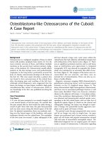
Báo cáo y học: "Inflammatory myofibroblastic tumor of the lung- a case report" pdf
Bạn đang xem bản rút gọn của tài liệu. Xem và tải ngay bản đầy đủ của tài liệu tại đây (5.71 MB, 4 trang )
CAS E REP O R T Open Access
Inflammatory myofibroblastic tumor of the
lung- a case report
Chien-Kuang Chen
1
, Chia-Ing Jan
2
, Jian-Shun Tsai
1
, Hsu-Chih Huang
1
, Pin-Ru Chen
1
, Yu-Sen Lin
1
, Chih-Yi Chen
1
,
Hsin-Yuan Fang
1*
Abstract
A 45-year-old man presented with a six-month history of progressive dyspnea with productive cough and wheez-
ing. The patient was a heavy smoker and had a history of tongue cancer, hypertension, and asthma. Chest X-ray
and computed tomography showed a mass lesion in the left hilar region and total collapse of the upper left lobe
of the lung. Bronchoscopy revealed a whitish solid tumor obstructing the left upper lobe bronch us. Positron emis-
sion tomography showed increased tracer uptake in the lesion. A thoracoscopic lobectomy of the left upper lobe
of the lung was performed. The final pathologic diagnosis was inflammatory myofibroblastic tumor.
Introduction
Inflammatory myofib roblastic tumor (IMT) of the lung,
also known as plasma cell granuloma or inflammatory
pseudotumor, is a rare disease entity [1]. Diagnosis of
IMT is difficult to establish before surgery because of its
diversified radiologic manifestations. This tumor can be
cystic or homogeneous, endobronchial or parenchymal
with or w ithout clear margins [2]. Complete surgical
resection is the treat ment of choice not only to exclude
malignancy but also to achieve a goo d prognosis [3,4].
We report a case of inflammatory myofibroblastic
tumor t hat was successfully removed by thoracoscopic
lobectomy.
Case report
A 45-year-old man presented with a 6-month history
of progressive dyspnea with productive cough and
wheezing. The patient had a history of smoking (1
pack per day for 20 years), hypertension and asthma,
which was under regular medical control. He also had
a history of tongue cancer (squamous cell carcinoma,
pT2N0M0, stage II) for which he underwent wide exci-
sion of the right side of the tongue and modified neck
lymph node dissection five years prior to this presenta-
tion. Chest plain film showed a protruding mass sha-
dow in the left hilar region. Cos todia ph ragm atic angles
were clear. There was increased density over left lung
field with elevation of the left side of the diaphragm.
Thesefeatureswereindicativeofahilarmassobstruct-
ing the bronchus with collapse of the upper left lobe of
the lung (Fig. 1A). Contrast enhanced computed tomo-
graphy (CT) showed a hilar mass measuring approxi-
mately 35 mm × 28 mm × 15 mm and a collapsed left
upper lobe of the lung. There was weak enhancement in
the arterial phase. The endobronchial part of the tumor
had clear margins along the bronchus of the upper left
lobe of the lung . The distal par t of the tum or had indis-
tinct margins along the lung parenchyma. The distal
bronchus was dilated and filled with secretions. There
was no mediastinal lymphadenopathy (Fig. 1B).
Bronchoscopy revealed a whitish tumor obstructing the
left upper bronchus (Fig. 2). Biopsy specimens of the
tumor taken during the bronchoscopic examination
showed evidence of smooth muscle cell proliferation
with focal abnormal mitosis. A smooth muscle cell
tumor of malignant potential was considered. Positron
emission tomography (PET) showed increased fluoro-
deoxyglucose (FDG) uptake in the lesion (Fig. 1C).
The tumor involved the upper left lobe of the lung
and obstructed the bronchus. The patient underwent a
thoracoscopic lobectomy under general anesthesia with
double lumen endotra cheal tube placement. The vessels
of the left upper lobe were divided and ligated using an
endoscopic autostapling d evice. The bronchus of the
upper left lobe was opened by endoscissor. The cutting
margin was checked by examination of frozen sections
* Correspondence:
1
Division of Thoracic Surgery, Department of Surgery, China Medical
University Hospital, China Medical University, Taichung, Taiwan
Chen et al. Journal of Cardiothoracic Surgery 2010, 5:55
/>© 2010 Chen et al; licensee BioMed Central Ltd. This is an Open Access article distributed under the terms of the Creative Commons
Attribution License (http://creativecom mons.org/licenses/by/2 .0), which permits u nrestricted use, distribution, and reproduction in
any medium, provided the original work is properly cited.
to ensure that the resection was clear. The upper left
lobe of lung was removed through a port with ext ended
skin incision 5 cm at t he anterior 5th intercostal space.
The orifice of the bronchus was sutured w ith standard
instrumentation through the utility incision.
The res ected tumor was white and elastic, measuring
3.5 cm × 2.5 cm × 1.5 cm in size. It impacted the whole
bronchus of the left upper lobe (Fig. 2). Microscopic
examination revealed a mixture of spindle cells showing
fibroblastic and myo fibroblastic differentiation arrayed
in fascicles, or with storiform architecture. The spindle
cells had oval nuclei, fine chromatin, inconspicuous
nucleoli, and bipolar, lightl y eosinophilic cytoplasm
(Fig. 2A). Admixed with the spindle proliferation was an
inflammatory infiltrat e containing lymphocytes, plasma
cells, and eosinophils. Immunohistochemical analysis
showed positive staining for vimentin (Fig. 2B) and des-
min, and focal positive staining for smooth mu scle actin
and cytokeratin (Fig. 2C). The tumor had a low Ki-67
proliferative index. In contrast, the tumor cells were not
reactive to CD34, CD99, or S-100 antibo dies. The surgi-
cal resection margins and all regional lymph no des were
tumor free. Inflammatory myofibroblastic tumor was
diagnosed. At the most recent follow-up (12 months
after operation), the patient was symptom free and there
was no evidence of tumor recurrence on chest CT scan.
Discussion
IMT is an uncommon pulmonary disease. The incidence
rate of IMT among patients with lung resection is
0.04%, and 26% of patients are less than 18 years old
[5]. Airway obstruction in IMT, although rare, normally
presents at an early stage due to obstructive respiratory
symptoms [6]. Mo st patients are symptomatic. There
are respiratory symptoms, such as cough, dyspnea, fever,
fatigue, and hemoptysis.
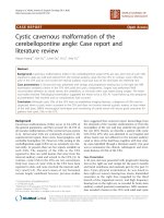
Figure 1 (A ) Chest plain film. A protruding mass shadow is seen in the left hilar region. The shadow of the left bronchus stop s at the mass.
Costodiaphragmatic angles are clear. There is increased density over the left lung field with elevation of the left side of the diaphragm. These
findings are indicative of a hilar mass obstructing the bronchus with collapse of the left upper lobe of lung. (B) Contrast computed tomography
(CT) image, distal part of the tumor. The distal bronchus is dilated and filled with secretions. The margin between the lung parenchyma and
tumor is indistinct. (C) Positron emission tomography (PET) and CT, proximal part of the tumor. An endobronchial tumor with high tracer uptake
and clear margins is visible.
Chen et al. Journal of Cardiothoracic Surgery 2010, 5:55
/>Page 2 of 4
Diagnosis of IMT is difficult to establish before sur-
gery because of its diversified radiologic manifestations
and because it can be difficult to distinguish from malig-
nant tumors on small tissue samples obtained from
bronchoscopic examination or needle biopsy. In fact,
only 6.3% of IMT cases are diagnosed based on analysis
of biopsy specimens alone [6]. In addition, I MT is often
difficult to differentiate from other neoplasms on PET
scan because of the high uptake of trac er in IMT. The
prognosis of IMT is dependent on tumor size (less than
or equal to 3 cm) and complete surgical resection. The
overall 3-year survival rate is about 82% and the overall
5-year survival rate is about 74% [3]. In our case, the
tumor was an endobronchial lesion with clear margins.
We were unable to prove whether the tum or involved
the lung parenchyma.
Surgical management of lesions in the major bronchi
is challenging. In our patient, we performed a thoraco-
scopic technique to cut the adhesion of the major fis-
sure, superior pulmonary vein and pulmonary artery
branches to upper lobe of the lung. We then opened the
left upper bronchu s to confirm that the cut end o f the
bronchus was free. The bronchus was closed with inter-
rupted sutures.
IMT is characterized histologically by spindle cell pro-
liferation. T he tumor is r eferred to by different names
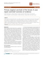
Figure 2 Bronchoscopic e xam shows a whitish tumor obstructing the left upper bronchus.Gross.Thetumorimpactedthewhole
bronchus with clear margins. Microscopically, the biopsy specimen is composed of spindle cells with fibroblastic and myofibroblastic
differentiation arrayed in fascicles. (A) The tumor is mostly limited within the bronchi. In a few foci, pushing of tumor margin to the lung
parenchyma is noted (×20; ×100). Immunohistochemical study demonstrated (B) vimentin (+) (×200), and (C) cytokeratin (focal +), (×200).
Chen et al. Journal of Cardiothoracic Surgery 2010, 5:55
/>Page 3 of 4
in the literature depending on the predominant cell type
encountered in the lesion: plasma cell granuloma or
tumor, xanthogranuloma, plasma cell/histiocytoma com-
plex, or post inflammatory pseudotumor [7]. Matsubara
et al. used the term inflammatory pseudotumor and
described three subgroups based on the cell type m ost
encountered in a mass: organizing pneumonia (44%),
fibrous histiocytoma (44%), and lymphoplasmocytic type
(12%) [8]. There are regions of organizing pneumonia in
all cases, and therefore, the current hypothesis is
that IMT might develop in individuals with a past his-
tory of upper respiratory infections or pneumonia. Some
studies, however, suggest that it might be a true
neoplasm as some mutations on chromosome 2p23 of
anaplastic lymphoma kinase are found to be related to
this tumor [9].
Conclusions
Although inflammatory myofibroblastic tumor is rare, it
should be considered in the differential diagnosis of pul-
monary lesions. It is generally a benign lesion, but has
potential for local invasion and recurrence. The diagno-
sis and prognosis are highly dependent on complete sur-
gical resection.
Consent
Written informed consent was obtained from the patient for publication of
this case report and accompanying images. A copy of the written consent is
available for review by the Editor-in-Chief of this journal.
Competing interests
The authors declare that they have no competing interests.
Authors’ contributions
CKC carried out the manuscript. HYF coordinated all authors. CIJ reported
pathologic findings and took the pathologic pictures. PRC and HCH
collected references; YSL and JST took the pictures of the case report. CYC
made conclusion. All authors read and approved the final manuscript.
Author details
1
Division of Thoracic Surgery, Department of Surgery, China Medical
University Hospital, China Medical University, Taichung, Taiwan.
2
Department
of Pathology, China Medical University Hospital, China Medical University,
Taichung, Taiwan.
Received: 18 April 2010 Accepted: 20 July 2010 Published: 20 July 2010
References
1. Pettinato G, Manivel JC, Derosa N, Dehner LP: Inflammatory
Myofibroblastic Tumor (Plasma-Cell Granuloma) - Clinicopathological
Study Of 20 Cases With Immunohistochemical And Ultrastructural
Observations. American Journal of Clinical Pathology 1990, 94:538-546.
2. Rasalkar DD, Chu WCW, To KF, Cheng FWT, Li CK: Radiological Appearance
of Inflammatory Myofibroblastic Tumour. Pediatric Blood & Cancer 2010,
54:1029-1031.
3. Melloni G, Carretta A, Ciriaco P, Arrigoni G, Fieschi S, Rizzo N, Bonacina E,
Augello G, Belloni PA, Zannini P: Inflammatory pseudotumor of the lung
in adults. Annals of Thoracic Surgery 2005, 79:426-432.
4. Sakurai H, Hasegawa T, Watanabe S, Suzuki K, Asamura H, Tsuchiya R:
Inflammatory myofibroblastic tumor of the lung. European Journal of
Cardio-Thoracic Surgery 2004, 25:155-159.
5. Cerfolio RJ, Allen MS, Nascimento AG, Deschamps C, Trastek VF, Miller DL,
Pairolero PC: Inflammatory pseudotumors of the lung. Annals of Thoracic
Surgery 1999, 67:933-936.
6. Lee HJ, Kim JS, Choi YS, Kim K, Shim YM, Han J, Kim J: Treatment of
inflammatory myofibroblastic tumor of the chest: The extent of
resection. Annals of Thoracic Surgery 2007, 84:221-224.
7. van den Heuvel DA, Keijsers RG, van Es HW, Bootsma GP, de Bruin PC,
Schramel FM, van Heesewijk JP: Invasive Inflammatory Myofibroblastic
Tumor of the Lung. Journal of Thoracic Oncology 2009, 4:923-926.
8. Matsubara O, Tanliu NS, Kenney RM, Mark EJ: Inflammatory Pseudotumors
Of The Lung - Progression From Organizing Pneumonia To Fibrous
Histiocytoma Or To Plasma-Cell Granuloma In 32 Cases. Human
Pathology 1988, 19:807-814.
9. Coffin CM, Hornick JL, Fletcher CDM: Inflammatory myofibroblastic tumor
- Comparison of clinicopathologic, histologic, and immunohistochemical
features including ALK expression in atypical and aggressive cases.
American Journal of Surgical Pathology 2007, 31:509-520.
doi:10.1186/1749-8090-5-55
Cite this article as: Chen et al.: Inflammatory myofibroblastic tumor of
the lung- a case report. Journal of Cardiothoracic Surgery 2010 5:55.
Submit your next manuscript to BioMed Central
and take full advantage of:
• Convenient online submission
• Thorough peer review
• No space constraints or color figure charges
• Immediate publication on acceptance
• Inclusion in PubMed, CAS, Scopus and Google Scholar
• Research which is freely available for redistribution
Submit your manuscript at
www.biomedcentral.com/submit
Chen et al. Journal of Cardiothoracic Surgery 2010, 5:55
/>Page 4 of 4