Báo cáo y học: "Synchronous primary intrapulmonary and mediastinal thymoma-A case report" pot
Bạn đang xem bản rút gọn của tài liệu. Xem và tải ngay bản đầy đủ của tài liệu tại đây (1.51 MB, 4 trang )
CAS E REP O R T Open Access
Synchronous primary intrapulmonary and
mediastinal thymoma-A case report
Zuoqing Song, Xiaohong Xu, Shujun Li, Sen Wei, Jun Chen
*
, Qinghua Zhou
*
Abstract
We report an extremely rare case of Synchronous primary intrapulmonary and mediastinal thymoma in a Chinese
patient. We describe the histological and radiological findings, which support the possibility of multicentric thy-
moma. Resection of the mass in the left anterior superior mediastinum and upper lobectomy of right lung were
performed, with lymph Nodes clearance, superior vena cava, left and right brachiocephalic veins resection, recon-
struction of left brachiocephalic vein to right auricle and reconstruction of right brachiocephalic vein to superior
vena cava.
Introduction
Thymomas are tumors derived from thymic epithelial
cells and have an incidence of 0.15 per 100000[1]. Pri-
mary intrapulmonary thymomas are d efined as thymo-
mas arising in an intrapulmonary location without an
associated mediastinal compon ent and are very rare[2].
Here we present a successfully resected case of synchro-
nous primary intrapulmonary and mediastinal thymom a
with vascular reconstruction.
Case report
A 55-year-old Chinese man was admitted with a history
of progressive exertional dyspnea of 55 days’ duration
and a radiological finding of an anterior mediastinal
mass for 7 days. The patient had no clinical features of
myasthenia gravis. An enhanced Chest computed tomo-
graphic scan revealed a 5.5 c m × 6.0 cm × 4.1 cm mass
in the anterior segment of the right upper lobe with
continuation to some mediastinal swelling lymph nodes.
Multiple swelling lymph nodes could be found in the
mediastinum (Figure 1A, B, Figure 2A, B, C). Three-D
reconstruction showed the superior vena cava, whose
lumen was unobstructed but deformated under the
compression of the mass (Figure 1C). A computed
tomographic scanning of the brain and bones were nor-
mal. An exploratory limited right thoracotomy was
undertaken through a median sternotomy. A soft
encapsulated mass(3.5 cm × 4.0 cm × 5 cm) was found
in the left anterior superior mediastinum, with invasion
to the left pericardium and visceral pleura, adhesive to
partial superior lobe of right lung and brachiocephalic
vein(Figure 1G, I). In the anterior segment of the right
upper lobe, a mass was 6 cm in diameter, invading the
junction of right and left brachiocephalic veins and
upper segment of superior vena cava (Figure 1F, H).
Both masses are solitary. Therefore resection of the
mass in the left anterior superior mediastinum and
upper lobectomy of right lung were performed, with
lymph Nodes clearance, superior vena cava, left and
right brachiocephalic veins r esection, reconstruction of
left brachiocephalic vein to right auricle and reconstruc-
tion of right brachio cephalic vein to superior vena cava.
Microscopically according to the WHO classification,
the mediastinal tumor(MT) was a B3/B2 primary thy-
moma and the mass in the upper lobe of right lung is
mainly a B3/B2 primary intrapulmonary thymoma(PIT)
with local A type tumors. Histologic evaluation indi-
cated that, CK5 & CK6 +, EMA + locally, CD5 -, CD99
+(Figure 2. No lymph metastasis was found. Warfarin
was applied to the patient as anticoagulation and 50 Gy
mediastinal irradiation was given a s adjuvant therapy.
The patient has since recovered uneventfully and is now
being followed up as an outpatient (Figure 1D, E). Af ter
follow-up of eight months, there w as no significant
metastasis or recurrence found by radiological
examinations.
* Correspondence: ;
Department of Lung Cancer Surgery, Tianjin Key Laboratory of Lung Cancer
Metastasis and Tumor Microenvironment, Tianjin Lung Cancer Institute,
Tianjin Medical University General Hospital, Tianjin 300052, China
Song et al. Journal of Cardiothoracic Surgery 2010, 5:69
/>© 2010 Song et al; licensee BioMed Central Ltd. This is an Open Access article distributed under the terms of the Creative Commons
Attribution Licens e ( which permits u nrestricted use, distribution, and reproduction in
any medium, provided the original work is properly cited.
Discussion
Primary intrapulmonary thymuses are very uncommon,
with 28 cases reported to date[2]. Even rarer cases were
reported for Synchronous primary intrapulmonary and
mediastinal thymoma. The incidence for lung cancer in
China increased by 1.63% from 1988 to 2005. Some
special thoracic malignancies should be paid attention
to in China[3]. Primary intrapulmonary thymomas
appear to fall into two group s: one is in the hilus of the
lung, in relation to the wall of a major bronchus or
attached to the pericardium, and the other is peripheral
in the lung and beneath the visceral pleura[4]. In the
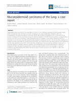
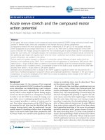
Figure 1 Chest computed tomographic scan. Figure 1A, 1B An enhanced Chest computed tomographic scan revealed a mass in the anterior
segment of the right upper lobe with continuation to some mediastinal swelling lymph nodes. Multiple swelling lymph nodes could be found
in the mediastinum. Figure C Three-D reconstruction showed the superior vena cava, whose lumen was unobstructed but deformated under the
compression of the mass. Figure 1D, 1E Postoperative enhanced Chest computed tomographic scan images. Figure 1F, 1G Surgical findings of
the mediastinal mass. Figure 1H Surgical findings of the intrapulmonary mass. Figure 1I Reconstruction of left brachiocephalic vein to right
auricle and reconstruction of right brachiocephalic vein to superior vena cava.
Song et al. Journal of Cardiothoracic Surgery 2010, 5:69
/>Page 2 of 4
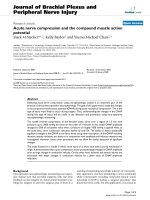
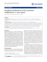
Figure 2 Macro and microscopic observations. Figure 2A,2B,2C The resected intrapulmonary and mediastinal tumors(3.5 cm × 4.0 cm × 5 cm and
6 cm × 6 cm × 6 cm, respectively). Figure 2D Histological findings of the primary intrapulmonary tumor(PIT), H&E × 100. Figure 2E PIT H&E × 400.
Figure 2F PIT CD99 Immunohistochemistry × 100. Figure 2G PIT CD99 Immunohistochemistry × 400. Figure 2H PIT CK 5& CK6 Immunohistochemistry
× 100. Figure 2I PIT CK 5& CK6 Immunohistochemistry × 400. Figure 2J Histological findings of the primary mediastinal tumor(MT), H&E × 100. Figure
2K MT, H&E × 400.
Song et al. Journal of Cardiothoracic Surgery 2010, 5:69
/>Page 3 of 4
case of hilar type, the notion that intrapulmonary thy-
momas are derived from mediastinal thymomas that
migrate into the lung with pinching off of the pleural
behin d them would be acceptable, but it fails to provide
a satisfactory explanation for the occurrence of the per-
ipheral[2]. Marchevsky[5] believed intrapulmonary thy-
momas arose from stem cells, uncommitted germinative
cells capable of differentiating along a variety of lines.
This theory is supported by the lar ge number of reports
of heterotopic, histologically mature tissues within the
lung parenchyma, such as t hyroid follicules, pancreas,
adrenal, liver, neuro-glial tissue and endometrium, or
tumors derived from ectopic tissue , such as melanoma,
meningioma, glomus or glomangioma, choriocarci noma,
terat oma, ependymoma and, of course, thymoma, which
could develop from such stem cells. Multiple thymomas
remain controversial as to whether multiple thymomas
involve intrathymic dissemination or represent multiple
primaries, which could be explained by Marchevsky’ s
theory. Although Bernatz et al. [6] reported 3 out of 138
(2.2%) thymomas to be multiple prim aries, it was diffi-
cult to clarify whether the multiple thymomas in their
cases involved double primary or dissemination, because
they did not mention any close histological characteris-
tics among the multiple thymomas. Since both our cases
were totally encapsulated tumors and did not have any
dissemination in the other portion, they were considered
to be multiple primaries[7]. Therefore our case provided
better evidence to support Marchevsky’s theory f or the
development of intrapulmonary thymomas.
The clinical course is that of a slow-growing lesion
that remains asymptomatic until it reaches a size caus-
ing problems due to local growth, such as pain, bron-
chial obstruction or hemoptysis. As with mediastinal
thymomas, they can be associated with paraneopla stic
syndromes, such as myasthen ia gravis or Good’ssyn-
drome. Resection appears sufficient in non-malignant
tumors. In incompletely resected patients, adjuvant
radiotherapy should be considered. Long-term regular
clinical follow-up is warranted, because of the risk of
late local recurrence.
Consent
Written informed consent was obtained from the patient
for publication of this case report and any accompany-
ing images. A co py of the written consent is available
for review by the Editor-in-Chief of this journal.
Acknowledgements
This study was partly supported by the grants from Key Project of National
Natural Science Foundation of China(No.30430300), National 973 Program
(No.2010CB529405), National 863 Program (No.2006AA02401) and S&T
Support Key Program of Tianjin (09ZCZDSF04100, 09ZCZDSF04000)
Authors’ contributions
ZS, JC and QZ were the primary caregiver for this patient and reviewed the
manuscript. SL and SW also cared for this patient. XX performed data
collection and drafted the manuscript. All authors read and approved the
final manuscript.
Competing interests
The authors declare that they have no competing interests.
Received: 12 July 2010 Accepted: 28 August 2010
Published: 28 August 2010
References
1. Engels EA, Pfeiffer RM: Malignant thymoma in the United States:
demographic patterns in incidence and associations with subsequent
malignancies. Int J Cancer 2003, 105:546-551.
2. Ishibashi H, Takahashi S, Tomoko H, Shibuya J, Suzuki S, Handa M: Primary
intrapulmonary thymoma successfully resected with vascular
reconstruction. Ann Thorac Surg 2003, 76:1735-1737.
3. Chen WQ, Zhang SW, Zou XN: Evaluation on the incidence, mortality and
tendency of lung cancer in China. Thoracic Cancer 2010, 1:35-40.
4. Kalish PE: Primary intrapulmonary thymoma. N Y State J Med 1963,
63:1705-1708.
5. Marchevsky AM: Lung tumors derived from ectopic tissues. Semin Diagn
Pathol 1995, 12:172-184.
6. Bernatz PE, Harrison EG, Clagett OT: Thymoma: a clinicopathologic study. J
Thorac Cardiovasc Surg 1961, 42:424-444.
7. Okada M, Tsubota N, Yoshimura M, Miyamoto Y, Sakamoto T: Two cases of
synchronous multiple thymoma. Surg Today 1998, 28:1323-1325.
doi:10.1186/1749-8090-5-69
Cite this article as: Song et al.: Synchronous primary intrapulmonary
and mediastinal thymoma-A case report. Journal of Cardiothoracic Surgery
2010 5:69.
Submit your next manuscript to BioMed Central
and take full advantage of:
• Convenient online submission
• Thorough peer review
• No space constraints or color figure charges
• Immediate publication on acceptance
• Inclusion in PubMed, CAS, Scopus and Google Scholar
• Research which is freely available for redistribution
Submit your manuscript at
www.biomedcentral.com/submit
Song et al. Journal of Cardiothoracic Surgery 2010, 5:69
/>Page 4 of 4