Báo cáo y học: "Wound contraction and macro-deformation during negative pressure therapy of sternotomy wounds" doc
Bạn đang xem bản rút gọn của tài liệu. Xem và tải ngay bản đầy đủ của tài liệu tại đây (960.17 KB, 6 trang )
RESEA R C H ART I C L E Open Access
Wound contraction and macro-deformation
during negative pressure therapy of sternotomy
wounds
Christian Torbrand
1
, Martin Ugander
2
, Henrik Engblom
2
, Håkan Arheden
2
, Richard Ingemansson
3
, Malin Malmsjö
1*
Abstract
Background: Negative pressure wound therapy (NPWT) is believed to initiate granulation tissue formation via
macro-deformation of the wound edge. However, only few studies have been performed to evaluate this
hypothesis. The present study was performed to investigate the effects of NPWT on wound contraction and
wound edge tissue deformation.
Methods: Six pigs underwent median sternotomy followed by magnetic reso nance imaging in the transverse
plane through the thorax and sternotomy wound du ring NPWT at 0, -75, -125 and -175 mmHg. The lateral width
of the wound and anterior-posterior thickness of the wound edge was measured in the images.
Results: The sternotomy wound decreased in size following NPWT . The lateral width of the wound, at the level of
the sternum bone, decreased from 39 ± 7 mm to 30 ± 6 mm at -125 mmHg (p = 0.0027). The greatest decrease
in wound width occurred when switching from 0 to -75 mmHg. The level of negative pressure did not affect
wound contraction (sternum bone: 32 ± 6 mm at -75 mmHg and 29 ± 6 mm at -175 mmHg, p = 0.0897). The
decrease in lateral wound width during NPWT was greater in subcutaneous tissue (14 ± 2 mm) than in ster num
bone (9 ± 2 mm), resulting in a ratio of 1.7 ± 0.3 (p = 0.0423), suggesting macro-deformation of the tissue. The
anterior-posterior thicknesses of the soft tissue, at 0.5 and 2.5 cm laterally from the wound edge, were not affected
by negative pressure.
Conclusions: NPWT contracts the wound and causes macro-deformation of the wound edge tissue. This shearing
force in the tissue and at the wound-foam interface may be one of the mechanisms by which negative pressure
delivery promotes granulation tissue formation and wound healing.
Introduction
Cardiac surgery is complicated by post-sternotomy med-
iastinitis in 1% to 5% of all procedures [1] and is a life-
threatening complication [2]. The reported early mortal-
ity in post-sternotomy mediastinitis following coronary
arter y bypass graft sur gery is between 8% and 25% [ 3,4].
Conventional treatment of post-sternotomy mediastinitis
includes surgical debridement, drainage, irrigation, and
reconstruction using pectoral muscle flap or omentum
transposition. In 1999, Obdeijn and colleagues described
a new method of treatment for post-sternotomy medias-
tinitis using a vacuum-assisted closure technique [5],
which is based on the principle of applying subatmo-
spher ic pressur e by co ntrolled suction through a porou s
dressing. The technique, also known as negative pres-
sure wound therapy (NPWT), has resulted in reduced
mortality in post-sternotomy mediastinitis [6].
Scientific evidence regarding the mechanisms by which
NPWT promotes wound healing has started to emerge.
NPWT results in the drainage of excessive fluid and deb-
ris, removal of wound edema, reduction in bacterial
counts and stimulation of wound edge microvascular
blood flow [7-10]. However, it is now believed that one of
the major driving forces that generate granulation tissue
formation is the macro-deformation of the wound edge
tissue that results from the suction force created by the
negative pressure. To our knowledge, there is only sparse
* Correspondence:
1
Department of Ophthalmology, Lund University and Skåne University
Hospital, Lund, Sweden
Full list of author information is available at the end of the article
Torbrand et al. Journal of Cardiothoracic Surgery 2010, 5:75
/>© 2010 Torbrand et al; licensee BioMed Central Ltd. This is an Open Acces s article distributed under the terms of the Creative
Commons Attribution License ( which permits unrestricted use, distribution, and
reprodu ction in any medium, provided the original work is properly cited.
scientific evidence for this instantaneous mechanical
effect by NPWT [11].
The present study was performed to in detail in vesti-
gate the effects of NPWT on wound contraction and
wound edge tissue deformation. Magnetic resonance
imaging (MRI) of the thorax was performed in a porcine
sternotomy wound model. The lateral width of the
wound and anterior-posterior thickness of the wound
edge was measured in the images taken be fore and after
initiation of NPWT at -75, -125 and -175 mmHg.
Materials and methods
Animals
An uninfected porcine sternotomy wound model was
used in the present study. Six dome stic landrace pigs of
both genders, with a mean body weight of 50 kg, were
fasted overnight with free access to water. The study
was approved by the Ethics Committee for Animal
Research, Lund University, Sweden. The investigation
complied with the “ GuidefortheCareandUseof
Laboratory Animals” as recommended by the U.S.
National Institutes of Health and published by the
National Academies Press (1996).
Anesthesia
Anesthesia was induced with ketamine hydrochloride
(Ketaminol Vet™ 100 mg/ml, Farmaceutici Gellini S.p.A,
Aprilia, Italy), 15 mg/kg intramuscularly, and xylazine
(Rompun Vet ™ 20 mg/mL, Bayer AG, Leverkusen, G er-
many), 2 mg/kg intramuscularly. The pigs were intu-
bated and mechanical ventilation was establi shed with a
Siemens-Elema 900B ventilator in the volume-controlled
mode. Anesthesia was maintained by continuous intra-
venous infusion of propofol (Diprivan™, Astra Zeneca,
Sweden), 0.1-0.2 mg/kg/min, in co mbination with fenta-
nyl (Leptanal™ , Lilly, France), 0.05 μg/kg/min, and at ra-
curium besylate (Tracrium™ , Glaxo, Täby, Sweden),
0.2-0.5 mg/kg/hour.
Surgical procedure
After a midline sternotomy, the pericardium was opened
and a polyurethane foam dressing was p laced between
the sternal edges. Two non-collapsible drainage tubes
were inserted into the foam. The open wound was then
sealed with a transparent adhesive drape. The drainage
tubes were connected to a purpose-built vacuum source
(VAC® pump unit, KCI, Copenhagen, Denmark), which
was set to deliver a continuous negative pressure of -75,
-125 or -175 mmHg.
Experimental procedure
MRI was first performed at baseline (0 mmHg). A negative
pressure was then applied and MRI was performed when
the target pressure had been reached. This procedure was
repeated for e ach negative pressure (-7 5, -125, and -1 75
mmHg). In order to eliminate time ef fects, the sequence
of application of the three different negative pressures was
varied between the animals using a 3 by 3 Latin square
design.
Magnetic resonance imaging
MRI was conducted using a 1.5T system (Intera CV,
Philips Medical Systems, Best, the Netherlands) with a
five-element cardiac coil and the pig in the supine posi-
tion. The images were acquired during ventilator-
controlled end expiratory apnea at the functional residual
lung capacity. Images were acquired i n the transverse
and sagittal planes, covering the entire thoracic cavity
using a steady-state free precession sequence. Typical
imaging para meters were: spatia l resolution 1.1 × 1.1
mm, slice thickness 5 mm, slice gap 0 mm, repetition
time 3.1 ms, echo time 1.6 ms, flip angle 60°, no ECG
triggering, sensitivity-encoding factor 2.
Image analysis
All images were evaluated using freely available software
(Segment 1.699, availab le at http://segment .heiberg.se)
[12]. Measurements of wound contraction and soft tis-
sue macro-deformation were performed in the same
transverse image at the cardiac midventricular level that
were acquired before (0 mmHg) and after the applica-
tion of -75, -125 and -175 mmHg. The distance between
the two wound edges of subcutaneous tissue, muscle tis-
sueandsternumboneweremeasured(lateralwound
width). The anterior-posterior thickness of the soft tis-
sue, including the subcutaneous and muscle tissue, was
measured at a distance of 0.5 cm and 2.5 cm from the
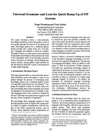
wound edge (Figure 1).
Calculations and statistics
Stati stical analysis was performed using paired S tudent’s
t-test. Significance w as defined as p < 0.05. The results
are presented as mean values ± the standard error of
the mean (S.E.M.).
Results
The s ternotomy wound changed in appearance and the
lateral wound width decreased when negative pressure
was applied (Figure 2). The lateral wound width
decreasedfrom39±7mmto30±6mm,forsternum
bone, upo n application of -125 mmHg (p = 0.0027, n =
6, Figure 3). The greatest decrease in lateral wound
width, as measured between the sternum bon e edges,
occurred when switching from 0 mmHg to -75 mmHg,
and the level of negative pressure did not play a role for
the degree of wound contraction ( 32 ± 6 mm at -75
mmHg and 29 ± 6 mm at -175 mmHg, for the sternum
bone, p = 0.0897, n = 6, Figure 3).
Torbrand et al. Journal of Cardiothoracic Surgery 2010, 5:75
/>Page 2 of 6
The wound edge tissue was also deformed upon applica-
tion of NPWT. The decrease in lateral wound width dur-
ing NPWT was greater in subcutaneous tissue (14 ± 2
mm) than in sternum bone (9 ± 2 mm), which resulted in
a ratio of subcutaneous to sternal decrease in wound
width of 1.7 ± 0.3 (p = 0.0423), suggesting macro-defor-
mation of the wound edg e tissue. The major decrease in
lateral wound width occurred when switching from 0 to
-75 mmHg and the level of negative pressure did not play
a significant role for the degree of wound contraction
(23 ± 4 mm at -75 mmHg and 19 ± 2 mm at -175 mmHg,
for muscle tissue p = 0.0982, n = 6, Figure 3).
The anterior-posterior thickness of the soft tissue,
including subcutaneous and muscle tissue, at 0.5 and
2.5 cm laterally from the wound edge, was not affected by
negative pressure (13 ± 2 mm at 0 mmHg and 14 ± 2 mm
Foam
Adhesive drape
0.5 cm
2.5 cm
Subcutaneous
Muscle
Sternum bone
Figure 1 Schematic illustration showing a transverse section through a sternotomy wound and the location of the wound dimension
measurements. The thick bracketed horizontal lines illustrate the lateral wound width at the level of subcutaneous tissue, muscle tissue and
sternum bone. The thick bracketed vertical lines illustrate the anterior-posterior thickness of the soft tissue, including the muscle and
subcutaneous tissue, at a lateral distance of 0.5 cm and 2.5 cm from the wound edge.
Figure 2 Transverse magnetic resonance images at the cardiac midventricular level illustrating the wound contraction upon negative
pressure wound therapy application. The images were obtained before (0 mmHg) and after the application of -125 mmHg. The lower panels
are enlargements of the insets in the upper panels and illustrate the position of the measurements taken. Note how negative pressure wound
therapy pulls the two sternotomy wound edges closer together.
Torbrand et al. Journal of Cardiothoracic Surgery 2010, 5:75
/>Page 3 of 6
at -125 mmHg, 0.5 cm from the wound edge, p = 0.1111,
n = 6, Figure 4).
Discussion
The present study shows wound contraction upon appli-
cation of NPWT in a porcine sternotomy wound model.
Furthermore, it provides detailed evidence for the
Subcutaneous tissue
0 mmHg
-75 mmHg
-125 mmHg
-175 mmHg
0
10
20
30
40
50
60
**
*
A
Lateral wound width (mm)
Muscle tissue
0 mmHg
-75 mmHg
-125 mmHg
-175 mmHg
0
10
20
30
40
50
60
B
Lateral wound width (mm)
*
n.s.
Sternum bone
0 mmHg
-75 mmHg
-125 mmHg
-175 mmHg
0
10
20
30
40
50
60
C
Lateral wound width (mm)
**
n.s.
Figure 3 Graphs showi ng wound contraction upon negative
pressure application. The distance between the wound edges
(lateral wound width) in subcutaneous tissue (A), muscle tissue (B)
and sternum bone (C), measured in transverse magnetic resonance
images in sternotomized pigs before (0 mmHg) and after the
application of negative pressure wound therapy (NPWT) at -75, -125
and -175 mmHg. Results are presented as mean values ± S.E.M.
Statistical comparison was performed using Student’s paired t-test.
Significance is defined as p < 0.05 (*) and p < 0.01 (**) and n.s.
denotes non-significance. Note the decrease in lateral wound width
upon application of NPWT.
0.5 cm from the wound edge
0 mmHg
-75 mmHg
-125 mmHg
-175 mmHg
0
5
10
15
20
A
Wound thickness (mm)
n.s.
2.5 cm from the wound edge
0 mmHg
-75 mmHg
-125 mmHg
-175 mmHg
0
5
10
15
20
B
Wound thickness (mm)
n.s.
Figure 4 Graphs showing anterior-posterior thickness of
subcutaneous tissue and muscle tissue upon negative pressure
application. The anterior-posterior thickness of subcutaneous tissue
and muscle tissue at 0.5 cm (A) and 2.5 cm (B) from the wound
edge, measured in transverse magnetic resonance images in
sternotomized pigs before (0 mmHg) and after the application of
negative pressure wound therapy at -75, -125 and -175 mmHg.
Results are presented as mean values ± S.E.M. Statistical comparison
was performed using Student’s paired t-test. Significance is defined
as p < 0.05 and n.s. denotes non-significance.
Torbrand et al. Journal of Cardiothoracic Surgery 2010, 5:75
/>Page 4 of 6
deformation of the wound edge tissue. Pulling force s by
the negative pressure move the subcutaneous tissue
wound edges together to a greater extent than the
wound edges of the sternum bone. This presumably cre-
ates shearing forces in the tissue and at the wound-foam
interface. This so called macro-deformation of the tissue
is believed to be one of the fundamental mechanisms
by which NPWT results in wound healing [11]. This
mechanical effect of NPWT is thought to initiate a cas-
cade of inter-related biological effects in cluding the pro-
motion of wound edge microvascular blood flow,
removal of bacteria and stimulation of granulation tissue
formation [7,10,13,14].
Shearing forces at the foam-wound interface
Contraction of the wound and macro-deformation of
the wound edge tissue upon NPWT, as shown in the
present study, causes mechanical stress in the tissue.
Mechanical stress is known to promo te the expression
of growth factors (e.g., vascular endothelial growth fac-
tor and fibroblast growth factor-2) and to stimulate
granulation tissue formation and angio genesis [15-17].
In a computerized model of negative pressure-induced
wound deformation, most elements were stretched five
to twenty percent by NPWT [11], which is similar to
in vitro strain levels shown to promote cellular prolifera-
tion. The beneficial effects of NPWT on healing may
depend on these macro-mechanical effects and the
shearing forces at the foam-wound interface.
Blood flow
The mechanical effect of NPWT on the wound edge tis-
sue is also believed to alter microvascular blood flow.
Close to the wound edge there is contraction of the tis-
sue res ulting in hypoperfusion [18-20]. Factors released
in response to hypoperfusion are strong stimulators of
angiogenesis and granulation tissue formation, which
may be one of the mechanisms governing the positive
effects of NPWT. Pressure against the wound w all may
also be beneficial since it has been shown to tamponade
superficial bleedings during surgical procedures [18] and
reduce wound edge edema. Further away fro m the
wound edge, microvascular blood flow is increased upon
negative pressure application. It may be speculated that
the pulling forces on the wound edge tissue opens up
capillary beds and surges blood to the area. The present
study shows differences in the wound edge tissue defor-
mation when comparing subcutaneous and muscle tis-
sue. Similarly, blood flow effects by NPWT are different
in subcutaneous and muscl e tissue [19,20]. It may be
speculated that the mechanical effects that NPWT result
in depend on the density of the tissue and the tissue
composition of the treated wound.
Sternum stability
In sternotomy wounds, there are underlying vital struc-
tures and an important aspect during treatment of these
wounds is the heart and lung function and the recon-
struction of a stable thorax. The present study shows
that the sternotomy wound contracts during NPWT.
This is i n concordance with one of our previous studies
showing that the sternum is stabilised and can withstand
external forces during NPWT [21]. Stabilization of the
sternum enables early mobilization which is crucial for
the clinical outcome [22,23].
Heart and lung function
As shown by the present study, NPWT contracts the
wound and draws the two sternal edges together, thereby
resealing the thoracic cavity. NPWT thus largely restores
the macroscopic anatomical conditions in the thorax,
which may explain the clinical benefits of NPWT over
open-chest care, in cluding reduced n eed for me chanical
ventilation [24,25]. Sternotomy wound contraction and
resealing of the sternum also has effects on the heart
pumping function. The findings that cardiac output
decreases during NPWT [26,27] have been a reason for
concern. However, we now believe that cardiac output
increases and the energy eff iciency of cardiac pumping
decreases upon sternotomy and both these measures
return to pre-sternotomy levels when the thorax is
resealed by NPWT [28]. It is reassuri ng to know that the
effects on cardiac pumping function upon resealing of
the thorax is physiological since many patients with deep
sternal wound infections suffer impaired cardiac function
and heart failure and may thereby be especially vulner-
able to increased cardiac load.
Different levels of negative pressure
In the present study, the greatest change in wound dia-
meter was observed between 0 and -75 mmHg, and the
level of negative pressure did not play a significant role
for the degree of wound contraction. Similar findings
were shown in a study by Isago et al [29], carried out in
peripheral rat wounds and using polyurethane foam.
Negative pressures of -50, -75 and -125 mmHg caused
similar reduction in wound area. Furthermore, in a pig
sternotomy wound model [21], the wound contraction
upon NPWT application was similar in wounds treated
with low (-50 to -100 mmHg) and high (-150 to -200
mmHg) negative pressures. Thus, both low and high
levels of negative pressure will induce macro-mechanical
deformation during NPWT.
Conclusions
In conclusion, NPWT contracts the wound and causes
macro-deformation of the wound edge tissue. This
Torbrand et al. Journal of Cardiothoracic Surgery 2010, 5:75
/>Page 5 of 6
mechanical stress in the tissue and at the wound-foam
interface creates shearing forces that is known to pro-
mote granulation tissue formation and facilitate healing.
Acknowledgements
We thank Einar Heiberg, PhD, for valuable help and advice regarding image
analysis. This study was supported by the Swedish Medical Research Council,
Lund University Faculty of Medicine, the Swedish Government Grant for
Clinical Research, Lund University Hospital Research Grants, the Swedish
Medical Association, the Royal Physiographic Society in Lund, the Åke
Wiberg Foundation, the Anders Otto Swärd Foundation/Ulrika Eklund
Foundation, the Magnus Bergvall Foundation, the Crafoord Foundation, the
Anna-Lisa and Sven-Erik Nilsson Foundation, the Jeansson Foundation, the
Swedish Heart-Lung Foundation, Anna and Edvin Berger’s Foundation, the
Märta Lundqvist Foundation, and the Lars Hierta Memorial Foundation.
Author details
1
Department of Ophthalmology, Lund University and Skåne University
Hospital, Lund, Sweden.
2
Department of Clinical Physiology, Lund University
and Skåne University Hospital, Lund, Sweden.
3
Department of Cardiothoracic
Surgery, Lund University and Skåne University Hospital, Lund, Sweden.
Authors’ contributions
CT performed the image analysis, data analysis and drafted the manuscript.
MU participated in the design of the study, image acquisition and analysis,
data analysis and drafting the manuscript. HE participated in the design of
the study and image acquisition. HA participated in the design of the study.
RI participated in the design of the study and performed the surgical
procedures. MM conceived of the study, participated in the surgical
procedures, data analysis, drafting the manuscript and participated in its
design and coordination. All authors critically revised the manuscript for
important intellectual content, and approved the final manuscript.
Competing interests
The authors declare that they have no competing interests.
Received: 5 August 2010 Accepted: 30 September 2010
Published: 30 September 2010
References
1. Raudat CW, Pagel J, Woodhall D, et al: Early intervention and aggressive
management of infected median sternotomy incision: a review of 2242
open-heart procedures. Am Surg 1997, 63(3):238-41, discussion 241-2.
2. El Oakley RM, Wright JE: Postoperative mediastinitis: classification and
management. Ann Thorac Surg 1996, 61(3):1030-6.
3. Crabtree TD, Codd JE, Fraser VJ, et al: Multivariate analysis of risk factors
for deep and superficial sternal infection after coronary artery bypass
grafting at a tertiary care medical center. Semin Thorac Cardiovasc Surg
2004, 16(1):53-61.
4. Lu JC, Grayson AD, Jha P, et al: Risk factors for sternal wound infection
and mid-term survival following coronary artery bypass surgery. Eur J
Cardiothorac Surg 2003, 23(6):943-9.
5. Obdeijn MC, de Lange MY, Lichtendahl DH, et al: Vacuum-assisted closure
in the treatment of poststernotomy mediastinitis. Ann Thorac Surg 1999,
68(6):2358-60.
6. Sjogren J, Gustafsson R, Nilsson J, et al: Clinical outcome after
poststernotomy mediastinitis: vacuum-assisted closure versus
conventional treatment. Ann Thorac Surg 2005, 79(6):2049-55.
7. Argenta LC, Morykwas MJ: Vacuum-assisted closure: a new method for
wound control and treatment: clinical experience. Ann Plast Surg 1997,
38(6):563-76, discussion 577.
8. Clare MP, Fitzgibbons TC, McMullen ST, et al: Experience with the vacuum
assisted closure negative pressure technique in the treatment of non-
healing diabetic and dysvascular wounds. Foot Ankle Int 2002,
23(10):896-901.
9. Domkowski PW, Smith ML, Gonyon DL Jr, et al: Evaluation of vacuum-
assisted closure in the treatment of poststernotomy mediastinitis. J
Thorac Cardiovasc Surg 2003, 126(2):386-90.
10. Morykwas MJ, Argenta LC, Shelton-Brown EI, et al: Vacuum-assisted
closure: a new method for wound control and treatment: animal studies
and basic foundation. Ann Plast Surg 1997, 38(6):553-62.
11. Saxena V, Hwang CW, Huang S, et al: Vacuum-assisted closure:
microdeformations of wounds and cell proliferation. Plast Reconstr Surg
2004, 114(5):1086-96, discussion 1097-8.
12. Heiberg E, Sjögren J, Ugander M, et al
: Design and Validation of Segment
- freely available software for cardiovascular image analyses. BMC Med
Imaging 2010, 10:1.
13. Morykwas MJ, Simpson J, Punger K, et al: Vacuum-assisted closure: state
of basic research and physiologic foundation. Plast Reconstr Surg 2006,
117(7 Suppl):121S-126S.
14. Malmsjo M, Ingemansson R, Sjogren J: Mechanisms governing the effects
of vacuum-assisted closure in cardiac surgery. Plast Reconstr Surg 2007,
120(5):1266-75.
15. Quinn TP, Schlueter M, Soifer SJ, et al: Cyclic mechanical stretch induces
VEGF and FGF-2 expression in pulmonary vascular smooth muscle cells.
Am J Physiol Lung Cell Mol Physiol 2002, 282(5):L897-903.
16. Rivilis I, Milkiewicz M, Boyd P, et al: Differential involvement of MMP-2 and
VEGF during muscle stretch- versus shear stress-induced angiogenesis.
Am J Physiol Heart Circ Physiol 2002, 283(4):H1430-8.
17. Urschel JD, Scott PG, Williams HT: The effect of mechanical stress on soft
and hard tissue repair; a review. Br J Plast Surg 1988, 41(2):182-6.
18. Sjögren J, Gustafsson R, Koul B, et al: Selective mediastinal tamponade to
control coagulopathic bleeding. Ann Thorac Surg 2003, 75(4):1311-3.
19. Wackenfors A, Gustafsson R, Sjogren J, et al: Blood flow responses in the
peristernal thoracic wall during vacuum-assisted closure therapy. Ann
Thorac Surg 2005, 79(5):1724-30, discussion 1730-1.
20. Wackenfors A, Sjogren J, Gustafsson R, et al: Effects of vacuum-assisted
closure therapy on inguinal wound edge microvascular blood flow.
Wound Repair Regen 2004, 12(6):600-6.
21. Mokhtari A, Petzina R, Gustafsson L, et al: Sternal stability at different
negative pressures during vacuum-assisted closure therapy. Ann Thorac
Surg 2006, 82(3):1063-7.
22. Gustafsson RI, Sjogren J, Ingemansson R: Deep sternal wound infection: a
sternal-sparing technique with vacuum-assisted closure therapy. Ann
Thorac Surg 2003, 76(6):2048-53, discussion 2053.
23. Hersh RE, Jack JM, Dahman MI, et al: The vacuum-assisted closure device
as a bridge to sternal wound closure. Ann Plast Surg 2001,
46(3):250-4.
24. Kutschka I, Frauendorfer P, Harringer W: [Vacuum assisted closure therapy
improves early postoperative lung function in patients with large sternal
wounds]. Zentralbl Chir 2004, 129(Suppl 1):S33-4.
25. Ramnarine IR, McLean A, Pollock JC: Vacuum-assisted closure in the
paediatric patient with post-cardiotomy mediastinitis. Eur J Cardiothorac
Surg 2002, 22(6):1029-31.
26. Conquest AM, Garofalo JH, Maziarz DM, et al: Hemodynamic effects of the
vacuum-assisted closure device on open mediastinal wounds. J Surg Res
2003, 115(2):209-13.
27. Petzina R, Ugander M, Gustafsson L, et al: Hemodynamic effects of
vacuum-assisted closure therapy in cardiac surgery: assessment using
magnetic resonance imaging. J Thorac Cardiovasc Surg 2007,
133(5):1154-62.
28. Torbrand C, Ugander M, Engblom H, et al: Changes in cardiac pumping
efficiency and intra-thoracic organ volume during negative pressure
wound therapy of sternotomy wounds, assessment using magnetic
resonance imaging. Int Wound J 7(4):305-11.
29. Isago T, Nozaki M, Kikuchi Y, et al: Effects of different negative pressures
on reduction of wounds in negative pressure dressings. J Dermatol 2003,
30(8):596-601.
doi:10.1186/1749-8090-5-75
Cite this article as: Torbrand et al.: Wound contraction and macro-
deformation during negative pressure therapy of sternotomy wounds.
Journal of Cardiothoracic Surgery 2010 5:75.
Torbrand et al. Journal of Cardiothoracic Surgery 2010, 5:75
/>Page 6 of 6