Báo cáo y học: "Myocardial contractile function in survived neonatal piglets after cardiopulmonary bypass" pptx
Bạn đang xem bản rút gọn của tài liệu. Xem và tải ngay bản đầy đủ của tài liệu tại đây (623.95 KB, 6 trang )
RESEARC H ARTIC LE Open Access
Myocardial contractile function in survived
neonatal piglets after cardiopulmonary bypass
Theodor Tirilomis
*
, Oliver J Liakopoulos, K Oguz Coskun, Marc Bensch, Aron-Frederik Popov, Jan D Schmitto,
Friedrich A Schoendube
Abstract
Background: Hemodynamic function may be depressed in the early postoperative stages after cardiac surgery.
The aim of this study was the analysis of the myocardial contractility in neonates after cardiopulmonary bypass
(CPB) and mild hypothermia.
Methods: Three indices of left ventricular myocardial contractile function (dP/dt, (dP/dt)/P, and wall thickening)
were studied up to 6 hours after CPB in neonatal piglets (CPB group; n = 4). The contractility data were analysed
and then compared to the data of newborn piglets who also underwent median thoracotomy and
instrumentation for the same time intervals but without CPB (non-CPB group; n = 3).
Results: Left ventricular dP/dt
max
and (dP/dt
max
)/P remained stable in CPB group, while dP/dt
max
decreased in
non-CPB group 5 hours postoperatively (1761 ± 205 mmHg/s at baseline vs. 1170 ± 205 mmHg/s after 5 h; p <
0.05). However, with regard to dP/dt
max
and (dP/dt
max
)/P there were no statistically significant differences between
the two groups. Comparably, although myocardial thickening decreased in the non-CPB group the differences
between the two groups were not statistically significant.
Conclusions: The myocardial contractile function in survived neonatal piglets remained stable 6 hours after
cardiopulmonary bypass and mild hypothermia probably due to regional hypercontractility.
Introduction
The postoperative course after cardiac surgery in infants
and children is in most cases uneventful. However, in
some cases hemodynamic deterioration w as observed
early after surgery. The first characteristic change is
regarding systemic blood pressure. The cause may be
hypovolemia or reduced cardiac output. In clinical stu-
dies a significant reduction of cardiac index and stroke
work index started at least two hours after cardiopul-
monary bypass [1]. Management of hypovolemia
requires infusions t o maintain fluid balance. A fall in
car diac index results in inotropic support. Neverth eless,
a hemodynamic unstable situation may result in com-
bined treatment with blood, colloid, and crystalloid infu-
sions and use of catecholamines with the goal to prevent
further h emodynamic deterioration and to restore a de-
quate organ perfusion.
Extracorporeal perfusion, hypothermia, myocardial
ischemia, and reperfusion are some of the factors identi-
fied to be responsible for postoperative hemodynamic
depression [2]. Very often the terms hemodynamics and
hemodynamic instability ar e incorrect used equal to the
terms contractility and contractile depression. Keeping
this condition in mind, is the following questi on very
important: is the cardiopulmonary bypass with mild
hypothermia responsible for possible postoperative
impairment of myocardial contractility in neonates? The
aim of present study was the analysis of indices regard-
ing myocardial contractility of the left ventricle.
Materials and methods
The experimental protocol was approved by the Animal
Care and Use Committees of the University of Göttingen
and of the Gover nment of the District of Braunsch weig,
Germany. All animals were handled according to the
Federal Laws and to the guidelines of the American Phy-
siological Society. Experimental preparation and protocol
were performed under sterile conditions. Newborn piglets
* Correspondence:
Department for Thoracic, Cardiac, and Vascular Surgery, Goettingen
University, Goettingen, Germany
Tirilomis et al. Journal of Cardiothoracic Surgery 2010, 5:98
/>© 2010 Tirilomis et al; licensee BioMed Central Ltd. This is an Open Access article distributed under the terms of t he Creative Commons
Attribution License ( which permits unrestricted use, distribution, and reproduction in
any medium, provided the orig inal work is properly cited.
(younger than seven days of age) were examined. The
mean body weight of the piglets was 2.9 ± 0.4 kg.
Anaesthesia was induced with azaperon (4 mg/kg;
i.m.), ketamine (10 mg/kg; i.m.), and maintained with
ketamin (6 mg/kg/h; i.v.), pentobarbital (5-10 mg/kg/h;
i.v.), and inhaled isoflurane. Mechanical ventilation was
performed through tracheostomy. After median sternot-
omy, exposure of the heart, and systemic application of
heparin (300 U/kg), first a Millar pressure transducer-
tip catheter was placed into the left ventricle (SPC-
350,MillarInstrumentsInc.,Houston,TX,USA),and
then a sonomicrometric piezoelectric crystal was
implanted in the anterolateral left ventricular wall
(Hugo-Sachs Elektronik-Harvard Apparatus, March-
Hugstetten, Germany).
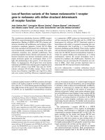
In the first group (CPB group) piglets were placed on
CPB. In the second group (non-CPB group) three new-
born animals were studied forthesametimeinterval
without cardiopulmonary perfusion (Figure 1).
Extracorporeal circuit was composed of a roller pump
(Stöckert, Munich, Germany), a blood reservoir with
pediatric membrane oxygenator (Babysafe, Jostra,
Hirrlingen, Germany), and an arterial line blood filter
(Capiox AFO2, Terumo Corp., Tokyo, Japan). The
priming volume (300 ml) consisted of fresh whole neo-
natal piglet blood (two sibling animals per study animal),
NaCl 0.9%, and 1000 units heparin. Cardiopulmonary
bypass was initiated with a flow rate of 2.5 l/min/m
2
.
Activated clotting time was maintained at a value >400
seconds throughtout duration of CPB.
On CPB, animals were cooled to 32°C core tempera-
ture. After 30 minutes the ascending aorta was cross-
clamped and cold Bretschneider’s crystalloid cardiopleg ic
solution (Custodiol HTK, Köhl er Chemie, Alsbach-
Hähnlein, Germany) was infused into the aortic root
(30 ml/kg). Following 90 minutes of cardioplegic arrest,
the aortic crossclamp was released and piglets were
rewarmed to 37°C. After a total duration of 180 minutes
animals were separated from CPB, cannulae were
removed, and anticoagulation was reversed by protamine
administration.
Thereafter piglet s were observed for up to another six
hours and data were registered.
No inotropic support was given throughout the proto-
col. Postoperative volume treatment was restrictive; cen-
tral venous pressure (CVP) and left atrial pressure
Figure 1 Schematic presentation of time intervals in both groups.
Tirilomis et al. Journal of Cardiothoracic Surgery 2010, 5:98
/>Page 2 of 6
(LAP) were kept at the base line levels (mean CVP <5
mmHg and mean LAP <2 mmHg, respectively).
Animals with complete observation time of six hours
after t ermi nation of CPB were euthanized with an over-
dose of pentobarbital.
Contractility data analysis and calculations for CPB
group (n = 4) and non-CPB group (n = 3) were per-
formed regarding the following contractility parameters:
1) Left ventricular dP/dt
max
2) Left ventricular contractility index (dP/dt
max
)/P,
and
3) Changes in regional left ventricular myocardial
thickening.
Twenty subsequent values were calculated for each
time point per piglet. Data were expressed as mean ±
standard deviation and processed with Statistica 6.1 soft-
ware (StatSoft (Europe) GmbH, Hamburg, Germany).
ThedatawereanalyzedbyANOVA,followedbyFish-
er’s LSD procedure for post hoc repeated measurements.
Differences were considered statistical significant at
P < 0.05.
Results
In the non-CPB group the left ventricular dP/dt
max
decreased from 1761 at baseline to 1170 mmH g/s at the
endpoint (P < 0.05) (Table 1). The dP/dt
max
remained
stable in the CPB group during follow up of six hours
after the end of CPB and was similar to the baseline
values (Table 1).
The performance of contractility index (dP/dt
max
)/P
was in both groups more stable (Table 2).
Myocardial thicke ning decreased significantly i n non-
CPB controls after the 2
nd
hour “post-bypass” while it
remained constant in CPB group (Table 3).
The differences between the CPB and non-CPB group
were not statistically significant regarding left ventricular
dP/dt
max
(Figure 2), contractility index (dP/dt
max
)/P
(Figure 3), and regional wall thickening (Figure 4).
Discussion
Thecurrentstudyemployedanin vivo neonatal piglet
model in which clinical standard techniques used at our
institution were applied. Many studies that examined
myocardial contractility were performed on isolated
hearts (modified Langendorff preparations) [3-6]. Extra-
polation of results from these studies to the clinical
situation should be viewed with caution. Therefore , pre-
sent study provides more relevant information about
myocardial contractility of the neonatal heart in a clini-
cal setting. Furthermore, all piglets were within the age
of the first week, before transition from the neonatal to
the adult situation may result [7].
Additionally, the changes at birth consist of conver-
sion f rom the fetal cardiovascular system to c losure of
low-resistance vascular pathways [8]. Functional closure
oftheductusarteriosusoccurswithin4hoursafter
birth [9]. At autopsy, we carefully examined the atrial
septum and the ductus arteriosus, and they were never
open.
The contractility parameter dP/dt
max
is a function of
the contractile element power, the elastici ty constant,
and the ventricular dimensions [10]. Inotropic interven-
tions (positive and negative) at constant end-diastolic
volumes reflect changes in maximal contractile element
power. In the present study there was no application of
any positive inotropic drug avoiding pharmacological
increase of myocardial contractility. The only drugs used
were the anesthetics without differences in dosages
between the two groups.
Increasing ventricular filling has two opposing effects
on dP/dt
max
; (1) the volume increase tends to increase
dP/dt
max
, according to Frank-Starling mechanism and
(2) the greater volume tends to decrease it, in accor-
dance to La Place effect. At physiological filling
Table 1 Values of left ventricular dP/dt
max
[mmHg/s] before and after CPB (up to 6 hours) or time equivalent in non-
CPB group
group pre CPB (baseline) CPB end 1 h post CPB 2 h post CPB 3 h post CPB 4 h post CPB 5 h post CPB 6 h post CPB
CPB (n = 4) 1495 ± 159 1679 ± 159 1838 ± 159 1708 ± 159 1609 ± 159 1412 ± 159 1730 ± 180 1400 ± 180
non-CPB (n = 3) 1761 ± 205 1566 ± 205 1544 ± 205 1519 ± 205 1455 ± 205 1340 ± 205 1170 ± 205 * 1151 ± 205 *
* p < 0.05 vs. baseline.
Table 2 Left ventricular contractility index ((dP/dt
max
)/P) [/s] before and after CPB (up to 6 hours) or time equivalent
in non-CPB group
group pre CPB (baseline) end CPB 1 h post CPB 2 h post CPB 3 h post CPB 4 h post CPB 5 h post CPB 6 h post CPB
CPB (n = 4) 60.5 ± 4.1 67.1 ± 7.6 65.2 ± 10.6 63.9 ± 11.6 63.3 ± 12.1 62.8 ± 12.0 65.7 ± 11.7 65.4 ± 12.5
non-CPB (n = 3) 65.7 ± 3.5 78.3 ± 7.5 74.3 ± 6.7 75.0 ± 8.3 74.4 ± 9.1 71.5 ± 10.6 66.4 ± 21.3 65.2 ± 23.1
Tirilomis et al. Journal of Cardiothoracic Surgery 2010, 5:98
/>Page 3 of 6
pressures, the first mechanism predominates [11]. In the
current study volume treatment was restrictive; central
venous and left atrial pressures were kept at the level
before procedure; mean central venous pressure was less
than 5 mmHg and mean left atrial pressure less than 2
mmHg.
However, application of dP/dt
max
maybelimited,
because of it load dependence. In this study, also the
maximal values of the ratio of the first derivative of left
ventricular pressure to instantaneous pressure (that is
(dP/dt
max
)/P; so-called contractility index) have been
considered. Peak values of (dP/dt
max
)/P were essentially
independent of preload and afterload [ 12]. Nevertheless,
extreme elevations of preload and afterload may
decrease contractility index. Decrease of (dP/dt
max
)/P
has been demonstrated for end-diastolic pressures >25
mmHg [13]. At aortic diastolic pressures of less than
120 mmHg, contractility index is independent of after-
load [12]. In the present study preload and afterload
remained within physiological range.
On a cellular level myocardial contractility depends on
many factors such as sarcoplasmic reticulum calcium
handling and myofilament calcium sensivity [14]. The
sarcoplasmic reticulum seems to play a key role; the pri-
mary function of it is to accumulate and store calcium
during diastole and release that calcium rapidly at the
onset of systole, e nabling the cardiomyoc yte to develop
rapid contraction [15]. Neonatal hearts reperfused after
the development of peak ischemic contracture have
shown negligible postischemic functional and metabolic
recovery [16]. Our findings suggest that in a clinically
relevant setting ischemic contr acture and subsequent
metabolic response could be avoided. The performan ce
of wall thickening indicates in some degree of
Table 3 Changes in (left ventricular) myocardial thickening [mm/s] before and after CPB (or time equivalent in non-
CPB group)
group pre CPB (baseline) end CPB 1 h post CPB 2 h post CPB 3 h post CPB 4 h post CPB 5 h post CPB 6 h post CPB
CPB (n = 4) 1.21 ± 0.08 1.00 ± 0.08 1.00 ± 0.08 1.13 ± 0.08 1.08 ± 0.08 0.99 ± 0.08 1.03 ± 0.09 1.05 ± 0.09
non-CPB (n = 3) 1.45 ± 0.10 1.30 ± 0.10 1.23 ± 0.10 0.98 ± 0.10 * 1.01 ± 0.10 * 1.04 ± 0.10 * 1.01 ± 0.10 * 0.99 ± 0.10 *
* p < 0.05 vs. baseline.
Figure 2 Performanc e of the left ventricular dP/dt
max
in survived neonatal piglets in % of baseline value.*P < 0.05 in comparison to
the baseline value. No statistically significant differences between the two groups.
Tirilomis et al. Journal of Cardiothoracic Surgery 2010, 5:98
/>Page 4 of 6
Figure 3 Changes of the left ventricular contractility index (dP/dt
max
)/P in survived newborn piglets in % of baseline value.No
statistically significant differences between both groups.
Figure 4 Presentation of the changes of the left ventricu lar myocardial wall thick ening (WT
amp
) in survived newborn piglets in % of
baseline value.*P < 0.05 in comparison to the baseline value. No statistically significant differences between the two groups.
Tirilomis et al. Journal of Cardiothoracic Surgery 2010, 5:98
/>Page 5 of 6
hypercontractiliy after CPB. This hypercontractility may
be the result of t he systemic inflammatory response on
myocardial level.
The present study has two important limitations; (1)
the inclusio n of survived piglets only and (2) the dura-
tion of the post-bypass observation time of six hours,
then decrease of myocardial contractility may result at
least theoretically even later than six hours after CPB
termination. Nevertheless, Burrows et al.[1] found dete-
rioration of cardiac performance four hours after cardio-
pulmonary bypass for ventricular septal defect repair,
Mustard’s operation, and repair of Tetralogy of Fallot.
In general, the results of present study are surprising.
From the theoretical point of view the response of neo-
natal myocardium to the effects of anaesthetic drugs
may be modified after cardiopulmonary bypass resulting
in this paradox of d ecreased myocardial contractility in
the control group (non-CPB group). Additionally, the
effect of the cardioplegic solution is not clear. The role
of the applied cardioplegic Bretschneider ’s solution has
to be elucidated in further studies comparing d ifferent
types of myocardial protection.
Conclusions
Applying an in vivo neonatal piglet model closely
mimicking the clinical setting of car diopulmonary
bypass with mild hypothermia (and crystalloid cardiople-
gic myocardial protection) but without postoperative
inotropic support, we found that the myocardial con-
tractility of the neonatal heart remained in survived ani-
mals at the baseline values after cardiopulmonary
bypass, probably due to some degree of regional
hypercontractility.
Authors’ contributions
TT conceived the study, participated in design and coordination, participated
in acquisition, analysis and interpretation of the data and drafted the
manuscript. OJL participated in the design of the study and performed the
statistical analysis. KOC participated in data analysis and helped to draft the
manuscript. MB participated in the design of the study and helped in
acquisition of the data. AFP participated in data analysis and helped to draft
the manuscript. JDS participated in data analysis and helped to draft the
manuscript. FAS participated in the design and coordination, and revised
manuscript critically. All authors read and approved the final manuscript.
Competing interests
The authors declare that they have no competing interests.
Received: 16 May 2010 Accepted: 2 November 2010
Published: 2 November 2010
References
1. Burrows FA, Williams WG, Teoh KH, Wood AE, Burns J, Edmonds J,
Barker GA, Trusler GA, Weisel RD: Myocardial performance after repair of
congenital cardiac defects in infants and children. J Thorac Cardiovasc
Surg 1988, 96:548-556.
2. Wessel DL: Managing low cardiac output syndrome after congenital
heart surgery. Crit Care Med 2001, 29:S220-S230.
3. Grice WN, Konishi T, Apstein CS: Resistance of neonatal myocardium to
injury during normothermic and hypothermic ischemic arrest and
reperfusion. Circulation 1987, 76(Suppl V):V150-V155.
4. Downing ES, Chen V: Myocardial hibernation in the ischemic neonatal
heart. Circ Res 1990, 66:763-772.
5. Rebeyka IM, Hanan SA, Borges MR, Lee KF, Yeh T Jr, Tuchy GE, Abd-
Elfattah AS, Williams WG, Wechsler AS: Rapid cooling contracture of the
myocardium. J Thorac Cardiovasc Surg 1990, 100:240-249.
6. Shum-Tim D, Tchervenkov CI, Hosseinzadeh T, Chiu RCJ: Contracture of the
newborn myocardium after prolonged prearrest cooling. J Thorac
Cardiovasc Surg 1993, 106:643-650.
7. Penefsky ZJ, Buckley NM, Robinson TF, Sorensen AL: Mechanical properties
of developing swine myocardium. Comp Biochem Physiol A 1985,
80:399-409.
8. Gootman PM: Cardiovascular system. In Biology of the domestic pig. Edited
by: Pond WG, Mersmann HJ. Ithaka and London: Cornell University Press;
2001:533-559.
9. Haworth SG, Hislop AA: Adaptation of the pulmonary circulation to extra-
uterine life in the pig and its relevance to the human infant. Cardiovasc
Res 1981, 15:108-119.
10. Taylor RR: Theoretical analysis of the isovolumic phase of left ventricular
contraction in terms of cardiac muscle mechanisms. Cardiovasc Res 1970,
4:429-435.
11. Taylor RR, Ross J, Covell JW, Sonnenblick EH: A quantitative analysis of left
ventricular myocardial function in the intact, sedated dog. Circ Res 1967,
21:99-115.
12. Nejad NS, Klein MD, Mirky I, Lown B: Assessment of myocardial
contractility from ventricular pressure recordings. Cardiovasc Res 1971,
5:15-23.
13. Krayenbühl HP, Rutishauser W, Wirz P, Amende I, Mehmel H: High-fidelity
left ventricular pressure measurements for the assessment of cardiac
contractility in man. Amer J Cardiol 1973, 31:415-427.
14. Pieske B, Schlotthauer K, Schattman J, Beyersdorf F, Martin J, Just H,
Hasenfuss G: Ca
2+
-dependent and Ca
2+
-independent regulation of
contractility in isolated human myocardium. Basic Res Cardiol 1997,
92(Suppl 1):75-86.
15. Klautz RJM, Baan J, Teitel DF: The effect of sarcoplasmic reticulum
blockade on the force/frequency relationship and systolic contraction
patterns in the newborn pig heart. Pflügers Arch - Eur J Physiol 1997,
435:130-136.
16. Torrance SM, Belanger MP, Wallen WJ, Wittnich C: Metabolic and
functional response of neonatal pig hearts to the development of
ischemic contracture: is recovery possible? Pediatr Res 2000, 48:191-199.
doi:10.1186/1749-8090-5-98
Cite this article as: Tirilomis et al.: Myocardial contractile function in
survived neonatal piglets after cardiopulmonary bypass. Journal of
Cardiothoracic Surgery 2010 5:98.
Submit your next manuscript to BioMed Central
and take full advantage of:
• Convenient online submission
• Thorough peer review
• No space constraints or color figure charges
• Immediate publication on acceptance
• Inclusion in PubMed, CAS, Scopus and Google Scholar
• Research which is freely available for redistribution
Submit your manuscript at
www.biomedcentral.com/submit
Tirilomis et al. Journal of Cardiothoracic Surgery 2010, 5:98
/>Page 6 of 6