Báo cáo y học: "Isolated cardiophrenic angle node metastasis from ovarian primary. report of two case" pptx
Bạn đang xem bản rút gọn của tài liệu. Xem và tải ngay bản đầy đủ của tài liệu tại đây (394.1 KB, 3 trang )
CAS E REP O R T Open Access
Isolated cardiophrenic angle node metastasis
from ovarian primary. report of two cases
Mark Ragusa
1
, Jacopo Vannucci
1*
, Rosanna Capozzi
1
, Niccolò Daddi
1
, Nicola Avenia
2
, Francesco Puma
1
Abstract
Ovarian cancer is the most lethal gynaecologic malignancy. It usually spreads out of the abdomen involving
thoraco-abdominal organs and serosal surface. This disease is poorly curable and surgery, at early stage, is
supposed to achieve the best survival outcome. In systemic dissemination, chemiotherapy is indicated, sometimes
with neoadjuvant aim. The most common clinical expressions of advanced ovarian carcinoma are multiple
adenopathy, neoplastic pleuritis, peritoneal seeding and distant metastasis, mainly hepatic and pulmonary. Isolated
adenopathy of the mediastinum is rare and isolated bilateral have never been described before . We report two
cases of isolated bilateral cardiophrenic angle lymphnode metastasis from ovarian carcinoma, without peritoneal
and pleural involvement. Both patients were successfully resected through minimally invasive thoracic surgery.
About the role of surgery, few data are available but survival seems to be longer after resection thus, more
investigation is required to make the indication to surgery more appropriate in advanced cases.
Background
The cardiophrenic angle lymphnodes (CPLN) were cl as-
sified by Rouviere into two groups: anterior prepericar-
diac and middle latero-pericardiac. The afferent
lymphatics of CPLN drain areas from the diaphragm,
liver, pleura and anterior abdominal wall and they
empty into the internal mammary chain. Malignant lym-
phoma and metastases of abdominal or thoracic neo-
plasms have been mentioned to be possible causes of
CPLN enlargement. Most of the times the disease is
unilateral.
CPLN involvement may represent a staging and prog-
nostic indicator for ovarian cancer [1]. Natural history
of ovarian cancer entails extensive tumor dissemination
on the peritoneal and pleural surface, with possible
intrathoracic lymphnodes metastasis.
In the present paper we report two patients with iso-
lated bilateral CPLN metastasis from previously resected
ovarian carcinoma, with no peritoneal and pleural
involvement.
Case 1
A 50-year-old woman was referred to our service for
bilateral cardiophrenic angle mass. Two months earlier,
the patient had undergone laparoscopic left ovariectomy
with incidental diagnosis of cancer. Postoperative
CA-125 value was within the normal range. Thoraco-
abdominal computed tomography (CT) scan revealed
bilateral neoplasms in the cardiophrenic angles, 2.5 and
1.5 cm in diameter. Fluorine 18-fluoro-2-deoxy-glucose-
positron emission tomography (FDG-PET) scan evi-
denced enhanced uptake in the above mentioned sites
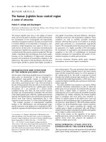
(Figure 1a). The case was discussed at the multidisci-
plinary oncology round and indication for surgery was
established. Videothoracoscopic complete removal of a
capsulated yellowish cardiophrenic tumor was per-
formed bilaterally. Pathology disclosed metastatic node
colonization by papillary ovarian cancer in both speci-
mens. T he patient had an uneventful recovery and was
discharged four days after the procedure. Two weeks
later she underwent chemotherapy.
Case 2
A 50-year-old woman, was admitted to our Hospital for
bilateral cardiophrenic angle tumor. The patient had
been submitted to laparotomic hysterectomy with bilat-
eral salpingo-oophorectomy two years earlier, for ovar-
ian papillary serous adenocarcinoma (pT 1bN0Mx).
* Correspondence:
1
Thoracic Surgery Unit. University of Perugia Medical School. Ospedale S.
Maria della Misericordia Perugia, Italy
Full list of author information is available at the end of the article
Ragusa et al . Journal of Cardiothoracic Surgery 2011, 6:1
/>© 2011 Ragusa et al; licensee BioMed Central Ltd. This is an Open Access article distributed under the terms of the Creative Commons
Attribution License (htt p://creativecommons.org/licenses/by/2.0), which permits unrestricted use, distribution, an d reproduction in
any medium, provided the original work is properly cited.
Preoperative Ca-125 value was 14.00 U/ml. Three cycles
of adjuvant chemotherapy with carboplatin and taxol
were administered. During follow-up, FDG-PET scan
revea led an increased uptake in a bilateral 1.5 cm cardi-
ophrenic tumor, not recognized on CT scan (Figure 1b).
After discussion w ith the referring oncologist and the
patient, she underwent a sequential bilateral videothora-
coscopy with complete cardiophrenic tumor removal.
Pathology disclosed metastatic node colonization
by papillary ovarian cancer in both specimens. After an
uneventful recovery, the patient was discharged five
days after surgery and returned to the oncologist for
chemotherapy.
Conclusion
Ovarian carcinoma remains the most lethal gynaecologic
malignancy. It usually spreads out of the abdomen along
different routes: lymphatic, haematogenous and trans-
caelomic. One of its hallmarks is the possible peritoneal
and pleural dissemination. Mediastinal lymphnode
metastasis (stage IV) entails a definite w orsening of
prognosis [2]. CPLN colonization is frequently asso-
ciated with extensive intrathoracic disease, typically
represented by right-sided pleural effusion [1]. Such
behaviour is explained by the anatomic arrangement of
abdominal lymphatic drainage, which follows a cloc k-
wise route, involving first the thoracic lymphatic stations
on the right side.
Isolated bilateral metastas is is to be considered anecdo-
tical and, to our knowledge, bilateral involvement with-
out pleural effusion was never reported in the English
Literature. In our patients FDG-PET scan facilitated
identification and proper diagnosis of CPLN metastasis.
In one patient CT scan did not clearly demonstrate nodal
disease. The possible anatomical pathway for tumor
spread in the cases herein reported is an unanswered
question, considering that serosal surfaces and intra-
abdominal viscera were apparently unaffected by disease.
An interesting, potentially misleading, feature of meta-
static supradiaphragmatic nodes from ovarian primary,
is calcification. Although notobservedinourcases,
such aspect is reported with an incidence up to 35%,
and must not be overlooked. Calcified intrathoracic
nodes in patients with previous ovarian serous adeno-
carcinoma cannot be ruled out as granulomatous dis-
ease, but metastatic deposits must be excluded. A hint
to the latter hypothesis is the progressive growth of the
involved station [3], also considering that, in such cir-
cumstances, FDG-PET scan is not entirely reliable
because granulomatous lymphadenitis as well may show
an increased FDG-uptake.
Surgery is carried out in order to achieve histologic
diagnosis, disease staging, and prolonged survival.
Vid eothoraco scopy is specifically fit for such procedure,
as recently stated by Lim et Al. [1]. The minimally inva-
sive approach enables thorough exploration of the entire
pleural cavity, easy resection of even small nodes deeply
sited within the pericardial fat, and the one-stage
removal of bilateral CPLN growths. Resection of isolated
node metastases can improve outlook, particularly for
slow growing tumors. In such setting, progression-free
survival before relapse does not appear to be a reliable
indicator of prognosis, as it is for many other cancers.
Tumor growth rate seems a more sound parameter [2].
Treatment of recurrent epithelial ovarian c ancer is
based on various considerations: recurrence site, general
conditions of the patient, disease-free interval (with the
above-mentioned caveat), growt h rate, response to first-
line chemotherapy.
In presence of isolated CPLN relapse, the patient may
be included in the Isolated Lymph Node Relapse group,
a subset appearing to gain from surgery in terms of sur-
vival [4]. On the other hand, only one series of video-
assisted transthoracic resection of lymph node and
pleural metastasis from ovarian cancer is available,
therefore further data are required to clarify the role of
surgery in downstaging ovarian cancer diffusion to the
mediastinum and thoracic cavity [1].
Written informed consent was obtained from the
patients for publication of this case report and any
accompanying images. A copy of the written consent is
available for review by the Editor-in-chief of this
journal.
List of abbreviations
CPLN: cardiophrenic angle lymphnodes; CT: computed tomography; FDG-
PET: Fluorine 18-fluoro-2-deoxy-glucose-positron emission tomography.
Author details
1
Thoracic Surgery Unit. University of Perugia Medical School. Ospedale S.
Maria della Misericordia Perugia, Italy.
2
Endocrine and Soft Tissue of the Neck
Surgery Unit. University of Perugia Medical School. Ospedale S. Maria Terni,
Italy.
Figure 1 PET-CT appearance of bilateral cardiophrenic angle
node metastasis in the two cases reported.
Ragusa et al . Journal of Cardiothoracic Surgery 2011, 6:1
/>Page 2 of 3
Authors’ contributions
MR, JV and FP wrote the article, RC and ND collected the clinical
information and selected the images, MR and NA analyzed the English
Literature. FP drafted the final manuscript. All authors approved the final
manuscript to be published.
Competing interests
The authors declare that they have no competing interests.
Received: 11 July 2010 Accepted: 5 January 2011
Published: 5 January 2011
References
1. Lim MC, Lee HS, Jung DC, Choi JY, Seo SS, Park SY: Pathological diagnosis
and cytoreduction of cardiophrenic lymph node and pleural metastasis
in ovarian cancer patients using video-assisted thoracic surgery. Ann
Surg Oncol 2009, 16:1990-6.
2. Blanchard P, Plantade A, Pagés C, Afchain P, Louvet C, Tournigand C, de
Gramont A: Isolated lymph node relapse of epithelial ovarian carcinoma:
Outcomes and prognostic factors. Gynecol Oncol 2007, 104:41-5.
3. Patel SV, Spencer JA, Wilkinson N, Perren TJ: Supradiaphragmatic
manifestations of papillary serous adenocarcinoma of the ovary. Clin
Radiol 1999, 54:748-54.
4. Uzan C, Morice P, Rey A, Pautier P, Camatte S, Lhommè C, Haie-Meder C,
Duvillard P, Castaigne D: Outcomes after combined therapy including
surgical resection in patients with epithelial ovarian cancer recurrence(s)
exclusively in lymph nodes. Ann Surg Oncol 2004, 11(7):658-64.
doi:10.1186/1749-8090-6-1
Cite this article as: Ragusa et al.: Isolated cardiophrenic angle node
metastasis from ovarian primary. report of two cases. Journal of
Cardiothoracic Surgery 2011 6:1.
Submit your next manuscript to BioMed Central
and take full advantage of:
• Convenient online submission
• Thorough peer review
• No space constraints or color figure charges
• Immediate publication on acceptance
• Inclusion in PubMed, CAS, Scopus and Google Scholar
• Research which is freely available for redistribution
Submit your manuscript at
www.biomedcentral.com/submit
Ragusa et al . Journal of Cardiothoracic Surgery 2011, 6:1
/>Page 3 of 3