Báo cáo y học: "Effects on heart pumping function when using foam and gauze for negative pressure wound therapy of sternotomy wounds" pptx
Bạn đang xem bản rút gọn của tài liệu. Xem và tải ngay bản đầy đủ của tài liệu tại đây (622.32 KB, 6 trang )
RESEARC H ARTIC L E Open Access
Effects on heart pumping function when using
foam and gauze for negative pressure wound
therapy of sternotomy wounds
Malin Malmsjö
1*
, Sandra Lindstedt
2
, Richard Ingemansson
2
Abstract
Background: Negative pressure wound therapy (NPWT ) has remarkable effects on the healing of poststernotomy
mediastinitis. Foam is presently the material of choice for NPWT in this indication. There is now increasing interest
in using gauze, as this has proven successful in the treatment of peripheral wounds. It is important to determine
the effects of NPWT using gauze on heart pumping function before it can be used for deep sternotomy wounds.
The aim was to examine the effects of NPWT when using gauze and foam on the heart pumping function during
the treatment of a sternotomy wound.
Methods: Eight pigs underw ent median sternotomy followed by NPWT at -40, -70, -120 and -160 mmHg, using
foam or gauze. The heart frequency, cardiac output, mean systemic arterial pressure, mean pulmonary artery
pressure, central venous pres sure and left atrial pressure were recorded.
Results: Cardiac output was not affected by NPWT using gauze or foam. Heart frequency decreased during NPWT
when using foam, but not gauze. Treatment with foam also lowered the central venous pressure and the left atrial
pressure, while gauze had no such effects. Mean systemic arterial pressure, mean pulmonary artery pressure and
systemic vascular resistance were not affected by NPWT. Similar haemodynamic effects were observed at all levels
of negative pressure studied.
Conclusions: NPWT using foam results in decreased heart frequency and lower right and left atrial filling pressures.
The use of gauze in NPWT did not affect the haemodynamic parameters studied. Gauze may thus provide an
alternative to foam for NPWT of sternotomy wounds.
Background
Cardiac surger y is complicated by poststernotomy med-
iastinitis in 1-5% of all procedures [1]; a complication
that is life-threatening [2]. The reported early mortality
using conventiona l therapy is between 8 and 25% [3,4].
In 1999, Obdeijn and colleagu es described the treatment
of poststernotomy mediastinitis using vacuum-assisted
closure [5], now called negative pressure wound therapy
(NPWT). The technique entails the application of nega-
tive pressure to a sealed wound. NPWT has remarkable
effects on the healing of poststernotomy mediastinitis,
and has reduced the rate of mortality considerably [6].
The organs in the mediastinum are haemodynamically
crucial, and both vulnerable bypass grafts and he art
function should be taken into consideration when per-
forming NPWT after cardiac surgery.
NPWT is known to affect the heart pumping function,
although the results from different studies on the sub-
ject are not consistent [7-11]. Using sonometry, Con-
quest and colleagues showed that subatmospheric
pressure decreased the left ventricular volume and car-
diac output in pigs by approximately 30% [7], but that
this could be p revented by rotati ng a rectus muscle flap
over the mediastinal wound. In a subsequent study by
Petzina et al., using magnet ic resonance imaging,
NPWT of sternotomy wounds in pigs was shown to
decrease the cardiac output to a lesser extent, 13% [10].
In studies by Sjögren et al. and Steigelman et al., using
thermodilution, cardiac output was found not to be
affected by negative pressure [8,11].
* Correspondence:
1
Department of Ophthalmology, Lund University Hospital, Lund, Sweden
Full list of author information is available at the end of the article
Malmsjö et al. Journal of Cardiothoracic Surgery 2011, 6:5
/>© 2011 Malmsjö et al; licensee BioMed Central Ltd. This is an Open Access articl e distributed under the terms of the Creative
Commons Attribu tion License ( which permits unrestricted use, distribution, and
reproduction in any medium, provided the origina l work is properly cited.
Foam is presently the material of choice when apply-
ing NPWT to deep sternotomy wounds. However, inter-
est in the use of gauze has increased [12,13], as this has
proven successful in the treatment of other kinds of per-
ipheral wounds [14]. The advantages with g auze include
its conformability and ease of application to large and
irregular wounds [18]. It has been suggested that gauze
under negative pressure can tamponade superficial
bleedings in sternotomy wounds [19]. Furthermore,
there are no reported problems with ingrowth of granu-
lation tissue into gauze in NPWT [20]. Before gauze is
used for NPWT of sternotomy wounds, it is important
to determine the effects on heart pumping function.
Hitherto, the effect of NPWT on the pumping function
of the heart has only been examined using foam [7-10].
The aim of the present study was therefore to compare
the haemodynamic e ffects of NPWT using gauze and
foa m. Eight pigs underwent median sternotomy and the
wounds were treated with NPWT at negative pressures
of -40, -70, -120 or -160 mmHg, using foam or gauze.
Haemodynamic parameters, including heart frequency,
cardiac output, mean systemic arterial pressure, mean
pulmonary artery pressure, central venous pressure and
left atrial pressure, were recorded.
Methods
Animals
A porcine sternotomy wound model was used. Eight
domestic landrace pigs with a mean body weight of
70 kg were fasted overnight with free access to water.
The study was approved by the Ethics Committee for
Animal Research, Lund Univer sity, Sweden. The investi-
gation complied with the “GuidefortheCareandUse
of Laboratory Animals” as recommended by the U.S.
National Institutes of Health and published by the
National Academies Press (1996).
Anaesthesia and surgery
Premedication was performed with an intramuscular
injection of xylazine (Rompun
®
vet. 20 mg/ml; Bayer
AG, Leverkusen, Germany; 2 mg/kg) mixed with keta-
mine (Ket aminol
®
vet. 100 mg/ml; Farmaceutici Gellini
S.p.A, Aprilia, Italy; 20 mg/kg). Before surgery, a tra-
cheotomy was performed and an endo-tracheal tube was
inserted. Anaesthesia was maintained with a cont inuous
infusion of ketamine (Ketaminol
®
vet. 50 mg/ml; 0.4-
.6 mg/kg/h). Complete neuromuscular blockade was
achieved with a continuous infusion of pancuronium
bromide (Pavulon; N.V. Organon, OSS, the Nether lands;
0.3-0.5 mg/kg/h). Fluid loss was compensated for by
continuous infusion of Ringer’ s acetate at a rate of
300 ml/kg/h. Mechanical ventilation was established
with a Siemens-Elema ventil ator (Servo Vent ilator 300,
Siemens, Solna, Sweden) in the volume-controlled mod e
(65% nitrous oxide , 35% oxygen). Ventilatory settings
were identical for all animals (respiratory rate: 15
breaths/min; minute ventilation: 8 l/min). A positive
end-expiratory pre ssure of 5 cmH
2
O was applied. A
Foley catheter was inserted into the urinary bladder
through a suprapubic cystostomy. Upon completion of
the experiments, the animals were euthanized with a
lethal dose (60 mmol) of intravenous potassium
chloride.
Wound preparation for NPWT
A midline sternotomy was performed. T hree layers of
paraffin gauze Jelonet
®
(Smith & Nephew, Hull, UK)
were placed over the anterior surface of the h eart to
protect it from the sternal edges. The wound was filled
with saline-soaked AMD gauze (RENASYS-G, St Peters-
berg, FL), or one of two kinds of foam: VAC foam size
18 × 12.5 × 3.3 cm (VAC
®
black GranuFoam
®
,KCI,
SanAntonio,TX),orRENASYS-Ffoam,20×12.5×3
cm (RENASYS-F, St Petersberg, FL). One layer of foam
or two rolls of gauze were placed between the sternal
edges. A second layer of foam or two rolls of gauze
were placed over the first la yer, between the soft tissue
wound edges, and secured to the surrounding skin. Two
drains were inserted into the wound filler. One drain
was placed between two layers of wound filler (foam or
gauze) and the other was placed at the top of the
wound filler The wound was sealed with a transparent
adhesive drape and connected to a vacuum source
(RENASYS-EZ, Smith & Nephew St Petersburg, FL).
The vacuum source was s et to deliver negative continu-
ous pressures of -40, -70, -120 and -160 mmHg.
Haemodynamic assessment
The mean systemic arterial pressure was monitored via a
catheter in the left carotid artery. The mean pulmonary
artery pressure was monitored via a catheter in the pul-
monary artery. Double-lumen central venous catheters
were inserted into the left external jugular vein and the left
atrium to record the central venous pressure and left atrial
pressure, respectively. A flow probe (CardioMed TraCe
System, Medistim, Norway) was placed around the pul-
monary artery to record the cardiac output. The catheter
was connected to a cardiac output monitor (Oximetrix 3,
Abbot Laboratories, North Chicago, IL, USA). The haemo-
dynamic data were collected in a data acquisition sys tem
(PowerLab, AD Instruments Ltd., Castle Hill, Australia).
Systemic vascular resistance and pulmonary vascular resis-
tance were calculated from th e above p arameters.
After surgical preparation, the animal was allowed to
stabilize for one hour. Baseline measurements of the
above mentioned haemodynamic parameters were then
recorded before applying NPWT. Negative pressures of
-40, -70, -120 or -160 mmHg were applied, and the
Malmsjö et al. Journal of Cardiothoracic Surgery 2011, 6:5
/>Page 2 of 6
effects on the haemodynamic parameters were recorded
after 2½ and 5 min of NPWT. The negative pressures
were applied in a random order, with intervals of
10 min without any pressure.
Limitations
The study was performed using an acute sternotomy
wound model in pigs where the organs in the thoracic
cavity can move f reely. In patient s with mediastinitis
there a re typically adhesions around the heart that may
restric t movement. In addition, following a short period
of NPWT, the mediastinum becomes fixed and rigid
with the development of granulation tissue and fibrous
ingrowth. The hemodynamic effects of NPWT may dif-
fer between an acute porcine sternotomy wound model
and patients with mediastinitis.
Calculations and statistics
Calculations and statistical analysis were performed using
GraphPad 5.0 software (San Diego, CA, USA). The effect
of NPWT on each haemodynamic parameter was calcu-
lated as a percent of the baseline value. Statistical analysis
was performed using the Mann-Whitney test. Significance
was defined as P < 0.05 (*), P < 0.01 (**), P < 0.001 (***)
and P > 0.05 (not significant, n.s.). Values are presented
as means ± the standard error on the mean (S.E.M.)
unless otherwise stated.
Results
Cardiac output was not a ffected by NPWT when using
gauze or foam (p = n.s., Figure 1). Heart frequency
was d ecreased (-9%, p < 0.05) when NPWT was
applied using foam, but not when using gauze (p = n.s.,
Figure 1). Neither the mean arterial pressure nor the mean
pulmonary artery pressure was affected by NPWT (p =
n.s., Figur e 1). NPWT did not alter the systemic vascular
resistance. The pulmonary vas cul ar resistance increased,
as a result of calculating it by a formula with the left atrial
pressure (Figure 2). Negative pressure treatment with
foam lowered the central venous pressure (-20%, p < 0.05)
and the left atrial pressure (-11%, p < 0.01), while gauze
had no such effects (p = n.s., Figure 3).
The effects of NPWT on the haemodynamic parameters
were immediate, and the recordings after 2½ and 5 min of
therapy were similar (Figures 1, 2 and 3). Comparable hae-
modynamic effects were observed at all levels of negative
pressure studied (-40, -70, -120 and -160 mmHg). There
was no significant difference in the haemodynamic effects
when using the two different types of foam.
Discussion
Cardiac output
The present findings regarding cardiac output agree lar-
gely with those in previ ous studies, although the results
in these studies vary somewhat. Mohktari et al. reported
that the haemodynamics in pigs with a sternotomy
wound treated with NPWT at -75 mmHg was unaltered,
using an ultraflow probe to measure the flow through
the pulmonary artery [9]. Fur thermore, in a study by
Steigelman et al., using theromdilution, cardiac output
was unaltered by a negative pressure of -125 mmHg
[11]. I n a study by Sjögren et al., using thermodilution,
the cardiac output was found to be slightly increased
when a sternotomy wound in pigs was treated with a
negative pressure of -75, while other negative pressures
(-50, -100, -125, -15 0 and -175 mmHg) had no effect
[8]. In a study by Petzina et al., cardiac output was
reported to be slig htly re duced during NPWT (-75, -125
and -175 mmHg) of sternotomy wounds in pigs, when
measured using magnetic resonance imaging [10].
The effect of NPWT on cardiac output is especially
important since many patients with deep sternal wound
infections have impaired cardiac function and heart fail-
ure due to ischaemic heart disease. Their ability to com-
pensate for a decrease in cardiac output during NPWT
may thus be limited. It has been suggested that haemo-
dynamic parameter s should be carefully monitored in
patients undergoing sternal NPWT [7]. However, when
considering the data from the present study and other
reports [8-10], it appears that NPWT has no major
effects on cardiac output, and invasive monitoring may
therefore be unnecessary.
Heart rate
The results of the present study show that the heart fre-
quency decreased when NPWT was applied using foam.
Similar results have been found in an experimental
study on NPWT of a sternoto my wound using foam,
demonstrating a tendency towards decreased heart rated
upon the application of negative pressures of -75, -125
and -175 mmHg [10]. Interestingly, the present study
shows that gauze has no such effects. The reason for
this cannot be deduced from the present study, but may
be due to differences in the mechanical effects by the
two materials on the heart [17]. The decrease in heart
rate when using foam is only slight (~20%) and the
heart pumping function may only be affected when
compensatory mechanisms are compromised as a result
of advanced heart failure.
Left and right atrial filling pressure
This study shows that the use of foam in NPWT lowers
the central venous pressure and t he left atrial pressure.
We suggest that this is also the result of differences in
the mechanical effects on the heart and large intrathor-
acic vessels when using foam and gauze. Suction in a
sternotomy wound results in the heart being displaced
outwards, towards the sternum [17]. Foam allows a
Malmsjö et al. Journal of Cardiothoracic Surgery 2011, 6:5
/>Page 3 of 6
C
ardiac output
VAC foam, 0 min
VAC foam, 2.5 min
VAC foam, 5 min
RENASYS-F foam, 0 min
RENASYS-F foam, 2.5 min
RENASYS-F foam, 5 min
Gauze, 0 min
Gauze, 2.5 min
Gauze, 5 min
60
80
100
120
n.s.n.s.n.s.
CO (% change)
Heart
f
requency
VAC foam, 0 min
VAC foam, 2.5 min
VAC foam, 5 min
RENASYS-F foam, 0 min
RENASYS-F foam, 2.5 min
RENASYS-F foam, 5 min
Gauze, 0 min
Gauze, 2.5 min
Gauze, 5 min
60
80
100
120
* **
n.s.
HF (% change)
Mean systemic arterial pressure
VAC foam, 0 min
VAC foam, 2.5 min
VAC foam, 5 min
RENASYS-F foam, 0 min
R
ENASYS-F foam, 2.5 min
RENASYS-F foam, 5 min
Gauze, 0 min
Gauze, 2.5 min
Gauze, 5 min
60
80
100
120
n.s.n.s.n.s.
MAP (% change)
Mean pulmonary artery pressure
VAC foam, 0 min
VAC foam, 2.5 min
VAC foam, 5 min
RENASYS-F foam, 0 min
RENASYS-F foam, 2.5 min
RENASYS-F foam, 5 min
Gauze, 0 min
Gauze, 2.5 min
Gauze, 5 min
60
80
100
120
n.s.n.s.n.s.
MPAP (% change)
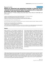
Figure 1 Cardiac output, heart frequency, mean systemic arterial pressure and mean pulmonary artery pressure before NPWT is
applied (0 min) and 2½ and 5 min after a negative pressure of -120 mmHg was applied. Measurements were made during NPWT using
gauze or two different types of foam. The results are shown as means ± the standard error on the mean of eight experiments. Each of the 8
pigs was subjected to NPWT using all 3 different dressings (n = 8). Statistical analysis was performed using the Mann-Whitney test. Significance
was defined as P < 0.05 (*), P < 0.01 (**), P < 0.001 (***) and P > 0.05 (not significant, n.s.). Before negative pressure was applied, baseline heart
frequency was 114 ± 1 bpm, cardiac output was 3.0 ± 0.1 L/min, mean systemic arterial pressure was 85 ± 3 mmHg and mean pulmonary artery
pressure was 17.5 ± 0.5 mmHg. Note that heart frequency decreases during NPWT using foam, but not gauze. Results were similar at the other
negative pressures studied (not shown).
S
ystemic vascular resistance
VAC foam, 0 min
VAC foam, 2.5 min
VAC foam, 5 min
RENASYS-F foam, 0 min
R
ENASYS-F foam, 2.5 min
RENASYS-F foam, 5 min
Gauze, 0 min
Gauze, 2.5 min
Gauze, 5 min
60
80
100
120
n.s.n.s.n.s.
SVR (% change)
Pulmonary vascular resistance
VAC foam, 0 min
VAC foam, 2.5 min
VAC foam, 5 min
RENASYS-F foam, 0 min
R
ENASYS-F foam, 2.5 min
RENASYS-F foam, 5 min
Gauze, 0 min
Gauze, 2.5 min
Gauze, 5 min
60
80
100
120
n.s.
**
PVR (% change)
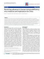
Figure 2 Change in systemic vascular resistance and pulmonary vascular resistance upon the applicati on of NPWT at -120 mmHg ,
using foam and gauze (n = 8). Before negative pressure was applied, baseline systemic vascular resistance was 1957 ± 43 dynes*sec/cm
5
and
pulmonary vascular resistance was 172 ± 3 dynes*sec/cm
5
. Results were similar at the other negative pressures studied (not shown).
Malmsjö et al. Journal of Cardiothoracic Surgery 2011, 6:5
/>Page 4 of 6
greater volume reduction than gauze, as the latter is a
denser material. The heart may therefore be drawn out-
wards to a greater extent when using foam than when
using gauze. NPWT using foam may relieve tension on
the caval and lung veins in the bottom of the thoracic
cavity, thereby decreasing the filling pressure in the
right and left atria. This is haemodynamically beneficial
as the load o n the heart is reduced. The suggested
mechanism is illustrated in Figure 4.
Clinical implications
The results of this and previous studies indicate that
careful consideration should be g iven to the choice o f
material used in NPWT, as both foam and gauze may
have advantages and disadvantages [21]. Both result in
the formation of granulation tissue; foam leads to the
growth of a thick layer of tissue, which may penetrate
the foam, making it difficult to remove after NPWT.
Gauze leads to the growth of thin, dense granulation
Central venous pressure
VAC foam, 0 min
VAC foam, 2.5 min
VAC foam, 5 min
RENASYS-F foam, 0 min
R
ENASYS-F foam, 2.5 min
RENASYS-F foam, 5 min
Gauze, 0 min
Gauze, 2.5 min
Gauze, 5 min
60
80
100
120
*
n.s.n.s.
CVP (% change)
Left atrial pressure
VAC foam, 0 min
VAC foam, 2.5 min
VAC foam, 5 min
RENASYS-F foam, 0 min
R
ENASYS-F foam, 2.5 min
RENASYS-F foam, 5 min
Gauze, 0 min
Gauze, 2.5 min
Gauze, 5 min
60
80
100
120
** **
n.s.
LAP (% change)
Figure 3 Change in central venous pressur e and left atrial pressure upon the applicat ion of NPWT at -120 mmHg , using foam and
gauze (n = 8). Before negative pressure was applied, baseline left atrial pressure was 6.3 ± 0.2 mmHg and central venous pressure was 4.8 ±
0.1 mmHg. Results were similar at the other negative pressures studied (not shown).
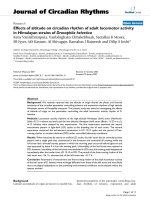
Figure 4 Illustrations of the cross-section of the thoracic cavity showing an open sternotomy, the heart, and caval and lung veins.
Suction in a sternotomy wound by NPWT results in the heart being displaced outwards, towards the sternum. There is a greater reduction in
the volume of the foam than the gauze, as the latter is a denser material. The heart may therefore be drawn outwards to a greater extent when
using foam than when using gauze. NPWT using foam may relieve tension on the caval and lung veins at the back of the thoracic cavity,
thereby decreasing the filling pressure in the right and left atria. This may be haemodynamically beneficial, as the load on the heart is reduced.
Malmsjö et al. Journal of Cardiothoracic Surgery 2011, 6:5
/>Page 5 of 6
tissue without ingrowth, commonly referred to by physi-
cians as high-quality tissue. NPWT using gauze has pro-
ven to be successful in the treatment of other kinds of
wounds [14].
Conclusions
This study was performed to compare the effects of
NPWT of a sternotomy wound using foam and gauze
on heart pumping function. Foam is presently the mate-
rial of choice for NPWT in cardiac surgery. NPWT
using foam results in a slight decrease in heart fre-
quency and lower right and left atrial filling pressures.
NPWT using gauze does not affect the heart pumping
function. Gauze may provide an alternative to foam for
the treatment of sternotomy wounds using NPWT.
Acknowledgements
This study was funded by Åke-Wieberg Foundation, the Magn Bergvall
Foundation, the Swedish Medical Association, the Royal Physiographic
Society in Lund, the Crafoord Foundation, the Swedish Heart-Lung
Foundation, the Swedish Hypertension Society and Smith & Nephew Wound
Management.
Author details
1
Department of Ophthalmology, Lund University Hospital, Lund, Sweden.
2
Department of Cardiothoracic Surgery, Lund Universi ty Hospital, Lund,
Sweden.
Authors’ contributions
MM; designed study and wrote the manuscript. SL; collected and analysed
the data. RI; designed the study. All authors have read and approved the
final manuscript.
Competing interest
The authors declare that they have no competing interests.
Received: 4 August 2010 Accepted: 13 January 2011
Published: 13 January 2011
References
1. Raudat CW, Pagel J, Woodhall D, Wojtanowski M, Van Bergen R: Early
intervention and aggressive management of infected median
sternotomy incision: a review of 2242 open-heart procedures. Am Surg
1997, 63:238-241, discussion 241-232.
2. El Oakley RM, Wright JE: Postoperative mediastinitis: classification and
management. Ann Thorac Surg 1996, 61:1030-1036.
3. Crabtree TD, Codd JE, Fraser VJ, Bailey MS, Olsen MA, Damiano RJ Jr:
Multivariate analysis of risk factors for deep and superficial sternal
infection after coronary artery bypass grafting at a tertiary care medical
center. Semin Thorac Cardiovasc Surg 2004, 16:53-61.
4. Lu JC, Grayson AD, Jha P, Srinivasan AK, Fabri BM: Risk factors for sternal
wound infection and mid-term survival following coronary artery bypass
surgery. Eur J Cardiothorac Surg 2003, 23:943-949.
5. Obdeijn MC, de Lange MY, Lichtendahl DH, de Boer WJ: Vacuum-assisted
closure in the treatment of poststernotomy mediastinitis. Ann Thorac
Surg 1999, 68:2358-2360.
6. Sjogren J, Gustafsson R, Nilsson J, Malmsjo M, Ingemansson R: Clinical
outcome after poststernotomy mediastinitis: vacuum-assisted closure
versus conventional treatment. Ann Thorac Surg 2005, 79:2049-2055.
7. Conquest AM, Garofalo JH, Maziarz DM, Mendelson KG, Su Sun Y,
Wooden WA, Meadows WM, Nifong W, Chitwood WR: Hemodynamic
effects of the vacuum-assisted closure device on open mediastinal
wounds. J Surg Res 2003, 115:209-213.
8. Sjögren J, Gustafsson R, Wackenfors A, Malmsjö M, Algotsson L,
Ingemansson R: Effects of vacuum-assisted closure on central
hemodynamics in a sternotomy wound model. ICVTS 2004, 3:666-671.
9. Mokhtari A, Gustafsson R, Sjogren J, Nilsson J, Lindstedt S, Malmsjo M,
Ingemansson R: Haemodynamic effects of -75 mmHg negative pressure
therapy in a porcine sternotomy wound model. Int Wound J 2009,
6:48-54.
10. Petzina R, Ugander M, Gustafsson L, Engblom H, Sjogren J, Hetzer R,
Ingemansson R, Arheden H, Malmsjo M: Hemodynamic effects of vacuum-
assisted closure therapy in cardiac surgery: assessment using magnetic
resonance imaging. J Thorac Cardiovasc Surg 2007, 133:1154-1162.
11. Steigelman MB, Norbury KC, Kilpadi DV, McNeil JD: Cardiopulmonary
effects of continuous negative pressure wound therapy in swine. Ann
Thorac Surg 2009, 88:1277-1283.
12. Campbell PE: Surgical wound case studies with the versatile 1 wound
vacuum system for negative pressure wound therapy. J Wound Ostomy
Continence Nurs 2006, 33:176-185, discussion 185-190.
13. Miller MS, Lowery CA: Negative pressure wound therapy: “a rose by any
other name”. Ostomy Wound Manage 2005,
51:44-46, 48-49.
14. Campbell PE, Smith GS, Smith JM: Retrospective clinical evaluation of
gauze-based negative pressure wound therapy. Int Wound J 2008,
5:280-286.
15. Abu-Omar Y, Naik MJ, Catarino PA, Ratnatunga C: Right ventricular rupture
during use of high-pressure suction drainage in the management of
poststernotomy mediastinitis. Ann Thorac Surg 2003, 76:974, author reply
974-975.
16. Sartipy U, Lockowandt U, Gabel J, Jideus L, Dellgren G: Cardiac rupture
during vacuum-assisted closure therapy. Ann Thorac Surg 2006,
82:1110-1111.
17. Malmsjo M, Petzina R, Ugander M, Engblom H, Torbrand C, Mokhtari A,
Hetzer R, Arheden H, Ingemansson R: Preventing heart injury during
negative pressure wound therapy in cardiac surgery: assessment using
real-time magnetic resonance imaging. J Thorac Cardiovasc Surg 2009,
138:712-717.
18. Jeffery LC: Advanced wound therapies in the management of severe
military lower limb trauma: a new perspective. Eplasty 2009, 9:e28.
19. Sjogren J, Gustafsson R, Koul B, Ingemansson R: Selective mediastinal
tamponade to control coagulopathic bleeding. Ann Thorac Surg 2003,
75:1311-1313.
20. Borgquist O, Gustafsson L, Ingemansson R, Malmsjo M: Tissue ingrowth
into foam but not into gauze during negative pressure wound therapy.
Wounds 2009.
21. Malmsjö M, Borgquist O: NPWT settings and dressing choices made easy.
Wounds International 2010, 1(3).
doi:10.1186/1749-8090-6-5
Cite this article as: Malmsjö et al.: Effects on heart pumping function
when using foam and gauze for negative pressure wound therapy of
sternotomy wounds. Journal of Cardiothoracic Surgery 2011 6:5.
Submit your next manuscript to BioMed Central
and take full advantage of:
• Convenient online submission
• Thorough peer review
• No space constraints or color figure charges
• Immediate publication on acceptance
• Inclusion in PubMed, CAS, Scopus and Google Scholar
• Research which is freely available for redistribution
Submit your manuscript at
www.biomedcentral.com/submit
Malmsjö et al. Journal of Cardiothoracic Surgery 2011, 6:5
/>Page 6 of 6